Embed Size (px)
Citation preview
Ch 6: Tour of the Cell2016
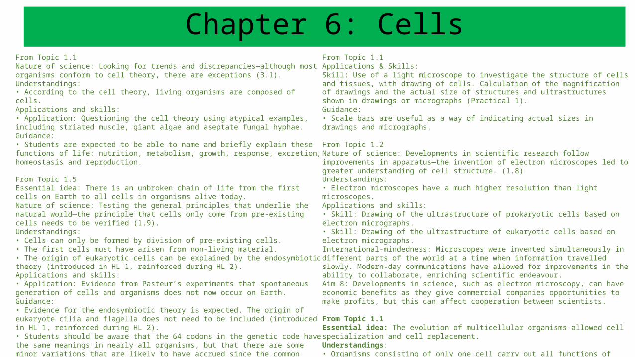
Chapter 6: CellsFrom Topic 1.1Nature of science: Looking for trends and discrepancies—although most organisms conform to cell theory, there are exceptions (3.1).Understandings:• According to the cell theory, living organisms are composed of cells.Applications and skills:• Application: Questioning the cell theory using atypical examples, including striated muscle, giant algae and aseptate fungal hyphae.Guidance:• Students are expected to be able to name and briefly explain these functions of life: nutrition, metabolism, growth, response, excretion, homeostasis and reproduction.
From Topic 1.5Essential idea: There is an unbroken chain of life from the first cells on Earth to all cells in organisms alive today.Nature of science: Testing the general principles that underlie the natural world—the principle that cells only come from pre-existing cells needs to be verified (1.9).Understandings:• Cells can only be formed by division of pre-existing cells.• The first cells must have arisen from non-living material.• The origin of eukaryotic cells can be explained by the endosymbiotic theory (introduced in HL 1, reinforced during HL 2).Applications and skills:• Application: Evidence from Pasteur’s experiments that spontaneous generation of cells and organisms does not now occur on Earth.Guidance:• Evidence for the endosymbiotic theory is expected. The origin of eukaryote cilia and flagella does not need to be included (introduced in HL 1, reinforced during HL 2).• Students should be aware that the 64 codons in the genetic code have the same meanings in nearly all organisms, but that there are some minor variations that are likely to have accrued since the common origin of life on Earth (introduced in HL 1, reinforced during HL 2).Aim 6: Pasteur’s experiment can be repeated using modern apparatus.
From Topic 1.1Applications & Skills: Skill: Use of a light microscope to investigate the structure of cells and tissues, with drawing of cells. Calculation of the magnification of drawings and the actual size of structures and ultrastructures shown in drawings or micrographs (Practical 1).Guidance:• Scale bars are useful as a way of indicating actual sizes in drawings and micrographs.
From Topic 1.2Nature of science: Developments in scientific research follow improvements in apparatus—the invention of electron microscopes led to greater understanding of cell structure. (1.8)Understandings:• Electron microscopes have a much higher resolution than light microscopes.Applications and skills:• Skill: Drawing of the ultrastructure of prokaryotic cells based on electron micrographs.• Skill: Drawing of the ultrastructure of eukaryotic cells based on electron micrographs.International-mindedness: Microscopes were invented simultaneously in different parts of the world at a time when information travelled slowly. Modern-day communications have allowed for improvements in the ability to collaborate, enriching scientific endeavour.Aim 8: Developments in science, such as electron microscopy, can have economic benefits as they give commercial companies opportunities to make profits, but this can affect cooperation between scientists.
From Topic 1.1Essential idea: The evolution of multicellular organisms allowed cell specialization and cell replacement.Understandings:• Organisms consisting of only one cell carry out all functions of life in that cell.• Surface area to volume ratio is important in the limitation of cell size.• Multicellular organisms have properties that emerge from the interaction of their cellular components.Applications and skills:• Application: Investigation of functions of life in Paramecium and one named photosynthetic unicellular organism.
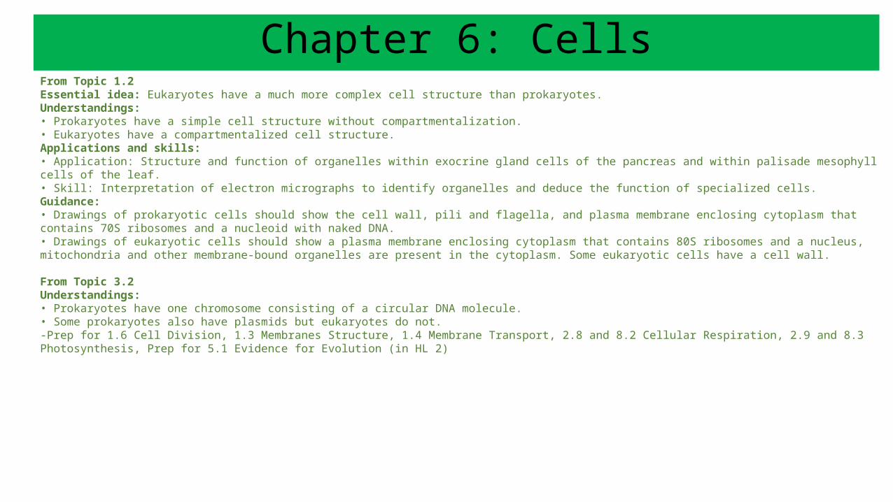
From Topic 1.2Essential idea: Eukaryotes have a much more complex cell structure than prokaryotes.Understandings:• Prokaryotes have a simple cell structure without compartmentalization.• Eukaryotes have a compartmentalized cell structure.Applications and skills:• Application: Structure and function of organelles within exocrine gland cells of the pancreas and within palisade mesophyll cells of the leaf.• Skill: Interpretation of electron micrographs to identify organelles and deduce the function of specialized cells.Guidance:• Drawings of prokaryotic cells should show the cell wall, pili and flagella, and plasma membrane enclosing cytoplasm that contains 70S ribosomes and a nucleoid with naked DNA.• Drawings of eukaryotic cells should show a plasma membrane enclosing cytoplasm that contains 80S ribosomes and a nucleus, mitochondria and other membrane-bound organelles are present in the cytoplasm. Some eukaryotic cells have a cell wall. From Topic 3.2Understandings:• Prokaryotes have one chromosome consisting of a circular DNA molecule.• Some prokaryotes also have plasmids but eukaryotes do not.-Prep for 1.6 Cell Division, 1.3 Membranes Structure, 1.4 Membrane Transport, 2.8 and 8.2 Cellular Respiration, 2.9 and 8.3 Photosynthesis, Prep for 5.1 Evidence for Evolution (in HL 2)
Chapter 6: Cells
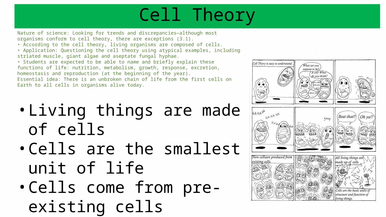
Cell TheoryNature of science: Looking for trends and discrepancies—although most organisms conform to cell theory, there are exceptions (3.1).• According to the cell theory, living organisms are composed of cells.• Application: Questioning the cell theory using atypical examples, including striated muscle, giant algae and aseptate fungal hyphae.• Students are expected to be able to name and briefly explain these functions of life: nutrition, metabolism, growth, response, excretion, homeostasis and reproduction (at the beginning of the year).Essential idea: There is an unbroken chain of life from the first cells on Earth to all cells in organisms alive today.
• Living things are made of cells• Cells are the smallest unit of life• Cells come from pre-existing
cells
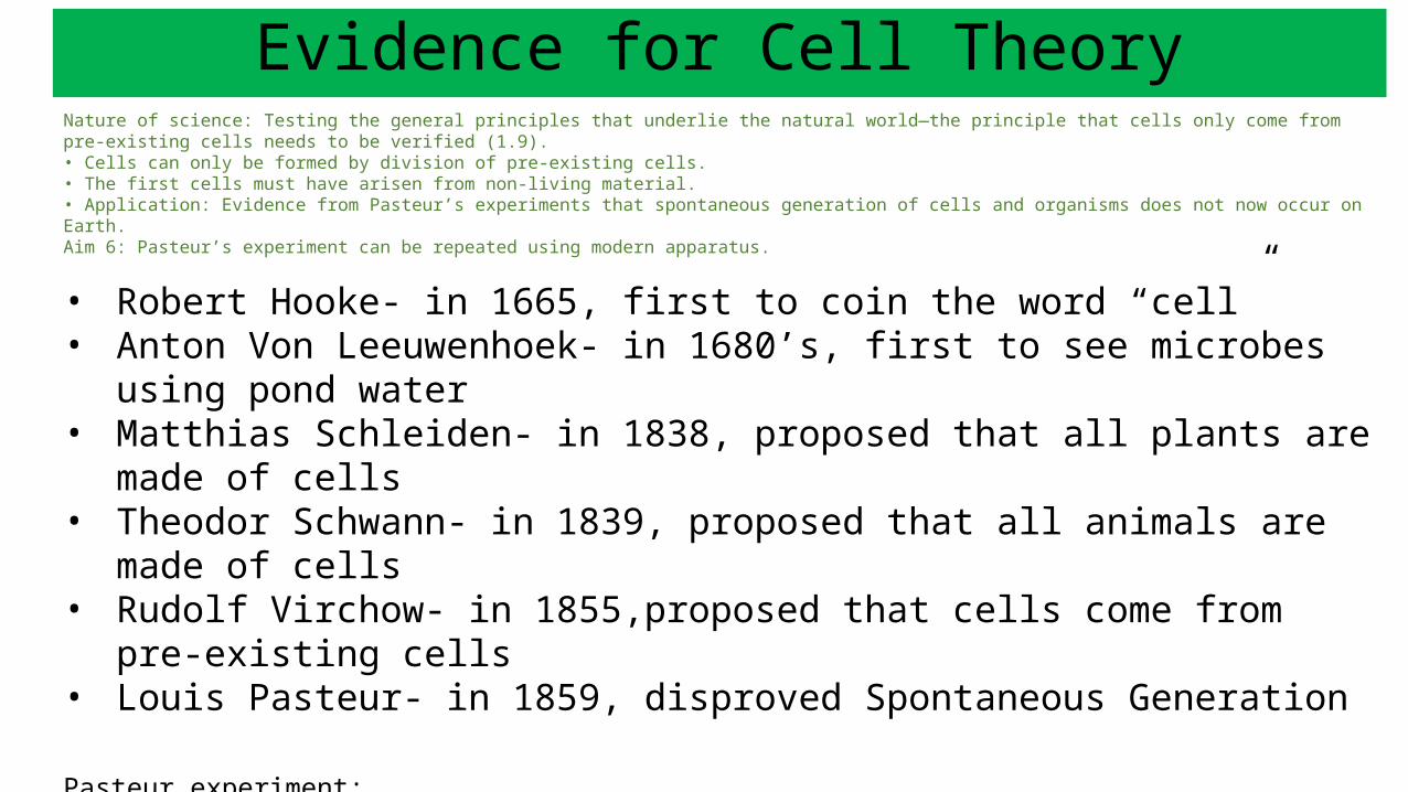
Evidence for Cell TheoryNature of science: Testing the general principles that underlie the natural world—the principle that cells only come from pre-existing cells needs to be verified (1.9).• Cells can only be formed by division of pre-existing cells.• The first cells must have arisen from non-living material.• Application: Evidence from Pasteur’s experiments that spontaneous generation of cells and organisms does not now occur on Earth.Aim 6: Pasteur’s experiment can be repeated using modern apparatus.
• Robert Hooke- in 1665, first to coin the word “cell” • Anton Von Leeuwenhoek- in 1680’s, first to see microbes using pond water• Matthias Schleiden- in 1838, proposed that all plants are made of cells• Theodor Schwann- in 1839, proposed that all animals are made of cells• Rudolf Virchow- in 1855,proposed that cells come from pre-existing cells• Louis Pasteur- in 1859, disproved Spontaneous Generation
Pasteur experiment: http://bcs.whfreeman.com/thelifewire/content/chp03/0302003.html
Cell theory: : http://ed.ted.com/lessons/the-wacky-history-of-cell-theory
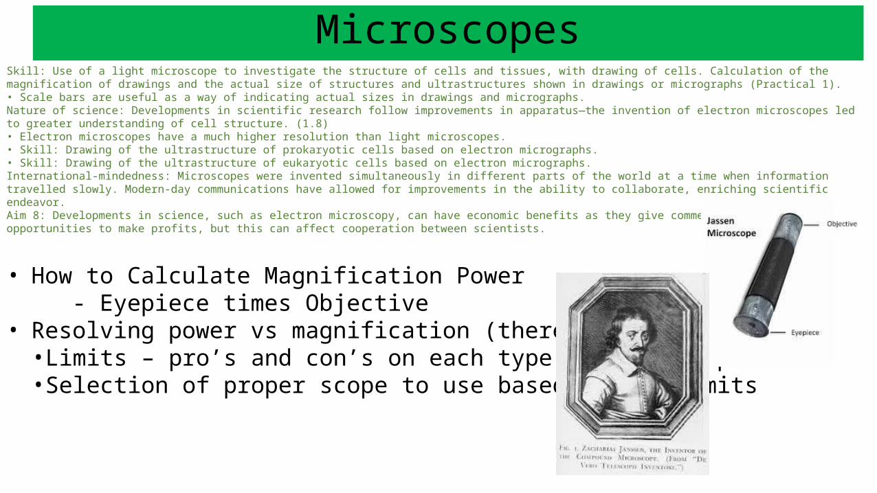
MicroscopesSkill: Use of a light microscope to investigate the structure of cells and tissues, with drawing of cells. Calculation of the magnification of drawings and the actual size of structures and ultrastructures shown in drawings or micrographs (Practical 1).• Scale bars are useful as a way of indicating actual sizes in drawings and micrographs.Nature of science: Developments in scientific research follow improvements in apparatus—the invention of electron microscopes led to greater understanding of cell structure. (1.8)• Electron microscopes have a much higher resolution than light microscopes.• Skill: Drawing of the ultrastructure of prokaryotic cells based on electron micrographs.• Skill: Drawing of the ultrastructure of eukaryotic cells based on electron micrographs.International-mindedness: Microscopes were invented simultaneously in different parts of the world at a time when information travelled slowly. Modern-day communications have allowed for improvements in the ability to collaborate, enriching scientific endeavor.Aim 8: Developments in science, such as electron microscopy, can have economic benefits as they give commercial companies opportunities to make profits, but this can affect cooperation between scientists.
• How to Calculate Magnification Power- Eyepiece times Objective
• Resolving power vs magnification (there’s a difference)•Limits – pro’s and con’s on each type of microscopes•Selection of proper scope to use based on the limits
Types of Microscopes
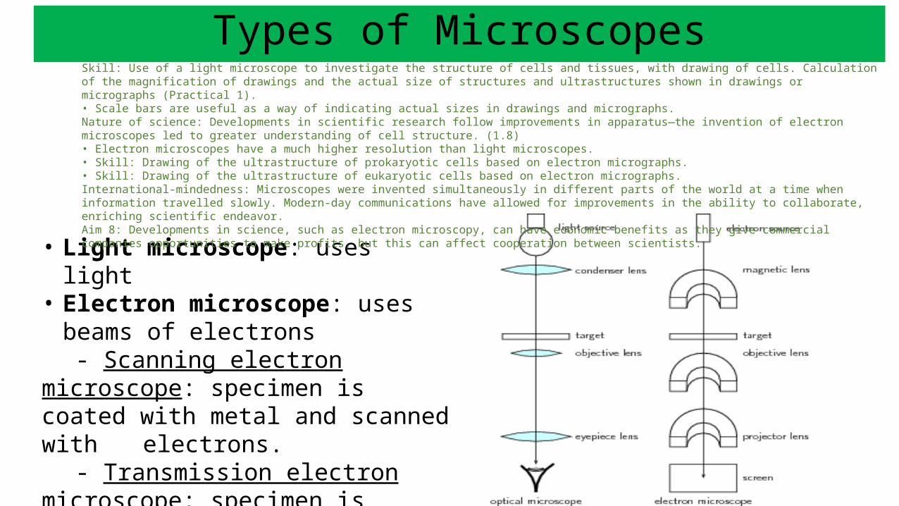
• Light microscope: uses light• Electron microscope: uses beams of
electrons- Scanning electron microscope:
specimen is coated with metal and scanned with electrons.
- Transmission electron microscope: specimen is sliced thin and stained; bombarded with electrons.
Skill: Use of a light microscope to investigate the structure of cells and tissues, with drawing of cells. Calculation of the magnification of drawings and the actual size of structures and ultrastructures shown in drawings or micrographs (Practical 1).• Scale bars are useful as a way of indicating actual sizes in drawings and micrographs.Nature of science: Developments in scientific research follow improvements in apparatus—the invention of electron microscopes led to greater understanding of cell structure. (1.8)• Electron microscopes have a much higher resolution than light microscopes.• Skill: Drawing of the ultrastructure of prokaryotic cells based on electron micrographs.• Skill: Drawing of the ultrastructure of eukaryotic cells based on electron micrographs.International-mindedness: Microscopes were invented simultaneously in different parts of the world at a time when information travelled slowly. Modern-day communications have allowed for improvements in the ability to collaborate, enriching scientific endeavor.Aim 8: Developments in science, such as electron microscopy, can have economic benefits as they give commercial companies opportunities to make profits, but this can affect cooperation between scientists.
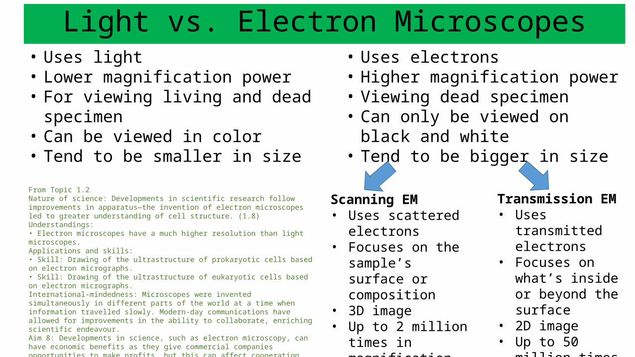
Light vs. Electron Microscopes• Uses light• Lower magnification power• For viewing living and dead specimen• Can be viewed in color• Tend to be smaller in size
From Topic 1.2Nature of science: Developments in scientific research follow improvements in apparatus—the invention of electron microscopes led to greater understanding of cell structure. (1.8)Understandings:• Electron microscopes have a much higher resolution than light microscopes.Applications and skills:• Skill: Drawing of the ultrastructure of prokaryotic cells based on electron micrographs.• Skill: Drawing of the ultrastructure of eukaryotic cells based on electron micrographs.International-mindedness: Microscopes were invented simultaneously in different parts of the world at a time when information travelled slowly. Modern-day communications have allowed for improvements in the ability to collaborate, enriching scientific endeavour.Aim 8: Developments in science, such as electron microscopy, can have economic benefits as they give commercial companies opportunities to make profits, but this can affect cooperation between scientists.
• Uses electrons• Higher magnification power• Viewing dead specimen• Can only be viewed on black and
white• Tend to be bigger in size
Scanning EM• Uses scattered
electrons• Focuses on the
sample’s surface or composition
• 3D image• Up to 2 million times
in magnification
Transmission EM• Uses transmitted
electrons• Focuses on what’s
inside or beyond the surface
• 2D image• Up to 50 million
times in magnification
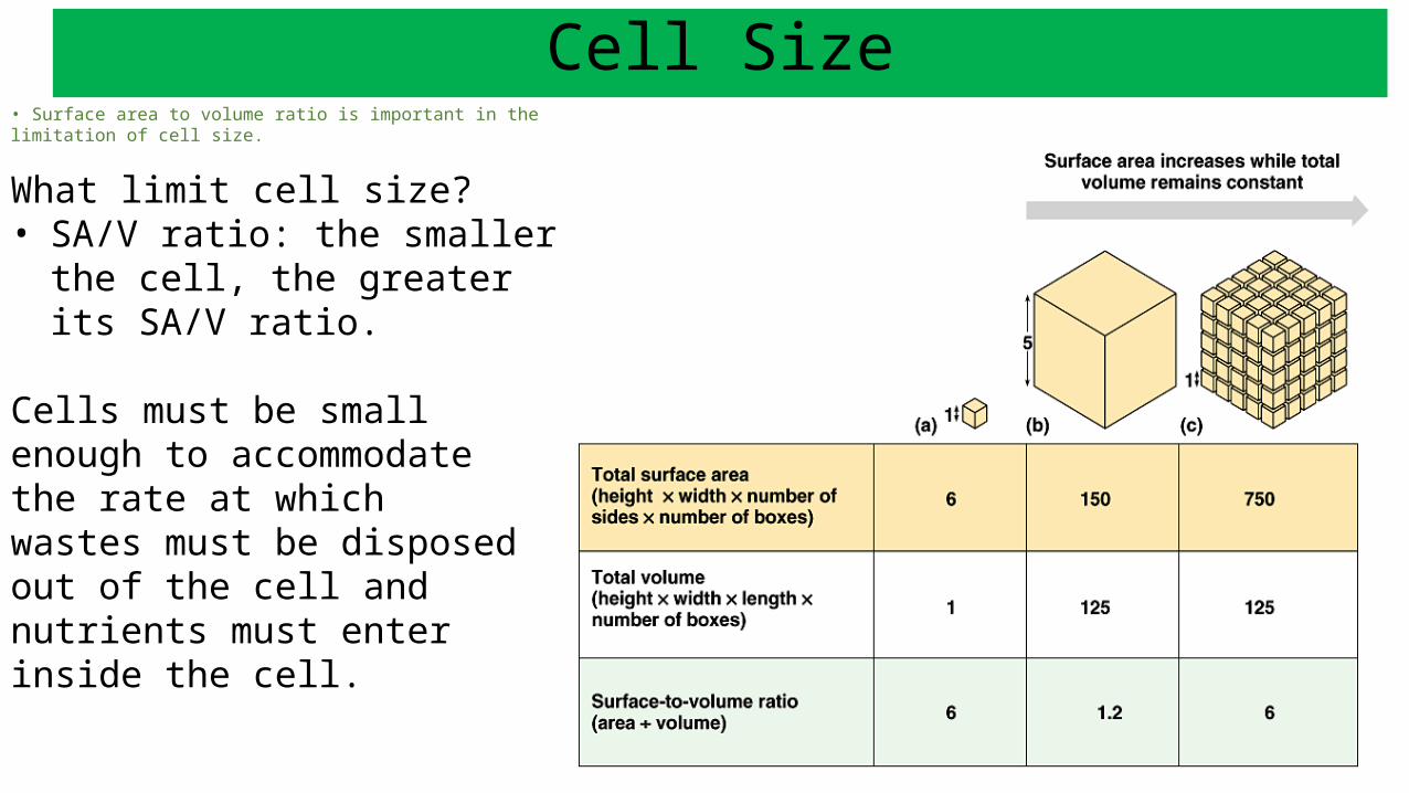
• Surface area to volume ratio is important in the limitation of cell size.
What limit cell size?• SA/V ratio: the smaller the cell,
the greater its SA/V ratio.
Cells must be small enough to accommodate the rate at whichwastes must be disposed out of the cell and nutrients must enter inside the cell.
Cell Size
Prokaryotic Cells
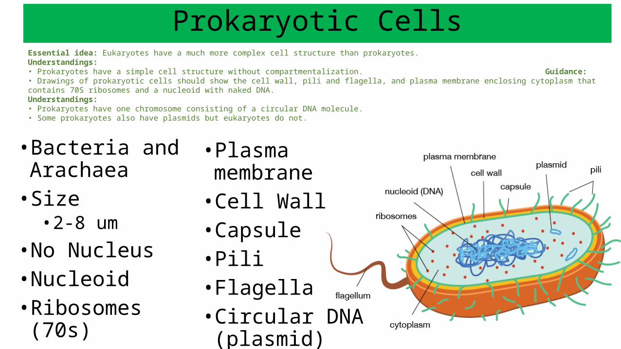
• Bacteria and Arachaea
• Size• 2-8 um
• No Nucleus• Nucleoid• Ribosomes (70s)
• Plasma membrane• Cell Wall• Capsule• Pili• Flagella• Circular DNA
(plasmid)
Essential idea: Eukaryotes have a much more complex cell structure than prokaryotes.Understandings:• Prokaryotes have a simple cell structure without compartmentalization.
Guidance:• Drawings of prokaryotic cells should show the cell wall, pili and flagella, and plasma membrane enclosing cytoplasm that contains 70S ribosomes and a nucleoid with naked DNA.Understandings:• Prokaryotes have one chromosome consisting of a circular DNA molecule.• Some prokaryotes also have plasmids but eukaryotes do not.
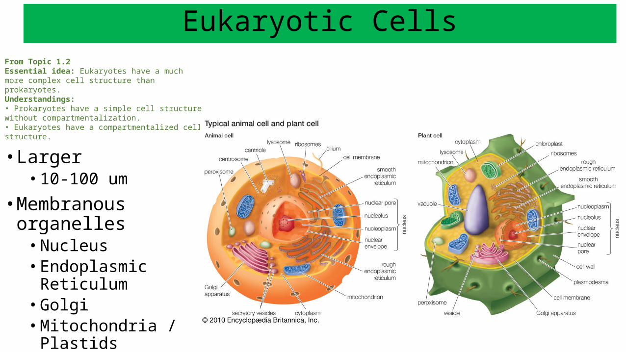
Eukaryotic CellsFrom Topic 1.2Essential idea: Eukaryotes have a much more complex cell structure than prokaryotes.Understandings:• Prokaryotes have a simple cell structure without compartmentalization.• Eukaryotes have a compartmentalized cell structure.
• Larger• 10-100 um
• Membranous organelles• Nucleus• Endoplasmic Reticulum• Golgi• Mitochondria / Plastids• Lysosomes• Peroxisomes
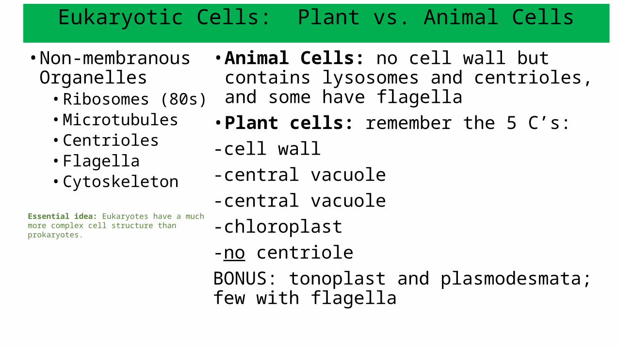
Eukaryotic Cells: Plant vs. Animal Cells• Non-membranous
Organelles• Ribosomes (80s)• Microtubules• Centrioles• Flagella• Cytoskeleton
Essential idea: Eukaryotes have a much more complex cell structure than prokaryotes.
• Animal Cells: no cell wall but contains lysosomes and centrioles, and some have flagella
• Plant cells: remember the 5 C’s: -cell wall-central vacuole-central vacuole-chloroplast-no centrioleBONUS: tonoplast and plasmodesmata; few with flagella
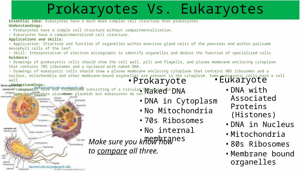
Prokaryotes Vs. Eukaryotes
• Prokaryote• Naked DNA• DNA in Cytoplasm• No Mitochondria• 70s Ribosomes• No internal membranes
• Eukaryote• DNA with Associated
Proteins (Histones)• DNA in Nucleus• Mitochondria• 80s Ribosomes• Membrane bound
organellesMake sure you know how to compare all three.
Essential idea: Eukaryotes have a much more complex cell structure than prokaryotes.Understandings:• Prokaryotes have a simple cell structure without compartmentalization.• Eukaryotes have a compartmentalized cell structure.Applications and skills:• Application: Structure and function of organelles within exocrine gland cells of the pancreas and within palisade mesophyll cells of the leaf.• Skill: Interpretation of electron micrographs to identify organelles and deduce the function of specialized cells.Guidance:• Drawings of prokaryotic cells should show the cell wall, pili and flagella, and plasma membrane enclosing cytoplasm that contains 70S ribosomes and a nucleoid with naked DNA.• Drawings of eukaryotic cells should show a plasma membrane enclosing cytoplasm that contains 80S ribosomes and a nucleus, mitochondria and other membrane-bound organelles are present in the cytoplasm. Some eukaryotic cells have a cell wall. Understandings:• Prokaryotes have one chromosome consisting of a circular DNA molecule.• Some prokaryotes also have plasmids but eukaryotes do not.
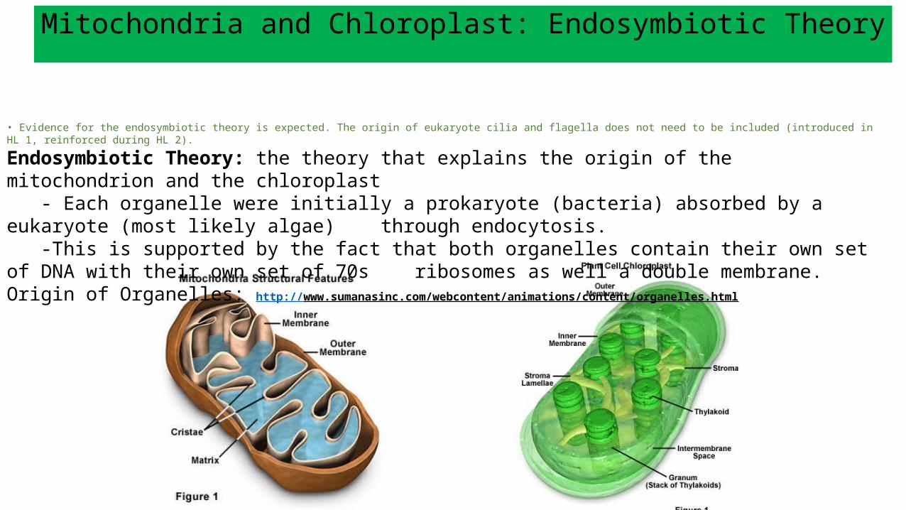
Mitochondria and Chloroplast: Endosymbiotic Theory
• Evidence for the endosymbiotic theory is expected. The origin of eukaryote cilia and flagella does not need to be included (introduced in HL 1, reinforced during HL 2).
Endosymbiotic Theory: the theory that explains the origin of the mitochondrion and the chloroplast- Each organelle were initially a prokaryote (bacteria) absorbed by a eukaryote (most likely algae)
through endocytosis. -This is supported by the fact that both organelles contain their own set of DNA with their own set of
70s ribosomes as well a double membrane.Origin of Organelles: http://www.sumanasinc.com/webcontent/animations/content/organelles.html
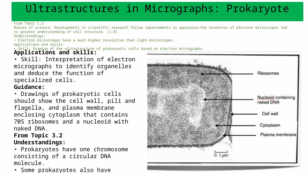
Ultrastructures in Micrographs: ProkaryoteFrom Topic 1.2Nature of science: Developments in scientific research follow improvements in apparatus—the invention of electron microscopes led to greater understanding of cell structure. (1.8)Understandings:• Electron microscopes have a much higher resolution than light microscopes.Applications and skills:• Skill: Drawing of the ultrastructure of prokaryotic cells based on electron micrographs.
Applications and skills:• Skill: Interpretation of electron micrographs to identify organelles and deduce the function of specialized cells. Guidance:• Drawings of prokaryotic cells should show the cell wall, pili and flagella, and plasma membrane enclosing cytoplasm that contains 70S ribosomes and a nucleoid with naked DNA. From Topic 3.2Understandings:• Prokaryotes have one chromosome consisting of a circular DNA molecule.• Some prokaryotes also have plasmids but eukaryotes do not.
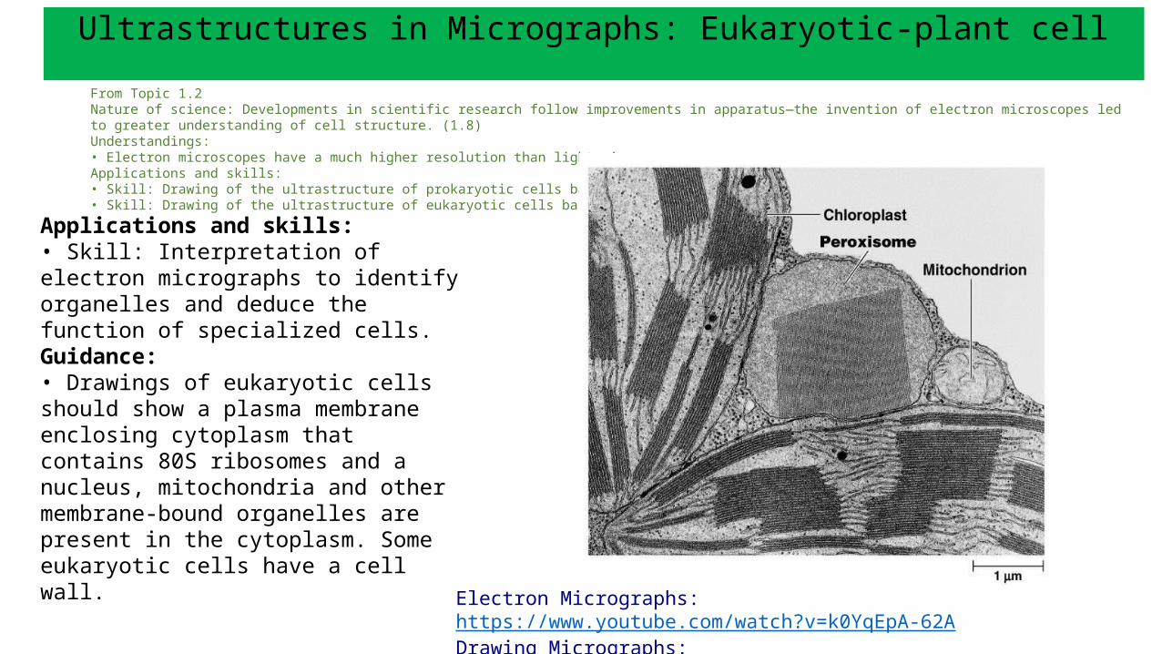
Ultrastructures in Micrographs: Eukaryotic-plant cellFrom Topic 1.2Nature of science: Developments in scientific research follow improvements in apparatus—the invention of electron microscopes led to greater understanding of cell structure. (1.8)Understandings:• Electron microscopes have a much higher resolution than light microscopes.Applications and skills:• Skill: Drawing of the ultrastructure of prokaryotic cells based on electron micrographs.• Skill: Drawing of the ultrastructure of eukaryotic cells based on electron micrographs.
Applications and skills:• Skill: Interpretation of electron micrographs to identify organelles and deduce the function of specialized cells.Guidance:• Drawings of eukaryotic cells should show a plasma membrane enclosing cytoplasm that contains 80S ribosomes and a nucleus, mitochondria and other membrane-bound organelles are present in the cytoplasm. Some eukaryotic cells have a cell wall.
Electron Micrographs: https://www.youtube.com/watch?v=k0YqEpA-62A
Drawing Micrographs: https://www.youtube.com/watch?v=vLORO1KKY74
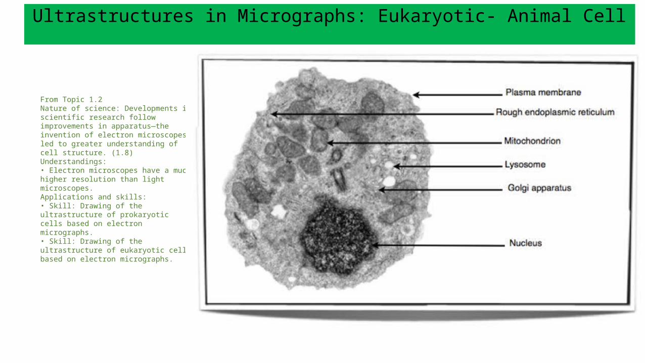
Ultrastructures in Micrographs: Eukaryotic- Animal Cell
From Topic 1.2Nature of science: Developments in scientific research follow improvements in apparatus—the invention of electron microscopes led to greater understanding of cell structure. (1.8)Understandings:• Electron microscopes have a much higher resolution than light microscopes.Applications and skills:• Skill: Drawing of the ultrastructure of prokaryotic cells based on electron micrographs.• Skill: Drawing of the ultrastructure of eukaryotic cells based on electron micrographs.

Categories:• Manufacture• Breakdown• Energy
Processing• Support and
Movement• Other- specify!
• Nucleus
• Nucleolus
• Plasma membrane
• Ribosomes
• Golgi apparatus
• Lysosomes
• Mitochondrion
• Peroxisomes
• Cytoskeleton (microfilaments –AKA actin, intermediate filaments, microtubules
• Cystol
• Centrosome (full function ?)
• Cilia/ Flagellum
• Rough ER• Smooth ER• Central Vacuole• tonoplast• Chloroplasts• Cell wall• Plasmodesmata• Gap junctions• Desomosomes
Major Functions of OrganellesFrom Topic 1.1Essential idea: The evolution of multicellular organisms allowed cell specialization and cell replacement.Understandings:• Organisms consisting of only one cell carry out all functions of life in that cell.• Surface area to volume ratio is important in the limitation of cell size.• Multicellular organisms have properties that emerge from the interaction of their cellular components.
Can you categorize what major function(s) each organelle performs?
• Students should be aware that the 64 codons in the genetic code have the same meanings in nearly all organisms, but that there are some minor variations that are likely to have accrued since the common origin of life on Earth (introduced in HL 1, reinforced during HL 2).Aim 6: Pasteur’s experiment can be repeated using modern apparatus.
International-mindedness: Microscopes were invented simultaneously in different parts of the world at a time when information travelled slowly. Modern-day communications have allowed for improvements in the ability to collaborate, enriching scientific endeavour.Aim 8: Developments in science, such as electron microscopy, can have economic benefits as they give commercial companies opportunities to make profits, but this can affect cooperation between scientists.
From Topic 3.2Understandings:• Prokaryotes have one chromosome consisting of a circular DNA molecule.• Some prokaryotes also have plasmids but eukaryotes do not.-Prep for 1.6 Cell Division, 1.3 Membranes Structure, 1.4 Membrane Transport, 2.8 and 8.2 Cellular Respiration, 2.9 and 8.3 Photosynthesis, Prep for 5.1 Evidence for Evolution (in HL 2)