Embed Size (px)
Citation preview
3/17/2015
1
Remind 101
Introduction to Microbiology CH 1
Microbiology 101
SO…what is microbiology?
What is a microbe?
2 dimensions of microbiology:
1. Types of microbes
2. What microbiologists do
3
Roles of Microbes
Pathogens
Food chain
Autotrophs
Decomposers
Digestive
Foods and fermentation
Antibiotics
Biotechnology
Bioremediation
Disease Research
Biological Research Uses Microbes
1. Size/ Structure
2. Large populations
3. Rapid Growth Rate
4. Research Benefits
Vaccines
Antibiotics
Overview of Microbes 6 Viruses
Prokaryotes Unicellular, no nucleus
Kingdoms Archaebacteria & Eubacteria
Eukaryotes Unicellular & multicellular, have a nucleus
Kingdoms Protista (algae & protozoa)
Fungi
Animalia (helminths & arthropods)
3/17/2015
2
Microbes: Viruses
Nonliving because it doesn’t
display characteristics of life
until it has a host
Simple structure
Capsid
Nucleic acid
Smaller relatives
Viroids
Prions
Microbes: Other Non-living
Infectious Agents
Viroid—smallest known particle that can cause
infections
Circular, single strands of RNA
No capsid (no protein coat)
Microbes: Other Non-living
Infectious Agents
Prions—proteins that do not have any nucleic acids, but instead cause other proteins to fold incorrectly.
responsible for many animal diseases like mad cow disease and the human equivalent Creutzfeldt-Jakob disease.
ex. Kuru—occurred in
many tribal places
Microbes: Prokaryotes
Structure
No nucleus
Organelles
Cell wall
Two Kingdoms:
Archaebacteria
Eubacteria
10
Microbes: Eukaryotic Parasites Protists
Ex. amoeba
Fungi
Helminths
Ex. Worms (flat, round, & segmented)
Arthropods
Ex. Ticks, insects, fleas
Cause/transmit disease in their
microscopic stages or as carriers
Health-Related Fields of Study
Immunology
Epidemiology
Etiology
Bioremediation
3/17/2015
3
Fields of Study using Application
Infection Control
Chemotherapy
Industrial Microbiology
Biotechnology
Brain Check…
1. How can microorganisms be beneficial?
2. Why are viruses not considered living?
3. What is the difference between epidemiology and
etiology?
4. Name 5 bacterial diseases.
5. Name 5 viral diseases.
Spontaneous Generation versus Biogenesis
Definitions
Redi
Spallanzani
? Role of oxygen
Pasteur
Biogenesis wins!
Fermentation
Pasteurization
Germ Theory
Theory definition:
Microorganisms can invade
other organisms and cause
disease
Important Contributors:
Koch
Semmelweiss
Lister
Koch’s Postulates
1. The bacteria must be present in every case of the
disease.
2. The bacteria must be isolated from the host with
the disease and grown in pure culture.
3. The specific disease must be reproduced when a
pure culture of the bacteria is inoculated into a
healthy susceptible host.
4. The bacteria must be recoverable from the
experimentally infected host.
17
Please Wash Your Hands!!!
Semmelweiss: work demonstrated that hand-washing
could drastically reduce the number of women dying
after childbirth
Lister: father of aseptic technique in surgery
18
3/17/2015
4
Immunology
Edward Jenner:
pioneer of smallpox vaccine,
the world's first vaccine.
"the father of immunology“
Pasteur
Pasteurization
Rabies vaccine
Metchnikoff: discovered phagocytes and their role in the immune
system
Future Trends
Recombinant Microbes
Drugs
Hormones
Vaccines
Gene therapy
Bacteriophage therapy
Genomics
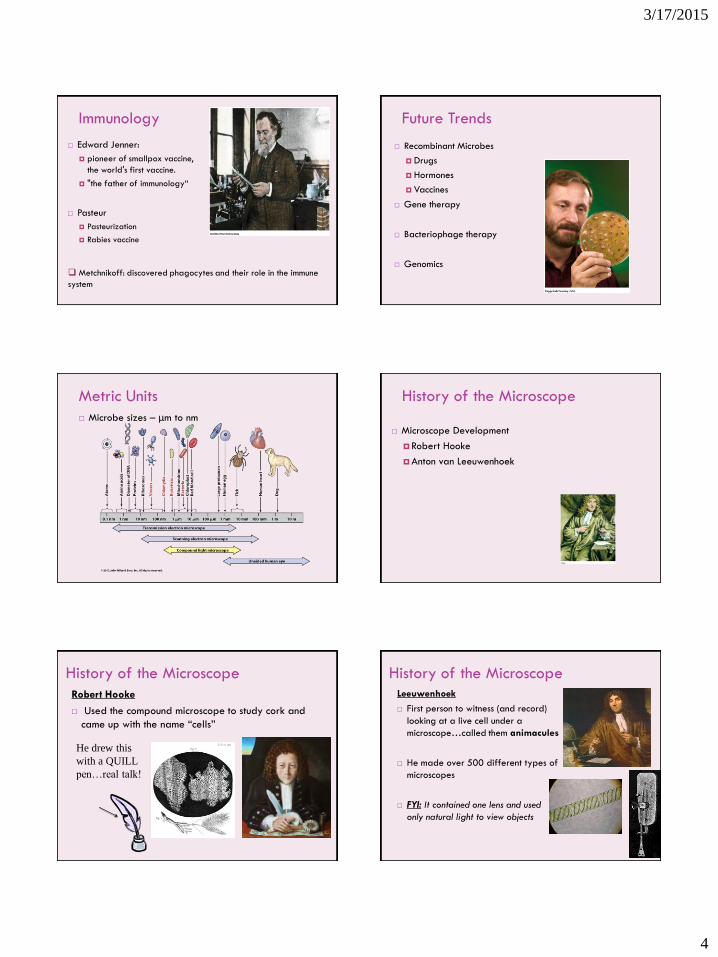
Metric Units
Microbe sizes – μm to nm
History of the Microscope
Microscope Development
Robert Hooke
Anton van Leeuwenhoek
Robert Hooke
Used the compound microscope to study cork and
came up with the name “cells”
He drew this
with a QUILL
pen…real talk!
History of the Microscope Leeuwenhoek
First person to witness (and record)
looking at a live cell under a
microscope…called them animacules
He made over 500 different types of
microscopes
FYI: It contained one lens and used
only natural light to view objects
History of the Microscope
3/17/2015
5
Compound Light Microscopy Condenser
Iris diaphragm
Objective lenses
Ocular lens(es)
Monocular or binocular
Stage
Focusing knobs
Total Magnification
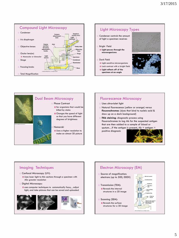
Light Microscopy Types
Condenser controls the amount
of light a specimen receives
Bright Field
Light passes through the
microorganisms
Dark Field
Light-sensitive microorganisms
Lack contrast with a bright field
Light reflects off of the
specimen at an angle
Dual Beam Microscopy Phase Contrast
for organisms that would be
killed by stains
Changes the speed of light
so that you have different
degrees of brightness
Nomarski
Uses a higher resolution to
make an almost 3D picture
Fluorescence Microscopy Uses ultraviolet light
Natural fluorescence (yellow or orange) versus
flouorochromes (dyes that bind to nucleic acid &
show up on a dark background)
FAb staining: diagnostic process using
flouorochromes to tag Ab for the suspected antigen
that are then added to a sample of blood or
sputum…if the antigen is present, Ab + antigen =
positive diagnosis
Imaging Techniques
Confocal Microscopy (UV):
Uses laser light to thin sections through a specimen with
40x greater resolution
Digital Microscopy:
uses computer techniques to automatically focus, , adjust
light, and take pictures that can be saved and uploaded
Electron Microscopy (EM)
Source of magnification:
electrons (up to 500, 000X)
Transmission (TEM):
Reveals the internal
structures in a 2D image
Scanning (SEM):
Reveals the surface
structures for a 3D image
3/17/2015
6
EM Images EM Images
Light Microscope Specimen Preparation:
Wet Mounts
Wet Mounts
Helps show motility in
microorganisms
Shows if specimen
are even present
fresh cultures must be
used for maximum
motility
NO STAINS!!
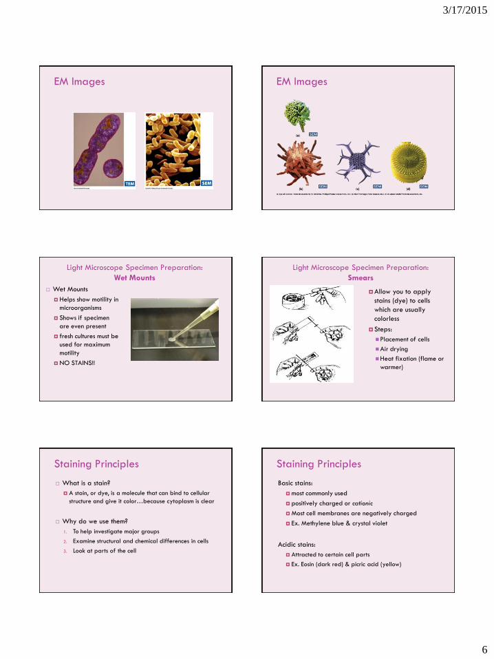
Light Microscope Specimen Preparation:
Smears
Allow you to apply
stains (dye) to cells
which are usually
colorless
Steps:
Placement of cells
Air drying
Heat fixation (flame or
warmer)
Staining Principles
What is a stain?
A stain, or dye, is a molecule that can bind to cellular
structure and give it color…because cytoplasm is clear
Why do we use them?
1. To help investigate major groups
2. Examine structural and chemical differences in cells
3. Look at parts of the cell
Staining Principles
Basic stains:
most commonly used
positively charged or cationic
Most cell membranes are negatively charged
Ex. Methylene blue & crystal violet
Acidic stains:
Attracted to certain cell parts
Ex. Eosin (dark red) & picric acid (yellow)
3/17/2015
7
Staining Principles 1. Simple Stains:
Uses a single dye
Reveals basic cell structures and arrangements
Ex. Methylene blue, crystal violet, & carbolfuchsin
2. Differential Stains: Gram & Ziehl-Neelsen
Using 2 or more dyes
Distinguishes between 2 kinds of organisms or 2 different
parts of it
3. Special Stains: Negative, Flagellar, & Endospore
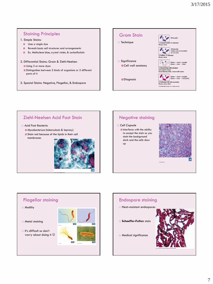
Gram Stain
Technique
Significance
Cell wall anatomy
Diagnosis
Ziehl-Neelsen Acid Fast Stain
Acid Fast Bacteria
Mycobacterium (tuberculosis & leprosy)
Stain red because of the lipids in their cell
membranes
Negative staining
Cell Capsule
Interferes with the ability
to accept the stain so you
stain the background
dark and the cells show
up
Flagellar staining
Motility
Metal staining
It’s difficult so don’t
worry about doing it
Endospore staining
Heat-resistant endospores
Schaeffer-Fulton stain
Medical significance
3/17/2015
8
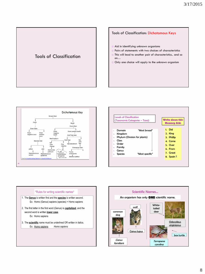
Tools of Classification
Aid in identifying unknown organisms
Pairs of statements with two choices of characteristics
This will lead to another pair of characteristics.. and so on…
Only one choice will apply to the unknown organism
Tools of Classification: Dichotomous Keys
45
Levels of Classification (Taxonomic Categories – Taxa)
1. Domain “Most broad” 2. Kingdom 3. Phylum (Division for plants) 4. Class 5. Order 6. Family 7. Genus 8. Species “Most specific”
1. Did
2. King
3. Phillip
4. Come
5. Over
6. From
7. Great
8. Spain ?
Write down this Memory Aid:
*Rules for writing scientific names*
1. The Genus is written first and the species is written second.
Ex: Homo (Genus) sapiens (species) = Homo sapiens
2. The first letter in the first word (Genus) is capitalized, and the
second word is written lower case.
Ex: Homo sapiens
3. The scientific name must be underlined OR written in italics.
Ex: Homo sapiens Homo sapiens
Scientific Names…
An organism has only ONE scientific name.
common dog
Canus familiaris
wolf
Canus lupus
box turtle
Terrapene carolina
Odocoileus virginianus
White-tailed deer
3/17/2015
9
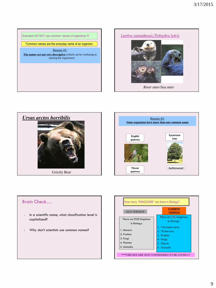
Scientists DO NOT use common names of organisms !!!
*Common names are the everyday name of an organism.
Reason #1:
The names are not very descriptive (which can be confusing in
naming the organisms)
Lontra canadensis/Enhydra lutris
River otter/Sea otter
Ursus arctos horribilis
Grizzly Bear
Reason #2: Some organisms have more than one common name
English sparrow
House sparrow
Sycamore tree
buttonwood
Brain Check…
1. In a scientific name, what classification level is
capitalized?
2. Why don’t scientists use common names?
How many “KINGDOMS” are there in Biology?
There are FIVE kingdoms
in Biology:
1. Monera
2. Protista
3. Fungi
4. Plantae
5. Animalia
There are SIX kingdoms
in Biology:
1. *Archaebacteria
2. *Eubacteria
3. Protista
4. Fungi
5. Plantae
6. Animalia
OLD VERSION CURRENT VERSION
***VIRUSES ARE NOT CONSIDERED TO BE LIVING!!!
3/17/2015
10
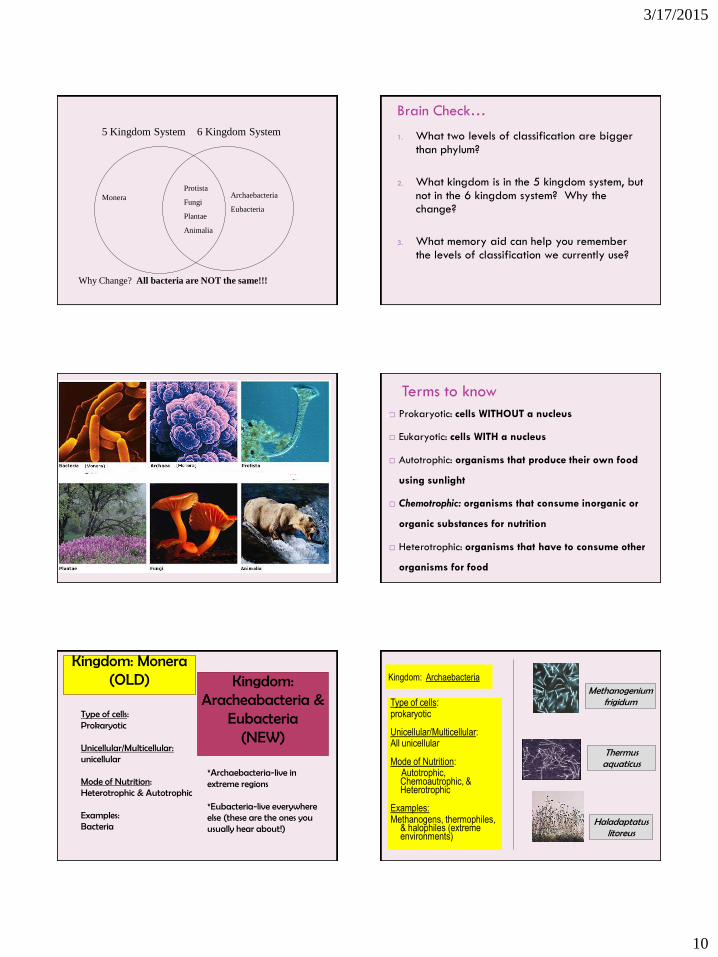
Protista
Fungi
Plantae
Animalia
Monera Archaebacteria
Eubacteria
5 Kingdom System 6 Kingdom System
Why Change? All bacteria are NOT the same!!!
Brain Check…
1. What two levels of classification are bigger than phylum?
2. What kingdom is in the 5 kingdom system, but not in the 6 kingdom system? Why the change?
3. What memory aid can help you remember the levels of classification we currently use?
Terms to know
Prokaryotic: cells WITHOUT a nucleus
Eukaryotic: cells WITH a nucleus
Autotrophic: organisms that produce their own food
using sunlight
Chemotrophic: organisms that consume inorganic or
organic substances for nutrition
Heterotrophic: organisms that have to consume other
organisms for food
58
Type of cells: Prokaryotic Unicellular/Multicellular: unicellular Mode of Nutrition: Heterotrophic & Autotrophic Examples: Bacteria
*Archaebacteria-live in extreme regions *Eubacteria-live everywhere else (these are the ones you usually hear about!)
Kingdom: Monera (OLD)
Kingdom: Aracheabacteria &
Eubacteria (NEW)
Kingdom: Archaebacteria
Type of cells: prokaryotic
Unicellular/Multicellular: All unicellular
Mode of Nutrition: Autotrophic,
Chemoautrophic, & Heterotrophic
Examples: Methanogens, thermophiles,
& halophiles (extreme environments)
Methanogenium frigidum
Thermus aquaticus
Haladaptatus litoreus
3/17/2015
11
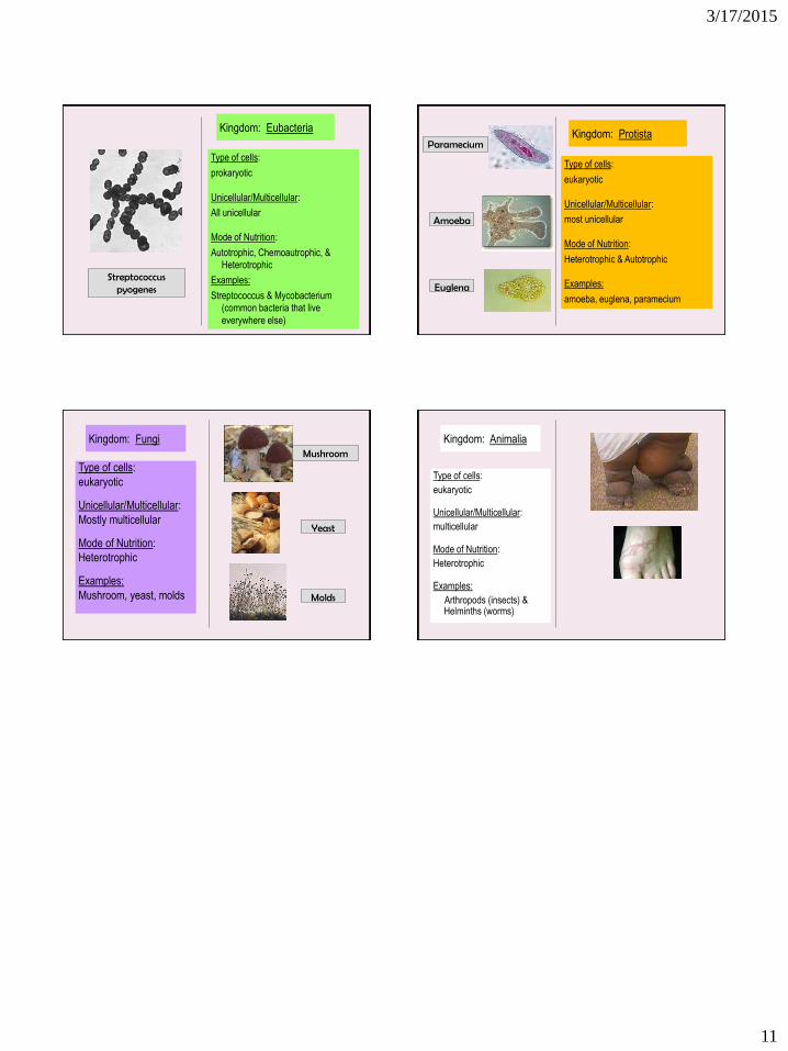
Kingdom: Eubacteria
Type of cells:
prokaryotic
Unicellular/Multicellular:
All unicellular
Mode of Nutrition:
Autotrophic, Chemoautrophic, &
Heterotrophic
Examples:
Streptococcus & Mycobacterium
(common bacteria that live
everywhere else)
Streptococcus pyogenes
Kingdom: Protista
Type of cells:
eukaryotic
Unicellular/Multicellular:
most unicellular
Mode of Nutrition:
Heterotrophic & Autotrophic
Examples:
amoeba, euglena, paramecium
Paramecium
Amoeba
Euglena
Kingdom: Fungi
Type of cells:
eukaryotic
Unicellular/Multicellular:
Mostly multicellular
Mode of Nutrition:
Heterotrophic
Examples:
Mushroom, yeast, molds
Mushroom
Yeast
Molds
Kingdom: Animalia
Type of cells:
eukaryotic
Unicellular/Multicellular:
multicellular
Mode of Nutrition:
Heterotrophic
Examples:
Arthropods (insects) & Helminths (worms)