Embed Size (px)
Citation preview
JOURNAL OF BACTERIOLOGY, July 2007, p. 5060–5067 Vol. 189, No. 140021-9193/07/$08.00�0 doi:10.1128/JB.00131-07Copyright © 2007, American Society for Microbiology. All Rights Reserved.
CfaD-Dependent Expression of a Novel Extracytoplasmic Proteinfrom Enterotoxigenic Escherichia coli�
M. Carolina Pilonieta, Maria D. Bodero, and George P. Munson*Department of Microbiology and Immunology, University of Miami Miller School of Medicine, Miami, Florida
Received 26 January 2007/Accepted 2 May 2007
H10407 is a strain of enterotoxigenic Escherichia coli (ETEC) that utilizes CFA/I pili to adhere tosurfaces of the small intestine, where it elaborates toxins that cause profuse watery diarrhea in humans.Expression of the CFA/I pilus is positively regulated at the level of transcription by CfaD, a member of theAraC/XylS family. DNase I footprinting revealed that the activator has two binding sites upstream of thepilus promoter cfaAp. One site extends from positions �23 to �56, and the other extends from positions�73 to �103 (numbering relative to the transcription start site of cfaAp). Additional CfaD binding siteswere predicted within the genome of H10407 by computational analysis. Two of these sites lie upstream ofa previously uncharacterized gene, cexE. In vitro DNase I footprinting confirmed that both sites aregenuine binding sites, and cexEp::lacZ reporters demonstrated that CfaD is required for the expression ofcexE in vivo. The amino terminus of CexE contains a secretory signal peptide that is removed duringtranslocation across the cytoplasmic membrane through the general secretory pathway. These studiessuggest that CexE may be a novel ETEC virulence factor because its expression is controlled by thevirulence regulator CfaD, and its distribution is restricted to ETEC.
Enterotoxigenic Escherichia coli (ETEC) is a noninvasivepathogen that colonizes the small intestine, and it is one of themost common causes of bacterial diarrhea worldwide (37).Adherence to the intestinal epithelium is mediated by pili (15),although nonpilus adherence factors have also been reported(9, 12, 32). Currently, there are 22 known ETEC pilus sero-types, although a typical strain expresses only two or three. Theexpression of some of the most common pilus serotypes isdependent upon a transcriptional activator. This includes theCFA/I pilus, which is regulated by CfaD (GenBank accessionno. P25393) (6, 31), and the CS1, CS2, CS3, and CS4 pili, whichare regulated by Rns (accession no. P16114) (4, 5, 11). CfaDhas alternatively been named CfaR (6), and 251 of its 265residues are identical to Rns. Given their identity to eachother, it is not surprising that CfaD and Rns are functionallyinterchangeable with each other (3, 6) and recognize the sameDNA binding sites (23).
CfaD and Rns are members of the AraC/XylS family oftranscriptional regulators (16). Like other family members,Rns is not sufficiently soluble for in vitro characterization.However, the limitation of protein solubility was overcome bythe addition of maltose binding protein (MBP) to the aminoterminus of Rns, which significantly increases the solubility ofthe regulator without disrupting its function (24). DNase Ifootprinting of MBP-Rns bound to the CS1 pilus promotercooBp revealed two binding sites: one extending into the �35hexamer and a second site between positions �93 and �129(numbering relative to the transcription start site) (24). Singlenucleotide substitutions within each site reduced Rns-depen-
dent expression from cooBp in vivo, demonstrating that bothsites are required for the full activation of the CS1 pilus pro-moter.
Rns also positively regulates its own expression by binding toa distal upstream site, from positions �243 to �210, and twosites downstream of the transcription start site (25). The up-stream site and at least one downstream site are essential forpositive autoregulation (25). Like most AraC/XylS familymembers, Rns has two helix-turn-helix motifs in its carboxy-terminal domain, and uracil interference assays have shownthat it binds in two adjacent regions of the DNA major groove(24, 25). Thus, it is likely that each helix-turn-helix motif placesa recognition helix in the major groove similar to MarA, an-other AraC/XylS family member for which there is a MarA-DNA cocrystal (28).
A recent study has shown that Rns and CfaD can also func-tion as repressors by binding to a site immediately downstreamof the transcription start site for nlpA, encoding an inner mem-brane lipoprotein, where they prevent the formation of RNApolymerase open complexes (3). Repression of nlpA may de-crease the production of outer membrane vesicles, as wasshown for mutants carrying nlpA::kan insertions (20). In thisstudy, we sought to further our understanding of the CfaD andRns regulons. We began by characterizing CfaD’s binding sitesupstream of the CFA/I pilus promoter. We then compiled thisinformation with previously identified Rns binding sites andused bioinformatics to locate additional CfaD binding sites inthe genome of ETEC strain H10407 (10). Two of these sites lieupstream of a previously uncharacterized gene, separate fromthe CFA/I pilus operon, that encodes a protein that is trans-ported across the cytoplasmic membrane via the generalsecretory pathway. Because the expression of this protein isregulated by CfaD, we have given it the mnemonic cexE, forCfaD-dependent expression, extracytoplasmic protein.
* Corresponding author. Mailing address: Department of Microbiologyand Immunology, University of Miami Miller School of Medicine, P.O.Box 016960 (R-138), Miami, FL 33101. Phone: (305) 243-5317. Fax: (305)243-4623. E-mail: [email protected].
� Published ahead of print on 11 May 2007.
5060
Dow
nloa
ded
from
http
s://j
ourn
als.
asm
.org
/jour
nal/j
b on
21
Dec
embe
r 20
21 b
y 18
6.14
8.24
0.58
.
MATERIALS AND METHODS
Strains and plasmids. Genomic DNA from ETEC strain H10407 (O78:H11CfaD� CFA/I� STIa� STIb� LT�) (10) was used as the template to amplifycexEp(�501 to �312) with primers aat-1for (GCTGGATCCCGAGCGGCGTATAAAA) and aat-1rev (GCGGAATTCCACTTGATTGTATGGAAT),cexEp(�421 to �312) with primers aat-4for (GCGGGATCCAGTCCAGGAAATCGAACG) and aat-1rev, and cexEp(�174 to �312) with primers aat-5for(GCGGGATCCTGTTAATCTCTATCAATACAT) and aat-1rev. Numbering ofthe cexE promoter is relative to its transcription start site (this study). Under-lining in primer sequences indicates primer-template mismatches that add sitesfor restriction endonucleases. Plasmid pHKLac1 is a promoterless lacZ reporterplasmid with a pir-dependent origin of replication that can be integrated into thechromosome of E. coli at attBHK022 (3). Each cexEp PCR product was digestedwith BamHI and EcoRI and then ligated into the same sites of pHKLac1 toconstruct pCexELac1 [cexEp(�501 to �312)::lacZ], pCexELac2 [cexEp(�421 to�312)::lacZ], and pCexELac3 [cexEp(�174 to �312)::lacZ]. Alternatively, thePCR product carrying cexEp(�501 to �312) was cloned into the BamHI andEcoRI sites of pNEB193 (New England Biolabs) to construct pGPM1043. Thisplasmid was then subjected to oligonucleotide-directed mutagenesis to introducepoint mutations within CfaD/Rns binding sites upstream of cexEp. Mutationswere confirmed by DNA sequencing, and the mutagenized promoter fragmentswere then cloned into pHKLac1 as BamHI-EcoRI fragments. Plasmid pCex-ELac4 carries cexEo1-1, which has four point mutations: �44T to C, �43G to C,�42T to G, and �41T to G. Plasmid pCexELac5 carries cexEo2-1, which also hasfour point mutations: �482T to G, �481A to G, �479C to A, and �478G to C.With the exception of the point mutations noted above, pCexELac4 and pCex-ELac5 carry the same cexEp promoter fragment upstream of lacZ as pCexELac1.Each reporter plasmid was integrated into the chromosome of MC4100 [F�
araD139 �(argF-lac)U169 rpsL150 relA1 flhD5301 deoC1 ptsF25 rbsR] (7) aspreviously described (3) to produce strains GPM1070 (attBHK022::pCexELac1),GPM1096 (attBHK022::pCexELac2), GPM1097 (attBHK022::pCexELac3), GPM1113(attBHK022::pCexELac4), and GPM1114 (attBHK022::pCexELac5). Single integrantswere verified by colony PCR as previously described (17).
Primers aat-1for (GCTGGATCCCGAGCGGCGTATAAAA) and rrsP-XbaI-Rev (CGCTCTAGAAACATTTTACATAATGTAATCA) were used toamplify the cexE locus from human ETEC strains 27D (O126:nonmotile CfaD�
CFA/I� STIb�) (21) and G427 (O28:H12 CfaD� CFA/I� STIa� LT�) (data notshown) (isolated in the Middle East during Operation Desert Shield ca. 1990).The 1-kb PCR products were digested with BamHI and XbaI and then ligatedinto the same sites of pNEB193 to construct pGPM1039-27D and pGPM1039-G427. Plasmid pGPM1034 expresses CexE-His6 from a T7 RNA polymerase-dependent promoter and was constructed by amplifying cexE from H10407 withprimers aat-1for (see above) and aat-XhoRev (GGACCTCGAGTTTATACCAATAAGGGGTGTCAC). The PCR product was digested with BamHI and XhoIand then ligated into the same sites of pET33b (Novagen).
Plasmid pMBPRns1 (3) was used for the expression of the fusion proteinMBP-Rns in KS1000 [F� lacIq lac� pro�/ara�(lac-pro) �prc::kan eda51::Tn10gyrA rpoB thi-1 argI(Am)] (New England Biolabs)/pRare2 (Novagen) (used toprovide rare tRNAs). Plasmid pGPMRns (3) expresses Rns from lacp and is aderivative of pNEB193 (New England Biolabs). Transposon mutagenesis ofpGPMRns produced pGPMRns�Tn�2, which carries rns::kan. PlasmidpNTP503 (36) expresses CfaD and is a derivative of cloning vector pBR322.Cloning vector pHSG576 (33) (GenBank accession no. D88215) has an expectedcopy number of �5 per cell due to its pSC101-derived replicon (30, 35) and wasused to construct pEU2035 (13), which expresses Rns from lacp.
DNA sequencing of the ETEC cexE locus. The genome of ETEC strain H10407(10) is being sequenced by the Sanger Institute in collaboration with Ian Hen-derson and Mark Pallen. H10407 preliminary sequence data were obtained fromhttp://www.sanger.ac.uk/Projects/E_coli_H10407/.
The cexE locus was cloned from two CFA/I� strains of ETEC, 27D (21) andG427 (data not shown). For each clone (pGPM1039-27D and pGPM1039-G427), both strands of the cexE locus were sequenced. The 990-bp locus wasfound to be identical among ETEC strains 27D, G427, and H10407.
Software. Software for the analysis of DNA sequences using Regular Expres-sions was designed and written by G. P. Munson. The Web server SignalP wasused to evaluate the amino terminus of CexE (GenBank accession no.ABM92275) and pCoo087 (accession no. CAI79570) for potential secretorysignal peptides (http://www.cbs.dtu.dk/services/SignalP/) (2).
Purification of RNA. Strain GPM1070/pGPMRns was cultured aerobically in10 ml of Luria-Bertani (LB) medium at 37°C. After the optical absorbance at 550nm reached 1.0, 2 ml of a 5% (vol/vol) phenol–95% (vol/vol) ethanol solution wasadded to the culture, and the cells were pelleted. The cell pellet was suspended
in 10 ml of RNA wash buffer (0.75% [vol/vol] NaCl, 0.8% [vol/vol] phenol, 15.8%[vol/vol] ethanol) and then centrifuged. The resulting pellet was suspended in 500�l 0.9% NaCl and 500 �l water-saturated, nonbuffered phenol and then shakenat room temperature for 30 min. Subsequently, 50 �l of a 24:1 chloroform-isoamyl alcohol solution was added and then incubated for an additional 15 min.The solution was then chilled on ice for 5 min and centrifuged in a microcen-trifuge at the maximum rpm for 5 min. RNA was ethanol precipitated from thesupernatant and then suspended in 22 �l of RNase-free water.
Primer extension. Two picomoles of 32P-end-labeled oligonucleotide (rrsP3rev[GCAGAATTCGCGGAGAGAGACCCCATAG]) was combined with 5 �g oftotal RNA and 0.8 mM deoxynucleoside triphosphates. The solution was heatedto 65°C for 5 min and then chilled on ice for 2 min. The annealed primer was thenextended with SuperScript III reverse transcriptase according to the supplier’sprotocol (Invitrogen). Heat-denatured aliquots were separated on DNA se-quencing gels alongside dideoxy chain-terminated sequencing ladders (30).
Purification of MBP-Rns. MBP-Rns was purified from strain KS1000/pRare2/pMBPRns1 as previously described (3). In brief, the strain was grown aerobicallyat 37°C in LB medium containing 0.2% (wt/vol) glucose, 30 �g/ml chloramphen-icol, and 100 �g/ml ampicillin. Upon reaching mid-log phase, the culture wascooled to 30°C. Expression of MBP-Rns was induced for several hours by theaddition isopropyl--D-1-thiogalactopyranoside (IPTG) to a final concentrationof 300 �M. Bacterial cells were then collected by centrifugation and concen-trated �100-fold in cold lysis buffer (10 mM Tris Cl [pH 7.6, at room tempera-ture], 200 mM NaCl, 1 mM EDTA, 0.5 mM CaCl2, 10 mM -mercaptoethanol,100 �g/ml DNase I). Cells were lysed by passage through a French press. Insol-uble material was removed from the lysate by centrifugation. MBP-Rns was thenbound to an amylose column equilibrated with buffer A (10 mM Tris Cl [pH 7.6,at room temperature], 200 mM NaCl, 1 mM EDTA, 15% [vol/vol] glycerol, and10 mM -mercaptoethanol). The fusion protein was eluted from the amylosecolumn with buffer B (buffer A with 10 mM maltose).
Purification and sequencing of CexE-His6. Strain BL21(DE3)/pGPM1034 wasgrown aerobically at 37°C in LB medium containing 0.2% (wt/vol) glucose and 50�g/ml kanamycin. Expression of CexE-His6 was induced during mid-log phase bythe addition of IPTG to a final concentration of 500 �M. After 4 h of induction,approximately 325 ml of spent culture medium was cooled to 4°C and thenpassed through a nickel-Sepharose column equilibrated with IMAC-A buffer (50mM Tris Cl [pH 7.6, at room temperature], 300 mM NaCl, 10 mM imidazole).The column was washed with several column volumes of IMAC-A buffer, andCexE-His6 was then eluted by increasing the concentration of imidazole to 154mM. Approximately 50 picomoles of purified protein was transferred onto apolyvinylidene fluoride membrane from a 15% sodium dodecyl sulfate-poly-acrylamide gel by electroblotting in 10 mM N-cyclohexyl-3-aminopropanesulfo-nic acid (pH 11.0)–10% (vol/vol) methanol buffer. The immobilized protein wasvisualized on the membrane with Coomassie brilliant blue R-250 and submittedto the W. M. Keck Foundation Biotechnology Resource Laboratory at YaleUniversity for amino-terminal sequencing by Edman degradation.
DNase I footprinting. Purified MBP-Rns was equilibrated at 37°C for 30 minwith cexE promoter DNA uniquely labeled with 32P on the 5� terminus of thecoding or noncoding strand in footprinting buffer [10 mM Tris Cl (pH 7.6, atroom temperature), 50 mM KCl, 1 mM dithiothreitol, 0.4 mM MgCl2, 0.2 mMCaCl2, 2 ng/�l poly(dI-dC), 10 �g/ml bovine serum albumin]. The samples werethen treated with DNase I at a final concentration of 100 ng/�l for 1 min at 37°C.DNase I cleavage reactions were terminated by the addition of 10 volumes ofDNase I stop buffer (570 mM ammonium acetate, 50 �g/ml tRNA, 80% [vol/vol]ethanol) and then precipitated on dry ice. DNA pellets were rinsed with 70%(vol/vol) ethanol and then dried. DNA samples were separated on DNA se-quencing gels after heat denaturation in 4 �l of loading buffer (80% [vol/vol]formamide, 50 mM Tris-borate [pH 8.3], 1 mM EDTA, 0.1% [wt/vol] xylenecyanol, and bromophenol blue). The Maxam-Gilbert method was used to gen-erate GA and TC sequence ladders (30).
�-Galactosidase assays. Reporter strains GPM1070, GPM1096, GPM1097,GPM1113, and GPM1114 transformed with pGPMRns (Rns�), pGPMRns�Tn�2(rns::kan), pNTP503 (CfaD�), or vector pBR322 were grown in LB medium with100 �g/ml ampicillin. Reporter strains transformed with low-copy-number plasmidpEU2035 (Rns�) or vector pHSG576 were grown in LB medium with 30 �g/mlchloramphenicol. All strains were grown aerobically at 37°C. Cells were harvestedduring the log phase of growth, lysed, and assayed for -galactosidase activity aspreviously described (22).
Accession numbers. The sequence of the cexE locus from strain 27D has beensubmitted to the GenBank database under accession number EF205439. Theamino acid sequence of CexE is available from GenBank under accession num-ber ABM92275.
VOL. 189, 2007 CfaD-DEPENDENT EXPRESSION OF cexE 5061
Dow
nloa
ded
from
http
s://j
ourn
als.
asm
.org
/jour
nal/j
b on
21
Dec
embe
r 20
21 b
y 18
6.14
8.24
0.58
.
RESULTS
Identification of CfaD’s binding sites at the CFA/I piluspromoter. Previous studies have shown that CfaD and Rns arefully interchangeable with each other (3, 6) and recognize thesame DNA binding sites (23). Therefore, for our in vitro DNAbinding studies, we utilized a previously characterized MBP-Rns fusion (24, 25) to identify the CfaD/Rns binding sites on aDNA fragment carrying the CFA/I pilus promoter cfaAp frompositions �469 to �91 (numbering relative to the previouslyreported transcription start site) (18). The DNase I footprint ofMBP-Rns bound to the cfaAp fragment revealed two distinctregions of protection (Fig. 1A). We have designated the foot-print encompassing positions �23 to �56 as site cfaAo1 andthe footprint extending from positions �73 and �103 as sitecfaAo2. DNase I is a large enzyme that overestimates DNAbinding sites due to steric occlusion; however, sites cfaAo1 andcfaAo2 each contain a run of 12 nucleotides (Fig. 1B) that aresimilar to sequences within other CfaD/Rns binding sites (Fig.2B). Although Rns may contact DNA beyond this central core
FIG. 1. Locations of CfaD/Rns binding sites upstream of the CFA/Ipilus promoter, cfaAp. Numbering is relative to the CfaD-dependenttranscription start site of cfaAp, whose location is indicated by a wavyarrow. (A) DNase I protection of MBP-Rns bound to the noncodingstrand of cfaAp. CfaD/Rns binding sites are indicated as cfaAo1 andcfaAo2. DNase I was not added to the DNA in the lane labeled control.Lanes labeled GA and TC contain Maxam-Gilbert sequencing ladders.(B) Sequence of cfaAp and upstream region. The underlined sequenceindicates the extent of MBP-Rns DNase I footprints on the noncodingstrand. Boxed sequences indicate nucleotides that are conservedamong various CfaD/Rns binding sites. Probable �35 and �10 hex-amers are shown in boldface type.
FIG. 2. Positions and alignment of CfaD/Rns binding sites.(A) CfaD/Rns binding sites are represented as filled rectangles. Theposition of the rectangle above or below the line indicates whether theconserved sequence occurs on the coding or noncoding strand, respec-tively. Numbering is relative to the transcription start sites, which arerepresented by wavy arrows. ETEC promoters rnsp, cooBp (CS1 pilus),cfaAp (CFA/I pilus), and cexEp are activated by Rns or CfaD. Incontrast, nlpA is common to both ETEC and K-12 strains and itspromoter, nlpAp, is repressed by Rns or CfaD. It is not known if theoccupancy of binding site yicSo affects expression from yicS. (B) Op-timal alignment of the conserved core sequence of each CfaD/Rnsbinding site. Although CfaD and Rns may contact DNA beyond thenucleotides shown, the greatest sequence conservation occurs withinthis core of 12 bp.
5062 PILONIETA ET AL. J. BACTERIOL.
Dow
nloa
ded
from
http
s://j
ourn
als.
asm
.org
/jour
nal/j
b on
21
Dec
embe
r 20
21 b
y 18
6.14
8.24
0.58
.
of 12 bp, the sequence conservation within the core suggeststhat it contains most of the base-specific contacts for Rns. Thisis supported by uracil interference assays, which identified thy-mine C-5 methyl groups critical for Rns binding (24, 25). Atthe five DNA binding sites examined, thymine C-5 methylgroups beyond the central core of 12 nucleotides were notrequired for Rns binding, but three within the core were.
We note a sequence discrepancy within site cfaAo2 betweenour DNA sequencing results and the CFA/I sequence file in
GenBank (accession no. M55661). In M55661, nucleotides 741to 750 are reported as being “GATACCAAAT,” but we havefound the sequence to be “GATACAAAAAT” (correctionsand additions are underlined). These changes have been in-corporated into the sequence shown in Fig. 1B and 2B.
Prediction and verification of additional CfaD binding sites.A recent study has shown that CfaD also represses the tran-scription of nlpA, which encodes an inner membrane protein,in addition to activating the expression of the CFA/I pilin
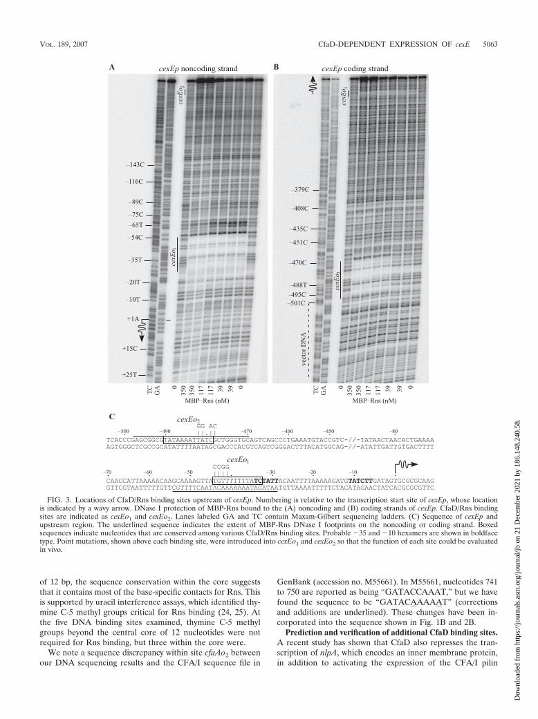
FIG. 3. Locations of CfaD/Rns binding sites upstream of cexEp. Numbering is relative to the transcription start site of cexEp, whose locationis indicated by a wavy arrow. DNase I protection of MBP-Rns bound to the (A) noncoding and (B) coding strands of cexEp. CfaD/Rns bindingsites are indicated as cexEo1 and cexEo2. Lanes labeled GA and TC contain Maxam-Gilbert sequencing ladders. (C) Sequence of cexEp andupstream region. The underlined sequence indicates the extent of MBP-Rns DNase I footprints on the noncoding or coding strand. Boxedsequences indicate nucleotides that are conserved among various CfaD/Rns binding sites. Probable �35 and �10 hexamers are shown in boldfacetype. Point mutations, shown above each binding site, were introduced into cexEo1 and cexEo2 so that the function of each site could be evaluatedin vivo.
VOL. 189, 2007 CfaD-DEPENDENT EXPRESSION OF cexE 5063
Dow
nloa
ded
from
http
s://j
ourn
als.
asm
.org
/jour
nal/j
b on
21
Dec
embe
r 20
21 b
y 18
6.14
8.24
0.58
.
genes (3). This suggests that the CfaD regulon may be largerthan previously realized. Since CfaD is a DNA binding protein,we sought to identify additional CfaD-regulated genes bysearching the genome of ETEC strain H10407 for sites similarto known CfaD/Rns binding sites. We first aligned the twobinding sites located upstream of cfaAp to previously reportedRns binding sites at the promoters for the CS1 pilus (24), nlpA(3), and rns (25) (Fig. 2). Because CfaD/Rns binding sites arenot palindromic, the sequences shown are found on the codingor noncoding strands of DNA as stated. Only the central coreof 12 nucleotides within each binding site were used in thealignment because these nucleotides have the greatest conserva-tion. The alignment was then used to write a text search string,“[CTG][AG][TA][TA][TA][TAG][ATC][ATG]TAT[CT],” inthe form of Regular Expression (26), where each position isrepresented by a nucleotide set, enclosed by brackets, or asingle character. At each position, potential binding sites mustmatch one character within each enclosed set as well as exactmatches to “TAT” at positions 9, 10, and 11. With this ap-proach, two potential binding sites were identified upstream ofa previously uncharacterized gene, cexE (Fig. 2). The sitemost proximal to cexE, cexEo1, is identical to the CfaD/Rnsbinding site cfaAo1 upstream of the CFA/I pilus promoter(Fig. 1B and 2B).
We next determined that MBP-Rns binds to both of the pre-dicted CfaD/Rns binding sites by in vitro DNase I footprinting(Fig. 3). The predicted site centered at position �38, cexEo1, wasfully encompassed by the MBP-Rns DNase I footprint, whichextends from positions �29 to �54 (Fig. 3A) (numbering relativeto the transcription start site of cexE) (see below). A secondDNase I footprint was observed between positions �470 and�498, which corresponds to site cexEo2 (Fig. 3B). AdditionalDNase I footprinting experiments revealed that cexEo1 andcexEo2 are the only MBP-Rns binding sites within the regionexamined, from position �501 through �7.
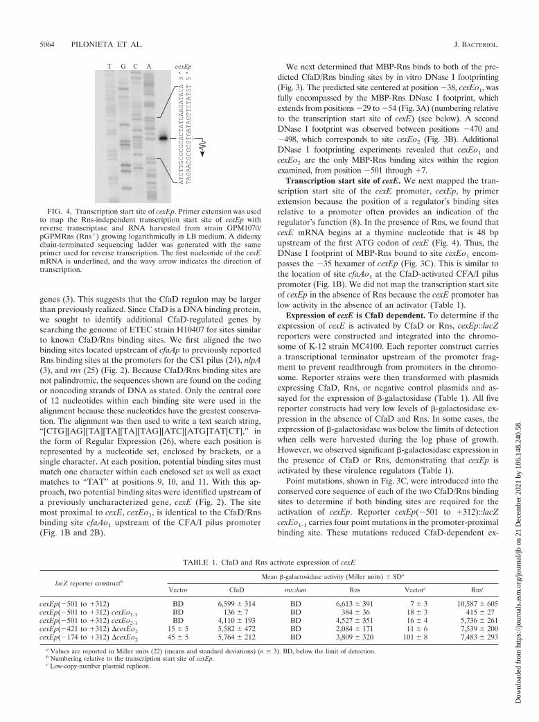
Transcription start site of cexE. We next mapped the tran-scription start site of the cexE promoter, cexEp, by primerextension because the position of a regulator’s binding sitesrelative to a promoter often provides an indication of theregulator’s function (8). In the presence of Rns, we found thatcexE mRNA begins at a thymine nucleotide that is 48 bpupstream of the first ATG codon of cexE (Fig. 4). Thus, theDNase I footprint of MBP-Rns bound to site cexEo1 encom-passes the �35 hexamer of cexEp (Fig. 3C). This is similar tothe location of site cfaAo1 at the CfaD-activated CFA/I piluspromoter (Fig. 1B). We did not map the transcription start siteof cexEp in the absence of Rns because the cexE promoter haslow activity in the absence of an activator (Table 1).
Expression of cexE is CfaD dependent. To determine if theexpression of cexE is activated by CfaD or Rns, cexEp::lacZreporters were constructed and integrated into the chromo-some of K-12 strain MC4100. Each reporter construct carriesa transcriptional terminator upstream of the promoter frag-ment to prevent readthrough from promoters in the chromo-some. Reporter strains were then transformed with plasmidsexpressing CfaD, Rns, or negative control plasmids and as-sayed for the expression of -galactosidase (Table 1). All fivereporter constructs had very low levels of -galactosidase ex-pression in the absence of CfaD and Rns. In some cases, theexpression of -galactosidase was below the limits of detectionwhen cells were harvested during the log phase of growth.However, we observed significant -galactosidase expression inthe presence of CfaD or Rns, demonstrating that cexEp isactivated by these virulence regulators (Table 1).
Point mutations, shown in Fig. 3C, were introduced into theconserved core sequence of each of the two CfaD/Rns bindingsites to determine if both binding sites are required for theactivation of cexEp. Reporter cexEp(�501 to �312)::lacZcexEo1-1 carries four point mutations in the promoter-proximalbinding site. These mutations reduced CfaD-dependent ex-
FIG. 4. Transcription start site of cexEp. Primer extension was usedto map the Rns-independent transcription start site of cexEp withreverse transcriptase and RNA harvested from strain GPM1070/pGPMRns (Rns�) growing logarithmically in LB medium. A dideoxychain-terminated sequencing ladder was generated with the sameprimer used for reverse transcription. The first nucleotide of the cexEmRNA is underlined, and the wavy arrow indicates the direction oftranscription.
TABLE 1. CfaD and Rns activate expression of cexE
lacZ reporter constructbean -galactosidase activity (Miller units) � SDa
Vector CfaD rns::kan Rns Vectorc Rnsc
cexEp(�501 to �312) BD 6,599 � 314 BD 6,613 � 391 7 � 3 10,587 � 605cexEp(�501 to �312) cexEo1-1 BD 136 � 7 BD 384 � 36 18 � 3 415 � 27cexEp(�501 to �312) cexEo2-1 BD 4,110 � 193 BD 4,527 � 351 16 � 4 5,736 � 261cexEp(�421 to �312) �cexEo2 15 � 5 5,582 � 472 BD 2,084 � 171 11 � 6 7,539 � 200cexEp(�174 to �312) �cexEo2 45 � 5 5,764 � 212 BD 3,809 � 320 101 � 8 7,483 � 293
a Values are reported in Miller units (22) (means and standard deviations) (n � 3). BD, below the limit of detection.b Numbering relative to the transcription start site of cexEp.c Low-copy-number plasmid replicon.
5064 PILONIETA ET AL. J. BACTERIOL.
Dow
nloa
ded
from
http
s://j
ourn
als.
asm
.org
/jour
nal/j
b on
21
Dec
embe
r 20
21 b
y 18
6.14
8.24
0.58
.
pression by 98% and Rns-dependent expression by 94% buthad no detectable effect upon CfaD- and Rns-independentexpression (Table 1). Mutations within the distal upstream site,cexEo2-1, also reduced CfaD- and Rns-dependent expressionfrom cexEp, but in each case, the reduction was less than 40%.These effects were also observed when Rns was expressed froma plasmid with a low copy number (Table 1). This suggests thatcexEo2 is functional in vivo despite its distance from the pro-moter and the fact that it appears to be a low-affinity site invitro. We did observe higher levels of expression fromcexEp(�501 to �312)::lacZ when Rns was expressed from alow-copy-number replicon versus a high-copy-number repliconeven though it was expressed from the same promoter, lacp, inboth cases. However, strains with low-copy-number repliconscould not be cultured in the same medium as those with high-copy-number replicons, and therefore, the absolute levels of-galactosidase expression are not directly comparable.
The distal upstream binding site, cexEo2, was deleted fromreporters cexEp(�421 to �312)::lacZ and cexEp(�174 to�312)::lacZ. These deletions reduced both CfaD- and Rns-dependent expression of -galactosidase, although we ob-served that the various strains had considerable variations inexpression (Table 1). The reasons for this variation are un-clear, but the reduction in CfaD- and Rns-dependent expres-sion from cexEp upon the deletion of cexEo2 is consistent withthe effect of point mutations in that site. Taken together, theseresults demonstrate that CfaD and Rns activate the expressionof cexE and that both DNA binding sites are required for fullactivation.
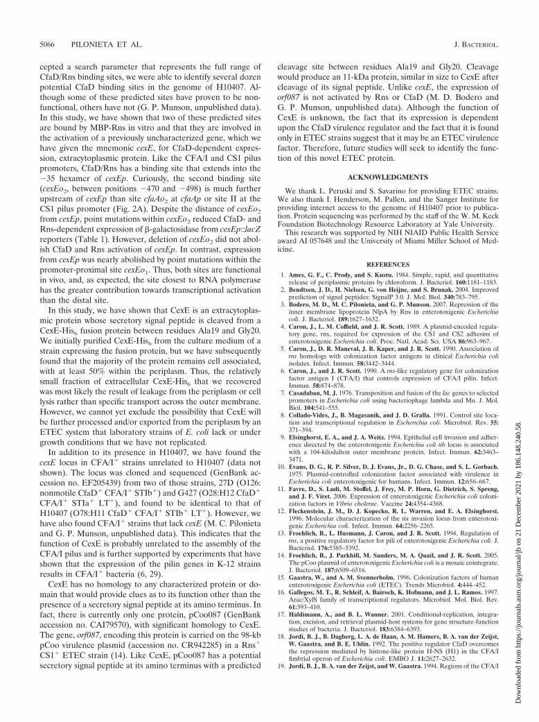
CexE is exported from the cytoplasm. CexE (GenBank ac-cession no. ABM92275) is a 120-amino-acid, 12.6-kDa protein.The first 19 amino acids of CexE have many of the features ofa signal peptide: lysines at residues 2 and 3, which wouldimpart a net positive charge; an �-helical hydrophobic regionat residues 4 through 12; and residues 13 through 19, compris-ing the recognition site for a signal peptidase (2, 34). Thesefeatures suggest that CexE is transported across the cytoplas-mic membrane via the general secretory pathway (27). Todetermine if CexE is an exported protein, a hexahistidine tagwas added to its carboxy terminus, and the epitope-taggedprotein was expressed in strain BL21(DE3)/pGPM1034. After4 h of expression, the tagged protein was purified from thegrowth medium by immobilized metal ion affinity chromatog-raphy (Fig. 5). The first 10 amino acids of the purified proteinwere determined to be “GGGNSERPPS” by Edman degrada-tion, which is identical to residues 20 through 29 of CexE.Thus, CexE is transported from the cytoplasm by the generalsecretory pathway, and its signal peptide is removed by a signalpeptidase. Although we purified CexE-His6 from the culturemedium, analysis of the cell pellet revealed that the majority ofthe protein remained associated with the cell, and approxi-mately 50% of the cell-associated protein can be released fromthe periplasm by chloroform shock (1) (data not shown).
DISCUSSION
Pili function as the primary adherence factors of ETEC (15),and some of the most prevalent pilus serotypes require a tran-scriptional activator for the expression of pilin genes. Theseinclude the CFA/I pilus, which is regulated by CfaD (6, 32),
and the Rns-regulated CS1, CS2, CS3, and CS4 pili (4, 5, 11).CfaD and Rns are fully interchangeable and recognize thesame DNA binding sites (23). This allowed us to use an MBP-Rns fusion protein in lieu of CfaD to identify the activator’sbinding sites at the CFA/I pilus promoter. DNase I footprint-ing revealed two DNA binding sites, site cfaAo1, from positions�23 to �56, and site cfaAo2, from positions �73 to �103(numbering relative to the transcription start site) (18). Thearrangement of CfaD’s binding sites at cfaAp is very similar tothose of Rns at the CS1 pilus promoter, cooBp (Fig. 2A) (24).At both promoters, the activators bind to a site that extendsinto the �35 hexamer. However, Rns binds to a second site,site II, between positions �93 and �129, that is further up-stream from the CS1 promoter than site cfaAo2 at the CFA/Ipromoter. Site cfaAo2 is not essential for the activation of theCFA/I pilus promoter because it was fortuitously deleted fromcfaAp::lacZ reporters in two previous studies without abolish-ing CfaD-dependent expression of -galactosidase (18, 19).However, it is likely that CfaD binding to site cfaAo2 contrib-utes to the activation of cfaAp, as has been shown for Rnsbinding to the promoter-distal site at the CS1 promoter (24).
The identification of cfaAo1 and cfaAo2 has increased thenumber of known CfaD/Rns binding sites from seven to nine(Fig. 2). The recent availability of the genomic sequence fromETEC strain H10407 has made it possible to exploit thisknowledge for the identification of additional genes within theCfaD regulon. By using custom-designed software that ac-
FIG. 5. Coomassie-stained sodium dodecyl sulfate-polyacrylamidegel of purified CexE-His6. CexE-His6 was purified from the culturemedium of an E. coli strain expressing the fusion protein by immobi-lized metal ion affinity chromatography. The amino terminus of thepurified protein was determined to be “GGGNSERPPS” by Edmandegradation, demonstrating that the amino terminus of CexE containsa signal peptide and that its first 19 amino acids are removed duringtranslocation from the cytoplasm. The expected molecular mass ofCexE-His6 after removal of its signal peptide is 11.7 kDa. Std., proteinstandards.
VOL. 189, 2007 CfaD-DEPENDENT EXPRESSION OF cexE 5065
Dow
nloa
ded
from
http
s://j
ourn
als.
asm
.org
/jour
nal/j
b on
21
Dec
embe
r 20
21 b
y 18
6.14
8.24
0.58
.
cepted a search parameter that represents the full range ofCfaD/Rns binding sites, we were able to identify several dozenpotential CfaD binding sites in the genome of H10407. Al-though some of these predicted sites have proven to be non-functional, others have not (G. P. Munson, unpublished data).In this study, we have shown that two of these predicted sitesare bound by MBP-Rns in vitro and that they are involved inthe activation of a previously uncharacterized gene, which wehave given the mnemonic cexE, for CfaD-dependent expres-sion, extracytoplasmic protein. Like the CFA/I and CS1 piluspromoters, CfaD/Rns has a binding site that extends into the�35 hexamer of cexEp. Curiously, the second binding site(cexEo2, between positions �470 and �498) is much furtherupstream of cexEp than site cfaAo2 at cfaAp or site II at theCS1 pilus promoter (Fig. 2A). Despite the distance of cexEo2
from cexEp, point mutations within cexEo2 reduced CfaD- andRns-dependent expression of -galactosidase from cexEp::lacZreporters (Table 1). However, deletion of cexEo2 did not abol-ish CfaD and Rns activation of cexEp. In contrast, expressionfrom cexEp was nearly abolished by point mutations within thepromoter-proximal site cexEo1. Thus, both sites are functionalin vivo, and, as expected, the site closest to RNA polymerasehas the greater contribution towards transcriptional activationthan the distal site.
In this study, we have shown that CexE is an extracytoplas-mic protein whose secretory signal peptide is cleaved from aCexE-His6 fusion protein between residues Ala19 and Gly20.We initially purified CexE-His6 from the culture medium of astrain expressing the fusion protein, but we have subsequentlyfound that the majority of the protein remains cell associated,with at least 50% within the periplasm. Thus, the relativelysmall fraction of extracellular CexE-His6 that we recoveredwas most likely the result of leakage from the periplasm or celllysis rather than specific transport across the outer membrane.However, we cannot yet exclude the possibility that CexE willbe further processed and/or exported from the periplasm by anETEC system that laboratory strains of E. coli lack or undergrowth conditions that we have not replicated.
In addition to its presence in H10407, we have found thecexE locus in CFA/I� strains unrelated to H10407 (data notshown). The locus was cloned and sequenced (GenBank ac-cession no. EF205439) from two of those strains, 27D (O126:nonmotile CfaD� CFA/I� STIb�) and G427 (O28:H12 CfaD�
CFA/I� STIa� LT�), and found to be identical to that ofH10407 (O78:H11 CfaD� CFA/I� STIb� LT�). However, wehave also found CFA/I� strains that lack cexE (M. C. Pilonietaand G. P. Munson, unpublished data). This indicates that thefunction of CexE is probably unrelated to the assembly of theCFA/I pilus and is further supported by experiments that haveshown that the expression of the pilin genes in K-12 strainsresults in CFA/I� bacteria (6, 29).
CexE has no homology to any characterized protein or do-main that would provide clues as to its function other than thepresence of a secretory signal peptide at its amino terminus. Infact, there is currently only one protein, pCoo087 (GenBankaccession no. CAI79570), with significant homology to CexE.The gene, orf087, encoding this protein is carried on the 98-kbpCoo virulence plasmid (accession no. CR942285) in a Rns�
CS1� ETEC strain (14). Like CexE, pCoo087 has a potentialsecretory signal peptide at its amino terminus with a predicted
cleavage site between residues Ala19 and Gly20. Cleavagewould produce an 11-kDa protein, similar in size to CexE aftercleavage of its signal peptide. Unlike cexE, the expression oforf087 is not activated by Rns or CfaD (M. D. Bodero andG. P. Munson, unpublished data). Although the function ofCexE is unknown, the fact that its expression is dependentupon the CfaD virulence regulator and the fact that it is foundonly in ETEC strains suggest that it may be an ETEC virulencefactor. Therefore, future studies will seek to identify the func-tion of this novel ETEC protein.
ACKNOWLEDGMENTS
We thank L. Peruski and S. Savarino for providing ETEC strains.We also thank I. Henderson, M. Pallen, and the Sanger Institute forproviding internet access to the genome of H10407 prior to publica-tion. Protein sequencing was performed by the staff of the W. M. KeckFoundation Biotechnology Resource Laboratory at Yale University.
This research was supported by NIH NIAID Public Health Serviceaward AI 057648 and the University of Miami Miller School of Med-icine.
REFERENCES
1. Ames, G. F., C. Prody, and S. Kustu. 1984. Simple, rapid, and quantitativerelease of periplasmic proteins by chloroform. J. Bacteriol. 160:1181–1183.
2. Bendtsen, J. D., H. Nielsen, G. von Heijne, and S. Brunak. 2004. Improvedprediction of signal peptides: SignalP 3.0. J. Mol. Biol. 340:783–795.
3. Bodero, M. D., M. C. Pilonieta, and G. P. Munson. 2007. Repression of theinner membrane lipoprotein NlpA by Rns in enterotoxigenic Escherichiacoli. J. Bacteriol. 189:1627–1632.
4. Caron, J., L. M. Coffield, and J. R. Scott. 1989. A plasmid-encoded regula-tory gene, rns, required for expression of the CS1 and CS2 adhesins ofenterotoxigenic Escherichia coli. Proc. Natl. Acad. Sci. USA 86:963–967.
5. Caron, J., D. R. Maneval, J. B. Kaper, and J. R. Scott. 1990. Association ofrns homologs with colonization factor antigens in clinical Escherichia coliisolates. Infect. Immun. 58:3442–3444.
6. Caron, J., and J. R. Scott. 1990. A rns-like regulatory gene for colonizationfactor antigen I (CFA/I) that controls expression of CFA/I pilin. Infect.Immun. 58:874–878.
7. Casadaban, M. J. 1976. Transposition and fusion of the lac genes to selectedpromoters in Escherichia coli using bacteriophage lambda and Mu. J. Mol.Biol. 104:541–555.
8. Collado-Vides, J., B. Magasanik, and J. D. Gralla. 1991. Control site loca-tion and transcriptional regulation in Escherichia coli. Microbiol. Rev. 55:371–394.
9. Elsinghorst, E. A., and J. A. Weitz. 1994. Epithelial cell invasion and adher-ence directed by the enterotoxigenic Escherichia coli tib locus is associatedwith a 104-kilodalton outer membrane protein. Infect. Immun. 62:3463–3471.
10. Evans, D. G., R. P. Silver, D. J. Evans, Jr., D. G. Chase, and S. L. Gorbach.1975. Plasmid-controlled colonization factor associated with virulence inEscherichia coli enterotoxigenic for humans. Infect. Immun. 12:656–667.
11. Favre, D., S. Ludi, M. Stoffel, J. Frey, M. P. Horn, G. Dietrich, S. Spreng,and J. F. Viret. 2006. Expression of enterotoxigenic Escherichia coli coloni-zation factors in Vibrio cholerae. Vaccine 24:4354–4368.
12. Fleckenstein, J. M., D. J. Kopecko, R. L. Warren, and E. A. Elsinghorst.1996. Molecular characterization of the tia invasion locus from enterotoxi-genic Escherichia coli. Infect. Immun. 64:2256–2265.
13. Froehlich, B., L. Husmann, J. Caron, and J. R. Scott. 1994. Regulation ofrns, a positive regulatory factor for pili of enterotoxigenic Escherichia coli. J.Bacteriol. 176:5385–5392.
14. Froehlich, B., J. Parkhill, M. Sanders, M. A. Quail, and J. R. Scott. 2005.The pCoo plasmid of enterotoxigenic Escherichia coli is a mosaic cointegrate.J. Bacteriol. 187:6509–6516.
15. Gaastra, W., and A. M. Svennerholm. 1996. Colonization factors of humanenterotoxigenic Escherichia coli (ETEC). Trends Microbiol. 4:444–452.
16. Gallegos, M. T., R. Schleif, A. Bairoch, K. Hofmann, and J. L. Ramos. 1997.Arac/XylS family of transcriptional regulators. Microbiol. Mol. Biol. Rev.61:393–410.
17. Haldimann, A., and B. L. Wanner. 2001. Conditional-replication, integra-tion, excision, and retrieval plasmid-host systems for gene structure-functionstudies of bacteria. J. Bacteriol. 183:6384–6393.
18. Jordi, B. J., B. Dagberg, L. A. de Haan, A. M. Hamers, B. A. van der Zeijst,W. Gaastra, and B. E. Uhlin. 1992. The positive regulator CfaD overcomesthe repression mediated by histone-like protein H-NS (H1) in the CFA/Ifimbrial operon of Escherichia coli. EMBO J. 11:2627–2632.
19. Jordi, B. J., B. A. van der Zeijst, and W. Gaastra. 1994. Regions of the CFA/I
5066 PILONIETA ET AL. J. BACTERIOL.
Dow
nloa
ded
from
http
s://j
ourn
als.
asm
.org
/jour
nal/j
b on
21
Dec
embe
r 20
21 b
y 18
6.14
8.24
0.58
.
promoter involved in the activation by the transcriptional activator CfaD andrepression by the histone-like protein H-NS. Biochimie 76:1052–1054.
20. McBroom, A. J., A. P. Johnson, S. Vemulapalli, and M. J. Kuehn. 2006.Outer membrane vesicle production by Escherichia coli is independent ofmembrane instability. J. Bacteriol. 188:5385–5392.
21. McVeigh, A., A. Fasano, D. A. Scott, S. Jelacic, S. L. Moseley, D. C.Robertson, and S. J. Savarino. 2000. IS1414, an Escherichia coli insertionsequence with a heat-stable enterotoxin gene embedded in a transposase-likegene. Infect. Immun. 68:5710–5715.
22. Miller, J. H. 1972. Experiments in molecular genetics. Cold Spring HarborLaboratory, Cold Spring Harbor, NY.
23. Munson, G. P., L. G. Holcomb, and J. R. Scott. 2001. Novel group ofvirulence activators within the AraC family that are not restricted to up-stream binding sites. Infect. Immun. 69:186–193.
24. Munson, G. P., and J. R. Scott. 1999. Binding site recognition by Rns, avirulence regulator in the AraC family. J. Bacteriol. 181:2110–2117.
25. Munson, G. P., and J. R. Scott. 2000. Rns, a virulence regulator within theAraC family, requires binding sites upstream and downstream of its ownpromoter to function as an activator. Mol. Microbiol. 36:1391–1402.
26. Neuburg, M. 2001. REALbasic: the definitive guide, 2nd ed. O’Reilly, Se-bastopol, CA.
27. Pugsley, A. P. 1993. The complete general secretory pathway in gram-neg-ative bacteria. Microbiol. Rev. 57:50–108.
28. Rhee, S., R. G. Martin, J. L. Rosner, and D. R. Davies. 1998. A novelDNA-binding motif in MarA: the first structure for an AraC family tran-scriptional activator. Proc. Natl. Acad. Sci. USA 95:10413–10418.
29. Sakellaris, H., G. P. Munson, and J. R. Scott. 1999. A conserved residue inthe tip proteins of CS1 and CFA/I pili of enterotoxigenic Escherichia coli thatis essential for adherence. Proc. Natl. Acad. Sci. USA 96:12828–12832.
30. Sambrook, J., and D. W. Russell. 2001. Molecular cloning: a laboratory manual,3rd ed. Cold Spring Harbor Laboratory Press, Cold Spring Harbor, NY.
31. Savelkoul, P. H., G. A. Willshaw, M. M. McConnell, H. R. Smith, A. M.Hamers, B. A. van der Zeijst, and W. Gaastra. 1990. Expression of CFA/Ifimbriae is positively regulated. Microb. Pathog. 8:91–99.
32. Sherlock, O., R. M. Vejborg, and P. Klemm. 2005. The TibA adhesin/invasinfrom enterotoxigenic Escherichia coli is self recognizing and induces bacterialaggregation and biofilm formation. Infect. Immun. 73:1954–1963.
33. Takeshita, S., M. Sato, M. Toba, W. Masahashi, and T. Hashimoto-Gotoh.1987. High-copy-number and low-copy-number plasmid vectors for lacZalpha-complementation and chloramphenicol- or kanamycin-resistance se-lection. Gene 61:63–74.
34. von Heijne, G. 1990. The signal peptide. J. Membr. Biol. 115:195–201.35. Wadood, A., M. Dohmoto, S. Sugiura, and K. Yamaguchi. 1997. Character-
ization of copy number mutants of plasmid pSC101. J. Gen. Appl. Microbiol.43:309–316.
36. Willshaw, G. A., H. R. Smith, and B. Rowe. 1983. Cloning of regions encod-ing colonisation factor antigen 1 and heat-stable enterotoxin in Escherichiacoli. FEMS Microbiol. Lett. 16:101–106.
37. World Health Organization. 2006. Future directions for research on ente-rotoxigenic Escherichia coli vaccines in developing countries. Wkly. Epide-miol. Rec. 81:97–104.
VOL. 189, 2007 CfaD-DEPENDENT EXPRESSION OF cexE 5067
Dow
nloa
ded
from
http
s://j
ourn
als.
asm
.org
/jour
nal/j
b on
21
Dec
embe
r 20
21 b
y 18
6.14
8.24
0.58
.


![PCR CHARACTERIZATION OF ESCHERICHIA COLIcrcooper01.people.ysu.edu/microlab/pcr-ecoli.pdf · • Escherichia coli, isolated from the environment [abbreviated as ECENV] • Escherichia](https://img.pdfslide.us/doc/110x75/5e6ee29ee0ed112b0c6f544d/pcr-characterization-of-escherichia-a-escherichia-coli-isolated-from-the-environment.jpg)