Embed Size (px)
DESCRIPTION
Cervical Spondylotic MyelopathyDiagnosis and Treatment
Citation preview
Journal of the American Academy of Orthopaedic Surgeons376
Cervical spondylosis results fromthe nearly universal process of de-generation of the disks and joints ofthe cervical spine. These changes inthe spinal motion segments havedoubtless existed since the evolu-tion of man, but our understandingof the pathoanatomy and clinicalconditions associated with cervicalspondylosis is relatively recent.Classic anatomic studies by Brain etal1 and Payne and Spillane2 in the1950s began to clarify the diseaseprocess and its effect on the neuralelements. Surgical proceduresthrough a posterior approach fordecompression of the cervical spine
were available in the 1940s; how-ever, decompression from an ante-rior approach did not begin to beused until the late 1950s. As cross-sectional imaging evolved—withcomputed tomographic (CT) scansin the 1970s and later with magneticresonance (MR) imaging—a betterappreciation of the pathoanatomyemerged.
A thorough understanding of thepathology of cervical spondylosis,as well as the principles of clinicalexamination, radiologic evaluation,and surgical indications, is essentialfor optimal treatment planning.Complications as a consequence of
the treatment of cervical spondyloticmyelopathy are intimately relatedto the type and extent of surgicalprocedure selected.
Natural History
Spinal cord compression resultingfrom spondylotic changes in the cer-vical spine is typically a slowly pro-gressive process. Many patientshave evidence of significant com-pression on neuroradiologic imag-ing but are relatively asymptomatic.It can be surprising how muchchronic deformation the spinal cordcan tolerate without interfering withpatient function (Fig. 1).
The natural history of cervicalmyelopathy has been described inclassic papers by Lees and Turner3
and Clarke and Robinson.4 Leesand Turner described exacerbationof symptoms followed by oftenlong periods of static or worseningfunction or, in rare instances, im-provement. Very few patients had
Dr. Emery is Associate Professor, Departmentof Orthopaedics, University Hospitals ofCleveland Spine Institute, Cleveland, Ohio.
Reprint requests: Dr. Emery, UniversityHospitals of Cleveland Spine Institute, CaseWestern Reserve University, 11100 EuclidAvenue, Cleveland, OH 44106.
Copyright 2001 by the American Academy ofOrthopaedic Surgeons.
Abstract
The delineation of cervical spondylotic myelopathy as a clinical entity hasimproved with the development of high-quality cross-sectional neuroradiologicimaging. The natural history of this disorder is usually slow deterioration in astepwise fashion, with worsening symptoms of gait abnormalities, weakness,sensory changes, and often pain. The diagnosis can usually be made on thebasis of findings from the history, physical examination, and plain radiographs,but confirmation by magnetic resonance imaging or computed tomography andmyelography is necessary. Minimal symptoms without hard evidence of gaitdisturbance or pathologic reflexes warrant nonoperative treatment, but patientswith demonstrable myelopathy and spinal cord compression are candidates foroperative intervention. Both anterior and posterior approaches have been uti-lized for surgical treatment of cervical myelopathy. Anterior decompression fre-quently requires corpectomy at one or more levels and strut grafting with bonefrom the ilium or fibula. Multilevel laminectomies were initially used for poste-rior decompression but now are either combined with fusion or replaced bylaminoplasty. Any operative technique requires proper patient selection anddemands adequate decompression of the canal to effect neurologic improvement.Perioperative complications can be devastating in this group of high-riskpatients with cervical spondylotic myelopathy, but careful attention to detail,meticulous technique, and experience can result in excellent outcomes.
J Am Acad Orthop Surg 2001;9:376-388
Cervical Spondylotic Myelopathy: Diagnosis and Treatment
Sanford E. Emery, MD
Sanford E. Emery, MD
Vol 9, No 6, November/December 2001 377
steady progressive deterioration.Clarke and Robinson described asimilar stepwise pattern of decreas-ing function. Long periods of sta-ble neurologic function, sometimeslasting for years, were noted inabout 75% of their patients. In themajority, however, the conditiondeteriorated between quiescentstreaks. About 20% of patients hada slow, steady progression of symp-toms and signs without a stableperiod, and 5% had rapid deterio-ration of neurologic function.
Generally, once moderate signsand symptoms of myelopathy de-
velop, the ultimate prognosis ispoor. As cervical myelopathy hasbecome better understood, mostauthors have recommended surgi-cal intervention for patients withmoderate to severe myelopathy,taking into account both the clinicalstatus and the neuroradiologicfindings, to alter this unfavorablenatural history.
Pathology
The pathoanatomy of cervical spon-dylosis with myelopathy results
from the sequelae of the agingprocess in the spine (i.e., disk de-generation with hypertrophic os-seous and ligamentous changes).Disk desiccation is accompanied bybiochemical changes, with a relativeincrease in the ratio of keratan sul-fate to chondroitin sulfate. The lossof elasticity and total disk substanceresults in a decrease in disk heightwith annular bulging. This alteredbiomechanical environment stimu-lates formation of chondro-osseousspurs at the annular insertion nearthe end-plates. The uncovertebraljoints hypertrophy, which may lead
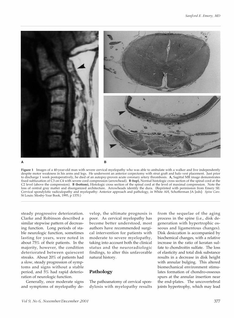
Figure 1 Images of a 40-year-old man with severe cervical myelopathy who was able to ambulate with a walker and live independentlydespite motor weakness in his arms and legs. He underwent an anterior corpectomy with strut graft and halo vest placement. Just priorto discharge 1 week postoperatively, he died of an autopsy-proven acute coronary artery thrombosis. A, Sagittal MR image demonstratesfixed subluxation of C3 on C4 with severe cord compression (arrowhead). B (top), Normal histologic cross section of the spinal cord at theC2 level (above the compression). B (bottom), Histologic cross section of the spinal cord at the level of maximal compression. Note theloss of central gray matter and disorganized architecture. Arrowheads identify the dura. (Reprinted with permission from Emery SE:Cervical spondylotic radiculopathy and myelopathy: Anterior approach and pathology, in White AH, Schofferman JA [eds]: Spine Care.St Louis: Mosby-Year Book, 1995, p 1370.)
A B
Cervical Spondylotic Myelopathy
Journal of the American Academy of Orthopaedic Surgeons378
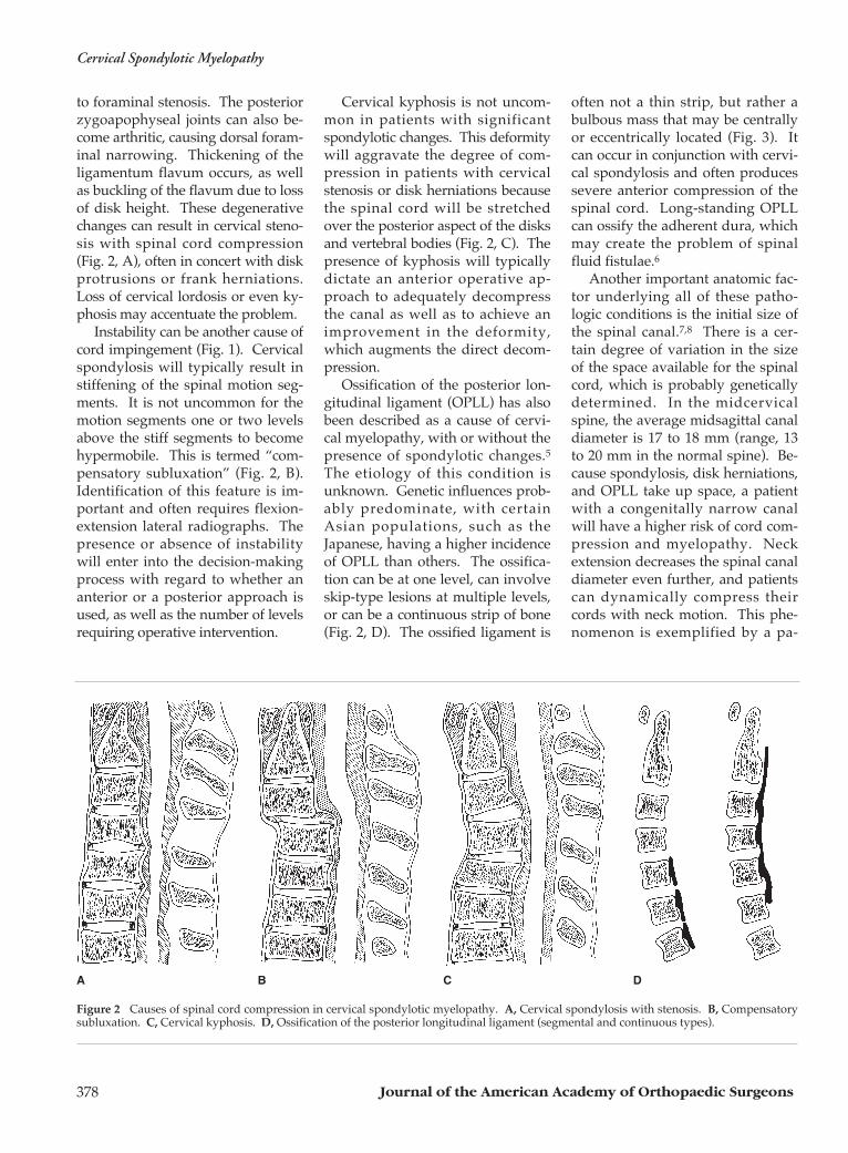
to foraminal stenosis. The posteriorzygoapophyseal joints can also be-come arthritic, causing dorsal foram-inal narrowing. Thickening of theligamentum flavum occurs, as wellas buckling of the flavum due to lossof disk height. These degenerativechanges can result in cervical steno-sis with spinal cord compression(Fig. 2, A), often in concert with diskprotrusions or frank herniations.Loss of cervical lordosis or even ky-phosis may accentuate the problem.
Instability can be another cause ofcord impingement (Fig. 1). Cervicalspondylosis will typically result instiffening of the spinal motion seg-ments. It is not uncommon for themotion segments one or two levelsabove the stiff segments to becomehypermobile. This is termed “com-pensatory subluxation” (Fig. 2, B).Identification of this feature is im-portant and often requires flexion-extension lateral radiographs. Thepresence or absence of instabilitywill enter into the decision-makingprocess with regard to whether ananterior or a posterior approach isused, as well as the number of levelsrequiring operative intervention.
Cervical kyphosis is not uncom-mon in patients with significantspondylotic changes. This deformitywill aggravate the degree of com-pression in patients with cervicalstenosis or disk herniations becausethe spinal cord will be stretchedover the posterior aspect of the disksand vertebral bodies (Fig. 2, C). Thepresence of kyphosis will typicallydictate an anterior operative ap-proach to adequately decompressthe canal as well as to achieve animprovement in the deformity,which augments the direct decom-pression.
Ossification of the posterior lon-gitudinal ligament (OPLL) has alsobeen described as a cause of cervi-cal myelopathy, with or without thepresence of spondylotic changes.5The etiology of this condition isunknown. Genetic influences prob-ably predominate, with certainAsian populations, such as theJapanese, having a higher incidenceof OPLL than others. The ossifica-tion can be at one level, can involveskip-type lesions at multiple levels,or can be a continuous strip of bone(Fig. 2, D). The ossified ligament is
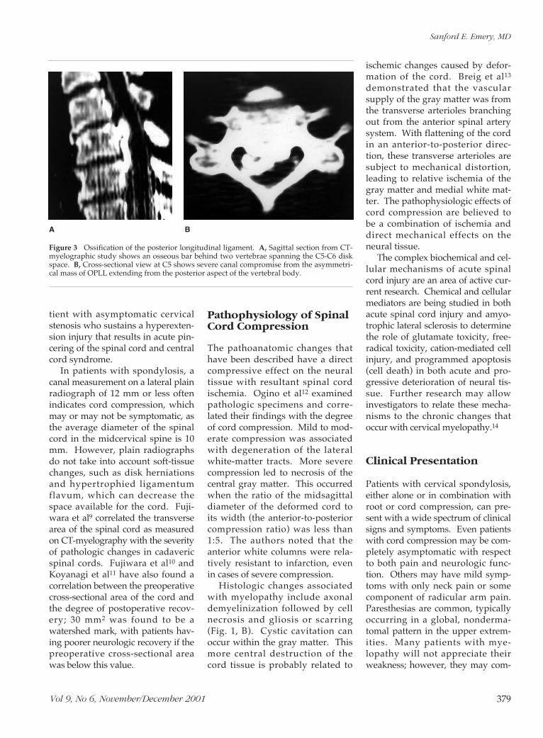
often not a thin strip, but rather abulbous mass that may be centrallyor eccentrically located (Fig. 3). Itcan occur in conjunction with cervi-cal spondylosis and often producessevere anterior compression of thespinal cord. Long-standing OPLLcan ossify the adherent dura, whichmay create the problem of spinalfluid fistulae.6
Another important anatomic fac-tor underlying all of these patho-logic conditions is the initial size ofthe spinal canal.7,8 There is a cer-tain degree of variation in the sizeof the space available for the spinalcord, which is probably geneticallydetermined. In the midcervicalspine, the average midsagittal canaldiameter is 17 to 18 mm (range, 13to 20 mm in the normal spine). Be-cause spondylosis, disk herniations,and OPLL take up space, a patientwith a congenitally narrow canalwill have a higher risk of cord com-pression and myelopathy. Neckextension decreases the spinal canaldiameter even further, and patientscan dynamically compress theircords with neck motion. This phe-nomenon is exemplified by a pa-
Figure 2 Causes of spinal cord compression in cervical spondylotic myelopathy. A, Cervical spondylosis with stenosis. B, Compensatorysubluxation. C, Cervical kyphosis. D, Ossification of the posterior longitudinal ligament (segmental and continuous types).
A B C D
Sanford E. Emery, MD
Vol 9, No 6, November/December 2001 379
tient with asymptomatic cervicalstenosis who sustains a hyperexten-sion injury that results in acute pin-cering of the spinal cord and centralcord syndrome.
In patients with spondylosis, acanal measurement on a lateral plainradiograph of 12 mm or less oftenindicates cord compression, whichmay or may not be symptomatic, asthe average diameter of the spinalcord in the midcervical spine is 10mm. However, plain radiographsdo not take into account soft-tissuechanges, such as disk herniationsand hypertrophied ligamentumflavum, which can decrease thespace available for the cord. Fuji-wara et al9 correlated the transversearea of the spinal cord as measuredon CT-myelography with the severityof pathologic changes in cadavericspinal cords. Fujiwara et al10 andKoyanagi et al11 have also found acorrelation between the preoperativecross-sectional area of the cord andthe degree of postoperative recov-ery; 30 mm2 was found to be awatershed mark, with patients hav-ing poorer neurologic recovery if thepreoperative cross-sectional areawas below this value.
Pathophysiology of SpinalCord Compression
The pathoanatomic changes thathave been described have a directcompressive effect on the neuraltissue with resultant spinal cordischemia. Ogino et al12 examinedpathologic specimens and corre-lated their findings with the degreeof cord compression. Mild to mod-erate compression was associatedwith degeneration of the lateralwhite-matter tracts. More severecompression led to necrosis of thecentral gray matter. This occurredwhen the ratio of the midsagittaldiameter of the deformed cord toits width (the anterior-to-posteriorcompression ratio) was less than1:5. The authors noted that theanterior white columns were rela-tively resistant to infarction, evenin cases of severe compression.
Histologic changes associatedwith myelopathy include axonaldemyelinization followed by cellnecrosis and gliosis or scarring(Fig. 1, B). Cystic cavitation canoccur within the gray matter. Thismore central destruction of thecord tissue is probably related to
ischemic changes caused by defor-mation of the cord. Breig et al13
demonstrated that the vascularsupply of the gray matter was fromthe transverse arterioles branchingout from the anterior spinal arterysystem. With flattening of the cordin an anterior-to-posterior direc-tion, these transverse arterioles aresubject to mechanical distortion,leading to relative ischemia of thegray matter and medial white mat-ter. The pathophysiologic effects ofcord compression are believed tobe a combination of ischemia anddirect mechanical effects on theneural tissue.
The complex biochemical and cel-lular mechanisms of acute spinalcord injury are an area of active cur-rent research. Chemical and cellularmediators are being studied in bothacute spinal cord injury and amyo-trophic lateral sclerosis to determinethe role of glutamate toxicity, free-radical toxicity, cation-mediated cellinjury, and programmed apoptosis(cell death) in both acute and pro-gressive deterioration of neural tis-sue. Further research may allowinvestigators to relate these mecha-nisms to the chronic changes thatoccur with cervical myelopathy.14
Clinical Presentation
Patients with cervical spondylosis,either alone or in combination withroot or cord compression, can pre-sent with a wide spectrum of clinicalsigns and symptoms. Even patientswith cord compression may be com-pletely asymptomatic with respectto both pain and neurologic func-tion. Others may have mild symp-toms with only neck pain or somecomponent of radicular arm pain.Paresthesias are common, typicallyoccurring in a global, nonderma-tomal pattern in the upper extrem-ities. Many patients with mye-lopathy will not appreciate theirweakness; however, they may com-
Figure 3 Ossification of the posterior longitudinal ligament. A, Sagittal section from CT-myelographic study shows an osseous bar behind two vertebrae spanning the C5-C6 diskspace. B, Cross-sectional view at C5 shows severe canal compromise from the asymmetri-cal mass of OPLL extending from the posterior aspect of the vertebral body.
A B
Cervical Spondylotic Myelopathy
Journal of the American Academy of Orthopaedic Surgeons380
plain of subtle changes in gait andbalance. This is often the first clue tothe presence of early myelopathy.
If the cord compression andmyelopathy are either moderate orsevere, patients complain of gaitand balance abnormalities involvingthe lower extremities. They alsohave numbness or paresthesias intheir upper extremities. Fine motorcontrol is usually affected as well,and they will note changes in theirhandwriting or ability to manipu-late buttons or zippers. Arm weak-ness is common in this group ofpatients, either unilaterally or bilat-erally. Leg weakness can occur, andpatients may notice problems mov-ing their body weight, such as isnecessary when rising out of a chairor going up stairs. In patients withcervical myelopathy, the proximalmotor groups of the legs are moreinvolved than the distal groups(which is the opposite of the patternwith lumbar stenosis); thus, presen-tation with foot-drop complaints israre. Changes in bowel or bladderfunction can occur in extremely se-vere cases of myelopathy, but this isquite rare. Although most patientswith cervical spondylotic myelopa-thy have neck pain, approximately15% with moderate to severe mye-lopathy do not. This may cause con-fusion or a delay in diagnosis.15
Spondylotic cord compressioncan predispose a patient to spinalcord injury (acute myelopathy) withminor trauma. This typically occursin elderly patients who sustain afall that results in a hyperextensionneck injury. A central cord syn-drome (motor weakness greater inthe arms than in the legs) oftenensues, with variable degrees ofparalysis. The patient may demon-strate obvious weakness, prompt-ing immediate evaluation and hos-pitalization. At times, however, thechanges in the patient’s functionare minimal, and only with in-depthhistory taking can one relate the de-terioration to minor trauma.
Physical Examination
The clinical evaluation should be-gin with an accurate description ofthe onset of symptoms and thetime course over which they devel-oped. Areas of neck tendernessand range of motion should thenbe evaluated. Neck extension isgenerally restricted and may bepainful for patients with cervicalstenosis or root compression. Thisis an important clinical feature andmay indicate a narrowed canal andfrank cord compression, whichmay be extremely important forpatients undergoing procedures re-quiring general anesthesia. Recog-nition of the decreased extension
and stenosis may prevent iatro-genic injury during intubation andoperative positioning.
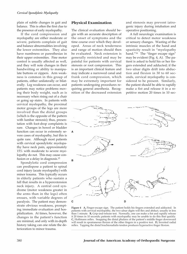
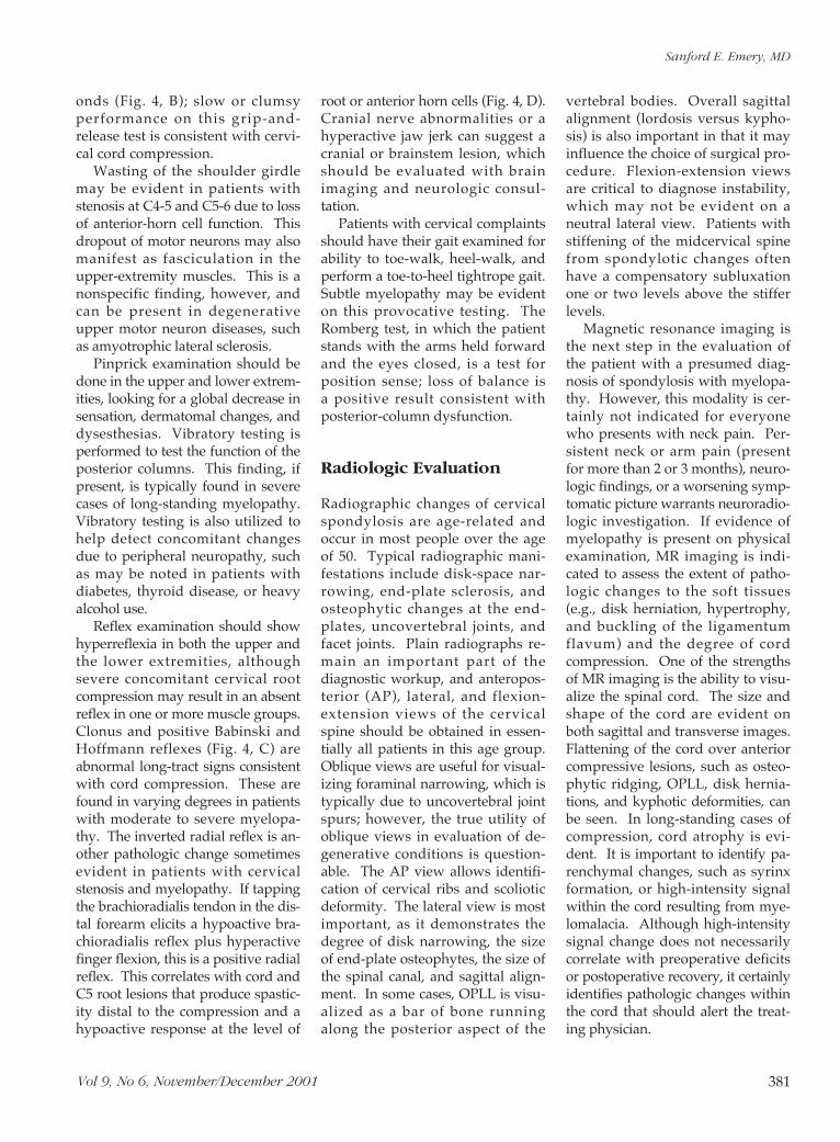
A full neurologic examination iscritical to detect motor weaknessor sensory changes. Wasting of theintrinsic muscles of the hand andspasticity result in “myelopathyhand.”16 The “finger escape sign”may be evident (Fig. 4, A). The pa-tient is asked to hold his or her fin-gers extended and adducted; if thetwo ulnar digits drift into abduc-tion and flexion in 30 to 60 sec-onds, cervical myelopathy is con-sidered to be present. Similarly,the patient should be able to rapidlymake a fist and release it in a re-petitive motion 20 times in 10 sec-
B
A
C D
Figure 4 A, Finger-escape sign. The patient holds his fingers extended and adducted. Inpatients with cervical myelopathy, the two ulnar digits will flex and abduct, usually in lessthan 1 minute. B, Grip-and-release test. Normally, one can make a fist and rapidly releaseit 20 times in 10 seconds; patients with myelopathy may be unable to do this that quickly.C, Hoffmann reflex. Snapping the distal phalanx of the patient’s middle finger downwardwill result in spontaneous flexion of the other fingers in a positive test. D, Inverted radialreflex. Tapping the distal brachioradialis tendon produces hyperactive finger flexion.
Sanford E. Emery, MD
Vol 9, No 6, November/December 2001 381
onds (Fig. 4, B); slow or clumsyperformance on this grip-and-release test is consistent with cervi-cal cord compression.
Wasting of the shoulder girdlemay be evident in patients withstenosis at C4-5 and C5-6 due to lossof anterior-horn cell function. Thisdropout of motor neurons may alsomanifest as fasciculation in theupper-extremity muscles. This is anonspecific finding, however, andcan be present in degenerativeupper motor neuron diseases, suchas amyotrophic lateral sclerosis.
Pinprick examination should bedone in the upper and lower extrem-ities, looking for a global decrease insensation, dermatomal changes, anddysesthesias. Vibratory testing isperformed to test the function of theposterior columns. This finding, ifpresent, is typically found in severecases of long-standing myelopathy.Vibratory testing is also utilized tohelp detect concomitant changesdue to peripheral neuropathy, suchas may be noted in patients withdiabetes, thyroid disease, or heavyalcohol use.
Reflex examination should showhyperreflexia in both the upper andthe lower extremities, althoughsevere concomitant cervical rootcompression may result in an absentreflex in one or more muscle groups.Clonus and positive Babinski andHoffmann reflexes (Fig. 4, C) areabnormal long-tract signs consistentwith cord compression. These arefound in varying degrees in patientswith moderate to severe myelopa-thy. The inverted radial reflex is an-other pathologic change sometimesevident in patients with cervicalstenosis and myelopathy. If tappingthe brachioradialis tendon in the dis-tal forearm elicits a hypoactive bra-chioradialis reflex plus hyperactivefinger flexion, this is a positive radialreflex. This correlates with cord andC5 root lesions that produce spastic-ity distal to the compression and ahypoactive response at the level of
root or anterior horn cells (Fig. 4, D).Cranial nerve abnormalities or ahyperactive jaw jerk can suggest acranial or brainstem lesion, whichshould be evaluated with brainimaging and neurologic consul-tation.
Patients with cervical complaintsshould have their gait examined forability to toe-walk, heel-walk, andperform a toe-to-heel tightrope gait.Subtle myelopathy may be evidenton this provocative testing. TheRomberg test, in which the patientstands with the arms held forwardand the eyes closed, is a test forposition sense; loss of balance is a positive result consistent withposterior-column dysfunction.
Radiologic Evaluation
Radiographic changes of cervicalspondylosis are age-related andoccur in most people over the ageof 50. Typical radiographic mani-festations include disk-space nar-rowing, end-plate sclerosis, andosteophytic changes at the end-plates, uncovertebral joints, andfacet joints. Plain radiographs re-main an important part of the diagnostic workup, and anteropos-terior (AP), lateral, and flexion-extension views of the cervicalspine should be obtained in essen-tially all patients in this age group.Oblique views are useful for visual-izing foraminal narrowing, which istypically due to uncovertebral jointspurs; however, the true utility ofoblique views in evaluation of de-generative conditions is question-able. The AP view allows identifi-cation of cervical ribs and scolioticdeformity. The lateral view is mostimportant, as it demonstrates thedegree of disk narrowing, the sizeof end-plate osteophytes, the size ofthe spinal canal, and sagittal align-ment. In some cases, OPLL is visu-alized as a bar of bone runningalong the posterior aspect of the
vertebral bodies. Overall sagittalalignment (lordosis versus kypho-sis) is also important in that it mayinfluence the choice of surgical pro-cedure. Flexion-extension viewsare critical to diagnose instability,which may not be evident on aneutral lateral view. Patients withstiffening of the midcervical spinefrom spondylotic changes oftenhave a compensatory subluxationone or two levels above the stifferlevels.
Magnetic resonance imaging isthe next step in the evaluation ofthe patient with a presumed diag-nosis of spondylosis with myelopa-thy. However, this modality is cer-tainly not indicated for everyonewho presents with neck pain. Per-sistent neck or arm pain (presentfor more than 2 or 3 months), neuro-logic findings, or a worsening symp-tomatic picture warrants neuroradio-logic investigation. If evidence ofmyelopathy is present on physicalexamination, MR imaging is indi-cated to assess the extent of patho-logic changes to the soft tissues(e.g., disk herniation, hypertrophy,and buckling of the ligamentumflavum) and the degree of cordcompression. One of the strengthsof MR imaging is the ability to visu-alize the spinal cord. The size andshape of the cord are evident onboth sagittal and transverse images.Flattening of the cord over anteriorcompressive lesions, such as osteo-phytic ridging, OPLL, disk hernia-tions, and kyphotic deformities, canbe seen. In long-standing cases ofcompression, cord atrophy is evi-dent. It is important to identify pa-renchymal changes, such as syrinxformation, or high-intensity signalwithin the cord resulting from mye-lomalacia. Although high-intensitysignal change does not necessarilycorrelate with preoperative deficitsor postoperative recovery, it certainlyidentifies pathologic changes withinthe cord that should alert the treat-ing physician.
Cervical Spondylotic Myelopathy
Journal of the American Academy of Orthopaedic Surgeons382
Although MR imaging providesoptimal visualization of soft tissues,CT-myelography offers better defi-nition of bone spurs and OPLL. Theexact degree of cord deformation inthe transverse plane is more sharplyvisualized with CT-myelographyas well. This modality is useful inevaluating whether marginal levelsneed to be included in an operativeprocedure.
Other forms of clinical evalua-tion include electrodiagnostic tech-niques. For patients with cervicalradiculopathy, electromyographic–nerve conduction studies may beuseful in considering the differen-tial diagnosis of carpal tunnel syn-drome, ulnar cubital tunnel syn-drome, or thoracic outlet syndrome.Electrodiagnostic modalities mayalso help elucidate the confusingclinical presentations of amyotrophiclateral sclerosis, multiple sclerosis,and severe peripheral neuropathy.
Somatosensory-evoked poten-tials and motor-evoked potentialsare of limited utility during thediagnostic evaluation but are usedintraoperatively. A preoperativebaseline study can be very helpful,especially in patients with severechanges in latency and amplitude.Some authors advocate the use ofintraoperative spinal-cord evokedpotentials to identify the level ofgreatest conduction delay and thenlimit surgery to that level17; how-ever, this approach risks leavingclinically significant pathologicchanges in untreated areas.
Nonoperative Treatment
Patients with neuroradiologic evi-dence of spinal cord compressionbut no symptoms or signs of mye-lopathy should generally be ob-served. One exception would be apatient with such severe compres-sion that even low-energy trauma,such as might occur with a rear-endmotor vehicle impact or a fall, could
predictably result in spinal cordinjury. It is extremely rare for a pa-tient with that degree of cord com-pression on imaging studies to betruly asymptomatic; nevertheless,these patients should be counseled toavoid high-risk situations in which ahyperextension injury might occur,as they are at some increased risk forcord impingement.
Patients with mild myelopathymay display findings such as slightgait disturbance and mild hyper-reflexia but may have no functionaldeficits and no weakness. The indi-vidual clinical course and especiallythe pattern of deteriorations shouldbe well understood by both physi-cian and patient. If the patient is ina plateau period without recent ex-acerbation, nonoperative treatmentmay be indicated. Reevaluationevery 6 to 12 months to look for de-terioration of neurologic function ora change in symptoms may be ap-propriate.
Indications for Surgery
The natural history of cervicalmyelopathy for most patients isslow deterioration over time. Typ-ically, this is in a stepwise fashionwith variable periods of stable neuro-logic function. If one assumes sig-nificant deterioration for all pa-tients with myelopathy, it can beargued that operative interventionis indicated for everyone with thisclinical and radiographic diagnosis.However, the decision making ismuch more complex, with the clini-cal severity of myelopathy beingthe most important issue.
The extent of myelopathy isreflected predominantly by physi-cal examination findings such asbalance deficits, gait, motor weak-ness, long-tract signs, and changesin function (e.g., decreased finemotor control). All of these clinicalfindings provide evidence of thedegree of cord dysfunction. Other
important factors involved in thedecision-making process includethe amount of pain the patient isexperiencing, the degree of changeof function that can be tolerated,and the evaluation of symptoms.Patients with rapid neurologic de-terioration should undergo earlieroperative intervention.
Consideration of the severity ofcompression evident on neuroradio-logic studies is important, as theseverity of cord compression gener-ally, but not always, correlates withthe level of function. For patientswith equivalent signs and symp-toms of moderate myelopathy,operative intervention would berecommended earlier if there weremore severe radiologic findings,such as smaller cord area, cord atro-phy, signal changes indicative ofmyelomalacia, or the presence of akyphotic deformity. Although notall neuroradiologic findings havebeen correlated with preoperativesymptoms or postoperative out-come, more severe compressionintuitively suggests more risk forthe spinal cord.
For patients with moderate to se-vere compression and myelopathy,surgical intervention is indicated toalter the natural history. Surgerycan be expected to halt progressionin neurologic function and mayimprove motor, sensory, and gaitdisturbance. The degree of recoverydepends largely on the severity ofthe myelopathy at the time of inter-vention.10,15 Other factors of posi-tive prognostic value include largertransverse area of the cord, youngerpatient age, shorter duration ofsymptoms, and single rather thanmultiple levels of involvement.10,11
Many patients with cervical spon-dylosis and myelopathy are elderly,but age alone is not a contraindica-tion to operative intervention.
Patients with chronic cervicalspondylosis who suffer acute minortrauma, particularly a hyperexten-sion injury, can sustain acute spinal
Sanford E. Emery, MD
Vol 9, No 6, November/December 2001 383
cord injuries of varying severitysuperimposed on the long-standingmyelopathy. Typically, this pre-sents as a central cord syndromewith greater weakness in the upperextremities than in the lower ex-tremities and proximal rather thandistal muscle involvement in eachextremity. This can occur with orwithout a prior history of myelo-pathic symptoms. Initial treatmentinvolves collar immobilization,high-dose methylprednisolone, anda neuroradiologic investigation. Ifneurologic function improves afterthe injury, the plateau functionallevel should be determined. If re-covery is complete or near com-plete, surgery is not necessary.Residual deficits, as evidenced bythe appearance of cord compressionon imaging studies, warrant opera-tive intervention to promote neuro-logic recovery. One recent long-term study of patients with centralcord syndrome treated nonopera-tively documented much poorerrecovery in patients over 50 years ofage compared with younger pa-tients.18 There are no data docu-menting a substantial difference inrecovery if diagnosis was earlyrather than late.
Surgical Approaches
The preferred approach for surgicaltreatment of cervical myelopathycontinues to be controversial, asboth anterior and posterior tech-niques have been used successfully.Posterior options include multilevellaminectomy,19 laminoplasty, andlaminectomy plus fusion proce-dures. Anterior options includemultiple anterior diskectomieswith fusion and corpectomy plusstrut fusion techniques with orwithout the use of anterior instru-mentation. The choice of approachis determined on the basis of theexisting lesion and surgeon experi-ence. Factors to be considered in-clude the number of involved lev-els, overall sagittal alignment, thedirection of compression, the pres-ence of instability, and clinicalsymptoms.
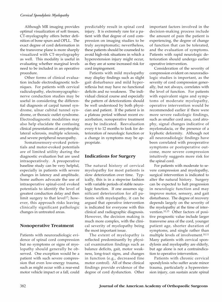
Posterior ApproachFor patients with diffuse canal
stenosis or dorsal cord compres-sion due to buckling of the liga-mentum flavum posteriorly, a pos-terior decompression technique maybe ideal to achieve adequate decom-pression (Fig. 5). However, most
patients with cervical spondylosisand certainly those with OPLLhave predominantly anterior com-pression of the cervical cord. Anyposterior decompressive procedureis an indirect technique that re-quires posterior shifting of the cordin the thecal sac to diminish theeffect of the anterior compression.For this to occur, the preoperativesagittal alignment of the cervicalspine must be at least straight orpreferably lordotic. A kyphotic spineis less likely to allow sufficient pos-terior translation of the spinal cordto diminish symptoms. This is akey point in choosing between pos-terior and anterior approaches forsurgical treatment of myelopathy,as is the presence of instability.Laminectomy alone will only wors-en preexisting instability. Fusionmust be added if the posterior ap-proach is the preferred route of de-compression.
Multilevel laminectomy was ini-tially the only procedure availableto treat cervical stenosis and maystill have a place for selected pa-tients. The results after that proce-dure deteriorate due to the devel-opment of late instability, such askyphosis or subluxation, although
A B C
Figure 5 Images of a 61-year-old man with moderate cervical spondylotic myelopathy, gait changes, upper-extremity neurologic signsand symptoms, and minimal neck pain. A, Sagittal MR image shows normal lordosis and suggests diffuse narrowing of the spinal canalover multiple levels. B, Axial CT-myelographic image at C5 shows severe stenosis that is causing circumferential, rather than focal anteri-or, cord impingement. C, The patient underwent a laminoplasty from C3 to C7 performed with use of the Chiba method. A postoperativeCT image demonstrates expansion of the spinal canal at C4. The clinical outcome at 3-year follow-up was rated as successful.
Cervical Spondylotic Myelopathy
Journal of the American Academy of Orthopaedic Surgeons384
the exact incidence of this problemis difficult to determine. The addi-tion of a multilevel fusion at thetime of laminectomy eliminates thepotential for development of latepostoperative kyphosis or instabil-ity. Although originally done withbone graft wired to the facets, it isnow more easily achieved by lateralmass plating and fusion.
Laminoplasty evolved as a methodto eliminate postoperative develop-ment of instability and kyphosis byexpanding the canal while retainingthe posterior elements.20,21 Severaltechniques for performing lamino-plasty have been devised, but alladhere to the concept of canal ex-pansion by opening the posteriorelements in a trapdoor fashion butnot completely removing the osse-ous posterior arch. By expandingthe size of the canal, the cord com-pression can be alleviated or less-ened, and the chance of postopera-tive instability is minimized becausethe posterior musculature can healto the residual posterior osseous ele-ments. Most methods are based oneither a unilateral hinge with a one-way trapdoor opening to expandthe canal20 or a midline spinousprocess–splitting procedure withbilateral hinges to expand the canalin a symmetrical fashion.22,23 Asmall amount of bone graft or spaceris often placed in the opening de-fects, but arthrodesis of the motionsegments is not desirable. Lamino-plasty results in a 30% to 50% loss of motion in the cervical spine,23,24
which is less than occurs with mul-tilevel arthrodesis.
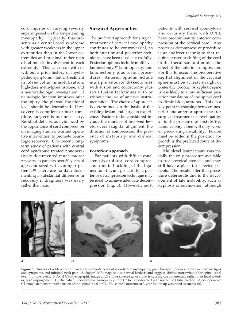
Anterior ApproachBecause the pathoanatomy of
cord compression in degenerativeconditions is typically anterior tothe spinal cord, an anterior ap-proach allows direct decompres-sion of the dura (Fig. 6). Two dif-ferent techniques can be utilized,with selection dependent on align-ment and the pathologic features.
If the cord compression is presentonly at the disks at one, two, orthree levels, an anterior cervicaldiskectomy with graft at each levelis appropriate. In most patientswith spondylotic myelopathy orOPLL, there is compression at thedisk as well as above and below thedisk space. Usually, this is causedby large osteophytes or ridging atthe vertebral end-plates. Ossifica-
tion of the posterior longitudinalligament occurs behind the verte-bral body and may be focal or mul-tifocal or may appear as a continu-ous long osseous bar. Because thesurgeon cannot safely reach poste-rior to the vertebral bodies throughthe disk space, it is necessary toremove part or all of the midpor-tion of the vertebral body to ade-quately decompress the canal.
A
C
B
D
Figure 6 A, Sagittal T2-weighted MR image demonstrates spondylotic changes withsevere spinal cord compression predominantly at two levels. B, Postoperative CT scandemonstrates decompression of the spinal canal and the fibular graft. C, Lateral radio-graph obtained immediately after two-level anterior cervical corpectomies and fibularstrut grafting (arrowheads). D, Lateral radiograph obtained 2 years later shows smoothbone remodeling, indicating a solid arthrodesis.
Sanford E. Emery, MD
Vol 9, No 6, November/December 2001 385
Hemicorpectomies may be per-formed for end-plate osteophyteslocated near the disk spaces; how-ever, full corpectomies are morecommonly performed to totallydecompress the canal at severaldisk levels as needed. The lateralwalls of the vertebral body are leftintact because they provide protec-tion against vertebral artery injury.The typical midline channel for acorpectomy is 16 to 18 mm, whichprovides adequate decompressionfor the entire canal if it is appropri-ately centered in the midline.
It is not uncommon for a patientwith cervical spondylotic myelopa-thy to require a two- or three-levelcorpectomy and then a strut graftfor fusion or to correct kyphosis.The degree of difficulty of the proce-dure, the risk of postoperative graftcomplications, and the potential forsoft-tissue complications increasewith the number of corpectomy lev-els. This limitation should enter intothe decision-making process regard-ing choice of approach.
Autograft, allograft, and evenmetal cages with cancellous graftshave been used as struts to main-tain alignment and promote ar-throdesis. Autografts provide thehighest union rate. Harvestinglarge iliac-crest grafts may be asso-ciated with local pain, fracture ofthe ilium, and injury to the lateralfemoral cutaneous nerve. Autol-ogous fibular grafts have been asso-ciated with less morbidity than longiliac grafts, although tibial stressfractures,25 pain, and muscle weak-ness26 have been described. Allo-graft iliac-crest or fibular grafts areused for single-level diskectomyand fusion, with good success ratesreported in most studies27 but lessoptimal results in others.28 Fibularstrut allografts have also been usedsuccessfully29 for reconstructionafter multilevel corpectomy but areslower to heal and have a higherrate of pseudarthrosis. Some sur-geons use cancellous chips from the
vertebrectomy to augment the allo-graft; others prefer supplementalposterior fixation combined withanterior allograft struts to promoteunion. Many surgeons utilize iliac-crest strut grafts for one- or two-level vertebrectomy procedures andfibular strut grafts for constructs tobe used at two or more levels.
Theoretically, the use of anteriorcervical plates provides additionalstability, maintains correction ofdeformity, and promotes arthrode-sis, especially in longer or multilevelconstructs. There is considerablecontroversy concerning the use ofplates for one-level anterior cervicaldiskectomy and fusion, unless thereare certain coexisting circumstances,such as a history of smoking or thepresence of adjacent segment fu-sions. Anterior plate fixation afterone-level corpectomy (two-levelfusion) with iliac-strut fusion pro-vides increased stability and mayallow less restrictive immobilizationpostoperatively.
The use of anterior plates formultilevel corpectomy and strut-graft procedures is more controver-sial. Because of the long lever armwith only two screws above andtwo screws below, a high rate ofloosening and displacement hasbeen described for these long-plateconstructs.30 Three-level corpectomyprocedures seem to be at higherrisk for this complication than two-level procedures. Also, plate fixa-tion does not allow settling of thegraft into the vertebral-body dock-ing sites, which may actually inhibitarthrodesis. Other authors haveutilized a small buttress-type plateat the inferior end of the strut-graftconstruct to help prevent graft dis-lodgment. Failures with this tech-nique have also been reported.31
Meticulous preparation of the ver-tebral bodies, including centralizingthe graft in the end-plate withsculpted mortices, will help mini-mize complications due to graft dis-lodgment.
Choice of ApproachFor each patient, the surgeon
should weigh the relative advan-tages and disadvantages of theanterior and posterior approaches.Neither is optimal for every patientwith cervical spondylotic myelopa-thy, although either may be appro-priate for some patients. The rela-tive pros and cons of laminoplastyversus anterior corpectomy andstrut grafting are summarized inTable 1.
Anterior decompression andarthrodesis is a more direct decom-pression method that allows cor-rection of deformity and stabiliza-tion with fusion. It is technicallydemanding, especially in multi-level cases, and one must be pre-pared to deal with graft-relatedcomplications. Rigid postoperativebracing is necessary with an ortho-sis or a halo vest.
The posterior approach is an in-direct method of decompression inmost cases and relies on the spinalcord being able to shift posteriorlyin an expanded canal. For this rea-son, patients with preoperativekyphosis are not good candidatesfor a posterior unroofing-type pro-cedure because the anterior im-pingement on the cord will remain.Compensatory subluxation or otherinstability may also worsen with aposterior approach if fusion is notperformed.
Laminoplasty techniques are notas technically demanding as multi-level anterior corpectomy and strut-grafting procedures. There is lessbracing required, as a soft collar willgenerally suffice for comfort afterlaminoplasty. Although some lossof motion is typical after lamino-plasty procedures, this would beexpected to be less than occurs withlong arthrodesis methods. More re-cent data have suggested that lami-noplasty techniques may not provideconsistent relief of axial neck pain,32
whereas anterior fusion proceduresprovide good axial pain relief.15
Cervical Spondylotic Myelopathy
Journal of the American Academy of Orthopaedic Surgeons386
The preoperative symptoms playa role in the decision-making pro-cess as well. Patients with diffusecanal stenosis and a congenitally nar-row canal may require decompres-sion of virtually the entire cervicalspine. This is more readily achievedwith laminoplasty techniques. Someauthors prefer the anterior approachfor patients with pathologic changesat one or two levels and posteriorsurgery for those with involvementat three or more levels.33 With prop-er patient selection, both anteriorand posterior techniques will pro-vide comparable rates of neurologicrecovery and improvement of func-tion.34,35
There are cases in which anteriormultilevel decompression and strutgrafting followed by posterior stabi-lization is indicated. Postlaminec-tomy kyphosis is perhaps the bestexample of this. Anterior decom-pression will typically be neededbecause of the degree of deformityand anterior-cord compression.Prior removal of the posterior ele-ments predisposes them to graftcomplications if only an anteriorapproach is performed36; immediateposterolateral mass plating will helpprotect the graft, maintain alignment,and promote successful arthrodesis.The circumferential approach is alsopreferable for patients with severe
osteoporosis, because it avoids frac-ture of the inferior end-plate due toloading by the graft. In multilevelcases requiring corpectomies atthree or more levels, supplementalposterior fixation may increasefusion rates and decrease compli-cations.
Complications
Complications can generally be cat-egorized as: (1) approach-related,(2) decompression-related, (3) graft-related, and (4) long-term. Risksincurred with the anterior approachto the cervical spine include stretchinjuries to the recurrent laryngealnerve, which produce hoarseness.The incidence of this injury is be-lieved to be approximately 1% to2%.37 Dysphagia is experiencedtransiently by most patients after ananterior surgical approach, but canbe a persistent problem for some.Upper airway compromise fromedema or hematoma formation ismore likely after multilevel corpec-tomy procedures.38 Drains shouldbe utilized, and patients frequentlyare monitored for 24 to 48 hourspostoperatively. The airway shouldbe evaluated before extubation to minimize the risk of airway ob-struction.
Both nerve root and spinal cordinjury may occur during the decom-pression. Good visualization, care-ful technique, and experience aremandatory to avoid devastatingresults. The incidence of neurologicinjury is approximately 1% to 2%.15
Spinal cord monitoring should beused for most, if not all, of theseprocedures. Corticosteroids may begiven prophylactically in high-riskcases. During the procedure, anychange in spinal cord monitoringconsidered to be significant shouldbe treated with the same dose ofmethylprednisolone used for trau-matic spinal cord injury (loadingdose of 30 mg per kilogram of bodyweight, followed by 5.4 mg/kg perhour for 23 hours).
Motor palsy of the C5 root inpatients who have undergone lami-noplasty procedures has been welldescribed. The etiology of this post-operative deficit is not clear but isthought to be related to the shortlength of the C5 root and the maxi-mal lordosis at that level; when thespinal cord shifts posteriorly afterdecompression, the C5 root is be-lieved to sustain a stretch injury.The incidence of this complication is1% to 3%,24 and slow but progres-sive recovery has been reported inmost, but not all, patients. C5 rootpalsy can also occur following ante-
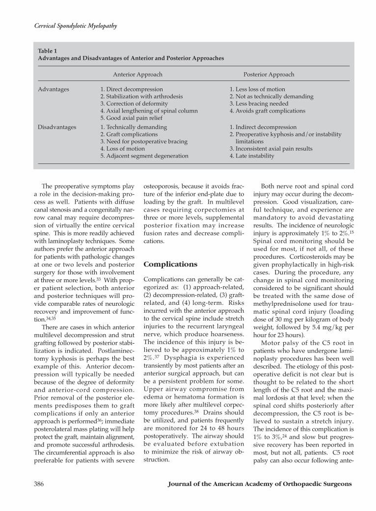
Table 1Advantages and Disadvantages of Anterior and Posterior Approaches
Anterior Approach Posterior Approach
Advantages 1. Direct decompression 1. Less loss of motion2. Stabilization with arthrodesis 2. Not as technically demanding3. Correction of deformity 3. Less bracing needed4. Axial lengthening of spinal column 4. Avoids graft complications5. Good axial pain relief
Disadvantages 1. Technically demanding 1. Indirect decompression2. Graft complications 2. Preoperative kyphosis and/or instability 3. Need for postoperative bracing limitations4. Loss of motion 3. Inconsistent axial pain results5. Adjacent segment degeneration 4. Late instability
Sanford E. Emery, MD
Vol 9, No 6, November/December 2001 387
rior decompression and fusion pro-cedures but is relatively rare.
Injury to the vertebral artery isalso possible during anterior verte-bral corpectomies.39 Strict orienta-tion to the midline is necessary tohelp avoid this complication. Man-agement includes exposure of theartery above and below the corpec-tomy, with ligation or microscopicrepair. Spinal fluid leaks can occurduring both anterior and posteriorprocedures. Because patients withlong-standing OPLL may have ero-sion of the dura,6 gelatin-foamsponge and fibrin glue, fascial patch-ing, or a lumbar cerebrospinal fluiddrain may be needed to prevent apersistent fistula.40
Graft-related complications afteran anterior strut-graft procedure mayinclude dislodgment, fracture, andsevere settling into the cancellousbone of the vertebral bodies. Withlong fibular grafts, the docking sitein the inferior vertebral body cansplit due to axial loading, and theinferior end of the graft can displaceanteriorly. If there is no longer bone
contact, if the esophagus is threat-ened, or if significant kyphosis en-sues, operative revision is indicated.
Long-term complications fromanterior decompression and ar-throdesis procedures include pseud-arthrosis and adjacent-segmentdegeneration. Patients with recur-rent myelopathy should be evaluatedfor pseudarthrosis and for compres-sion at levels adjacent to the longfusion that have undergone furtherdegenerative changes. It appears tobe true that fusions accelerate spon-dylotic changes at adjacent disk lev-els; however, a recent study sug-gests it may not be higher thanwould be attributable to the naturalhistory of spondylosis.41
Laminectomy procedures areassociated with an increased risk ofpostlaminectomy kyphosis, swan-neck deformity, or instability withlate neurologic deterioration. Lami-noplasty techniques decrease theserisks, but add the potential compli-cation of inadvertent closure of theopened lamina with recurrent ste-nosis. Incomplete decompression
may necessitate a second-stage ante-rior procedure.
Summary
Operative intervention for cervicalmyelopathy has consistently beenshown to improve the neurologicfunction of a high percentage ofpatients. Neurologic outcomes ap-pear to improve to a similar degree,regardless of whether anterior orposterior techniques are utilized,provided the guidelines discussedearlier are taken into consideration.
As with other types of spinesurgery, careful patient selectionremains the cornerstone of goodsurgical results. This, combinedwith high-quality imaging studiesand meticulous surgical technique,will result in gratifying results withrespect to neurologic recovery,function, and pain relief.
Acknowledgment: The author wishes tothank Val Schmedlen for her assistance inthe preparation of the manuscript.
References
1. Brain WR, Northfield D, Wilkinson M:The neurological manifestations of cer-vical spondylosis. Brain 1952;75:187-225.
2. Payne EE, Spillane JD: The cervicalspine: An anatomico-pathologicalstudy of 70 specimens (using a specialtechnique) with particular reference tothe problem of cervical spondylosis.Brain 1957;80:571-596.
3. Lees F, Turner JWA: Natural historyand prognosis of cervical spondylosis.BMJ 1963;2:1607-1610.
4. Clarke E, Robinson PK: Cervicalmyelopathy: A complication of cervi-cal spondylosis. Brain 1956;79:483-510.
5. Tsuyama N: Ossification of the poste-rior longitudinal ligament of the spine.Clin Orthop 1984;184:71-84.
6. Smith MD, Bolesta MJ, Leventhal M,Bohlman HH: Postoperative cere-brospinal-fluid fistula associated witherosion of the dura: Findings afteranterior resection of ossification of theposterior longitudinal ligament in the
cervical spine. J Bone Joint Surg Am1992;74:270-277.
7. Wolf BS, Khilnani M, Malis L: Thesagittal diameter of the bony cervicalspinal canal and its significance in cer-vical spondylosis. J Mt Sinai Hosp1956;23:283-292.
8. Arnold JG Jr: The clinical manifesta-tions of spondylochondrosis (spondy-losis) of the cervical spine. Ann Surg1955;141:872-889.
9. Fujiwara K, Yonenobu K, HiroshimaK, Ebara S, Yamashita K, Ono K:Morphometry of the cervical spinalcord and its relation to pathology incases with compression myelopathy.Spine 1988;13:1212-1216.
10. Fujiwara K, Yonenobu K, Ebara S,Yamashita K, Ono K: The prognosis ofsurgery for cervical compressionmyelopathy: An analysis of the factorsinvolved. J Bone Joint Surg Br 1989;71:393-398.
11. Koyanagi T, Hirabayashi K, Satomi K,
Toyama Y, Fujimura Y: Predictabilityof operative results of cervical com-pression myelopathy based on preop-erative computed tomographic myel-ography. Spine 1993;18:1958-1963.
12. Ogino H, Tada K, Okada K, et al:Canal diameter, anteroposterior com-pression ratio, and spondylotic mye-lopathy of the cervical spine. Spine1983;8:1-15.
13. Breig A, Turnbull I, Hassler O: Effectsof mechanical stresses on the spinalcord in cervical spondylosis: A studyon fresh cadaver material. J Neurosurg1966;25:45-56.
14. Fehlings MG, Skaf G: A review of thepathophysiology of cervical spondy-lotic myelopathy with insights forpotential novel mechanisms drawnfrom traumatic spinal cord injury.Spine 1998;23:2730-2737.
15. Emery SE, Bohlman HH, Bolesta MJ,Jones PK: Anterior cervical decom-pression and arthrodesis for the treat-
Cervical Spondylotic Myelopathy
Journal of the American Academy of Orthopaedic Surgeons388
ment of cervical spondylotic myelopa-thy: Two to seventeen-year follow-up.J Bone Joint Surg Am 1998;80:941-951.
16. Ono K, Ebara S, Fuji T, Yonenobu K,Fujiwara K, Yamashita K: Myelopathyhand: New clinical signs of cervicalcord damage. J Bone Joint Surg Br1987;69:215-219.
17. Tani T, Ishida K, Ushida T, YamamatoH: Intraoperative electroneurographyin the assessment of the level of opera-tion for cervical spondylotic myelopa-thy in the elderly. J Bone Joint Surg Br2000;82:269-274.
18. Newey ML, Sen PK, Fraser RD: Thelong-term outcome after central cordsyndrome: A study of the natural histo-ry. J Bone Joint Surg Br 2000;82:851-855.
19. Epstein JA, Janin Y, Carras R, LavineLS: A comparative study of the treat-ment of cervical spondylotic myelo-radiculopathy: Experience with 50 casestreated by means of extensive laminec-tomy, foraminotomy, and excision ofosteophytes during the past 10 years.Acta Neurochir (Wien) 1982;61:89-104.
20. Hirabayashi K, Satomi K: Operativeprocedure and results of expansiveopen-door laminoplasty. Spine 1988;13:870-876.
21. Kimura I, Shingu H, Nasu Y, ShiotaniA, Oh-hama M, Murata M: Long-termfollow-up of cervical spondyloticmyelopathy treated by canal-expan-sive laminoplasty. J Bone Joint Surg Br1995;77:956-961.
22. Tomita K, Kawahara N, Toribatake Y,Heller JG: Expansive midline T-Sawlaminoplasty (modified spinousprocess-splitting) for the managementof cervical myelopathy. Spine 1998;23:32-37.
23. Edwards CC II, Heller JG, Silcox DHIII: T-Saw laminoplasty for the man-agement of cervical spondylotic
myelopathy: Clinical and radiographicoutcome. Spine 2000;25:1788-1794.
24. Satomi K, Nishu Y, Kohno T, Hirabay-ashi K: Long-term follow-up studiesof open-door expansive laminoplastyfor cervical stenotic myelopathy. Spine1994;19:507-510.
25. Emery SE, Heller JG, Petersilge CA,Bolesta MJ, Whitesides TE Jr: Tibialstress fracture after a graft has beenobtained from the fibula: A report offive cases. J Bone Joint Surg Am 1996;78:1248-1251.
26. Vail TP, Urbaniak JR: Donor-site mor-bidity with use of vascularized autoge-nous fibular grafts. J Bone Joint SurgAm 1996;78:204-211.
27. Martin GJ Jr, Haid RW Jr, MacMillan M,Rodts GE Jr, Berkman R: Anterior cer-vical diskectomy with freeze-dried fibu-la allograft: Overview of 317 cases andliterature review. Spine 1999;24:852-859.
28. Zdeblick TA, Ducker TB: The use offreeze-dried allograft bone for anteriorcervical fusions. Spine 1991;16:726-729.
29. Macdonald RL, Fehlings MG, TatorCH, et al: Multilevel anterior cervicalcorpectomy and fibular allograft fu-sion for cervical myelopathy. J Neuro-surg 1997;86:990-997.
30. Vaccaro AR, Falatyn SP, Scuderi GJ, etal: Early failure of long segment ante-rior cervical plate fixation. J SpinalDisord 1998;11:410-415.
31. Riew KD, Sethi NS, Devney J, GoetteK, Choi K: Complications of buttressplate stabilization of cervical corpecto-my. Spine 1999;24:2404-2410.
32. Hosono N, Yonenobu K, Ono K: Neckand shoulder pain after laminoplasty:A noticeable complication. Spine1996;21:1969-1973.
33. Yonenobu K, Fuji T, Ono K, Okada K,Yamamoto T, Harada N: Choice ofsurgical treatment for multisegmental
cervical spondylotic myelopathy.Spine 1985;10:710-716.
34. Hukuda S, Mochizuki T, Ogata M,Shichikawa K, Shimomura Y: Opera-tions for cervical spondylotic mye-lopathy: A comparison of the results ofanterior and posterior procedures. JBone Joint Surg Br 1985;67:609-615.
35. Yonenobu K, Hosono N, Iwasaki M,Asano M, Ono K: Laminoplasty ver-sus subtotal corpectomy: A compara-tive study of results in multisegmentalcervical spondylotic myelopathy.Spine 1992;17:1281-1284.
36. Riew KD, Hilibrand AS, Palumbo MA,Bohlman HH: Anterior cervical cor-pectomy in patients previously man-aged with a laminectomy: Short-termcomplications. J Bone Joint Surg Am1999;81:950-957.
37. Flynn TB: Neurologic complicationsof anterior cervical interbody fusion.Spine 1982;7:536-539.
38. Emery SE, Smith MD, Bohlman HH:Upper-airway obstruction after multi-level cervical corpectomy for myelopa-thy. J Bone Joint Surg Am 1991;73:544-551.
39. Smith MD, Emery SE, Dudley A,Murray KJ, Leventhal M: Vertebralartery injury during anterior decom-pression of the cervical spine: A retro-spective review of ten patients. J BoneJoint Surg Br 1993;75:410-415.
40. Kitchel SH, Eismont FJ, Green BA:Closed subarachnoid drainage formanagement of cerebrospinal fluidleakage after an operation on thespine. J Bone Joint Surg Am 1989;71:984-987.
41. Hilibrand AS, Carlson GD, PalumboMA, Jones PK, Bohlman HH: Radicu-lopathy and myelopathy at segmentsadjacent to the site of a previous ante-rior cervical arthrodesis. J Bone JointSurg Am 1999;81:519-528.