Embed Size (px)
Citation preview
CERVICAL SPONDYLOSIS
DR T.P MOJASTEVE BIKO ACADEMIC HOSPITAL
CERVICAL SPONDYLOSIS
DR T.P MOJASTEVE BIKO ACADEMIC HOSPITAL
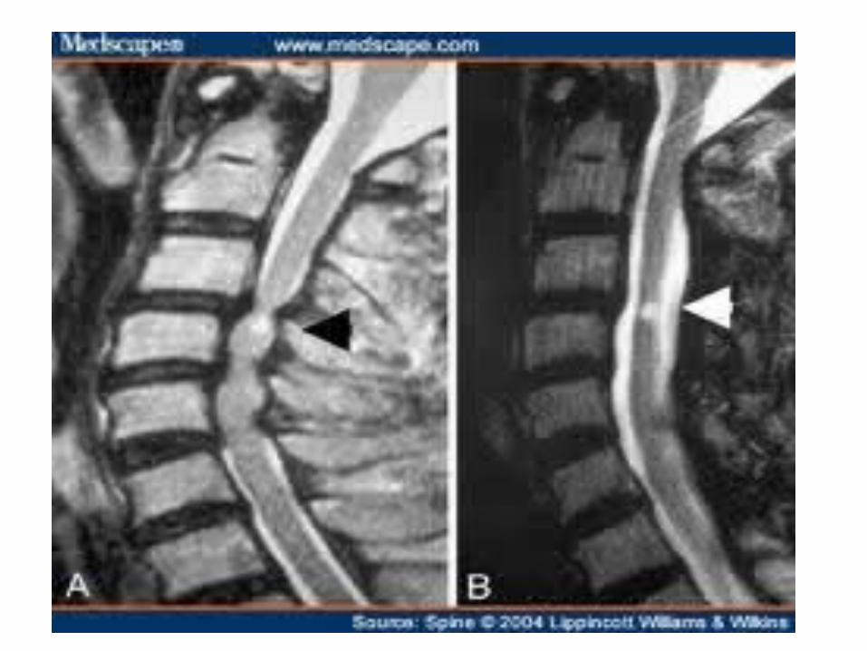
Pathophysiology Pathophysiology Disc degeneration
-nucleus pulposus loses water content, fissuring, loss of height and bulging annulus.
-acute rupture and herniation may occur
Secondary changes due to increased and uneven loading of forces
- Vertebral osteophytes
- Facet and uncovertebral joint osteoarthritis and hypertrophy
- Ligamentum flavum becomes thickened and may ossify
- Spine deformity due to segmental instability
Degenerative spondylolistheses
Degenerative kyphosis or scoliosis
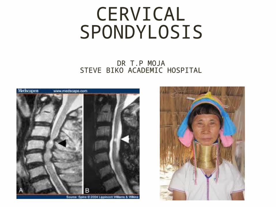
• Narrowing of the central canal, lateral recesses and foramina with subsequent neural and vascular compression
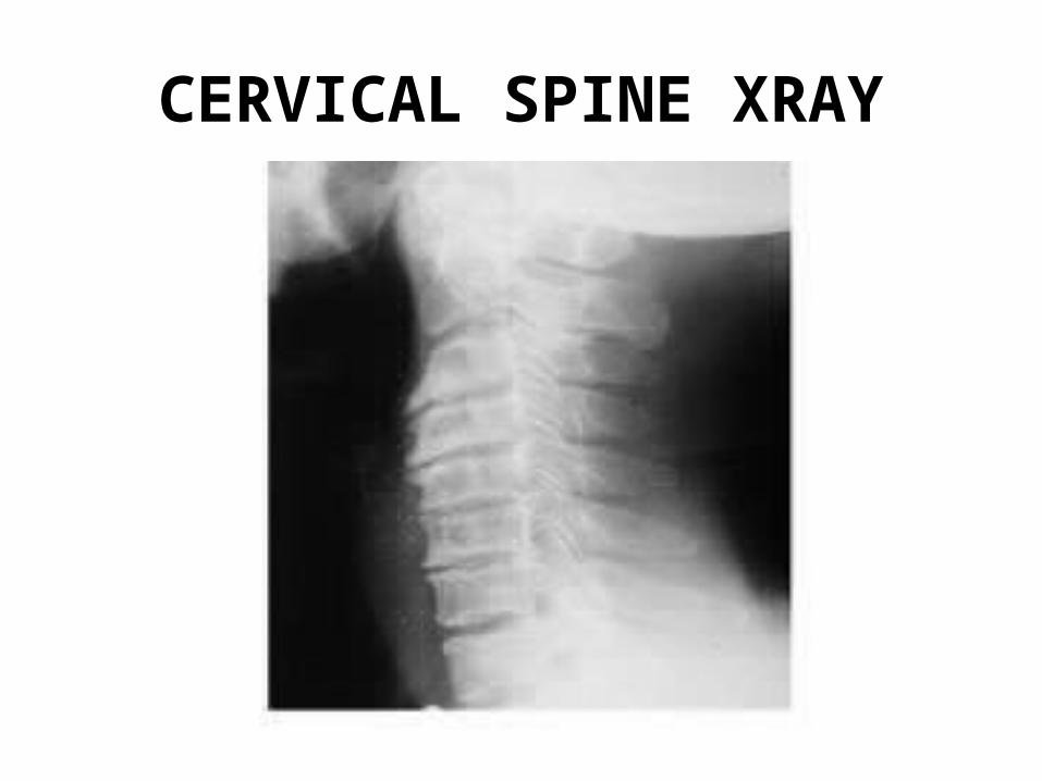
CERVICAL SPINE XRAY
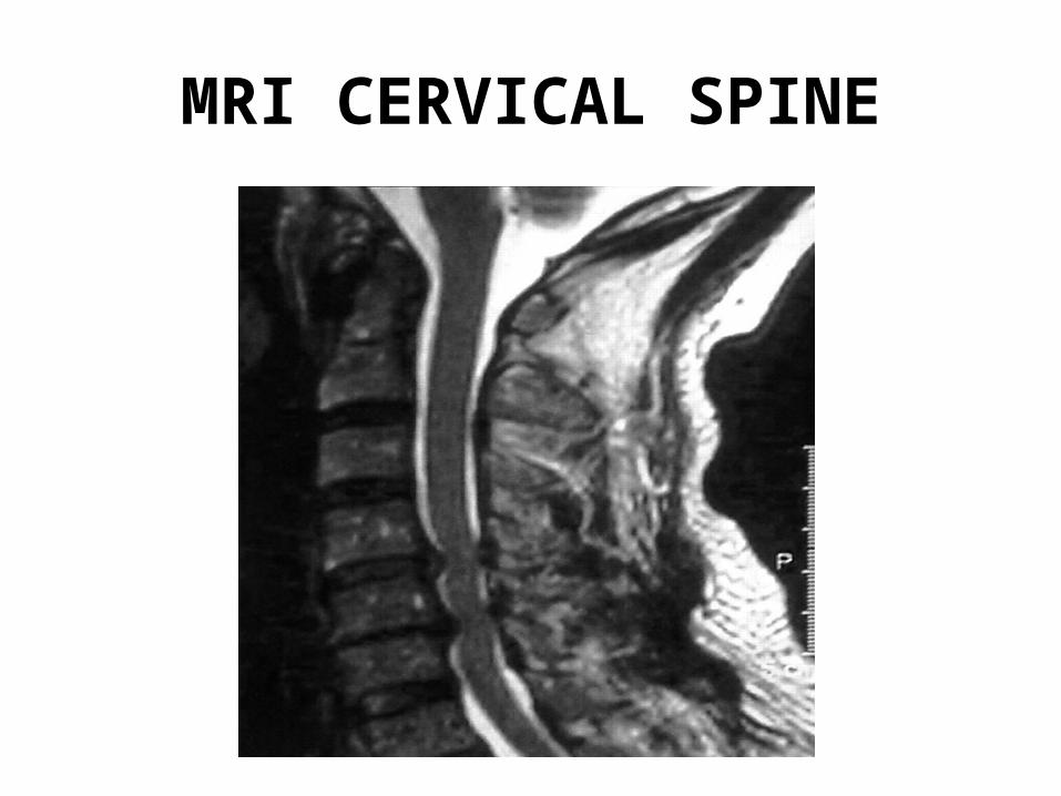
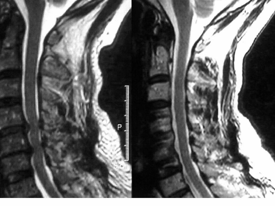
MRI CERVICAL SPINE
Clinical PresentationClinical PresentationAsymptomtic with incidental radiographic findings
Symptomatic - in most cases: onset is slow and insidious . However some cases may be acute eg hyperextension injury in minor trauma or acute disc herniation
Neck pain
Myelopathy
Radiculopathy
Neck pain• Occurs if there is a disc extrusion
• Nerve root compression
• Facet joint arthritis
• Segmental instability
• Often poorly localized
• May radiate to the occipital region, shoulders, interscapular.
• There may be associated stiffness of the neck from muscle spasm
Myelopathy• May be complex and variable
• Most cases seem to present with a central cord syndrome, rarely brown squard, or complete myelopathy
• Motor
• -upper limbs: LMN Weakness
•Clumsiness of the hands. Muscle wasting. Absent biceps reflex, inverted reflex, Triceps reflex may be brisk. Positive Hoffman reflex
• -Lower limbs: Spasticity, difficulty walking. No or slight weakness.
• Sphincters: usually no symptoms. Rarely mild bladder symptoms. ? Prostate
• Sensory
- No involvement
- Patchy sensory loss
- Paraesthesia in the hands, sometimes the feet and legs
- May be asymmetrical or symmetrical
- Different from radiculopathy in that it is not in a specific dermatomes
- Lhermittes’s sign
Radiculopathy•May be acute if due to a disc protrusion
•Slow and insidious if due to an osteophyte
•Most common nerve root is C6
•Neck pain and shoulder pain. Pain radiates down the biceps, then the lateral aspect of the forearm then the thumb and index finger.
•Head may be tilted to the affected side due to muscle spasm. Pain made worse by neck extension, relieved by neck flexion and shoulder abduction.
•Often numbness, more often hand and fingers
• Chronic cases – wasting and fasciculations of biceps and brachioradialis muscle.
• Weakness of elbow flexion (Thumb-nose), and wrist extension.
• Absent biceps and brachioradialis reflex
C5 nerve root radiculopathy
• - Neck pain
• - Shoulder pain, pain over the lateral aspect of the upper arm.
• - Numbness or paraesthesia over the lateral aspect of the upper arm.
• - Weakness of deltoid and biceps muscles, with absent biceps reflex
•In severe cases, wasting of the deltoid and biceps muscles
TreatmentTreatmentNeck Pain
-Conservative
-Rarely, surgery
Myelopathy
-Surgery in most cases
-Some may stabilize on conservative
Acute radiculopathy
-Conservative
-Surgery if indicated
Chronic radiculopathy
-Most cases, surgery
Conservative treatment
Conservative treatment• Medication: Analgesia
NSAIDS
Diazepam
Baclofen
Carbamazepine, Gabapentin, Lyrica
• Physiotherapy: Range of motion exercises
Isometric exercises
Heat and massage
• Traction: Continuous or intermittent halter traction
• Neck collar Soft neck collar < 1 week
• Facet and Medial branch block – Cortisone, L.A, Radiofrequency
SURGERY
• Anterior decompression– Anterior cervical discectomy and fusion– Corpectomy and fusion
• Posterior decompression– Laminaplasty– Laminectomy