Embed Size (px)
Citation preview

Cervical Spondylomyopathy (CSM) Wobbler Syndrome➤ Continued from Page X
www.livs.org 1
L O N G I S L A N D V E T E R I N A R Y S P E C I A L I S T S 163 SOUTH SERVICE ROAD • PLAINVIEW, NEW YORK 11803PH: 516-501-1700 • FAX: 516-501-1169 • WWW.LIVS.ORG
Inside This IssueMetabolic Encephalopathies (Part II): It’s More � an Just HE Ann Bilderback, DVM, DACVIM (Neurology)
A Note From the EditorLeonard J. Marino, MD, FAAP, LVT
Immunosuppressive � erapy - Part 1Joshua W. Tumulty, DVM, Diplomate ACVIM (SAIM), Dept. of Internal Medicine
Metabolic Encephalopathies (Part II): It’s More Than Just HE Ann Bilderback, DVM, DACVIM (Neurology)
LIVS In Plain ViewLIVS LIVS LIVS In Plain ViewIn Plain ViewIn Plain ViewIn Plain View LIVS In Plain ViewJULY/AUGUST 2019
VOLUME 12 • ISSUE 4
3
8
1
M etabolic encephalopathy is a clinical syndrome resulting from disorders of metabolism. Due to the extremely
high metabolic demands of the brain, systemic abnormalities that interfere with its energy metabolism may result in clinical signs of en-cephalopathy. The majority of metabolic dis-eases lead to diffuse, symmetrical forebrain signs because the cerebral cortical neurons are the most susceptible to dysfunction of energy metabolism. Clinical signs can be acute or chronic in onset and signs may wax and wane. Although the most well known metabolic en-cephalopathy is hepatic encephalopathy (HE), there are numerous other metabolic causes of encephalopathy. Treating the clinical signs of encephalopathy in these patients generally depends on treating the underlying metabolic disease. The topics of hepatic, renal and hypo-glycemic encephalopathies were introduced in Part I of this two part series. In this article electrolyte, endocrine, thiamine deficiency, hypertensive, organic aciduria, and mitochon-drial encephalopathies will be discussed. This article is meant as an introduction to the vari-ous causes of metabolic encephalopathies; an in-depth discussion, particularly diagnosis and treatment, is beyond the scope of this article.
Electrolyte-Associated EncephalopathySodium (Na+) and calcium (Ca++) imbalances can cause encephalopathy, with severity typi-cally correlating with how rapidly the imbal-ance develops. The prognosis for reversing the encephalopathy due to electrolyte dis-turbances is favorable. However, the overall
prognosis for the individual patient varies and depends on the underlying disease process re-sponsible for the electrolyte imbalance. There are many possible causes of these electrolyte disturbances and treatment for the underly-ing disorders which is beyond the scope of this article.
CalciumHypercalcemia can cause decreased excit-ability of neuronal cell membranes as well as direct toxic damage to neurons. Neurologic signs of hypercalcemia can include listless-ness, dullness, and lethargy.
Hypocalcemia may lead to increased excit-ability of neuronal cell membranes as well as abnormal neurotransmission. Neurologic signs of hypocalcemia include facial rubbing, muscle twitching, cramping, tremors, ataxia and seizures.
Continued on Page 4 ➤

Emergency Department
Open 24 Hours
CardiologyJonathan Goodwin, DVM, DACVIM
DermatologyMeghan Umstead, DVM, MS, DACVD
Emergency/Critical CareElizabeth Underwood, DVMJennifer Krawchuk, DVMLaura Fiore, DVMAmanda Pisano, DVMMeredith von Roedern, DVM, DACVECC
Integrative MedicineMichel Selmer, DVM, MS, CTCVMP
Internal MedicineJacqueline F. Gest, DVM, DACVIMJoshua W. Tumulty, DVM, DACVIMKimberly Golden, DVM
Neurology/NeurosurgeryAnn Bilderback, DVM, DACVIMCatherine A. Loughin, DVM, DACVS, DACCTCurtis W. Dewey, DVM, MS, CTCVMP, DACVS, DACVIMDominic J. Marino, DVM, DACVS, DACCT, CCRPFernando Leyva, DVM, DACVS-SAMatthew Morgan, DVM, DACVS-SAPatrick F. Roynard, DVM, MRCVS, DACVIMRobert Waddell, DVM, DACVS-SA
OncologyMaria Camps, DVM, DACVIM (SAIM, Oncology)Nicole Leibman, DVM, DACVIM (Oncology)
OphthalmologyJohn S. Sapienza, DVM, DACVOKay Kim, VMD, DACVO
Radioiodine Therapy (I-131)Dominic J. Marino, DVM, DACVS, DACCT, CCRPJoshua W. Tumulty, DVM, DACVIM
Rehabilitation TherapyDominic J. Marino, DVM, DACVS, DACCT, CCRPHeather Goodman, LVT, CCRP
SurgeryCatherine A. Loughin, DVM, DACVS, DACCTCurtis W. Dewey, DVM, MS, CVA, DACVS, DACVIMDominic J. Marino, DVM, DACVS, DACCT, CCRPFernando Leyva, DVM, DACVS-SAJaclyn Holdsworth, DVMRobert Waddell, DVM, DACVS-SA
163 South Service Road | Plainview, NY 11803516-501-1700 www.livs.org

www.livs.org 3
Last month, LIVS celebrated its twenty first year as the first specialty referral hospital on Long Island and it continues to uniquely offer Medical Infrared Imaging, 3 Tesla MRI, Brachytherapy and Nano/Micro Total Hip replacements among its services to the veterinary community. It is with great pride that we continue to offer these services in support of the regional veterinary community.
Summer brings on accidents and injuries of many kinds and LIVS remains open for any emergen-cies that may arise no matter the day or hour. Our regular appointment hours remain as before with each service ready to serve the needs of our clients and those patients referred to LIVS. Our contact number remains (516) 501-1700
A few days of temperature in the high nineties should remind us to be careful when walking our pets outside. If it feels hot enough to fry an egg on pavement, it probably can. When the air temperature is 90 degrees, the asphalt can reach a blistering 150 degrees — more than hot enough to cause burns and permanent damage with scarring in under one minute of contact. Hot sidewalks, pavement and parking lots can not only burn paws, they also reflect heat onto dogs’ bodies, increasing their risk of heatstroke. Fresh water should be provided to pets ad libitum, yet some still seem to find the water accumulated in a flower pot base quite attractive though it often contains toxic traces of the fertilizer found in plant food.
Additionally, both pets and children left in cars for even a few moments can be life threatening in 10 to 20 minutes as temperatures reach unbearable levels, as high as 160 degrees in 10 minutes when the temperature outside is 90. Law enforcement officials are permit-ted to break windows to rescue pets or children when found inside locked vehicles. If civilians come upon this situation, they should call the police first, and check to see if the car is unlocked before going for the glass.
We’ve seen the surge in flooding incidents and subsequent power outages right here on Long Island, especially after the June 30 “tornado” which laid waste to trees that came down on roads we all need to travel on get to our destinations. Electrical lines came down with the trees. Our generator at LIVS can supply the whole complex with electricity in any power emergency.
Our Jeanne O’Brien just participated in a visit to the local “Ronald McDonald House” where she entertained the children being treated at the children’s hospital next door, LIJ/Northwell. She brought lots of coloring books and stories to share and was a big hit. This month she will return with other LIVS’ members to bake cupcakes and cookies for those in residence. At a camp for kids (5-21) with special needs, the “Henry Kaufmann Campgrounds” in Wheatley Heights, Jeanne told the story of LIVS, what the different personnel do, and showed pictures of pets treated including birds, reptiles, dogs, even a Llama. All the participants were thoroughly enthralled.
The news is full of political stories and I quote from the “Letter from the Editor” during the last presidential primary in 2016—“Af-ter the fireworks celebrating the nation’s birth, things got even more fiery as the number of candidates for the presidency “exploded.”
The unexpected happened and now we are again seeing an “explosion” of candidates but only on the Democrat side. No lack of spirited commentary on either side but rivalling the ambient heat is the political scene with all candidates doing his, her or “their” best to demonize the others. A quote from the past is a propos…
“Under democracy one party always devotes its chief energies to trying to prove that the other party is unfit to rule - and both com-monly succeed, and are right.” — H.L. Mencken
Back to school preparations are in order and besides pens, pencils and notebooks or rather computers, backpacks and credit cards, we add inspecting and servicing the cars some of the college kids are taking back.
Dr. Meghan Umstead has extended hours to offer services to our clients and referring veterinarians and is available to consult in cases that need direction and appropriate allergic management. The Internal Medicine Department under Dr. Joshua Tumulty’s direc-tion, has expanded appointment availability for elective and emergency internal medicine consultations and ultrasound evaluations Monday through Saturdays. Dr. Curtis Dewey, associate professor and section head of Neurology/Neurosurgery at the College of Veteri-nary Medicine at Cornell is at LIVS regularly for consultation. Appointments can be made at 516 501-1700. Feel free to contact any of the aforementioned staff members about how they may be of service.
As before we welcome all comments, please submit them to [email protected]
A Note fromtheEditor
www.livs.org 3
Leonard J. Marino, MD, FAAP, LVT

www.livs.org 4
Metabolic Encephalopathies (Part II): It’s More Than Just HE➤ Continued from Front Cover
The underlying cause of both hyper- and hypocalcemia should be identified and treat-ed. However, the electrolyte imbalance itself should only be treated if clinical signs are present
SodiumMost sodium imbalances result from disorders of water balance and can be synonymous with hyperosmolality (hypernatremia) and hyposmolality (hyponatremia). In both in-stances neurological dysfunction can occur due to alterations in neuronal cell volume and function. Clinical signs typically indicate forebrain dysfunction (behavior changes, ob-tundation, head pressing, seizures, blindness) but can progress to involving the brainstem (ataxia, coma).
Hypernatremia can result in shrinkage of neurons due to intracellular dehydration (movement of water out of the neuron). Neu-ronal shrinkage can secondarily result in stretching and tearing of intracranial blood vessels and hemorrhage. Both intracellular dehydration and intracranial hemorrhage can contribute to clinical signs of encephalopathy. In cases of chronic hypernatremia (greater than 2-3 days), neurons compensate for the increased extracellular osmolality by creating intracellular substances, known as idiogenic osmoles, to prevent intracellular dehydration.
Hyponatremia can result in swelling of neurons (due to movement of water into the neuron) resulting in intracellular edema and subsequent brain edema. Neurons compen-sate for chronic hyponatremia (greater than 2-3 days) by extruding osmotically active in-tracellular components, such as potassium and amino acids, to prevent edema.
Small changes in sodium concentration that occur rapidly can cause more profound neurological dysfunction than larger more gradual changes, due to the subsequent pa-thology described previously (i.e. intracellular edema, intracellular dehydration, intracra-nial hemorrhage). Slow, gradual changes in sodium concentration can be tolerated, due to intracellular compensation described pre-viously, with the patient not showing any signs of encephalopathy until concentrations become extreme. Some patients may remain neurologically normal with their sodium im-balance; however, rapid correction of either chronic hypernatremia or hyponatremia can subsequently result in severe encephalopath-ic signs. In patients with chronic hypernatre-mia, rapid correction can result in neuronal edema, due to the presence of idiogenic os-moles and osmotic gradient reversal inducing water to move into neurons. In patients with
and coma. Hypothyroid encephalopathy can be caused
by a number of proposed mechanisms, includ-ing decreased neuronal oxygen consumption, accumulation of water-retaining extracellular mucopolysaccharide substances in the brain (myxedema), and atherosclerosis of major blood vessels resulting in vascular compromise to the brain. Encephalopathic signs include obtunded mental status and central vestibular signs. A rare form of hypothyroid encepha-lopathy can produce severe alterations of con-sciousness and even coma, called myxedema stupor or coma; Doberman Pinschers may be more predisposed to this syndrome than other breeds.
Both ketoacidotic and nonketotic hyper-osmolar diabetes mellitus can lead to clini-cal signs of encephalopathy. In both forms of the disease, the pathogenesis of diabetic encephalopathy is believed to be due to the hyperosmolarity present. The predominant encephalopathic sign is obtunded mentation but these patients can progress to becoming comatose.
Excessive circulating glucocorticoids in hy-peradrenocorticism may lead to neurotransmit-ter imbalance. Systemic hypertension is com-monly associated with hyperadrenocorticism and may also lead to vascular compromise of the brain. Hyperadrenocorticoid-related en-cephalopathy can result in either obtundation or hyperexcitability. Central vestibular signs or forebrain signs can also be present secondary to vascular compromise
Primary hyperparathyroidism (PHPTH) is characterized by elevated PTH concentration, hypercalcemia, hypophosphatemia, and hyper-phosphaturia. Clinical signs of encephalopathy secondary to PHPTH can include listlessness, depression, and lethargy. This is likely due to reduced CNS excitability secondary to the effects of hypercalcemia on the neuronal mem-brane. In a small percentage of patients with PHPTH, seizures may occur secondary to hy-percalcemia, cerebral thromboembolic event or vasospasm.
Diagnosis of an endocrine-related encepha-lopathy is made by documenting clinical signs of brain dysfunction in a patient with an en-docrinopathy that improves or resolves with treatment and control of the underlying endo-crine disorder. Treatment of endocrine-related encephalopathies involves controlling the un-derlying endocrine disorder. The prognosis varies and depends on the endocrinopathy. With the exception of hypothyroid myxedema stupor/coma syndrome, the overall prognosis
www.livs.org4
Continued on Page 6 ➤
chronic hyponatremia, rapid correction can result in neuronal and axonal shrinkage, due to the relative lack of intracellular osmolality inducing water to move out of the neurons/axons, and subsequent demyelination in the brainstem (especially the thalamus) which is similar to central pontine myelinolysis (CPM) in people. Clinical signs of CPM may no t be-come evident until days after the patient’s hy-ponatremia has been corrected (see Figure 1).
The underlying cause of the sodium im-balance should be identified and both the underlying cause and the sodium imbalance itself should be treated. How rapidly the im-balance is corrected depends on whether the imbalance is acute (less than 2 days duration) or chronic (longer than 2-3 days duration). If the sodium imbalance is acute, the imbalance can be corrected rapidly. If the sodium imbal-ance is chronic, the rate of sodium correction should not exceed 0.5 mEq/l/hour (usually corrected over 48-72 hours) with sodium con-centration monitored every 4-6 hours to avoid rapid correction and the secondary encepha-lopathy that can develop.
Endocrine-Associated EncephalopathiesThere are numerous endocrine disorders that can lead to brain dysfunction including parathyroid disease, hyperthyroidism, hypo-thyroidism, diabetes mellitus, and hyperad-renocorticism. Dogs and cats with endocrine-related encephalopathy typically exhibit signs of forebrain dysfunction.
Hyperthyroidism can cause signs of en-cephalopathy by altering the balance of neu-rotransmitters. Thyroid hormones may also directly increase neuronal membrane excit-ability. Systemic hypertension can be seen with hyperthyroidism and may also contribute to signs of encephalopathy (see “Hypertensive Encephalopathy” later in this article). Clinical signs of hyperthyroid encephalopathy can in-clude behavioral changes, agitation, seizures

For Comfurt Collar sales, contact:Sandi Garfinkel
[email protected] • 516-428-6000www.comfurtcollar.com
A great alternative to the plastic E-collar or inflatable collars.
• 5 sizes: Extra Small, Small, Medium, Large, and Extra Large• 3 different colors: Cheetah, Solid Black and Black/White Zebra• Soft micro-plush fabric for maximum comfort• Machine Washable• Stuffed with poly-fil which acts as a soft pillow for pet to rest on. It is not inflatable! • Pets are able to eat, drink, sleep, and move around freely• The neck is adjustable which provides for a more custom fit for each pet • BONUS leash attachment to walk the dog, which will not interfere with the Collar
“Outstanding! Great for comfort and
the pet’s ability to maneuver around.”Lorine A.
“My dog did very well with the collar, and it kept her away from the incision.”
Smith C.

Metabolic Encephalopathies (Part II): It’s More Than Just HE➤ Continued from Page 4
for the reversal of encephalopathic signs is good with appropriate treatment and control of the underlying endocrine disorder.
Thiamine DeficiencyThiamine (Vitamin B1) is an essential dietary requirement in small animals as it is a neces-sary component of carbohydrate metabolism. Thiamine deficiency results in impaired cere-bral energy metabolism, focal lactic acidosis, neuronal excitotoxicity, and blood-brain barrier breakdown. This can result in polioencepha-lomalacia (i.e. cerebrocortical necrosis) with bilaterally symmetrical edema, necrosis and hemorrhage of brainstem nuclei.
The clinical signs differ between dogs and cats with thiamine deficiency. Dogs typically present with dilated unresponsive pupils and mild ataxia progressing to abnormal menta-tion, hyperesthesia, tetraparesis, seizures and opisthotonus. Cats, on the other hand, present with central vestibular signs, head tremors, mydriasis, and cervical ventroflexion (e.g. ap-pearing as if they cannot lift their head).
Thiamine deficiency can be due to decreased intake, impaired absorption secondary to gas-trointestinal disease, hepatic disease, increased use of thiamine due to fever or infection, or increased urinary loss. Thiamine deficiency has been reported in dogs and cats fed thiamine deficient diets, fresh fish diets containing thi-aminase, or sulphur dioxide-preserved meat.
Thiamine deficiency can be suspected based on signalment, clinical signs, and dietary his-tory. Deficiency can be confirmed with thiamine metabolite assays of blood, by measuring eryth-rocyte transketolase activity or a presumptive
edema with resultant elevations in intracranial pressure (ICP) and hypertensive injury to the brain, especially in acute hypertension. The edema can be severe enough to result in brain herniation, which can be fatal. With chronic hypertension, intracranial blood vessels may be chronically vasoconstricted which can result in fibrous changes and degeneration of the vas-culature, predisposing to blood vessel rupture and microhemorrhages.
The most common clinical signs associated with hypertensive encephalopathy include de-pressed mentation, ataxia, blindness, seizures and abnormal behaviors (e.g. circling, head pressing, mentally inappropriate, etc). Diag-nosis is based on the documentation of consis-tently elevated systemic blood pressures with appropriate neurologic signs and MRI findings of either generalized edema or hemorrhagic/ischemic vascular event.
Treatment is aimed at addressing the un-derlying cause (if secondary hypertension) and symptomatic treatment of the hypertension (for both primary and secondary hypertension) with antihypertensive therapy. In severely neu-rologic patients who are rapidly deteriorating, hyperosmolar therapy (e.g. mannitol) may aid in reducing cerebral edema and ICP. However, it is possible that mannitol can result in a transient increase in ICP; therefore, it would be ideal to try and stabilize blood pressure if possible prior to mannitol therapy. Correction of hyperten-sion can result in improvement of clinical signs. Long-term prognosis is dependent on the ability to manage both the underlying disease process and the hypertension itself.
Mitochondrial EncephalopathyAbnormal mitochondrial function, due to a num-ber of potentially heritable defects, can result in various encephalopathies resulting in pro-gressive or episodic signs of CNS dysfunction. Several breeds have been reported including Alaskan Husky, Australian Cattle Dog, Shet-land Sheepdog, Yorkshire Terrier, Jack Russell Terrier, and English Springer Spaniel, although any breed can be affected. The majority of re-ported dogs begin to develop neurologic signs within the first year of life but there are reports of onset of neurologic signs as late as 2.5 or 6 years of age. Seizures or acute onset ataxia are typically the initial clinical signs but can subsequently progress (i.e. behavioral changes, visual deficits, tetraparesis, visual deficits, ves-tibular dysfunction, cerebellar dysfunction, etc) depending on the nature of the mitochondrial encephalopathy.
Antemortem diagnosis can be difficult to
www.livs.org6
diagnosis may be made with a urinary organic acid profile analysis. Resolution of clinical signs following treatment with thiamine supports the diagnosis. MRI findings in dogs and cats reveal non-contrast enhancing bilaterally symmetrical thalamic and brainstem nuclear hyperintensity on T2-weighted and FLAIR images.
The prognosis for resolution of neurologic signs with thiamine deficiency is good if the diagnosis is made early and treatment with thiamine supplementation is initiated rapidly. The long-term prognosis is dependent on the underlying disorder which should be ascer-tained and managed appropriately.
Hypertensive EncephalopathyHypertension is defined as consistently ele-vated blood pressures, i.e. sustained systolic pressure >160-180 mm Hg. Hypertension can be either primary or secondary (due to an un-derlying disease). The most common causes of secondary hypertension in dogs and cats include renal disease, hyperadrenocorticism, corticosteroid administration, diabetes mellitus, pheochromocytoma, hyperthyroidism, hyperal-dosteronism, and hepatic disease.
Hypertensive encephalopathy is believed to be caused by vasogenic edema, leakage of fluid from blood vessels into the brain tissue, due to impaired vascular autoregulation and endothelial/blood vessel injury. Autoregulatory mechanisms regulate intracranial blood flow and protect local microcirculation but can only function when blood pressure is maintained be-tween 50 mm Hg to approximately 150-180 mm Hg. Hypertension can result in hyperperfusion, breakdown of blood-brain barrier, subsequent
Figure 1: Brain MRI at level of thalamus (FLAIR sequence). A) Normal Brain. B) 2.5 yr MI Shetland Sheepdog with severe hyponatremia (secondary to GI foreign body) which was corrected within 24 hours. Patient was neurologically normal while hyponatremic and at time of correction. Within 3 days of resolution of hyponatremia he was neurologically inappropriate. MRI reveals bilaterally symmetrical hyperintense (bright) lesions within the thalamus secondary to CPM. Clinical signs resolved with time and patient was neurologically normal within 2 weeks.
Continued on Page 7 ➤

Seeing cases 6 days/week including Saturday- Spinal diseases- Intracranial diseases- Neuromuscular diseases- Movement disorders
Superior diagnostics- 3.0 Tesla MRI - top quality and shorter scan times - Brainsight® Stereotactic Brain Biopsy System - only MRI guided stereotactic neuronavigation system for veterinary patients on LI. Less invasive biopsies, 3D reconstruction of brain and skull for better outcomes- 64-slice CT - faster scans and better quality- Cavitron Ultrasonic Surgical Aspirator (CUSA) - improves the outcome for brain tumor removal - BAER testing - for hearing loss and brain function
Integrated treatment approach - Shortened recovery and improved outcomes with board-certified, full-time specialists joining forces in Neurology, Neurosurgery, Critical Care, and Rehabilitation
Ann Bilderback, DVM,DACVIM (Neurology)
Curtis W. Dewey, DVM, MSCTCVMP, DACVS, DACVIM (Neurology/Neurosurgery)
Patrick Roynard,DVM, MRCVS, DACVIM
(Neurology/Neurosurgery)
To refer your patients, please contact:
Long Island Veterinary Specialists163 South Service Road, Plainview, NY 11803516-501-1700 • www.livs.org
LIVS Neurology Department:

Metabolic Encephalopathies (Part II): It’s More Than Just HE➤ Continued from Page 6
alopathies, with the exception that organic acidurias produce identifi able accumulated organic acids in bodily fl uids. In some cases, an organic aciduria is an acquired disorder, due to a malabsorptive disorder (e.g. exocrine pancreatic insuffi ciency[EPI]) or a toxin (e.g. propylene glycol toxicity). Not only have dogs been reported with acquired organic acidurias but also cats with one cat developing D-lactic acidosis secondary to EPI and subsequent in-testinal bacterial overgrowth and another cat with organic aciduria secondary to cobalamin (vitamin B12) defi ciency.
With organic acidurias, encephalopathy de-velops secondary to abnormal cellular energy metabolism, toxic effects of the accumulated organic acid(s), or a combination of these two processes. Furthermore, altered oxidative cel-lular energy metabolism may lead to increased anaerobic energy pathways which can lead to other metabolic derangements (ketoacidosis, hypoglycemia, lactic acidosis, hyperammone-mia) which can contribute to encephalopathy. Clinical signs are variable with their severity and progression, even within a specifi c organic aciduria disorder. In general, multifocal or dif-fuse CNS dysfunction is present and may be
make in cases of mitochondrial encephalopathy, especially if not already suspected based on sig-nalment. MRI may reveal bilaterally symmetric, cavitary lesions in the brain and/or spinal cord. Cerebrospinal fl uid (CSF) analysis is generally normal in these dogs. Diagnosis of these dis-orders is based on gross and histopathologic features of CNS at necropsy. The prognosis with mitochondrial encephalopathy is poor with no known treatment. Most, if not all, cases are euthanized, usually within 2-7 months of onset of neurologic signs, due to either progression or recurrence of neurologic dysfunction.
Organic AciduriasOrganic acidurias represent a group of dis-eases in which there is a defect in cellular metabolism resulting in the accumulation of one or more organic acids detectable in the serum, CSF and/or urine. Most of these disorders are due to inherited defi ciencies of mitochondrial enzymes with reported breeds including Staffordshire Bull Terrier, West High-land White Terrier, Maltese, Standard Poo-dle, Cavalier King Charles Spaniel, Labrador Retriever, and Shetland Sheepdog. Organic acidurias overlap with mitochondrial enceph-
LIVS Radiation Therapy
To refer your clients for radiation therapy, call516-501-1700 or visit www.livs.org
163 South Service Road, Plainview, NY 11803
LIVS has pioneered the application of a specific type of radiation therapy called electronic brachytherapy (EB) to dogs and cats. EB allows the radioactivity to be administered to the surrounding cancer cells from a miniaturized radiation source, rather than a radioactive material as with conventional radiation therapy.
• Effective for a variety of tumor types• Early radiation “drop off” and direct treatment of
tumor bed results in less damage to surrounding tissues
• Treatment ranges from 3-8 days• Can be used in combination with surgery and/or
chemotherapy to provide permanent control or death of a tumor
• Up to 30% lower cost than traditional radiation therapy
Maria Camps, DVM, DACVIM (SAIM, Onc.) • Nicole Leibman, DVM, DACVIM Dominic J. Marino, DVM, DACVS, DACCT, CCRP
progressive and/or episodic. Organic acidurias are diagnosed by dem-
onstrating abnormally high levels of specifi c organic acids in urine, serum, and/or CSF. MRI lesions in these patients are similar to mito-chondrial encephalopathies with bilaterally symmetrical lesion noted. CSF protein con-centration and cytology are generally normal.
Treatment is based primarily on manipulat-ing diet and adding vitamin supplementation to compensate for abnormal metabolic path-ways. Although specifi c dietary changes are tailored to the needs of the patient, general recommendations include high-carbohydrate, low fat (mostly medium-chain triglycerides), low protein diet, and supplementation with L carnitine and several B-vitamins (cobalamin, thiamine, ribofl avin). In cases of acquired or-ganic aciduria, treatment involves addressing the underlying cause of the organic aciduria (e.g. EPI, toxin, cobalamin deficiency, etc). The prognosis is variable, depending on the specifi c organic aciduria, but overall guarded. Acquired organic acidurias have a better prog-nosis with the resolution of neurologic signs in some patients with treatment of the underly-ing disorder. ❏
www.livs.org8
F
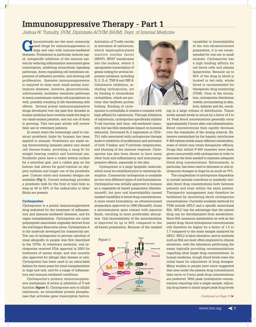
Immunosuppressive Therapy - Part 1Joshua W. Tumulty, DVM, Diplomate ACVIM (SAIM), Dept. of Internal Medicine
Glucocorticoids are the most commonly used drugs for immunosuppression in dogs and cats with immune-mediated
diseases. Prednisone in particular induces rap-id, nonspecifi c inhibition of the immune sys-tem by reducing infl ammation-associated gene transcription, inhibiting intracellular signaling pathways, down-regulating cell membrane ex-pression of adhesion proteins, and slowing cell proliferation. Systemic immunosuppression is required to treat most small-animal auto-immune diseases; however, glucocorticoids, unfortunately, modulate metabolic pathways in many nonimmune-system cell populations as well, possibly resulting in life-threatening side effects. Several potent immunosuppressive drugs developed over the past few decades in human medicine have recently made the leap to our small-animal patients, and our use of them is growing. This two-part article will review their use in veterinary patients.
In recent years the technology used to con-struct prosthetic limbs for humans has been applied to animals. Prosthetics are made us-ing thermosetting laminate plastic and closed cell thermo-foams, providing a snug fit for weight bearing comfort and functional use. Prosthetic paws have a rocker bottom surface for a smoother gait, and a rubber grip on the bottom that allows for good traction on slip-pery surfaces and longer use of the prosthetic paw. Custom colors and cosmetic designs are available (Fig 1). Current technology provides a prosthetic limb for the front or hind limb so long as 40 to 50% of the radius/ulna or tibia/fi bula are present.
CyclosporineCyclosporine is a potent immunosuppressive drug indicated for the treatment of infl amma-tory and immune-mediated diseases, and for organ transplantation. Cyclosporins are cyclic polypeptide macrolides originally derived from the soil fungus Beauveria nivea. Cyclosporine A is the molecule developed for commercial use. The use of cyclosporine to prevent rejection of renal allografts in people was fi rst described in the 1970s. In veterinary medicine, oral cy-closporine received FDA approval in 2003 for treatment of canine atopy, and was recently also approved for allergic skin disease in cats. Cyclosporine has been used in an extra-label fashion for many years for renal transplantation in dogs and cats, and for a range of infl amma-tory and immune-mediated conditions.
Cyclosporine’s primary immunosuppres-sive mechanism of action is inhibition of T-cell function (fi gure 1). Cyclosporine acts to inhibit calcineurin, an intracellular protein phospha-tase that activates gene transcription factors. Continued on Page 10 ➤
variability in bioavailability of the non-ultramicronized preparation, it is not recom-mended for oral use in small animals. Cyclosporine has a high binding affinity for red blood cells and plasma lipoproteins. Because up to 50% of the drug in blood is located in red cells, whole blood is recommended for therapeutic drug monitoring (TDM). Once in the circula-tion, cyclosporine distributes widely, accumulating in skin, liver, kidneys and fat, result-
ing in a large volume of distribution. Tissue levels exceed levels in serum by a factor of 3 to 14. Peak blood concentrations generally occur approximately 2 hours after oral administration. Blood concentrations then rapidly decrease over the remainder of the dosing interval. Ex-tensive metabolism by the hepatic cytochrome P-450 system yields many different metabolites, some of which may retain therapeutic effi cacy. Drugs that inhibit P-450 enzymes have been given concurrently with cyclosporine in order to decrease the dose needed to maintain adequate blood drug concentrations. Ketoconazole, in particular, has been used to decrease in oral cy-closporine dosages in dogs by as much as 75%.
The complexities of cyclosporine disposition in normal animals contribute to markedly vari-able blood drug concentrations both between patients and even within the same patient. Therapeutic management may therefore be facilitated by monitoring blood cyclosporine concentrations. Currently available methods for TDM include HPLC and a specifi c monoclonal RIA. HPLC has the advantage that the parent drug can be discriminated from metabolites. Since RIA measures metabolites as well as the parent drug, blood cyclosporine concentrations will therefore be higher by a factor of 1.5 to 1.7 compared to the same sample analyzed by HPLC. HPLC is labor intensive, and so methods such as RIA are most often employed in clinical situations, with the laboratory performing the assay typically providing recommendations regarding ideal target drug concentrations. In human medicine, trough blood levels were the initial basis for adjustment of drug dosages. Many studies in people have since suggested that area under the plasma drug concentration time curve or 2-hour peak drug concentrations are preferred. With peak cyclosporine concen-trations requiring only a single sample, adjust-ing drug doses to attain target peak drug levels
Figure 1
Activation of T-cells results in activation of calcineurin, which dephosphorylates inactive nuclear factor (NFAT). NFAT translocates into the nucleus, where it upregulates transcription of genes coding for several im-portant cytokines, including IL-2, IL-4, TNF-� and INF-�. Calcineurin inhibitors, in-cluding cyclosporine, act by binding to intracellular cyclophilins, which are pro-teins that facilitate protein folding. Binding of cyclo-sporine to cyclophilin A creates a complex with high affi nity for calcineurin. Through inhibition of calcineurin, cyclosporine specifi cally inhibits T-cell function and thus, cell-mediated immu-nity, but has little immediate impact on humoral immunity. Decreased IL-2 expression in CD4+ Th1 cells associated with cyclosporine therapy leads to inhibition of proliferation and activation of both T-helper and T-cytotoxic lymphocytes, and blunting of the immune response. Cyclo-sporine has also been shown to have many other local anti-infl ammatory and immunosup-pressive effects, especially in the skin.
Cyclosporine is a large lipophilic molecule, which must be solubilized prior to intestinal ab-sorption. Commercial cyclosporine is available as two very different types of oral formulations. Cyclosporine was initially approved in humans as a vegetable-oil based preparation (Sandim-mune®), but poor oral bioavailability caused marked variability in blood drug concentrations. A more recent formulation, an ultramicronized preparation approved in 1996 (Neoral®), forms a microemulsion upon contact with aqueous fl uids, resulting in more predictable absorp-tion. Oral bioavailability of the microemulsion is improved by up to 50% compared to the oil-based preparation. Because of the marked
www.livs.org 9

Immunosuppressive Therapy - Part 1➤ Continued from Page 9
has become the single best blood concentra-tion measurement for use during human organ transplantation. In veterinary medicine, mea-surement of trough cyclosporine concentrations also prevailed for many years based on initial work done in canine and feline renal transplant studies. Recommendations from laboratories of-fering TDM have often involved measurement of both peak and trough cyclosporine blood lev-els, although target peak concentrations have not been well established in small animals.
Pharmacodynamic assays investigate a drug’s effect on target cells. Many pharmaco-dynamic markers of the immunosuppressive effects of cyclosporine have been studied in hu-man medicine, including lymphocyte prolifera-tion, calcineurin enzyme activity, lymphocyte surface antigen expression, and intracellular cytokine quantifi cation. Through pharmacody-namic monitoring, human studies have shown individually distinct degrees of calcineurin inhibitor sensitivity in patients. Pharmacody-namic monitoring shows great promise for op-timizing cyclosporine therapy and delivering individualized therapy.
Cyclosporine is FDA-approved for the treat-ment of canine atopic dermatitis and feline al-lergic skin disease, and has also been used to prevent transplant rejection and to treat seba-ceous adenitis, pemphigus foliaceus, anal fu-runculosis, feline stomatitis, infl ammatory bowel disease (IBD), myasthenia gravis, noninfectious, infl ammatory meningoencephalitis, pure red cell aplasia, immune-mediated hemolytic anemia (IMHA), immune-mediated thrombocytopenia (IMT), and immune-mediated polyarthritis in dogs and cats. Recent pharmacodynamic re-search has confi rmed that canine responses to the drug are comparable to the response profi le that is well recognized in people: individual re-sponses are extremely variable from dog to dog, both in dogs receiving the same standard oral dose, and in dogs with oral doses adjusted to at-tain comparable blood levels. Given this high de-gree of variability of individual responsiveness to cyclosporine in dogs, dosing protocols should be tailored to allow for this patient-to-patient vari-ability. In my opinion, recommended dosing pro-tocols in dogs with chronic, non-life-threatening infl ammatory skin and gastrointestinal diseases should be quite different from the protocols used in dogs with more acute and life-threatening immune-mediated diseases.
In chronic diseases that are typically not life-threatening, such as skin conditions, anal furunculosis, and mild IBD, cyclosporine is often effective at a standard, relatively low starting dose. Cyclosporine is typically given long term, with doses adjusted upwards if needed ‘to ef-fect,’ based predominantly on clinical signs.
PCR within a week of starting therapy, with dose adjustments as needed, are the best methods that are currently avail-able to assess adequacy of therapy, and are strongly recommended in dogs with severe disease.
Side effects are uncommon with cy-closporine, with the exception of GI signs such as vomiting, diarrhea, anorexia and nausea. Giving the drug frozen and/or with food can reduce GI side effects, although such measures may also alter drug absorption profiles. Uncommonly, cyclosporine can cause an idiosyncratic hepatotoxicity, which does not seem to be dose dependent. Gingival hyperplasia and hypertrichosis have also occasionally been reported with cyclosporine. Chronic
cyclosporine therapy may also predispose to neoplasia such as lymphoma. One advantage of cyclosporine is that it is not myelosuppressive. Experimentally, cyclosporine has been shown to increase some markers of platelet activation in dogs, which may be a concern in patients with IMHA, where hypercoagulability and re-sultant pulmonary thromboembolism can be a major contributor to patient mortality. However, to date, it has not been demonstrated whether this phenomenon is clinically relevant in IMHA patients with naturally occurring disease.
Cyclosporine is an expensive drug, and clini-cians are therefore tempted to explore cheaper forms of the drug. There are many approved hu-man generic microemulsion preparations simi-lar to Neoral®, and these have been shown to have therapeutic equivalency in people. Studies investigating the pharmacokinetics of these preparations in dogs have not been performed, and it is not safe to assume that a generic for-mulation is therapeutically equivalent to the approved canine product (Atopica®). Neoral® currently costs around $2 for a 25-mg capsule and $6 for a 100-mg capsule, while the generic equivalents cost around $1 and $2 for the 25-mg and 100-mg capsules respectively. The veteri-nary product is priced comparably to the human proprietary products, but has the advantage of being FDA-approved and available in capsule sizes that are convenient for dosing accuracy (10 mg, 25 mg, 50 mg and 100 mg), as well as a 100 mg/ml oral suspension. Unfortunately, transdermal cyclosporine has been shown to be inadequately absorbed in cats.
Lefl unomideLefl unomide is an isoxazole derivative, im-munosuppressive drug that was developed within the past two decades for treatment of
Typically, however, starting doses do not need to be increased and, in the long term, the drug is tapered to the lowest effective dose needed to maintain remission. Recommended starting cyclosporine doses in dogs are 5 mg/kg once daily for most skin diseases and IBD, and 5 mg/kg once to twice daily for anal furunculosis. In cats with skin conditions such as allergic skin disease, eosinophilic granuloma complex and pemphigus foliaceus, a starting cyclosporine dose of around 5 mg/kg daily is recommended. Blood concentrations are usually not necessary for treatment of these conditions, as remis-sion of disease is the main criterion used to decide whether drug therapy is adequate. In fact, for many of these conditions, cyclosporine blood levels have been shown to have minimal correlation with remission, perhaps because the drug is selectively concentrated in tissues such as the skin. Even at standard low FDA-approved doses, some dogs can still develop signifi cant suppression of T-cell biomarkers of immunosuppression despite very low trough cyclosporine concentrations. This could explain the phenomenon reported by some dermatolo-gists, that individual dogs treated for atopy can develop severe secondary infections.
In dogs suffering from more life-threatening diseases such as severe IMHA and IMT, in con-trast, cyclosporine must be targeted to attain effective immunosuppression as rapidly as pos-sible. In these patients, starting cyclosporine at a low dose and adjusting doses upwards ‘to effect’ is not recommended. Attaining effective oral doses as rapidly as possible is essential for ensuring adequate immunosuppression whilst avoiding overdosage. Currently recommended starting cyclosporine doses for life-threatening diseases range from 5 mg/kg to 10 mg/kg twice daily. Subsequent measurement of blood drug levels and/or assessment of activated T-cell mRNA IL-2 and IFN-� expression using qRT-
Figure 2
www.livs.org10
Continued on Page 11 ➤
rheumatoid arthritis and prevention of trans-plant rejection. Lefl unomide is a prodrug for its primary active malononitriloamide metabolite, A77 1726 (terifl unomide). Malononitriloamides reversibly inhibit the mitochondrial enzyme dihydroorotate dehydrogenase, a key enzyme in pyrimidine synthesis, with resultant inhibi-tion of the pyrimidine ribonucleotide uridine monophosphate (rUMP), and decreased DNA and RNA synthesis and G1 cell cycle arrest (fi gure 2). Lefl unomide inhibits B- and T-cell function, suppresses antibody production and has anti-inflammatory effects, possibly via inhibition of de novo pyrimidine biosynthesis and cytokine-associated and IL-2-stimulated tyrosine kinase activity.
Prior to commercial development, leflun-omide was made available to the transplant research group at UC-Davis. This group de-scribed treatment of small number of canine patients with refractory infl ammatory and im-mune-mediated diseases such as IMHA, IMT, noninfectious infl ammatory meningoencepha-litis, systemic histiocytosis, immune-mediated polymyositis, immune-mediated polyarthritis, and pemphigus foliaceous with lefl unomide, typically with promising success rates. Unfor-tunately, when these results were reported in the late 1990s, the drug was no longer commer-cially available. When lefl unomide did become available as the proprietary product Arava®, drug cost limited its use in small-animal clinic patients. Only recently did an affordable ge-neric equivalent become available and, as a result, preliminary reports of lefl unomide use in small-animal patients are beginning to surface. There are still currently very few published re-ports discussing the use of lefl unomide in dogs and cats. Recently, a case series describing the use of lefl unomide in 14 dogs with immune-mediated polyarthritis reported a high response rate with few side effects.
Lefl unomide appears to be very well toler-ated in dogs although, if the drug attains more common usage, it is likely that less frequent but more serious side effects will be recog-nized. The most common side effect seen with lefl unomide in dogs is occasional inappetance, lethargy and vomiting. Serious side effects oc-casionally reported in people include myelosup-pression, cutaneous drug reactions and hepato-toxicity. In humans, traces of terifl unomide can persist for many months after drug discontinua-tion, and in the instance of drug reactions, cho-lestyramine or activated charcoal are needed to rapidly reduce drug levels. In dogs, the terminal half-life of terifl unomide is much shorter than in humans, so the potential for persistent side effects is probably signifi cantly less. Complete blood counts and serum biochemistry (espe-
mofetil, CellCept®, and the related mycopheno-late sodium, Myfortic®, were expensive, and as a result the products only achieved limited usage in small-animal medicine. However, recently, the availability of cheaper generic alternatives has led to increased usage of mycophenolate in small-animal patients. A 250-mg CellCept® capsule currently costs around $7, whereas the equivalent generic 250-mg capsule costs less than 50c. An oral suspension version of my-cophenolate mofetil (200 mg/ml) is available for more convenient dosing in smaller patients. The successful use of mycophenolate mofetil in a small-animal patient with naturally occur-ring disease was fi rst described in a dog with myasthenia gravis. Much of the subsequent anecdotal usage of mycophenolate for a variety of different immune-mediated diseases used dosing similar to that reported in this original
paper. Mycophenolate mofetil is also available in an injectable form, and its IV use has been described during the successful stabilization of 3 dogs with my-asthenia that could not tolerate oral medications. Ironically, a re-cent case report of 15 dogs with myasthenia gravis treated with mycophenolate mofetil reported that the drug was ineffective at attaining clinical remission.
A recommended starting dose for mycophenolate mofetil in dogs is 10–20 mg/kg once daily or divided twice daily, although sometimes GI signs (particularly vomiting and di-
arrhea) at the high end of the dose rate will necessitate dose reductions. Mycophenolate mofetil has variable oral bioavailability in dogs, so variability in response to therapy should also be expected. Mycophenolate has not been used widely enough to establish the frequency of se-rious side effects in dogs but, in people, GI signs and, less commonly, marked myelosuppression and a rare and fatal neurologic disease (pro-gressive multifocal leukoencephalopathy) have been reported. Complete blood counts should therefore probably be regularly monitored in dogs receiving mycophenolate. In humans, GI side effects can be reduced by replacing myco-phenolate mofetil with mycophenolate sodium. Mycophenolic acid in humans is primarily ex-creted conjugated to glucuronide and, since cats lack the glucuronyl transferases needed for glucuronidation, the drug should be used with caution in this species, although the use of mycophenolate mofetil has been described at a dose rate of 10 mg/kg twice daily, with no obvious side effects, in two cats with IMHA. ❏
Immunosuppressive Therapy - Part 1➤ Continued from Page 10
cially ALT) should be regularly monitored in small-animal patients on lefl unomide.
The initial recommended starting oral dose for lefl unomide in dogs is 2–4 mg/kg daily, with doses adjusted to attain a plasma trough terifl u-nomide levels of 20 µg/ml within a few weeks of commencing therapy. Measurement of lefl u-nomide levels is available through Auburn Uni-versity. For cats with immune-mediated polyar-thritis, a lefl unomide dose of 10 mg (total dose) orally, once daily, combined with methotrexate, has been suggested, with dose reductions to effect. Lefl unomide comes in tablet sizes (10 and 20 mg) that are convenient for dosing our smaller patients. Proprietary lefl unomide costs about $40 for a 10-mg or 20-mg tablet, while the generic equivalent is priced at around $1 for a 10-mg tablet and $1.50 for a 20-mg tablet. Lefl unomide generics have an ‘AB’ rating by the
FDA, meaning that the generic is ‘equivalent’ to Arava®. However, since ‘equivalence’ is often determined by pharmacokinetic data in healthy individuals, an AB rating does not guarantee identical performance in clinical patients.
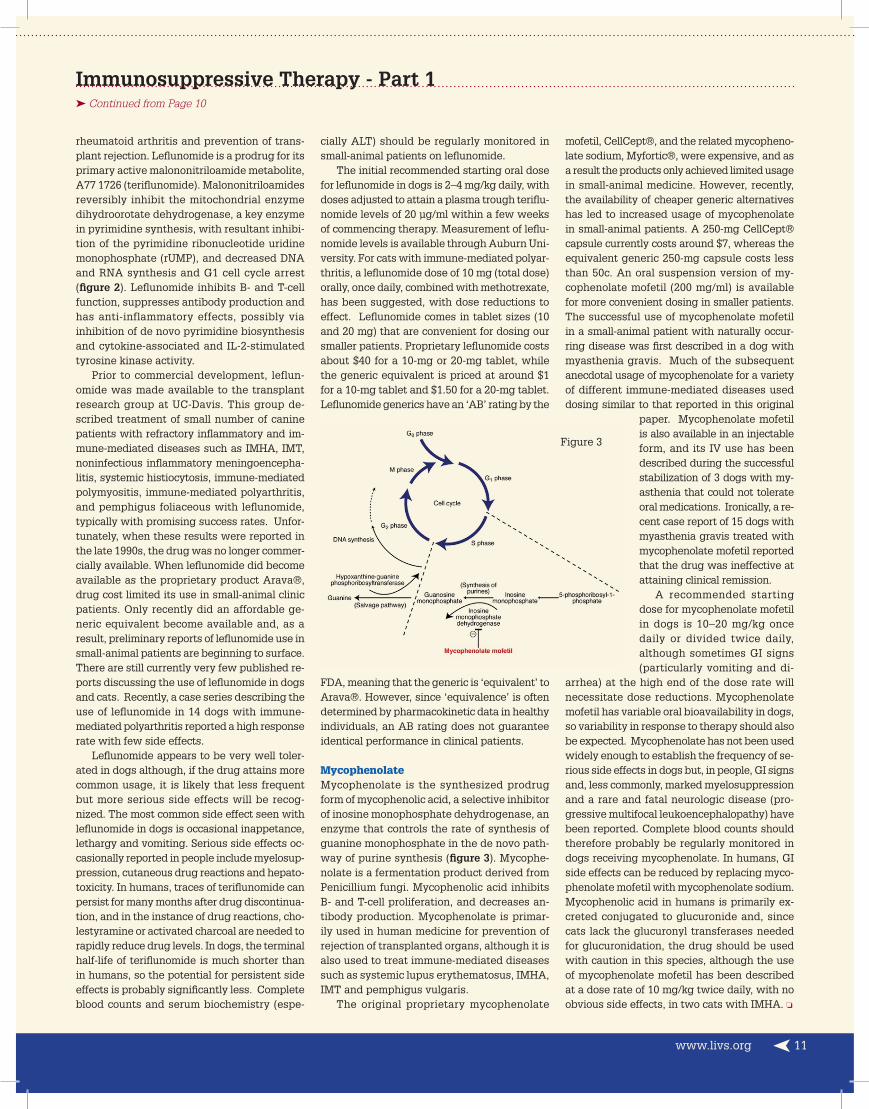
MycophenolateMycophenolate is the synthesized prodrug form of mycophenolic acid, a selective inhibitor of inosine monophosphate dehydrogenase, an enzyme that controls the rate of synthesis of guanine monophosphate in the de novo path-way of purine synthesis (fi gure 3). Mycophe-nolate is a fermentation product derived from Penicillium fungi. Mycophenolic acid inhibits B- and T-cell proliferation, and decreases an-tibody production. Mycophenolate is primar-ily used in human medicine for prevention of rejection of transplanted organs, although it is also used to treat immune-mediated diseases such as systemic lupus erythematosus, IMHA, IMT and pemphigus vulgaris.
The original proprietary mycophenolate
Figure 3
www.livs.org 11

East Meets West...One Medicine Dr. Michel Selmer is an Integrative Veterinarian and one of only a handful of Traditional Chinese Veterinary Medicine Practitioners that holds a Master’s Degree in the United States.
Dr. Selmer is a certified TCVM practitioner and provides the following Integrative Medical Therapies:
• Veterinary Acupuncture• Herbal Medicine• Veterinary Food Therapy• Veterinary Tui-na
To learn more about Dr. Selmer and Traditional Chinese Veterinary Medicine, check out his book: “The Best of Both Worlds, An Advanced Guide to Integrative Veterinary Care for Healthier, Happi-er Pups”
To refer your clients to Dr. Selmer, call516-501-1700 or visit www.livs.org
163 South Service Road, Plainview, NY 11803
163 South Service Road, Plainview, New York 11803