Embed Size (px)
Citation preview
Cervical Spine Mobility Deficits ICD-9-CM code: 723.1 Cervicalgia ICF codes: Activities and Participation Domain code: d4108 Changing a basic
body position, other specified - specified as: rotating the head and neck, such as in looking to the left or to the right
Body Structure code: s76000 Cervical vertebral column Body Functions code: b7101 Mobility of several joints
Common Historical Findings:
Neck pain, usually unilateral, pain referral from base of occiput to scapular region (location of pain referral is dependent upon which segment or segments are involved)
Strain; awkward, unguarded movement; or prolonged period of time in strained position ("Woke up with pain")
Common Impairment Findings - Related to the Reported Activity Limitation or Participation Restrictions:
Increase in pain at end range of rotation left or rotation right Symptoms reproduced with palpation of the involved facet Motion limitation and pain at end range of either anterior/superior glide or
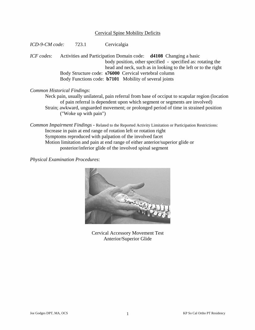
posterior/inferior glide of the involved spinal segment Physical Examination Procedures:
Cervical Accessory Movement Test Anterior/Superior Glide
Joe Godges DPT, MA, OCS KP So Cal Ortho PT Residency 1
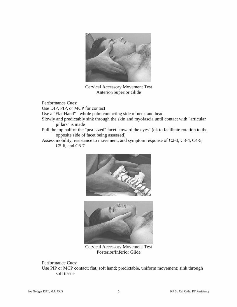
Cervical Accessory Movement Test
Anterior/Superior Glide Performance Cues: Use DIP, PIP, or MCP for contact Use a "Flat Hand" - whole palm contacting side of neck and head Slowly and predictably sink through the skin and myofascia until contact with "articular
pillars" is made Pull the top half of the "pea-sized" facet "toward the eyes" (ok to facilitate rotation to the
opposite side of facet being assessed) Assess mobility, resistance to movement, and symptom response of C2-3, C3-4, C4-5,
C5-6, and C6-7
Cervical Accessory Movement Test
Posterior/Inferior Glide Performance Cues: Use PIP or MCP contact; flat, soft hand; predictable, uniform movement; sink through
soft tissue
Joe Godges DPT, MA, OCS KP So Cal Ortho PT Residency 2
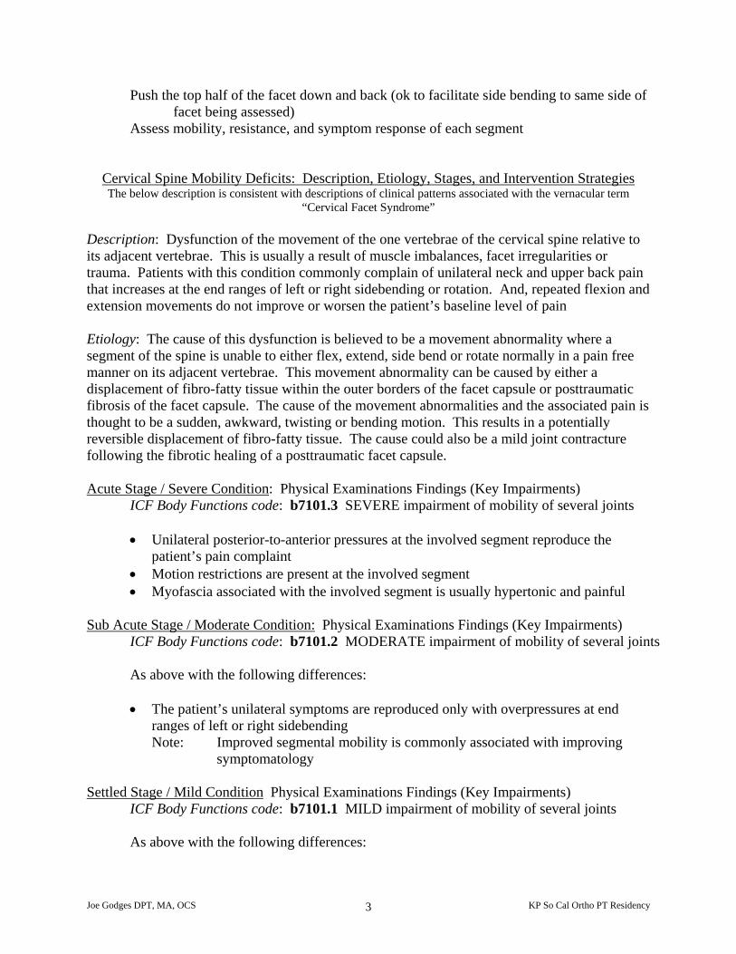
Push the top half of the facet down and back (ok to facilitate side bending to same side of facet being assessed)
Assess mobility, resistance, and symptom response of each segment
Cervical Spine Mobility Deficits: Description, Etiology, Stages, and Intervention Strategies The below description is consistent with descriptions of clinical patterns associated with the vernacular term
“Cervical Facet Syndrome” Description: Dysfunction of the movement of the one vertebrae of the cervical spine relative to its adjacent vertebrae. This is usually a result of muscle imbalances, facet irregularities or trauma. Patients with this condition commonly complain of unilateral neck and upper back pain that increases at the end ranges of left or right sidebending or rotation. And, repeated flexion and extension movements do not improve or worsen the patient’s baseline level of pain Etiology: The cause of this dysfunction is believed to be a movement abnormality where a segment of the spine is unable to either flex, extend, side bend or rotate normally in a pain free manner on its adjacent vertebrae. This movement abnormality can be caused by either a displacement of fibro-fatty tissue within the outer borders of the facet capsule or posttraumatic fibrosis of the facet capsule. The cause of the movement abnormalities and the associated pain is thought to be a sudden, awkward, twisting or bending motion. This results in a potentially reversible displacement of fibro-fatty tissue. The cause could also be a mild joint contracture following the fibrotic healing of a posttraumatic facet capsule. Acute Stage / Severe Condition: Physical Examinations Findings (Key Impairments)
ICF Body Functions code: b7101.3 SEVERE impairment of mobility of several joints
• Unilateral posterior-to-anterior pressures at the involved segment reproduce the patient’s pain complaint
• Motion restrictions are present at the involved segment • Myofascia associated with the involved segment is usually hypertonic and painful
Sub Acute Stage / Moderate Condition: Physical Examinations Findings (Key Impairments)
ICF Body Functions code: b7101.2 MODERATE impairment of mobility of several joints As above with the following differences: • The patient’s unilateral symptoms are reproduced only with overpressures at end
ranges of left or right sidebending Note: Improved segmental mobility is commonly associated with improving
symptomatology Settled Stage / Mild Condition Physical Examinations Findings (Key Impairments)
ICF Body Functions code: b7101.1 MILD impairment of mobility of several joints As above with the following differences:
Joe Godges DPT, MA, OCS KP So Cal Ortho PT Residency 3
• The patient’s unilateral symptoms are reproduced only with end range overpressures in either a combined extension and sidebending motion or a combined flexion and sidebending motion
Now when the patient is less acute examine for muscle flexibility and strength deficits that may be a predisposing factor for future injury. For example:
• Muscles that commonly exhibit flexibility deficits in patients with facet abnormalities
are middle and posterior scaleni, SCM, upper trapezius, and the myofascia associated with the involved cervical segment
• Muscles that are commonly weak are the cervical neck flexors (i.e., longus colli),
upper thoracic extensors and scapular retractors/adductors (i.e, middle and lower trapezius)
Intervention Approaches / Strategies Acute Stage / Severe Condition Goal: Restore painfree active spinal mobility
• Physical Agents Ice (or heat) to provide pain relief and reduce muscle guarding
• Manual Therapy
Soft tissue mobilization to the myofascia associated with the involved cervical segment
Isometric mobilization and contract/relax procedures to the involved segment to reduce muscle guarding
Passive stretching procedures to restore normal cervical segmental mobility
• Therapeutic Exercises Instruction in exercise and functional movements to maintain the improvements in
mobility gained with the soft tissue and joint manipulations Strengthening exercises for the neck flexors
Joe Godges DPT, MA, OCS KP So Cal Ortho PT Residency 4
• Re-injury Prevention Instruction
Instruct the patient in efficient, painfree, motor performance of movements that are related by the patient to be the cause of the current episode of neck pain
Sub Acute Stage / Moderate Condition: Goal: Restore normal, painfree response to overpressures at end ranges of cervical rotation and
sidebending
• Approaches / Strategies listed above – focusing on:
• Manual Therapy Soft tissue mobilization and joint mobilization/manipulation to normalize the segmental mobility
Note: Performing upper cervical joint mobilization/manipulations with the patients upper cervical spine at end ranges of extension or the end ranges of combined of extension/rotation movements is contraindicated due the potential disastrous effects that these manipulative procedures have been reported to have on some individual’s vertebral artery. Thus, all upper cervical manipulations are performed with the head and neck in the neutral or flexed position
• Therapeutic Exercises
Instruction in exercise and functional movements to maintain the improvements in mobility gained with the soft tissue and joint manipulations (e.g., towel SNAGs)
Settled Stage / Mild Condition: Goals: Restore normal, pain free responses to overpressures of combined extension and sidebending/rotation and/or combined flexion and sidebending/rotation
Normalize cervical and upper thoracic flexibility and strength deficits
• Approaches / Strategies listed above
• Therapeutic Exercises Stretching exercises to address the patient’s specific muscle flexibility deficits Strengthening exercises to address the patient’s specific muscle strength deficits
Joe Godges DPT, MA, OCS KP So Cal Ortho PT Residency 5
Intervention for High Performance / High Demand Functioning in Workers or Athletes Goal: Return to desired occupational or leisure time activities
• Approaches / Strategies listed above
• Therapeutic Exercises Encourage participation in regular low stress aerobic activities as a means to improve fitness, muscle strength and prevent recurrences
• Ergonomic Instruction
Provide body mechanics instructions and modify work area as indicated to prevent symptoms. This typically emphasizes neutral cervical position for sitting, driving, traveling as a passenger in a car, bus, or airplane, reading, eating, and resting/sleeping.
Selected References Di Fabio RP. Manipulation of the cervical spine: risks and benefits. Phys Ther. 1999;79:50-65. Jackson RP. The facet syndrome: myth or reality? Clin Orthop Rel Res. June, 1992. Taimela S, Takala E, Asklof T, Seppala K, Parvianen S. Active treatment of chronic neck pain. a prospective randomized intervention. Spine. 2000;25:1021-1027. Jull G, Trott P, Potter H, Zito G, Niere K. Shirley D, Emberson J, Marschner I, Richardson C. A randomized controlled trial of exercise and manipulative therapy for cervicogenic headache. Spine. 2002;27:1835-1843.
Joe Godges DPT, MA, OCS KP So Cal Ortho PT Residency 6
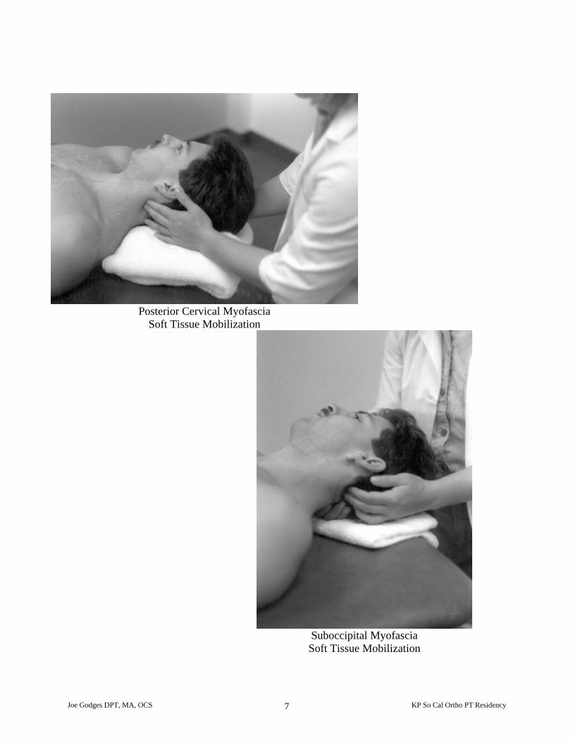
Posterior Cervical Myofascia
Soft Tissue Mobilization
Suboccipital Myofascia Soft Tissue Mobilization
Joe Godges DPT, MA, OCS KP So Cal Ortho PT Residency 7
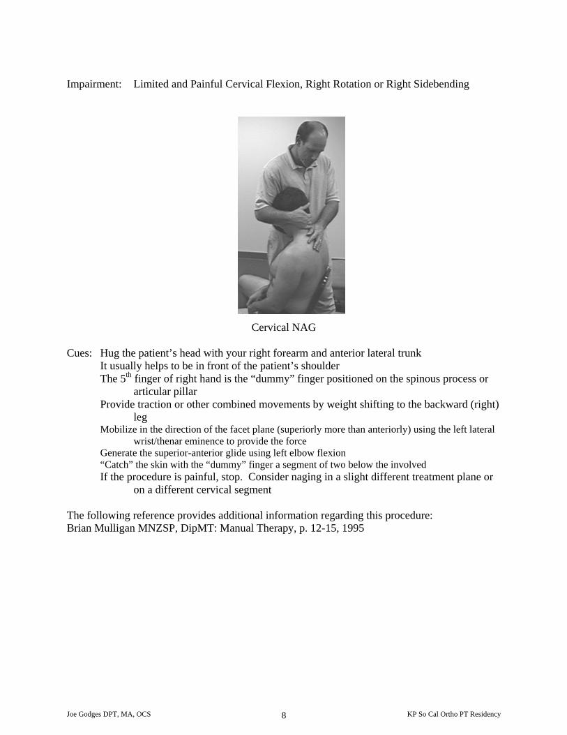
Impairment: Limited and Painful Cervical Flexion, Right Rotation or Right Sidebending
Cervical NAG Cues: Hug the patient’s head with your right forearm and anterior lateral trunk
It usually helps to be in front of the patient’s shoulder The 5th finger of right hand is the “dummy” finger positioned on the spinous process or
articular pillar Provide traction or other combined movements by weight shifting to the backward (right)
leg Mobilize in the direction of the facet plane (superiorly more than anteriorly) using the left lateral
wrist/thenar eminence to provide the force Generate the superior-anterior glide using left elbow flexion “Catch” the skin with the “dummy” finger a segment of two below the involved If the procedure is painful, stop. Consider naging in a slight different treatment plane or
on a different cervical segment The following reference provides additional information regarding this procedure: Brian Mulligan MNZSP, DipMT: Manual Therapy, p. 12-15, 1995
Joe Godges DPT, MA, OCS KP So Cal Ortho PT Residency 8
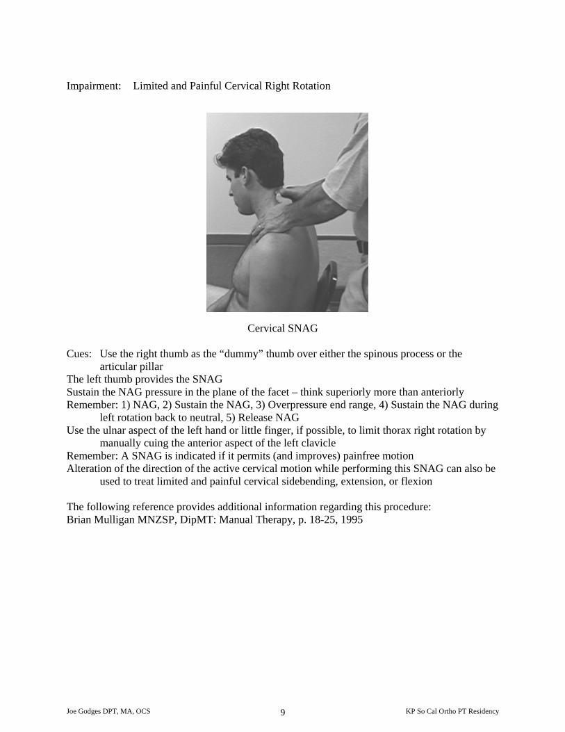
Impairment: Limited and Painful Cervical Right Rotation
Cervical SNAG Cues: Use the right thumb as the “dummy” thumb over either the spinous process or the
articular pillar The left thumb provides the SNAG Sustain the NAG pressure in the plane of the facet – think superiorly more than anteriorly Remember: 1) NAG, 2) Sustain the NAG, 3) Overpressure end range, 4) Sustain the NAG during
left rotation back to neutral, 5) Release NAG Use the ulnar aspect of the left hand or little finger, if possible, to limit thorax right rotation by
manually cuing the anterior aspect of the left clavicle Remember: A SNAG is indicated if it permits (and improves) painfree motion Alteration of the direction of the active cervical motion while performing this SNAG can also be
used to treat limited and painful cervical sidebending, extension, or flexion The following reference provides additional information regarding this procedure: Brian Mulligan MNZSP, DipMT: Manual Therapy, p. 18-25, 1995
Joe Godges DPT, MA, OCS KP So Cal Ortho PT Residency 9
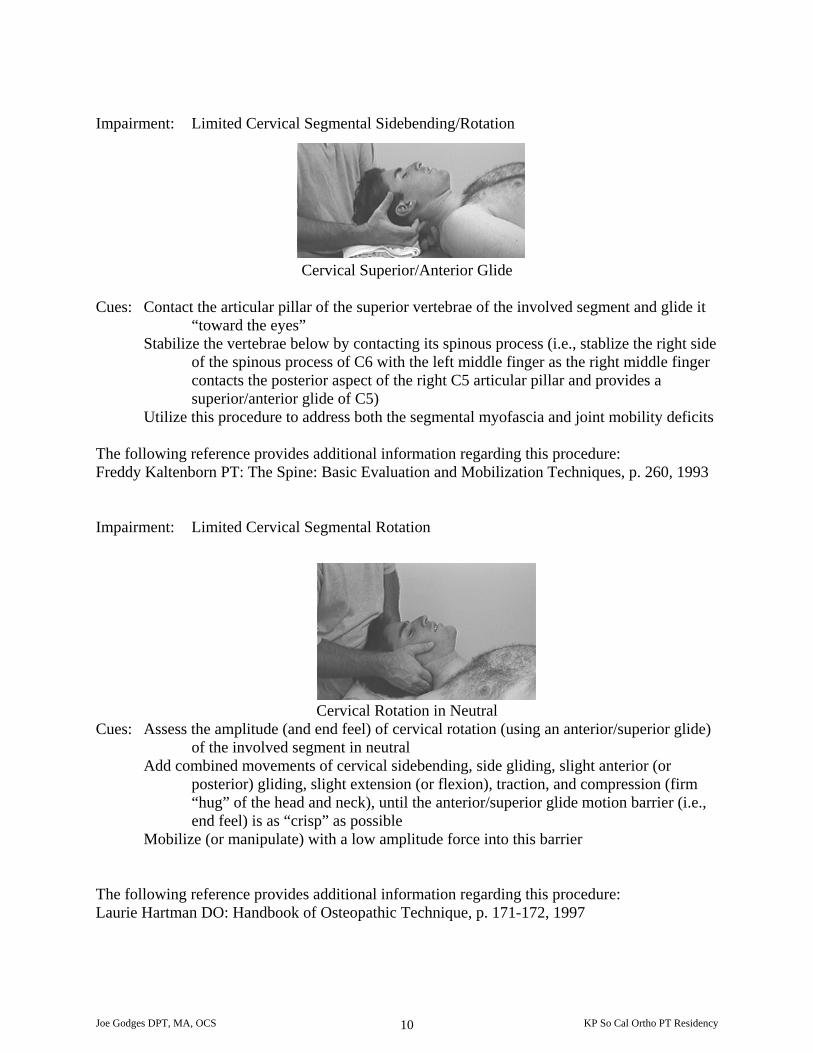
Impairment: Limited Cervical Segmental Sidebending/Rotation
Cervical Superior/Anterior Glide
Cues: Contact the articular pillar of the superior vertebrae of the involved segment and glide it “toward the eyes”
Stabilize the vertebrae below by contacting its spinous process (i.e., stablize the right side of the spinous process of C6 with the left middle finger as the right middle finger contacts the posterior aspect of the right C5 articular pillar and provides a superior/anterior glide of C5)
Utilize this procedure to address both the segmental myofascia and joint mobility deficits The following reference provides additional information regarding this procedure: Freddy Kaltenborn PT: The Spine: Basic Evaluation and Mobilization Techniques, p. 260, 1993 Impairment: Limited Cervical Segmental Rotation
Cervical Rotation in Neutral Cues: Assess the amplitude (and end feel) of cervical rotation (using an anterior/superior glide)
of the involved segment in neutral Add combined movements of cervical sidebending, side gliding, slight anterior (or
posterior) gliding, slight extension (or flexion), traction, and compression (firm “hug” of the head and neck), until the anterior/superior glide motion barrier (i.e., end feel) is as “crisp” as possible
Mobilize (or manipulate) with a low amplitude force into this barrier The following reference provides additional information regarding this procedure: Laurie Hartman DO: Handbook of Osteopathic Technique, p. 171-172, 1997
Joe Godges DPT, MA, OCS KP So Cal Ortho PT Residency 10
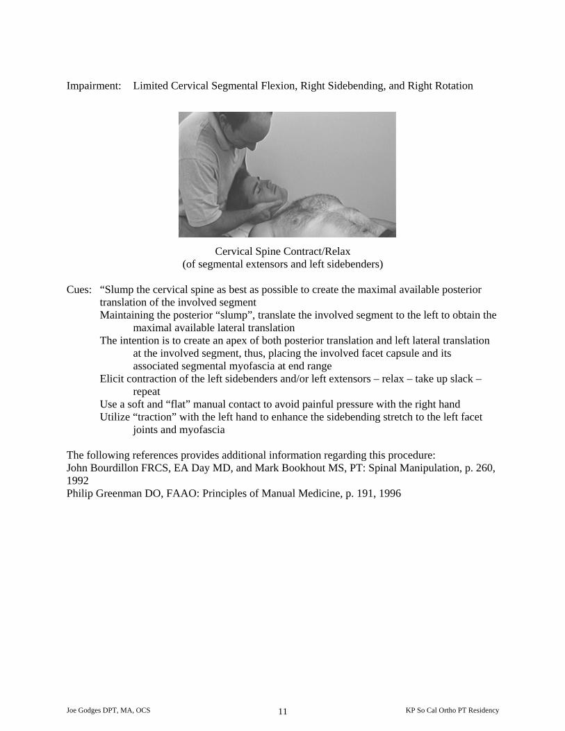
Impairment: Limited Cervical Segmental Flexion, Right Sidebending, and Right Rotation
Cervical Spine Contract/Relax (of segmental extensors and left sidebenders)
Cues: “Slump the cervical spine as best as possible to create the maximal available posterior
translation of the involved segment Maintaining the posterior “slump”, translate the involved segment to the left to obtain the
maximal available lateral translation The intention is to create an apex of both posterior translation and left lateral translation
at the involved segment, thus, placing the involved facet capsule and its associated segmental myofascia at end range
Elicit contraction of the left sidebenders and/or left extensors – relax – take up slack – repeat
Use a soft and “flat” manual contact to avoid painful pressure with the right hand Utilize “traction” with the left hand to enhance the sidebending stretch to the left facet
joints and myofascia The following references provides additional information regarding this procedure: John Bourdillon FRCS, EA Day MD, and Mark Bookhout MS, PT: Spinal Manipulation, p. 260, 1992 Philip Greenman DO, FAAO: Principles of Manual Medicine, p. 191, 1996
Joe Godges DPT, MA, OCS KP So Cal Ortho PT Residency 11
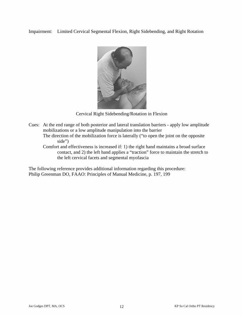
Impairment: Limited Cervical Segmental Flexion, Right Sidebending, and Right Rotation
Cervical Right Sidebending/Rotation in Flexion Cues: At the end range of both posterior and lateral translation barriers - apply low amplitude
mobilizations or a low amplitude manipulation into the barrier The direction of the mobilization force is laterally (“to open the joint on the opposite
side”) Comfort and effectiveness is increased if: 1) the right hand maintains a broad surface
contact, and 2) the left hand applies a “traction” force to maintain the stretch to the left cervical facets and segmental myofascia
The following reference provides additional information regarding this procedure: Philip Greenman DO, FAAO: Principles of Manual Medicine, p. 197, 199
Joe Godges DPT, MA, OCS KP So Cal Ortho PT Residency 12
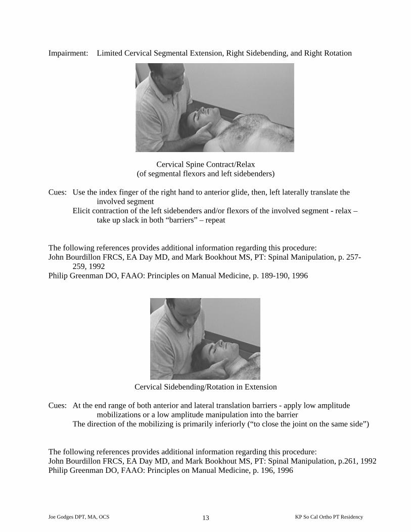
Impairment: Limited Cervical Segmental Extension, Right Sidebending, and Right Rotation
Cervical Spine Contract/Relax
(of segmental flexors and left sidebenders) Cues: Use the index finger of the right hand to anterior glide, then, left laterally translate the
involved segment Elicit contraction of the left sidebenders and/or flexors of the involved segment - relax –
take up slack in both “barriers” – repeat
The following references provides additional information regarding this procedure: John Bourdillon FRCS, EA Day MD, and Mark Bookhout MS, PT: Spinal Manipulation, p. 257-
259, 1992 Philip Greenman DO, FAAO: Principles on Manual Medicine, p. 189-190, 1996
Cervical Sidebending/Rotation in Extension
Cues: At the end range of both anterior and lateral translation barriers - apply low amplitude
mobilizations or a low amplitude manipulation into the barrier The direction of the mobilizing is primarily inferiorly (“to close the joint on the same side”)
The following references provides additional information regarding this procedure: John Bourdillon FRCS, EA Day MD, and Mark Bookhout MS, PT: Spinal Manipulation, p.261, 1992 Philip Greenman DO, FAAO: Principles on Manual Medicine, p. 196, 1996
Joe Godges DPT, MA, OCS KP So Cal Ortho PT Residency 13
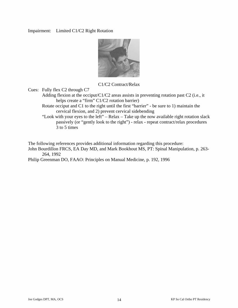
Impairment: Limited C1/C2 Right Rotation
C1/C2 Contract/Relax Cues: Fully flex C2 through C7
Adding flexion at the occiput/C1/C2 areas assists in preventing rotation past C2 (i.e., it helps create a “firm” C1/C2 rotation barrier)
Rotate occiput and C1 to the right until the first “barrier” - be sure to 1) maintain the cervical flexion, and 2) prevent cervical sidebending
“Look with your eyes to the left” – Relax – Take up the now available right rotation slack passively (or “gently look to the right”) - relax - repeat contract/relax procedures 3 to 5 times
The following references provides additional information regarding this procedure: John Bourdillon FRCS, EA Day MD, and Mark Bookhout MS, PT: Spinal Manipulation, p. 263-
264, 1992 Philip Greenman DO, FAAO: Principles on Manual Medicine, p. 192, 1996
Joe Godges DPT, MA, OCS KP So Cal Ortho PT Residency 14
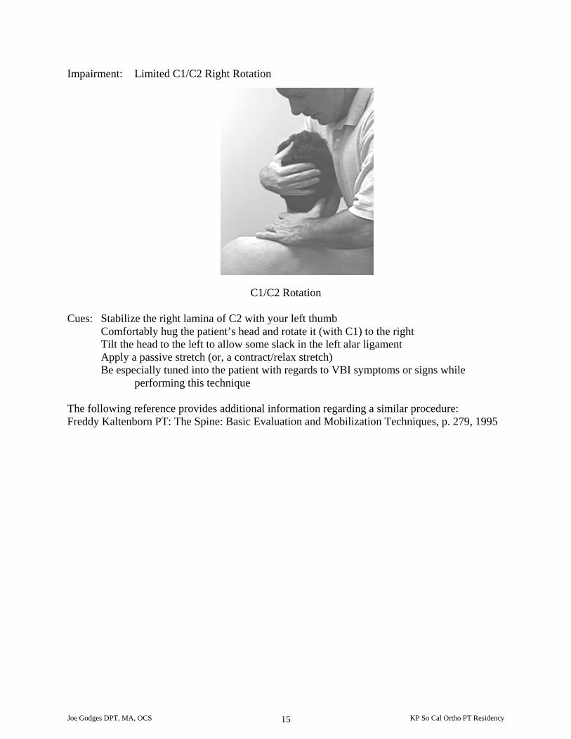
Impairment: Limited C1/C2 Right Rotation
C1/C2 Rotation Cues: Stabilize the right lamina of C2 with your left thumb
Comfortably hug the patient’s head and rotate it (with C1) to the right Tilt the head to the left to allow some slack in the left alar ligament Apply a passive stretch (or, a contract/relax stretch) Be especially tuned into the patient with regards to VBI symptoms or signs while
performing this technique The following reference provides additional information regarding a similar procedure: Freddy Kaltenborn PT: The Spine: Basic Evaluation and Mobilization Techniques, p. 279, 1995
Joe Godges DPT, MA, OCS KP So Cal Ortho PT Residency 15
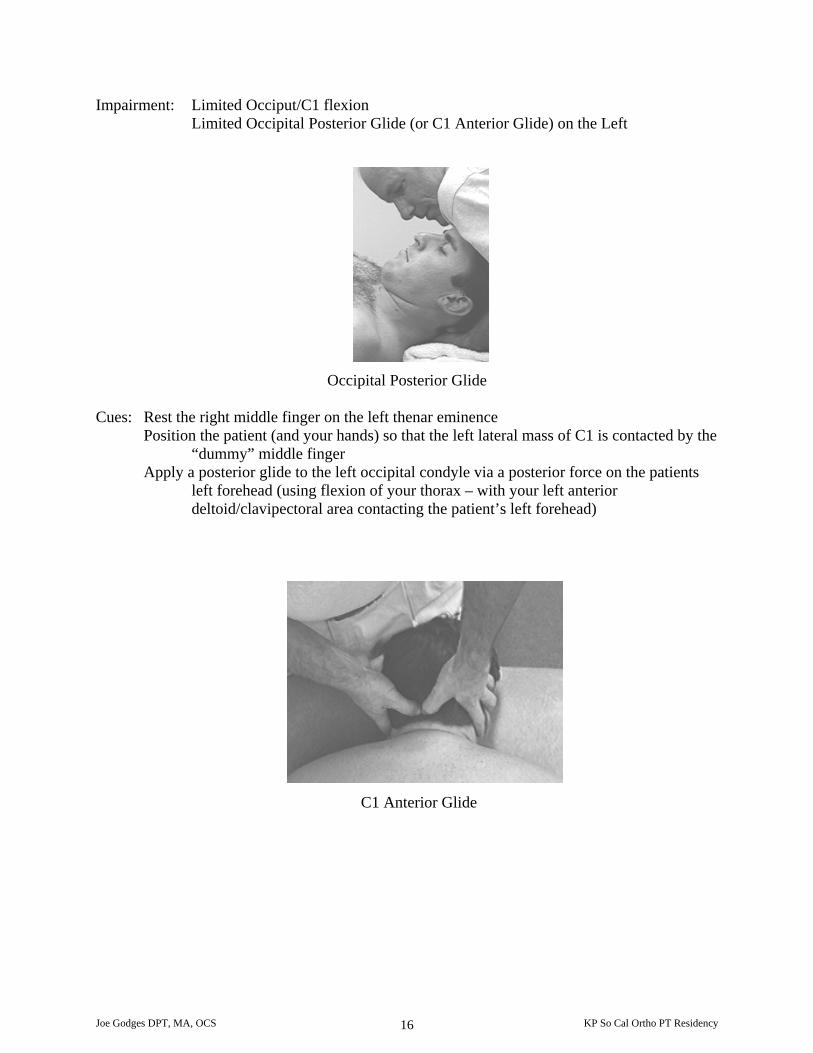
Impairment: Limited Occiput/C1 flexion Limited Occipital Posterior Glide (or C1 Anterior Glide) on the Left
Occipital Posterior Glide Cues: Rest the right middle finger on the left thenar eminence
Position the patient (and your hands) so that the left lateral mass of C1 is contacted by the “dummy” middle finger
Apply a posterior glide to the left occipital condyle via a posterior force on the patients left forehead (using flexion of your thorax – with your left anterior deltoid/clavipectoral area contacting the patient’s left forehead)
C1 Anterior Glide
Joe Godges DPT, MA, OCS KP So Cal Ortho PT Residency 16
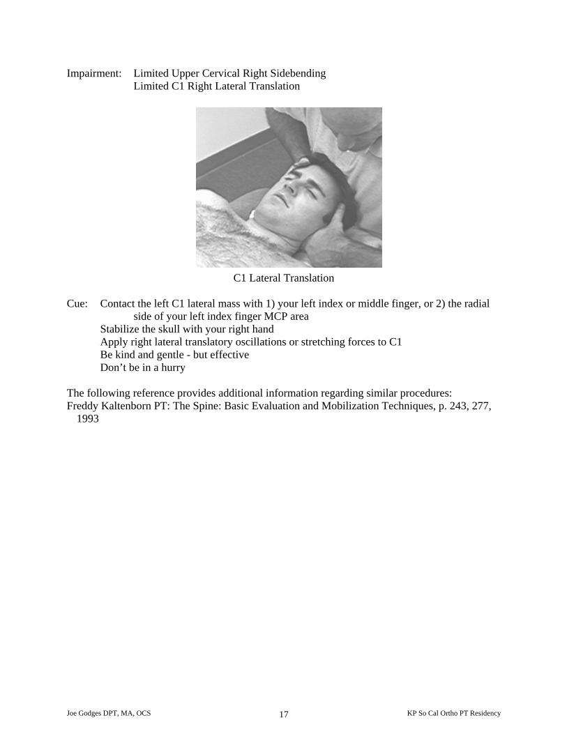
Impairment: Limited Upper Cervical Right Sidebending Limited C1 Right Lateral Translation
C1 Lateral Translation Cue: Contact the left C1 lateral mass with 1) your left index or middle finger, or 2) the radial
side of your left index finger MCP area Stabilize the skull with your right hand Apply right lateral translatory oscillations or stretching forces to C1 Be kind and gentle - but effective Don’t be in a hurry
The following reference provides additional information regarding similar procedures: Freddy Kaltenborn PT: The Spine: Basic Evaluation and Mobilization Techniques, p. 243, 277,
1993
Joe Godges DPT, MA, OCS KP So Cal Ortho PT Residency 17
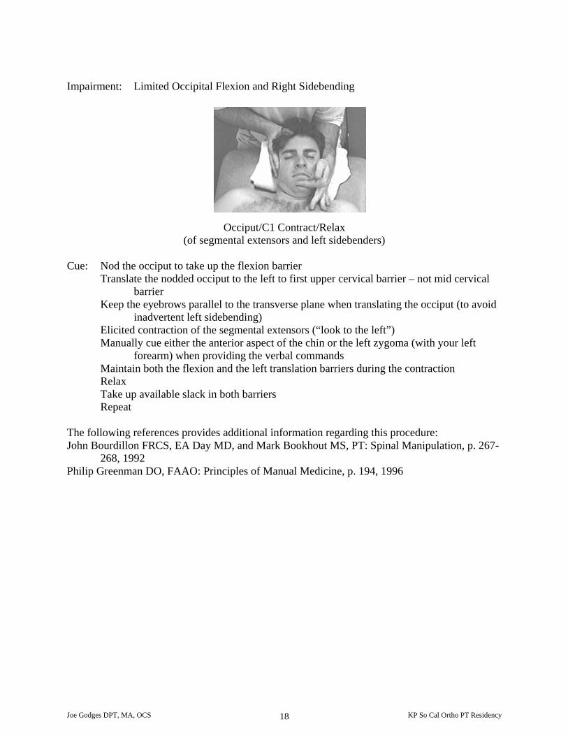
Impairment: Limited Occipital Flexion and Right Sidebending
Occiput/C1 Contract/Relax (of segmental extensors and left sidebenders)
Cue: Nod the occiput to take up the flexion barrier
Translate the nodded occiput to the left to first upper cervical barrier – not mid cervical barrier
Keep the eyebrows parallel to the transverse plane when translating the occiput (to avoid inadvertent left sidebending)
Elicited contraction of the segmental extensors (“look to the left”) Manually cue either the anterior aspect of the chin or the left zygoma (with your left
forearm) when providing the verbal commands Maintain both the flexion and the left translation barriers during the contraction Relax Take up available slack in both barriers Repeat
The following references provides additional information regarding this procedure: John Bourdillon FRCS, EA Day MD, and Mark Bookhout MS, PT: Spinal Manipulation, p. 267-
268, 1992 Philip Greenman DO, FAAO: Principles of Manual Medicine, p. 194, 1996
Joe Godges DPT, MA, OCS KP So Cal Ortho PT Residency 18
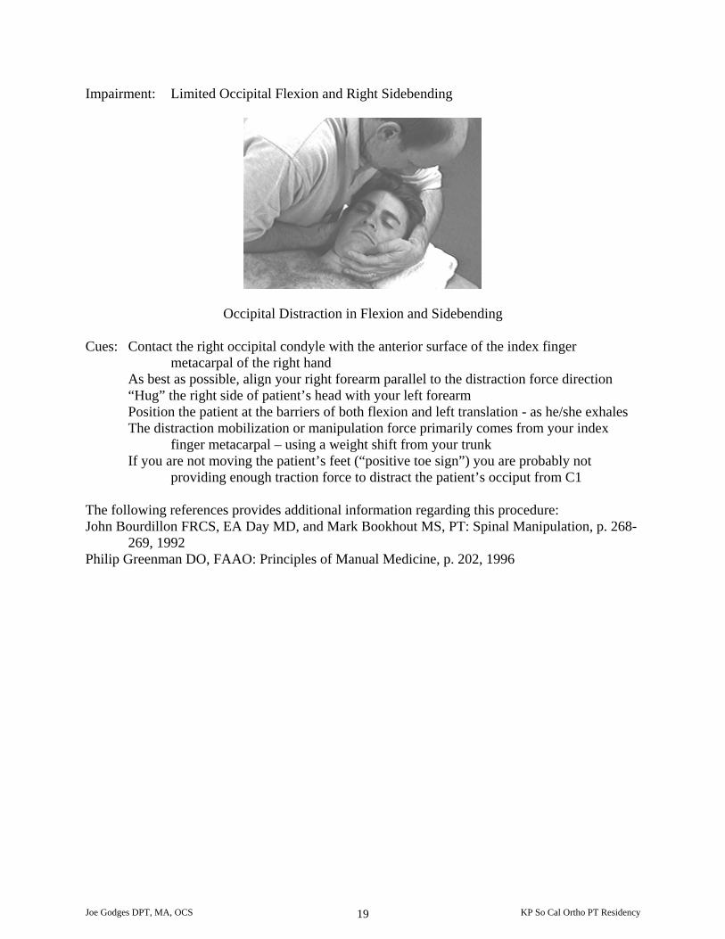
Impairment: Limited Occipital Flexion and Right Sidebending
Occipital Distraction in Flexion and Sidebending Cues: Contact the right occipital condyle with the anterior surface of the index finger
metacarpal of the right hand As best as possible, align your right forearm parallel to the distraction force direction “Hug” the right side of patient’s head with your left forearm Position the patient at the barriers of both flexion and left translation - as he/she exhales The distraction mobilization or manipulation force primarily comes from your index
finger metacarpal – using a weight shift from your trunk If you are not moving the patient’s feet (“positive toe sign”) you are probably not
providing enough traction force to distract the patient’s occiput from C1
The following references provides additional information regarding this procedure: John Bourdillon FRCS, EA Day MD, and Mark Bookhout MS, PT: Spinal Manipulation, p. 268-
269, 1992 Philip Greenman DO, FAAO: Principles of Manual Medicine, p. 202, 1996
Joe Godges DPT, MA, OCS KP So Cal Ortho PT Residency 19
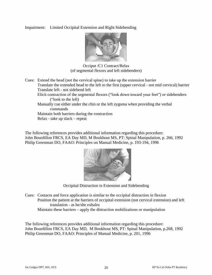
Impairment: Limited Occipital Extension and Right Sidebending
Occiput /C1 Contract/Relax (of segmental flexors and left sidebenders)
Cues: Extend the head (not the cervical spine) to take up the extension barrier
Translate the extended head to the left to the first (upper cervical - not mid cervical) barrier Translate left - not sidebend left Elicit contraction of the segmental flexors (“look down toward your feet”) or sidebenders
(“look to the left) Manually cue either under the chin or the left zygoma when providing the verbal
commands Maintain both barriers during the contraction Relax - take up slack – repeat
The following references provides additional information regarding this procedure: John Bourdillon FRCS, EA Day MD, M Bookhout MS, PT: Spinal Manipulation, p. 266, 1992 Philip Greenman DO, FAAO: Principles on Manual Medicine, p. 193-194, 1996
Occipital Distraction in Extension and Sidebending
Cues: Contacts and force application is similar to the occipital distraction in flexion Position the patient at the barriers of occipital extension (not cervical extension) and left
translation - as he/she exhales Maintain these barriers – apply the distraction mobilizations or manipulation
The following references provides additional information regarding this procedure: John Bourdillon FRCS, EA Day MD, M Bookhout MS, PT: Spinal Manipulation, p.268, 1992 Philip Greenman DO, FAAO: Principles of Manual Medicine, p. 201, 1996
Joe Godges DPT, MA, OCS KP So Cal Ortho PT Residency 20