Embed Size (px)
Citation preview
1
Articles
Cervical Dystonia Mimics: A Case Series and Review of the Literature
Srinivas Raju1, Amogh Ravi2 & LK Prashanth3,4*
1Department of Neurology, Vydehi Institute of Medical Science & Research Centre, Bangalore, IN, 2Stepping Hill Hospital, Stockport NHS Foundation Trust, Stockport, England, UK, 3Center for Parkinson’s Disease and Movement Disorders Clinic,
Vikram Hospitals, Bangalore, IN, 4Parkinson’s Disease and Movement Disorders Clinic, Bangalore, IN
IntroductionThe 2013 expert consensus panel by Albanese et al. defined dystonia as
“a movement disorder characterized by sustained or intermittent muscle contractions causing abnormal, often repetitive, movements, postures, or both. Dystonic movements are typically patterned, twisting, and may be tremulous. Dystonia is often initiated or worsened by voluntary action and associated with overflow muscle activation.”1 The goal of the revised defi-nition was to exclude possible conditions that may mimic dystonias, also known as “pseudodystonias.” These pseudodystonias are thought to be caused by etiologies that are presumed to be different from the broader dystonia group.1 It is critical to diagnose these pseudodystonias, as the
therapy and prognostication is significantly different from that of typical idiopathic dystonia. In this article, we review our cases of cervical pseu-dodystonias along with a review of the published literature.
MethodologyThe study involved retrospective analysis of subjects attending a
movement disorders clinic over a period of 7 years (January 2012 to December 2018). Clinical records of subjects who were diagnosed to have cervical pseudodystonias based on clinical and imaging findings were included in the study. Patients who had pseudodystonia involving other body parts/segments were excluded.
Columbia University Libraries
Freely available online
Tremor and Other Hyperkinetic Movementshttp://www.tremorjournal.org
AbstractBackground: Cervical dystonia is mostly idiopathic in nature. However, a small subset of cases are mimics, leading to diagnostic pitfalls. There is paucity of
literature on pseudodystonias affecting the cervical region.
Method: We performed a retrospective review of patients attending a movement disorders clinic over a period of 7 years (2012–2018). Among them, those who
were considered to have mimics of cervical dystonia based upon clinical and supportive investigations were included.
Results: Six out of 2,412 patients (0.24%) were diagnosed as cervical dystonia mimics and the causes included isolated neck extensor myopathy (2), craniovertebral
junction anomalies (2), sternocleidomastoid fibrosis (1) and post traumatic sequelae (1). Among these patients, three patients had received various treatments for
cervical dystonia, including botulinum toxin injections.
Discussion: Mimics of isolated cervical dystonia are rare. A high degree of suspicion and proper diligent clinical assessment assists management and
prognostication.
Keywords: Cervical dystonia, pseudodystonia, dystonia mimics, head drop, neck extensor myopathy
Citation: Raju S, Ravi A, Prashanth LK. Cervical dystonia mimics: A case series and review of the literature. Tremor Other Hyperkinet Mov. 2019; 9. doi: 10.7916/tohm.v0.707
*To whom correspondence should be addressed. E-mail: [email protected]
Editor: Elan D. Louis, Yale University, USA
Received: June 25, 2019; Accepted: November 4, 2019; Published: December 4, 2019
Copyright: © 2019 Raju et al. This is an open-access article distributed under the terms of the Creative Commons Attribution–Noncommercial–No Derivatives License, which permits
the user to copy, distribute, and transmit the work provided that the original authors and source are credited; that no commercial use is made of the work; and that the work is not altered
or transformed.
Funding: None.
Financial Disclosures: None.
Conflicts of Interest: The authors report no conflicts of interest.
Ethics Statement: All patients that appear on video have provided written informed consent; authorization for the videotaping and publication of the videotape was provided.
Raju S, Ravi A and Prashanth LK Cervical Pseudodystonias
Columbia University LibrariesTremor and Other Hyperkinetic Movementshttp://www.tremorjournal.org 2
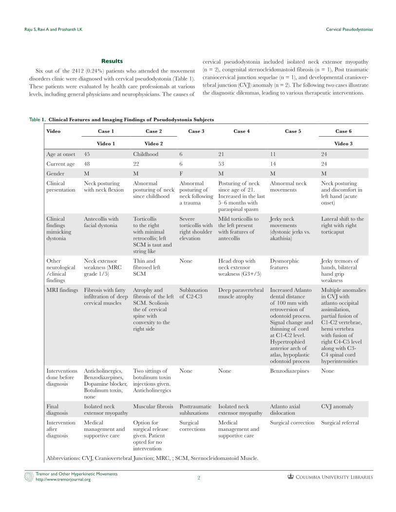
ResultsSix out of the 2412 (0.24%) patients who attended the movement
disorders clinic were diagnosed with cervical pseudodystonia (Table 1). These patients were evaluated by health care professionals at various levels, including general physicians and neurophysicians. The causes of
cervical pseudodystonia included isolated neck extensor myopathy (n = 2), congenital sternocleidomastoid fibrosis (n = 1), Post traumatic craniocervical junction sequelae (n = 1), and developmental craniover-tebral junction (CVJ) anomaly (n = 2). The following two cases illustrate the diagnostic dilemmas, leading to various therapeutic interventions.
Table 1. Clinical Features and Imaging Findings of Pseudodystonia Subjects
Video Case 1 Case 2 Case 3 Case 4 Case 5 Case 6
Video 1 Video 2 Video 3
Age at onset 45 Childhood 6 21 11 24
Current age 48 22 6 53 14 24
Gender M M F M M M
Clinical presentation
Neck posturing with neck flexion
Abnormal posturing of neck since childhood
Abnormal posturing of neck following a trauma
Posturing of neck since age of 21. Increased in the last 5–6 months with paraspinal spasm
Abnormal neck movements
Neck posturing and discomfort in left hand (acute onset)
Clinical findings mimicking dystonia
Antecollis with facial dystonia
Torticollis to the right with minimal retrocollis; left SCM is taut and string like
Severe torticollis with right shoulder elevation
Mild torticollis to the left present with features of antecollis
Jerky neck movements (dystonic jerks vs. akathisia)
Lateral shift to the right with right torticaput
Other neurological /clinical findings
Neck extensor weakness (MRC grade 1/5)
Thin and fibrosed left SCM
None Head drop with neck extensor weakness (G3+/5)
Dysmorphic features
Jerky tremors of hands, bilateral hand grip weakness
MRI findings Fibrosis with fatty infiltration of deep cervical muscles
Atrophy and fibrosis of the left SCM. Scoliosis the of cervical spine with convexity to the right side
Subluxation of C2-C3
Deep paravertebral muscle atrophy
Increased Atlanto dental distance of 100 mm with retroversion of odontoid process. Signal change and thinning of cord at C1-C2 level. Hypertrophied anterior arch of atlas, hypoplastic odontoid process
Multiple anomalies in CVJ with atlanto occipital assimilation, partial fusion of C1-C2 vertebrae, hemi vertebra with fusion of right C4-C5 level along with C3-C4 spinal cord hyperintensities
Interventions done before diagnosis
Anticholinergics, Benzodiazepines, Dopamine blocker, Botulinum toxin, none
Two sittings of botulinum toxin injections given. Anticholinergics
None None Benzodiazepines None
Final diagnosis
Isolated neck extensor myopathy
Muscular fibrosis Posttraumatic subluxations
Isolated neck extensor myopathy
Atlanto axial dislocation
CVJ anomaly
Intervention after diagnosis
Medical management and supportive care
Option for surgical release given. Patient opted for no intervention
Surgical corrections
Medical management and supportive care
Surgical correction Surgical referral
Abbreviations: CVJ, Craniovertebral Junction; MRC, ; SCM, Sternocleidomastoid Muscle.
Raju S, Ravi A and Prashanth LK Cervical Pseudodystonias
Columbia University LibrariesTremor and Other Hyperkinetic Movementshttp://www.tremorjournal.org 3
Video 1. Video Shows Neck Antecollis along with Lower Facial Movements. In the second part of the video, clinical examination shows difficulty in neck extension movements against gravity.
Case 1
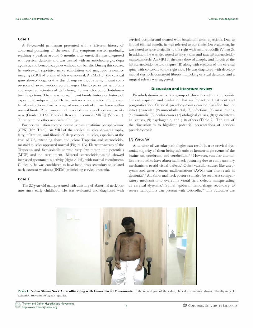
A 48-year-old gentleman presented with a 2.5-year history of abnormal posturing of the neck. The symptoms started gradually, reaching a peak at around 5 months after onset. He was diagnosed with cervical dystonia and was treated with an anticholinergic, dopa agonists, and benzodiazepines without any benefit. During this course, he underwent repetitive nerve stimulation and magnetic resonance imaging (MRI) of brain, which was normal. An MRI of the cervical spine showed degenerative disc changes without any significant com-pression of nerve roots or cord changes. Due to persistent symptoms and impaired activities of daily living, he was referred for botulinum toxin injections. There was no significant family history or history of exposure to antipsychotics. He had anterocollis and intermittent lower facial contractions. Passive range of movements of the neck was within normal limits. Power assessment revealed severe neck extensor weak-ness (Grade 0–1/5 Medical Research Council (MRC)) (Video 1). There were no other associated findings.
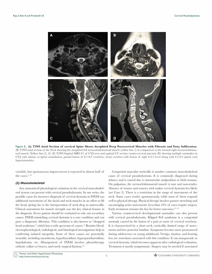
Further evaluation showed normal serum creatinine phosphokinase (CPK) (162 IU/dl). An MRI of the cervical muscles showed atrophy, fatty infiltration, and fibrosis of deep cervical muscles, especially at the level of C2, extending above and below. Trapezius and sternocleido-mastoid muscles appeared normal (Figure 1A). Electromyogram of the Trapezius and Semispinalis showed very few motor unit potentials (MUP) and no recruitment. Bilateral sternocleidomastoid showed increased spontaneous activity (right > left), with normal recruitment. Clinically, he was considered to have head drop secondary to isolated neck extensor weakness (INEM), mimicking cervical dystonia.
Case 2
The 22-year-old man presented with a history of abnormal neck pos-ture since early childhood. He was evaluated and diagnosed with
cervical dystonia and treated with botulinum toxin injections. Due to limited clinical benefit, he was referred to our clinic. On evaluation, he was noted to have torticollis to the right with mild retrocollis (Video 2). In addition, he was also noted to have a thin and taut left sternocleido-mastoid muscle. An MRI of the neck showed atrophy and fibrosis of the left sternocleidomastoid (Figure 1B) along with scoliosis of the cervical spine with convexity to the right side. He was diagnosed with develop-mental sternocleidomastoid fibrosis mimicking cervical dystonia, and a surgical release was suggested.
Discussion and literature reviewPseudodystonias are a rare group of disorders where appropriate
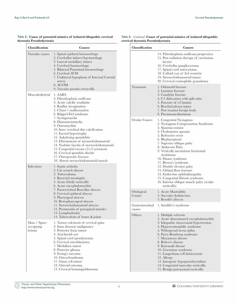
clinical suspicion and evaluation has an impact on treatment and prognostication. Cervical pseudodystonias can be classified further into (1) vascular, (2) musculoskeletal, (3) infections, (4) mass lesions, (5) traumatic, (6) ocular causes (7) otological causes, (8) gastrointesti-nal causes, (9) psychogenic, and (10) others (Table 2). The aim of the discussion is to highlight potential presentations of cervical pseudodystonia.
(1) Vascular
A number of vascular pathologies can result in true cervical dys-tonia, majority of them being ischemic or hemorrhagic events of the brainstem, cerebrum, and cerebellum.2–5 However, vascular anoma-lies are noted to have abnormal neck posturing due to compensatory mechanisms to aid visual defects.6 Other vascular causes like aneu-rysms and arteriovenous malformations (AVM) can also result in dystonia.6–9 An abnormal neck posture can also be seen as a compen-satory mechanism to overcome visual field defects masquerading as cervical dystonia.6 Spinal epidural hemorrhage secondary to severe hemophilia can present with torticollis.10 The outcomes are
Raju S, Ravi A and Prashanth LK Cervical Pseudodystonias
Columbia University LibrariesTremor and Other Hyperkinetic Movementshttp://www.tremorjournal.org 4
variable, but spontaneous improvement is reported in almost half of the cases.2–10
(2) Musculoskeletal
Any anatomical/physiological variations in the cervical musculoskel-etal system can present with cervical pseudodystonia. In our series, the possible cause for incorrect diagnosis of cervical dystonia in INEM was additional movements of the facial and neck muscles in an effort to lift the head, giving rise to the interpretation of neck drop as anterocollis. Clinical assessment for muscle strength was the key clinical feature in the diagnosis. Every patient should be evaluated to rule out secondary causes. INEM mimicking cervical dystonia is a rare condition and can pose a diagnostic dilemma. This condition is also known as “dropped head syndrome,” which has a wide spectrum of causes.11 Routine blood, electrophysiological, radiological, and histological investigations help in confirming isolated myopathy. Some of these causes are potentially treatable, including myasthenia, hypothyroidism, hyperparathyroidism, hypokalemia, etc. Management of INEM involves physiotherapy, orthotic collars or braces, and rarely surgical fixation.12
Congenital muscular torticollis is another common musculoskeletal cause of cervical pseudodystonia. It is commonly diagnosed during infancy and is caused due to intrauterine malposition or birth trauma. On palpation, the sternocleidomastoid muscle is taut and non-tender. Absence of tremor and sensory trick makes cervical dystonia less likely (see Case 2). There is a restriction in the range of movements of the neck. Some cases resolve spontaneously, while most of them respond well to physical therapy. Physical therapy involves passive stretching and encouraging active movements. Less than 10% of cases require surgery. Early treatment remains the key for better outcomes.13–15
Various craniocervical developmental anomalies can also present with cervical pseudodystonia. Klippel–Feil syndrome is a congenital anomaly caused by the fusion of a pair or more of cervical vertebrae. It is characterized by a short neck, torticollis, limited range of move-ments and low posterior hairline. Symptoms become more pronounced during adolescence or young adulthood. Vertigo, tinnitus, and hearing loss are sometimes associated with this condition. It can masquerade as cervical dystonia, which becomes apparent after radiological evaluation. Treatment is mostly symptomatic. Surgery may be needed if associated
Figure 1. (A) T2WI Axial Section of cervical Spine Shows Atrophied Deep Paracervical Muscles with Fibrosis and Fatty Infiltration. (B) T2WI axial section of the Neck showing the atrophied left sternocleidomastoid muscle (yellow line-1) in comparison to the normal right sternocleidomas-toid muscle (Yellow line-2). (C, D) T2WI Sagittal MRI (C) of CVJ area and sagittal CT section cranio-cervical junction (D) showing multiple anomalies in CVJ with atlanto occipital assimilation, partial fusion of C1-C2 vertebrae, hemi vertebra with fusion of right C4-5 level along with C3-C4 spinal cord hyperintensities.
Raju S, Ravi A and Prashanth LK Cervical Pseudodystonias
Columbia University LibrariesTremor and Other Hyperkinetic Movementshttp://www.tremorjournal.org 5
with scoliosis, radiculopathy, or myelopathy. Pseudodystonic posture secondary to Klippel–feil syndrome and diastometamyelia has been reported.16 Other CVJ anomalies like Chiari malformation and basilar invagination are known to cause torticollis.17–18 Syringomyelia associated with torticollis is a rare presentation. It requires a high degree of suspi-cion when head tilt is seen along with sensory loss. Dystonic movements of the rest of the body can also be seen occasionally. An MRI of the spine can confirm the diagnosis. Decompression of the syrinx relieves the symptoms.19
Acute, painful torticollis in children with a history of fall or trauma points toward atlantoaxial rotatory subluxation (AARS).20–22 It can also be due to a local inflammatory process, or connective tissue disorders.
A rare case of AARS was seen associated with familial Mediterranean fever (FMF) presenting with torticollis.23 FMF is an autosomal reces-sive autoinflammatory condition, presenting with episodes of fever and serositis. Cervical dystonia in ankylosing spondylitis resulting in AARS and craniocervical osseous fusion (CCOF) has been reported.24 If there is no history of trauma, it is important to look for a history of recent ENT surgeries or any inflammatory conditions of the neck, as it could be Grisel’s syndrome, which is a non-traumatic atlantoaxial sub-luxation resulting from an ongoing local inflammatory process.25–27 Grisel’s syndrome is more commonly seen in children due to laxity of ligaments along with the tenderness of the spinous process, and there may be unilateral occipital pain. A spinal cord injury should be
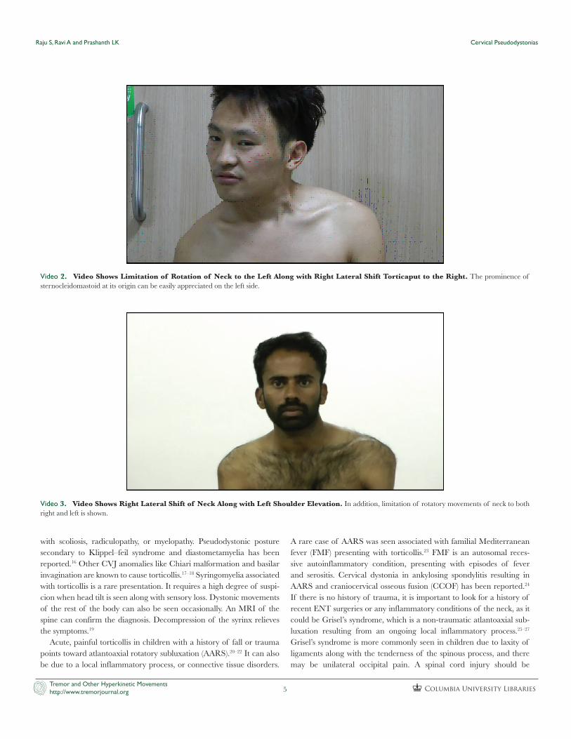
Video 3. Video Shows Right Lateral Shift of Neck Along with Left Shoulder Elevation. In addition, limitation of rotatory movements of neck to both right and left is shown.
Video 2. Video Shows Limitation of Rotation of Neck to the Left Along with Right Lateral Shift Torticaput to the Right. The prominence of sternocleidomastoid at its origin can be easily appreciated on the left side.
Raju S, Ravi A and Prashanth LK Cervical Pseudodystonias
Columbia University LibrariesTremor and Other Hyperkinetic Movementshttp://www.tremorjournal.org 6
Table 2. (Continued ) Cause of potential mimics of isolated idiopathic cervical dystonia Pseudodystonia
Table 2. Cause of potential mimics of isolated idiopathic cervical dystonia Pseudodystonia
Classification Causes
Vascular causes 1. Spinal epidural haemorrhage2. Cerebellar infarct/haemorrhage3. Lateral medullary infarct4. Cerebral haemorrhage5. Bilateral Putaminal haemorrhage6. Cerebral AVM7. Unilateral hypoplasia of Internal Carotid
artery8. ACOM9. Vascular pseudo retrocollis
Musculoskeletal 1. AARS
2. Fibrodysplasia ossificans3. Acute calcific tendinitis4. Basillar invagination5. Chiari 1 malformation6. Klippel-Fiel syndrome7. Syringomyelia8. Diastometomyelia9. Osteomyelitis10. Inter vertebral disc calcification11. Facetal hypertrophy12. Ankylosing spondylitis13. Fibromatosis of sternocleidomastoid14. Nodular fascitis of sternocleidomastoid15. Congenital oseous c2-c3 synostosis16. Cervical spondylo discitis17. Osteoporotic fracture18. Absent sternocleidomastoid muscle
Infections 1. Septic arthritis2. Cat scratch disease3. Tuberculoma4. Bacterial meningitis5. Acute febrile torticollis6. Acute encephalomyelitis7. Paravertebral Brucellar abscess8. Cervical epidural abscess9. Pharyngeal abscess10. Retropharyngeal abscess11. Sternocleidomastoid abscess12. Pyomyositis of paraspinal muscles13. Lymphadenitis14. Tuberculosis of bones & joints
Mass / Space occupying lesions
1. Tumor calcinosis of cervical spine2. Intra thoracic malignancy3. Posterior fossa tumor4. Arachnoid cyst5. Spinal cord ependymoma6. Cervical osteoblastoma7. Medullary tumor8. Posterior glioma9. Ewing’s sarcoma10. Osteochondroma11. Giant cell tumor12. Osteoid osteoma13. Cervical hemangioblastoma
Classification Causes
14. Fibrodysplasia ossificans progressiva15. Post radiation therapy of carcinoma
larynx16. Cerebellar gangliocytoma17. Spinal cord astrocytoma18. Colloid cyst of 3rd ventricle19. Sternocleidomastoid tumor20. Cervical eosinophilic granuloma
Traumatic 1. Odontoid fracture2. Laminar fracture3. Condylar fracture4. C1 dislocation with split atlas5. Fracture of c2 lamina6. Brachial plexus injury7. Post trauma foreign body8. Pneumomediastinum
Ocular Causes 1. Congenital Nystagmus2. Nystagmus Compensation Syndrome3. Spasmus nutans4. Oculomotor apraxia5. Refractive error6. Blepharoptosis7. Superior oblique palsy8. Abducens Palsy9. Vertically incomitant horizontal
strabismus10. Duane syndrome11. Brown’s syndrome12. Double elevator palsy13. Orbital floor fracture14. Endocrine ophthalmopathy15. Congenital fibrosis syndrome16. Inferior oblique muscle palsy (ocular
torticollis)
Otological Causes
1. Acute Mastoiditis2. Saccular dysfunction3. Bezold’s abscess
Gastrointestinal causes
1. Sandifer’s syndrome
Others 1. Multiple sclerosis2. Acute disseminated encephalomyelitis3. Idiopathic intracranial hypertension4. Hypereosinophilic syndrome5. Widespread nevus spilus6. Parry-Romberg syndrome7. Moyamoya disease8. Behcet’s disease9. Kawasaki disease10. Goeminne syndrome11. Langerhans cell histiocytosis12. Allergy13. Iatrogenic hypoparathyroidism14. Congenital muscular torticollis15. Benign paroxysmal torticollis
Raju S, Ravi A and Prashanth LK Cervical Pseudodystonias
Columbia University LibrariesTremor and Other Hyperkinetic Movementshttp://www.tremorjournal.org 7
suspected if there are associated neurological symptoms such as unsteady gait or hyperreflexia.25 Early diagnosis and management is the key. It is usually treated by manipulation under anesthesia and immobi-lization. For less severe cases, cervical soft collar with rest and analgesia may be sufficient. Delay in the diagnosis can lead to neurological com-plications and require surgery.28–30
Other causes like intervertebral disc calcification, acute calcific tendi-nitis of longus colli, facetal hypertrophy, and nodular fasciitis of sterno-cleidomastoid should be considered in the differential diagnosis of musculoskeletal causes of pseudodystonia.31–34
(3) Infections
Infections of the upper respiratory tract or soft tissues of the neck can cause torticollis to mimic cervical dystonia. These include cervical adeni-tis, lymphadenitis, retropharyngeal abscess, and sternocleidomastoid myo-sitis.35,36 Retropharyngeal abscess is a serious condition presenting with severe pain, fever, and difficulty in breathing and swallowing along with torticollis. It requires immediate treatment with antibiotics, non-steroidal anti-inflammatory drugs (NSAIDs), and sometimes surgical drainage of the abscess. Similarly, paravertebral brucellar abscess and sternocleido-mastoid abscess are documented to present as cervical pseudodystonia.37
Other reported causes of atypical infections presenting with cervical pseudodystonias include cat scratch disease, encephalomyeli-tis, bacterial meningitis, neuroborreliosis, tuberculoma, tuberculosis of the bone and joints, septic arthritis, spondylodiscitis, and osteomyelitis.38–46
(4) Mass lesions/space occupying lesions
Central nervous system lesions can also cause cervical pseudodysto-nia, along with symptoms like nausea, vomiting, headache, ataxia, visual disturbances, and cranial nerve deficits. These lesions include posterior fossa and infratentorial tumors (more commonly in the cerebellum, third ventricle, and brainstem) and spinal cord tumors like astrocytomas, medulloblastomas, and ependymomas.7,35,47,48 Cervical osteoblastoma, hemangioblastoma, osteochondroma, osteoid osteoma, fibrodysplasia ossificans progressiva, giant cell tumor, and Ewings sarcoma can all pres-ent with torticollis.7,49–54 Torticollis in children requires brain and spinal
cord imaging to avoid delay in diagnosis, which can be life threatening. Surgical removal of the tumor in most cases results in resolution of the symptoms.
Cervical pseudodystonia has been described in carcinoma larynx (following radiation therapy) due to fibrosis of neck muscles and in a Pancoast tumor of the lung due to possible segmental demyelination of the 11th cranial nerve.55,56
(5) Traumatic
Trauma resulting in odontoid fracture, laminar fracture, condylar fracture, osteoporotic fracture, C1 dislocation with split atlas due to var-ious causes can all present as dystonia mimics.57–61 Appropriate manage-ment will alleviate symptoms.
Other traumatic conditions presenting with a similar picture include brachial plexus injury, pneumomediastinum, foreign body, and rarely electrical injury.62–65
(6) Ocular causes
Ocular causes like congenital nystagmus, nystagmus compensation (blockage) syndrome, spasmus nutans, oculomotor apraxia, refractive error, blepharoptosis, superior oblique palsy, abducens palsy, vertically incomitant horizontal strabismus, Duane syndrome, Brown’s syn-drome, double elevator palsy, orbital floor fracture, endocrine ophthal-mopathy, congenital fibrosis syndrome, and inferior oblique muscle palsy may present with torticollis called ocular torticollis. Abnormal head position is assumed in order to maintain binocularity and/or to optimize visual acuity. Bielschowsky head tilt test is the primary test in the ocular torticollis workup. Treatment is usually surgical and depends on the underlying cause.66
(7) Otological causes
Cervical pseudodystonias secondary to vestibular dysfunctions Contribute to otological causes. These patients have ataxia, vertigo, or nystagmus along with torticollis. Acute mastoiditis, saccular dysfunc-tion, and Bezold’s abscess are few examples. This requires further assess-ment of the vestibular system and management.67–69
(8) Gastrointestinal causes
In a child presenting with torticollis or side-to-side head movements associated with vomiting, regurgitation, or epigastric pain, Sandifer’s syndrome can be suspected. The symptoms can be intermittent and are associated with meals in most cases. The symptoms are probably due to the patient assuming a position to minimize the painful acid reflux. As the early symptoms resemble dystonia, evaluation is focused on neuro-logical etiology, which is usually normal. Diagnosis is by monitoring esophageal pH and demonstrating reflux. Medical treatment for gastro-esophageal reflux usually resolves the symptoms, but sometimes this condition requires surgery.70,71
(9) Psychogenic dystonias
Psychogenic dystonia, also known as functional dystonia, is a contro-versial diagnosis commonly associated with psychiatric comorbidities
Table 2. (Continued ) Cause of potential mimics of isolated idiopathic cervical dystonia Pseudodystonia
Classification Causes
16. Grisel’s syndrome17. Complication of ventriculo-peritoneal
shunt18. Familial Mediterranean fever19. Foreign body
Psychogenic
Abbreviations: AVM - arteriovenous malformation, ACOM - anterior
communicating artery aneurysm, AARS - Atlanto Axial Rotatory
Subluxation.
Raju S, Ravi A and Prashanth LK Cervical Pseudodystonias
Columbia University LibrariesTremor and Other Hyperkinetic Movementshttp://www.tremorjournal.org 8
like anxiety, depression, and personality disorders. Variability of symp-toms suggests a psychogenic cause. The current classification has sug-gested categorization of psychogenic dystonias as one of the acquired dystonias.1 Although prognosis can be poor with long-term disability, mainstay of management involves communicating the diagnosis with the patient, physiotherapy, and behavioral therapy.72,73
(10) Others
There are various other causes of cervical pseudodystonias, which are either rarely reported or documented as single association. Among these, benign paroxysmal torticollis (BPT) of infancy is a self-limiting condition, characterized by periods of unusual, sustained posture of the head and neck, during which the head tilts to one side. Episodes are often accompanied by marked autonomic features, irritability, ataxia, apathy, and drowsiness. They last several hours to a few days and often recur every few weeks. They subside within the pre-school years. It is essential to recognize this condition and to reassure parents of its benign course and not to be misdiagnosed for other disorders, such as epileptic seizures. BPT of infancy has been linked to CACNA1A mutations and are likely to be associated with familial
hemiplegic migraine, episodic ataxia, and paroxysmal tonic upgaze.74,75 There is no approved medication for the disease. Some studies have demonstrated the uses of cyproheptadine and topiramate for BPT.76
Other rare neurological causes presenting as cervical pseudodys-tonia include idiopathic intracranial hypertension, acute demyelin-ating encephalomyelitis, multiple sclerosis, Guillain–Barre syndrome, hypereosinophilic syndrome, widespread nevus spilus, Parry–Romberg syndrome, moyamoya disease, atypical Kawasaki disease, Behcet’s disease, Langerhans cell histiocytosis, Goldenhar syndrome, Goeminne syndrome, iatrogenic hypoparathyroidism, and allergy.77–92 These conditions have been reported in isolated case reports.
ConclusionIn any case of presumed cervical dystonia, a diligent clinical evalua-
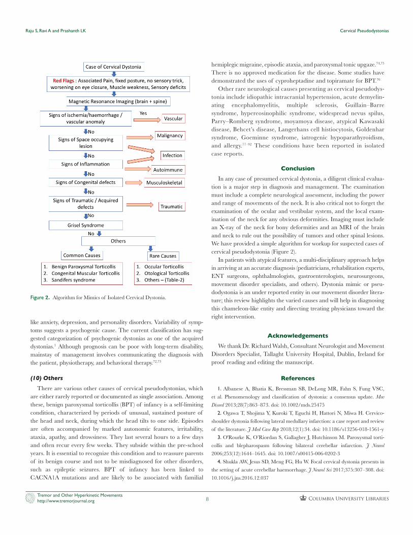
tion is a major step in diagnosis and management. The examination must include a complete neurological assessment, including the power and range of movements of the neck. It is also critical not to forget the examination of the ocular and vestibular system, and the local exam-ination of the neck for any obvious deformities. Imaging must include an X-ray of the neck for bony deformities and an MRI of the brain and neck to rule out the possibility of tumors and other spinal lesions. We have provided a simple algorithm for workup for suspected cases of cervical pseudodystonia (Figure 2).
In patients with atypical features, a multi-disciplinary approach helps in arriving at an accurate diagnosis (pediatricians, rehabilitation experts, ENT surgeons, ophthalmologists, gastroenterologists, neurosurgeons, movement disorder specialists, and others). Dystonia mimic or pseu-dodystonia is an under reported entity in our movement disorder litera-ture; this review highlights the varied causes and will help in diagnosing this chameleon-like entity and directing treating physicians toward the right intervention.
AcknowledgementsWe thank Dr. Richard Walsh, Consultant Neurologist and Movement
Disorders Specialist, Tallaght University Hospital, Dublin, Ireland for proof reading and editing the manuscript.
References1. Albanese A, Bhatia K, Bressman SB, DeLong MR, Fahn S, Fung VSC,
et al. Phenomenology and classification of dystonia: a consensus update. Mov
Disord 2013;28(7):863–873. doi: 10.1002/mds.25475
2. Ogawa T, Shojima Y, Kuroki T, Eguchi H, Hattori N, Miwa H. Cervico-
shoulder dystonia following lateral medullary infarction: a case report and review
of the literature. J Med Case Rep 2018;12(1):34. doi: 10.1186/s13256-018-1561-y
3. O'Rourke K, O’Riordan S, Gallagher J, Hutchinson M. Paroxysmal torti-
collis and blepharospasm following bilateral cerebellar infarction. J Neurol
2006;253(12):1644–1645. doi: 10.1007/s00415-006-0202-3
4. Shukla AW, Jesus SD, Meng FG, Hu W. Focal cervical dystonia presents in
the setting of acute cerebellar haemorrhage. J Neurol Sci 2017;375:307–308. doi:
10.1016/j.jns.2016.12.037
Figure 2. Algorithm for Mimics of Isolated Cervical Dystonia.
Raju S, Ravi A and Prashanth LK Cervical Pseudodystonias
Columbia University LibrariesTremor and Other Hyperkinetic Movementshttp://www.tremorjournal.org 9
5. Usmani N, Bedi GS, Sengun C, Pandey A, Singer C. Late onset of cervical
dystonia in a 39-year-old patient following cerebellar haemorrhage. J Neurol
2011;258:149–151. doi: 10.1007/s00415-010-5685-2
6. Giordano A, Tedeschi G, Tessitore A. Vascular-induced compensatory
pseudo-retrocollis. Neurology 2015;84(19):2005–2006. doi: 10.1212/WNL.000
0000000001568
7. Per H, Canpolat M, Tumturk A, Gumus H, Gokoglu A, Yikilmaz A, et al.
Different etiologies of acquired torticollis in childhood. Childs Nerv Syst 2014;
30(3):431–440. doi: 10.1007/s00381-013-2302-6
8. Bayrakci B, Aysun S, Firat M. Arteriovenous fistula: a cause of torticollis.
Pediatr Neurol 1999;20(2):146–147. doi: 10.1016/s0887-8994(98)00130-1
9. Lobo-Antunes J, Yahr MD, Hilal SK. Extrapyramidal dysfunction with
cerebral arteriovenous malformations. J Neurol Neurosurg Psychiatry 1974;37(3):
259–268. doi: 10.1136/jnnp.37.3.259
10. Agha BS, Tehan C, Perno J. Torticollis: not always the usual suspects.
Pediatr Emerg Care 2011;27:32–33. doi: 10.1097/PEC.0b013e3182045c2b
11. Martin AR, Reddy R, Fehlings MG. Dropped head syndrome: diagnosis
and management. Evid Based Spine Care J 2011;2(2):41–47. doi: 10.1055/s-
0030-1267104
12. Umapathi T, Chaudhry V, Cornblath D, Drachman D, Griffin J, Kuncl R.
Head drop and camptocormia. J Neurol Neurosurg Psychiatry 2002;73(1):1–7. doi:
10.1136/jnnp.73.1.1
13. Bredenkamp J, Hoover L, Berke G, Shaw A. Congenital muscular torticol-
lis: a spectrum of disease. Arch Otolaryngol Head Neck Surg 1990;116(2):212–216.
doi: 10.1001/archotol.1990.01870020088024
14. Cheng J, Tang S, Chen T. Sternocleidomastoid pseudotumor and congen-
ital muscular torticollis in infants: a prospective study of 510 cases. J Pediatr
1999;134(6):712–716. doi: 10.1016/s0022-3476(99)70286-6
15. Kuo A, Tritasavit S, Graham J. Congenital muscular torticollis and posi-
tional plagiocephaly. Pediatr Rev 2014;35(2):79–87. doi: 10.1542/pir.35-2-79
16. Lopez-Vicchi M, Da Prat G, Gatto EM. Pseudodystonic posture second-
ary to Klippel-Feil syndrome and diastematomyelia. Tremor Other Hyperkinet Mov
2015;5:325. doi: 10.7916/D8ZC820C
17. Alexiou GA, Prodromou N. Torticollis as an initial sign of Chiari I malfor-
mation. Pediatr Emerg Care 2009;25(3):215. doi: 10.1097/PEC.0b013e31819a8bd3
18. Souza PV, Pinto WB, Oliveira AS. Basilar invagination in headache asso-
ciated with physical exertion and recurrent torticollis. Arq Neuropsiquiatr
2014;72(11):902–903. doi: 10.1590/0004-282x20140163
19. Silva-Júnior FP, Santos JG, Sekeff-Sallem FA, Lucato LT, Barbosa ER.
Cervical and axial dystonia in a patient with syringomyelia. Arq Neuropsiquiatr
2012;70(9):742–743. doi: 10.1590/S0004-282X2012000900018
20. Hussain K, Abdo MM, AlNajjar FJ, Abbo M. Not your typical torticollis: a
case of atlantoaxial rotatory subluxation. BMJ Case Rep 2014;2014:bcr2013201023.
doi: 10.1136/bcr-2013-201023
21. Bagouri E, Deshmukh S, Lakshmanan P. Atlantoaxial rotatory subluxation
as a cause of torticollis in a 5-year-old girl. BMJ Case Rep 2014;2014:bcr2013202990.
doi: 10.1136/bcr-2013-202990
22. Sundseth J, Berg-Johnsen J, Skaar-Holme S, Züchner M, Kolstad F.
Atlantoaxial rotatory fixation-a cause of torticollis. Tidsskr Nor Legeforen 2013;133:
519. doi: 10.4045/tidsskr.11.1540
23. Gencpinar P, Bozkurt O, Karaali K, Gemici A, Kazan S, Haspolat S.
A rare coincidence of torticollis in Familial Mediterranean Fever: atlanto-axial
rotatory subluxation. Clin Neurol Neurosurg 2014;127:158–160. doi: 10.1016/j.
clineuro.2014.10.010
24. Weigel RM, Capelle HH, Krauss JK. Cervical dystonia in Bechterew dis-
ease resulting in atlantoaxial rotatory subluxation and cranio-cervical osseous
fusion. Spine 2007;32(25):E781–E784. doi: 10.1097/BRS.0b013e31815b7e35
25. Allegrini D, Autelitano A, Nocerino E, Fogagnolo P, De Cillà S, Rossetti L.
Grisel’s syndrome, a rare cause of anomalous head posture in children: a case
report. BMC Ophthalmol 2016;16:21. doi: 10.1186/s12886-016-0197-1
26. Deichmueller C, Welkoborsky HJ. Grisel’s syndrome—a rare complication
following “small” operations and infections in the ENT region. Eur Arch
Otorhinolaryngol 2010;267:1467–1473. doi: 10.1007/s00405-010-1241-z
27. Elyajouri A, Assermouh A, Abilkacem R, Agadr A, Mahraoui C. Grisel’s
syndrome: a rare complication following traditional uvulectomy. Pan Afr Med J
2015;20:62. doi: 10.11604/pamj.2015.20.62.5930
28. Martins J, Almeida S, Nunes P, Prata F, Lobo ML, Marques JG. Grisel
syndrome, acute otitis media, and temporo-mandibular reactive arthritis: a rare
association. Int J Pediatr Otorhinolaryngol 2015;79(8):1370–1373. doi: 10.1016/j.
ijporl.2015.05.030
29. Ortiz GL, Pratts I, Ramos E. Grisel’s syndrome: an unusual cause of tor-
ticollis. J Pediatr Rehabil Med 2013;6(3):175–180. doi: 10.3233/PRM-130253
30. Osiro S, Tiwari KJ, Matusz P, Gielecki J, Tubbs RS, Loukas M. Grisel’s
syndrome: a comprehensive review with focus on pathogenesis, natural history,
and current treatment options. Childs Nerv Syst 2012;28:821–825. doi: 10.1007/
s00381-012-1706-z
31. Kouamo EI, Nour M, Gennar JM, Guillaume JM, Choufani E, Merrot T,
et al. Case study of two children with intervertebral disc calcifications. Pan Afr
Med J 2016;25:34. doi: 10.11604/pamj.2016.25.34.10543
32. Cuevas Y, Schonhaut L, Espinoza A, Schonstedt V, Aird A, Castoldi F.
Pediatric intervertebral disc calcification: a rare cause of acquired torticollis.
Case report. Rev Chil Pediatr 2015;86(3):200–205. doi: 10.1016/j.rchipe. 2015.
03.002
33. Zibis AH, Giannis D, Malizos KN, Kitsioulis P, Arvanitis DL. Acute cal-
cific tendinitis of the longus colli muscle: case report and review of the literature.
Eur Spine J 2013;22(3):S434–S438. doi: 10.1007/s00586-012-2584-5
34. Hemmi S, Murakami T, Shirabe T, Sunada Y. A patient wsith muscular
torticollis caused by nodular fasciitis in the sternocleidomastoid muscle (SCM).
Rinsho Shinkeigaku 2002;42(9):864–867. PMID: 12710086.
35. Tomczak KK, Rosman NP. Torticollis. J Child Neurol 2013;28(3):365–367.
doi: 10.1177/0883073812469294
36. Lehtinen P, Ruuskanen O, Sonninen P. An unusual life threatening
cause of torticollis in a child. Arch Dis Child 2003;88(4):349. doi: 10.1136/
adc.88.4.349
37. Simsek S, Yigitkauli K, Kazanci AS, Beleu D, Bavbek M. Medically
treated paravertebral Brucella abscess presenting with acute torticollis: case
report. Surg Neurol 2007;67(2):207–210. doi: 10.1016/j.surneu.2006.06.061
38. Rafferty JR, Janopaul-Naylor E, Riese J. Torticollis and fever in a young
boy: a unique presentation of cat-scratch Disease with vertebral osteomyelitis
and epidural phlegmon. Pediatr Emerg Care 2017;33(12):e164–e166. doi: 10.1097/
PEC.0000000000001330
39. Mukherjee S, Sharief N. Bacterial meningitis presenting as acute
torticollis. Acta Paediatr 2004;93(7):1005–1006. doi: 10.1111/j.1651- 2227.2004.
tb02705.x
Raju S, Ravi A and Prashanth LK Cervical Pseudodystonias
Columbia University LibrariesTremor and Other Hyperkinetic Movementshttp://www.tremorjournal.org 10
40. van Breemen MS, van der Kuip M, Ang CW, van Furth AM, Wolf NI.
Torticollis and seizures due to neuroborreliosis in a child. Ned Tijdschr Geneeskd
2012;156(51):A5157. PMID: 23249509.
41. Tey HL, Seet RC, Lim EC. Tuberculomas causing cervical dystonia. Intern
Med J 2005;35(4):261–262. doi: 10.1111/j.1445-5994.2004.00793.x
42. Warpus JM. Tuberculosis of bones and joints. Torticollis. Foot disorders.
ONA J 1978;5(3):77–83. PMID: 246509.
43. Filleron A, L’Kaissi M, Cottalorda J, Jeziorski E, Rodiere M, Prodhomme
O, et al. Torticollis in children: a challenging diagnosis of C1-C2 septic arthritis.
Clin Pediatr 2016;55(5):459–462. doi: 10.1177/0009922815591888
44. Pizzol A, Bramuzzo M, Pillon R, Taddio A, Barbi E. Torticollis as the
presenting sign of cervical spondylodiscitis. Pediatr Emerg Care 2016;32(12):863–
864. doi: 10.1097/PEC.0000000000000643
45. McKnight P, Friedman J. Torticollis due to cervical epidural abscess and
osteomyelitis. Neurology 1992;42(3 Pt 1):696–697. doi: 10.1212/wnl.42.3.696-a
46. Dimaala J, Chaljub G, Oto A, Swischuk L. Odontoid osteomyelitis mas-
querading as a C2 fracture in an 18-month-old male with torticollis: CT and MRI
features. Emerg Radiol 2006;12(5):234–236. doi: 10.1007/s10140-006-0479-7
47. Krauss JK, Seeger W, Jankovic J Krauss JK, Seeger W, Jankovic J. Cervical
dystonia associated with tumors of the posterior fossa. Mov Disord 1997;12(3):
443–447. doi: 10.1002/mds.870120329
48. Debenedictis CN, Allen JC, Kodsi SR. Brainstem tumor presenting with
tearing, photophobia, and torticollis. J AAPOS 2010;14(4):369–370. doi:
10.1016/j.jaapos.2010.04.013
49. Patier de La Peña JL, Norman F, Rodríguez-Ramírez GI, Echániz-
Quintana A, Moreno-Cobo MA. Cervical pain, torticollis and polyglobulia as a
first manifestation of a cerebellar hemangioblastoma. Rev Clin Esp 2010;210(6):
e21–e23. doi: 10.1016/j.rce.2010.03.001
50. Akhaddar A, Boucetta M. Solitary osteochondroma of the cervical spine
presenting as recurrent torticollis. Pan Afr Med J 2014;17:271. doi: 10.11604/
pamj.2014.17.271.3977
51. Amirjamshidi A, Roozbeh H, Sharifi G, Abdoli A, Abbassioun K. Osteoid
osteoma of the first 2 cervical vertebrae. Report of 4 cases. J Neurosurg Spine
2010;13(6):707–714. doi: 10.3171/2010.5.SPINE09297
52. Moore RE, Dormans JP, Drummond DS, Shore EM, Kaplan FS,
Auerbach JD. Chin-on-chest deformity in patients with fibrodysplasia ossificans
progressive. A case series. J Bone Joint Surg Am 2009;91(6):1497–1502. doi:
10.2106/JBJS.H.00554
53. Karampalis C, Lenthall R, Boszczyk B. Solid variant of aneurysmal bone
cyst on the cervical spine of a child: case report, differential diagnosis and treat-
ment rationale. Eur Spine J 2013;22:523–531. doi: 10.1007/s00586-012-2548-9
54. Aydin R, Bilgici MC, Dagcinar A. A very rare cause of neck paIn: primary
Ewing sarcoma of the axis. Pediatr Emerg Care 2013;29(11):1197–1200. doi:
10.1097/PEC.0b013e3182aa11cf
55. Astudillo L, Hollington L, Game X, Benyoucef A, Boladeras AM, Delisle
MB, et al. Cervical dystonia mimicking dropped-head syndrome after radiother-
apy for laryngeal carcinoma. Clin Neurol Neurosurg 2003;106(1):41–43. doi:
10.1016/s0303-8467(03)00044-1
56. Landan I, Cullis PA. Torticollis following radiation therapy. Mov Disord
1987;2(4):317–319. doi: 10.1002/mds.870020410
57. Tsuji S, Inoue S, Tachibana T, Maruo K, Arizumi F, Yoshiya S. Post-
traumatic torticollis due to odontoid fracture in a patient with diffuse idiopathic
skeletal hyperostosis: a case report. Medicine 2015;94(36):e1478. doi: 10.1097/
MD.0000000000001478
58. Balpande DN, Agrawal A, Bhatlawande VK. Torticollis as a manifestation
of laminar fracture in an infant. J Pak Med Assoc 2008;58(10):591. PMID:
18998320.
59. Chou WC, Huang WC, Shih YH, Lee LS, Wu C, Cheng H. Occult occip-
ital condyle fracture with normal neurological function and torticollis. J Clin
Neurosci 2008;15(8)920–922. doi: 10.1016/j.jocn.2007.03.014
60. Ostrowski C, Ronan L, Sheridan R, Pearce V. An osteoporotic fracture
mimicking cervical dystonia in idiopathic Parkinson’s disease. Age Ageing
2013;42(5):658–659. doi: 10.1093/ageing/aft050
61. Tachibana A, Imabayashi H, Yato Y, Nakamichi K, Asazuma T, Nemoto
K. Torticollis of a specific C1 dislocation with split atlas. Spine 2010;35(14):E672
–E675. doi: 10.1097/BRS.0b013e3181dfcacb
62. Hervey-Jumper S, Justice D, Vanaman M, Nelson V, Yang L. Torticollis
associated with neonatal brachial plexus palsy. Pediatr Neurol 2011;45(5):305–310.
doi: 10.1016/j.pediatrneurol.2011.08.013
63. Dekel B, Paret G, Vardi A, Katz M, Barzilay Z. Torticollis: an usual pre-
sentation of spontaneous pneumomediastinum. Pediatr Emerg Care 1996;12(5):
352–353. PMID: 8897543.
64. Walton J, Darr A, George A. An unusual case of an oesophageal foreign
body presenting as torticollis. Ann R Coll Surg Engl 2016;98(3):e40–e42. doi:
10.1308/rcsann.2016.0076
65. Boonkongchuen P, Lees A. Case of torticollis occurring following electrical
injury. Mov Disord 1996;11(1):109–110. doi: 10.1002/mds.870110126
66. Rubin SE, Wagner RS. Ocular torticollis. Surv Ophthalmol 1986;30(6):366–
376. doi: 10.1016/0039-6257(86)90090-1
67. Bredenkamp JK, Maceri DR. Inflammatory torticollis in children. Arch
Otolaryngol Head Neck Surg 1990;116(3):310–313. doi: 10.1001/archotol.1990.018
70030074012
68. Hallberg A, Standring RT, Ahsan S. Congenital torticollis and saccular
dysfunction. JAMA Otolaryngol Head Neck Surg 2013;139(6):639–642. doi: 10.1001/
jamaoto.2013.3283
69. McMullan B. Bezold’s abscess: a serious complication of otitis media.
J Paediatr Child Health 2009;45:616–618. doi: 10.1111/j.1440-1754.2009.
01575.x.
70. Lehwald N, Krausch M, Franke C, Assmann B, Adam R, Knoefel W.
Sandifer syndrome – a multidisciplinary diagnostic and therapeutic challenge.
Eur J Pediatr Surg 2007;17(3):203–206. doi: 10.1055/s-2007-965145
71. Wasserman JK, Jimenez-Rivera C, Doja A. Refractory head movements
secondary to Sandifer syndrome treated with enteral feeding. Mov Disord
2010;25(11):1754–1755. doi: 10.1002/mds.23161
72. Schmerler DA, Espay AJ. Functional dystonia. Handb Clin Neurol
2016;139:235–245. doi: 10.1016/B978-0-12-801772-2.00020-5
73. Hallett M. Functional (psychogenic) movement disorders – clinical presen-
tations. Parkinsonism Relat Disord 2016;22(1):S149–S152. doi: 10.1016/j.
parkreldis.2015.08.036
74. Giffin NJ, Benton S, Goadsby PJ. Benign paroxysmal torticollis of infancy:
four new cases and linkage to CACNA1A mutation. Dev Med Child Neurol
2002;44(7):490–493. doi: 10.1017/S0012162201002407
75. Shin M, Douglass LM, Milunsky JM, Rosman NPJ. The genetics of
benign paroxysmal torticollis of infancy: is there an association with mutations in
Raju S, Ravi A and Prashanth LK Cervical Pseudodystonias
Columbia University LibrariesTremor and Other Hyperkinetic Movementshttp://www.tremorjournal.org 11
the CACNA1A gene? Child Neurol 2016;31(8):1057–1061. doi: 10.1177/
0883073816636226
76. Hadjipanayis A, Efstathiou E, Neubauer D. Benign paroxysmal torticollis
of infancy: an underdiagnosed condition. J Paediatr Child Health 2015;51(7):
674–678. doi: 10.1111/jpc.12841
77. Saifudheen K. Idiopathic intracranial hypertension presenting as
stiff neck and torticollis. Neurol India 2010;58(6):982. doi: 10.4103/0028-
3886.73765
78. Har-Gil M, Evrani M, Watemberg N. Torticollis as the only manifestation
of acute disseminated encephalomyelitis. J Child Neurol 2010;25(11):1415–1418.
doi: 10.1177/0883073810368995
79. Rüegg SJ, Bühlmann M, Renaud S, Steck AJ, Kappos L, Fuhr P. Cervical
dystonia as first manifestation of multiple sclerosis. J Neurol 2004;251(11):
1408–1410. doi: 10.1007/s00415-004-0544-7
80. Berger B, Rijntjes M, Stehlin L, Stich O. Corticosteroid-responsive
torticollis as a presenting symptom of multiple sclerosis. J Neurol Sci 2014;
340(1–2):239–240. doi: 10.1016/j.jns.2014.02.021
81. Planas AM, Occhiuzzo FP, Oferill JC, Marfa MP, Cubells CL. Torticollis
and spinal hyperextension. Unusual presentation of Guillain-Barré syndrome.
An Pediatr 2008;69(4):394–395. doi: 10.1157/13126575
82. Das JK, Gupta K, Deshmukh S, Shrivastava R. A rare case of hypereosin-
ophilic syndrome presenting with unilateral proptosis and torticollis. Indian J
Ophthalmol 2018;66(10):1508–1511. doi: 10.4103/ijo.IJO_316_18
83. Marti N, Jorda E, Martinez E, Gamez L, Ramon MD. Widespread nevus
spilus associated with torsion dystonia: a case report. Pediatr Dermatol 2010;
27(6):654–656. doi: 10.1111/j.1525-1470.2010.01325.x
84. Panda AK, Gopinath G, Singh S. Parry-Romberg syndrome with hemi-
masticatory spasm in pregnancy; A dystonia mimic. J Neurosci Rural Pract
2014;5(2):184–186. doi: 10.4103/0976-3147.131675
85. Yasutomo K, Hashimoto T, Miyazaki M, Kuroda Y. Recurrent Torticollis
As a presentation of Moyamoya disease. J Child Neurol 1993;8(2):187–188. doi:
10.1177/088307389300800215
86. Runel-Belliard C, Lasserre S, Quinet B, Grimprel E. Febrile torticollis: an
atypical presentation of Kawasaki disease. Arch Pédiatr 2009;16(2):115–117. doi:
10.1016/j.arcped.2008.11.015
87. Pellecchia MT, Cuomo T, Striano S, Filla A, Barone P. Paroxysmal
dystonia in Behçet’s disease. Mov Disord 1999;14(1):177–179. doi: 10.1002/
1531-8257(199901)14:1<177::aid-mds1037>3.0.co;2-n
88. Kostaridou S, Anastasopoulos J, Veliotis C, Nikas J, Stefanaki K,
Tzortzatou-Stathopoulou F. Recurrent torticollis secondary to Langerhans cell
histiocytosis: a case report. Acta Orthop Belg 2005;71(1):102–106. PMID: 15792216.
89. Al Kaissi A, Ben Chehida F, Ganger R, Klaushofer K, Grill F. Distinctive
spine abnormalities in patients with Goldenhar syndrome: tomographic assess-
ment. Eur Spine J 2015;24(3):594–599. doi: 10.1007/s00586-014-3204-3
90. Goeminne L. A New probably X-linked inherited syndrome: congenital
muscular torticollis, multiple keloids cryptorchidism and renal dysplasia. Acta
Genet Med Gemellol 1968;17(3):439–467. doi: 10.1017/s1120962300012634
91. Mancini F, Zangaglia R, Cristina S, Tassorelli C, Uggetti C, Nappi G, Pacchetti
C. Secondary cervical dystonia in iatrogenic hypoparathyroidism associated with
extensive brain calcifications. Funct Neurol 2006;21(3):165–166. PMID: 17049136.
92. Randolph TG. Allergy as a cause of Acute Torticollis. Am Pract Dig Treat
1950;1(10):1062–1067. PMID: 14771412.

![Pseudodystonia: a new perspective on an old phenomenon...axial weakness [4]. Acquired or congenital atlanto-axial displacements such as Klippel-Feil syndrome may mimic cervical dystonia](https://img.pdfslide.us/doc/110x75/60f85b05eb25954c136dc676/pseudodystonia-a-new-perspective-on-an-old-phenomenon-axial-weakness-4-acquired.jpg)