Embed Size (px)
Citation preview
1
Certification in Congenital Heart Disease Echocardiography
Syllabus
European Association of CardioVascular Imaging (EACVI))
Association for European Paediatric Cardiology (AEPC) Working Group on Grown-up Congenital Heart Disease of ESC (WG 22)
Introduction:
This syllabus is a guide for candidates wishing to pursue certification in congenital heart echocardiography. The candidate is expected to know and be skilled in basic principles of cardiac ultrasound. Additionally, they should know and understand the range of congenital heart defects in children and adults, and acquired heart defects in childhood. Although not all candidates will have the opportunity to train or work in cardiac surgical centres, they should be competent in the echocardiographic evaluation of unoperated congenital heart disease, the assessment of patients for surgical or catheter treatment, and post-procedural review. Intraoperative and cardiac catheter guidance for interventions is more advanced and the candidate is not expected to have experience of this, although an understanding of this is encouraged.
2
Part I. General Concepts ..................................................................................................................................... 4
The clinical role of echocardiography and Doppler in paediatric and congenital heart disease ............................ 4
Service provision ...................................................................................................................................................... 4
Relationship with patients ....................................................................................................................................... 4
Conscious sedation in children ................................................................................................................................ 4
Reporting and documentation of paediatric and congenital studies ...................................................................... 4
Safety of ultrasound ................................................................................................................................................ 4
Part II. Imaging physics & instrumentation in paediatric and congenital echocardiography .. 5
Concepts and terminology of cardiac ultrasound ................................................................................................... 5
Propagation of ultrasound through tissues ............................................................................................................. 5
Ultrasound Transducers .......................................................................................................................................... 5
Imaging physics ....................................................................................................................................................... 5
Echo Instrumentation .............................................................................................................................................. 5
Optimising Images ................................................................................................................................................... 6
Part III. Doppler physics & fluid dynamics in paediatric and congenital ......................................... 6
echocardiography ................................................................................................................................................ 6
Basic Fluid Dynamics ............................................................................................................................................... 6
Basic Principles of Doppler ...................................................................................................................................... 6
Spectral Doppler ...................................................................................................................................................... 6
Colour Flow Instrumentation ................................................................................................................................... 7
Measurements and calculations ............................................................................................................................. 7
Tissue Doppler Imaging (TDI) and Deformation ...................................................................................................... 7
Part IV. Cardiac anatomy and physiology for paediatric and adult congenital
echocardiography anatomy of the thorax ................................................................................................... 8
General thoracic anatomy ....................................................................................................................................... 8
General concepts of cardiac morphology and echo identification .......................................................................... 8
Terminology of congenital heart disease ................................................................................................................ 9
The cardiac cycle ..................................................................................................................................................... 9
Fetal and neonatal physiology ................................................................................................................................ 9
The physiology of congenital heart disease ............................................................................................................ 9
Part V. The different congenital heart defects and treatment for the paediatric and
congenital echocardiographer ..................................................................................................................... 10
A) Septation defects .................................................................................................................................................. 10
Ventricular septal defect (VSD) ............................................................................................................................. 10
Atrial septal defect (ASD) ...................................................................................................................................... 10
Atrioventricular septal defect (AVSD).................................................................................................................... 10
B. Shunt lesions not caused by septation defects ..................................................................................................... 10
Arterial duct ........................................................................................................................................................... 10
Partial anomalous pulmonary venous drainage (PAPVD) ..................................................................................... 11
Basic anatomy and echocardiographic features of other acyanotic shunts ......................................................... 11
C. Cyanotic congenital heart defects......................................................................................................................... 11
Ventriculoarterial discordance (transposition of the great arteries) .................................................................... 11
3
Tetralogy of Fallot (TOF)/pulmonary atresia with ventricular septal defect (PA/VSD) ............................................. 11
Pulmonary atresia with intact septum (PA/IVS) .................................................................................................... 11
Total anomalous pulmonary venous drainage (TAPVD) ....................................................................................... 12
D. Other complex lesions .......................................................................................................................................... 12
Common arterial trunk (CAT) ................................................................................................................................ 12
Lesions with single ventricle physiology ................................................................................................................ 12
Atrioventricular and ventriculoarterial discordance (AV/VA discordance) ........................................................... 13
Double outlet right ventricle (DORV) ..................................................................................................................... 13
E. Congenital valve disease ....................................................................................................................................... 13
The mitral valve ..................................................................................................................................................... 13
The aortic valve ..................................................................................................................................................... 13
The pulmonary valve ............................................................................................................................................. 14
The tricuspid valve (including Ebstein’s Malformation) ........................................................................................ 14
Prosthetic Valves ................................................................................................................................................... 14
F. Left outflow obstruction ........................................................................................................................................ 15
Subvalvular/supravalvar aortic stenosis ............................................................................................................... 15
Coarctation of the aorta ........................................................................................................................................ 15
Interrupted aortic arch .......................................................................................................................................... 15
G. Right outflow obstruction ..................................................................................................................................... 15
Subvalvular/supravalvar stenosis/peripheral branch stenosis.............................................................................. 15
H. Congenital coronary anomalies ............................................................................................................................ 16
Anomalous origin of the left coronary artery ........................................................................................................ 16
Coronary fistulae ................................................................................................................................................... 16
I. Miscellaneous lesions ............................................................................................................................................. 16
Cortriatriatum dexter and sinister ......................................................................................................................... 16
Intracardiac Masses .............................................................................................................................................. 16
Diseases of the aorta ............................................................................................................................................. 16
Pericardial Disease ................................................................................................................................................ 16
Part VI. Acquired heart disease in paediatrics and congenital heart disease ............................. 17
Kawasaki disease................................................................................................................................................... 17
Infective Endocarditis ............................................................................................................................................ 17
Rheumatic fever .................................................................................................................................................... 17
Intravascular thrombosis....................................................................................................................................... 17
Part VII. Cardiac functional evaluation in paediatric and adult congenital heart disease ...... 17
A. Ventricular Function ............................................................................................................................................. 17
Calculations ........................................................................................................................................................... 17
Diastolic function ................................................................................................................................................... 17
B. Pulmonary hypertension ....................................................................................................................................... 18
Pulmonary Hypertension ....................................................................................................................................... 18
C. Conditions associated with ventricular dysfunction ............................................................................................. 18
Dilated Cardiomyopathy ....................................................................................................................................... 18
Hypertrophic Cardiomyopathy .............................................................................................................................. 18
Restrictive Cardiomyopathy .................................................................................................................................. 18

4
PART I. GENERAL CONCEPTS The clinical role of echocardiography and Doppler in paediatric and congenital heart disease
Indications for echocardiography
Information that echocardiography can and cannot provide
‘Ruling out’ pathology (sensitivity, specificity & Baye’s theorem)
Likelihood of findings influencing patient management
Role of multimodality imaging: o Cardiac catheterisation o Multislice CT scan o Magnetic resonance imaging o Nuclear Cardiology
Service provision
Advantages and disadvantages of physiologist-led versus physician-led service
Specific requirements for paediatric and congenital echocardiography laboratories
Provision and indication for specialised techniques, e.g. Transoesphageal echocardiography
Availability and access
Controlling workload
Training & motivation of staff
Audit, Quality Control, Clinical Governance Relationship with patients
Explaining the procedure in terms relevant to the patient/parents
Respect for patients’ dignity and cultural backgrounds
Relationships with patient, parents and colleagues
Handling requests for information about the study findings Conscious sedation in children
Explaining the procedure in terms relevant to the patient/parents
Specific environment for performing studies in children/adults with CHD
Indications for conscious sedation
Precautions, dosage, follow-up Reporting and documentation of paediatric and congenital studies
Standard methods and terminology used for describing congenital heart disease (segmental sequential analysis)
Distinction between technical and clinical reports
Responsibility for reporting
Medico-legal considerations (Data Protection Act) Safety of ultrasound
Potential hazardous biological effects o Heating, resonance and cavitation effects
Measurement of beam intensity (Spatial Peak Temporal Average, [SPTA])
Practical precautions: power levels, use of colour and continuous wave Doppler
5
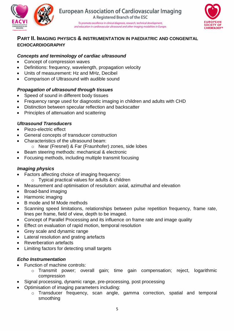
PART II. IMAGING PHYSICS & INSTRUMENTATION IN PAEDIATRIC AND CONGENITAL
ECHOCARDIOGRAPHY Concepts and terminology of cardiac ultrasound
Concept of compression waves
Definitions: frequency, wavelength, propagation velocity
Units of measurement: Hz and MHz, Decibel
Comparison of Ultrasound with audible sound Propagation of ultrasound through tissues
Speed of sound in different body tissues
Frequency range used for diagnostic imaging in children and adults with CHD
Distinction between specular reflection and backscatter
Principles of attenuation and scattering Ultrasound Transducers
Piezo-electric effect
General concepts of transducer construction
Characteristics of the ultrasound beam: o Near (Fresnel) & Far (Fraunhofer) zones, side lobes
Beam steering methods: mechanical & electronic
Focusing methods, including multiple transmit focusing Imaging physics
Factors affecting choice of imaging frequency: o Typical practical values for adults & children
Measurement and optimisation of resolution: axial, azimuthal and elevation
Broad-band imaging
Harmonic imaging
B mode and M Mode methods
Scanning speed limitations, relationships between pulse repetition frequency, frame rate, lines per frame, field of view, depth to be imaged.
Concept of Parallel Processing and its influence on frame rate and image quality
Effect on evaluation of rapid motion, temporal resolution
Grey scale and dynamic range
Lateral resolution and grating artefacts
Reverberation artefacts
Limiting factors for detecting small targets Echo Instrumentation
Function of machine controls: o Transmit power; overall gain; time gain compensation; reject, logarithmic
compression
Signal processing, dynamic range, pre-processing, post processing
Optimisation of imaging parameters including: o Transducer frequency, scan angle, gamma correction, spatial and temporal
smoothing
6
Optimising Images
Use of gel
Positioning of the subject
Standard views: o Parasternal, apical (4, 5 and 2-chamber), subcostal, suprasternal, right parasternal,
long and short axis
Use of non-standard views
Adapting for subjects with difficult windows
PART III. DOPPLER PHYSICS & FLUID DYNAMICS IN PAEDIATRIC AND CONGENITAL ECHOCARDIOGRAPHY Basic Fluid Dynamics
Fluid flow: significance of peak and mean velocities
Determination of volumetric flow
Continuity equation
Laminar & turbulent flow: Reynolds’ equation (qualitative)
Transition from Laminar to turbulent flow: inlet jet
Bernoulli equation Basic Principles of Doppler
Interaction of ultrasound waves with moving blood: the Doppler effect
The Doppler equation: factors influencing magnitude of Doppler shift
Spectral analysis: fast Fourier transform (qualitative)
The spectral Doppler display: determination of mean, modal and peak velocities
Limitation of CW Doppler caused by lack of depth discrimination
The effect of beam angle errors on Doppler velocities
Aliasing: o How it is caused and how it manifests in practice: the Nyquist limit o Influence on aliasing of: transducer frequency; sample depth (range x velocity
product); and beam angle
High pulse repetition frequency (extended range) PW Doppler
Relative advantages and disadvantages of CW, PW and HPRF modes
Concept of colour flow imaging as multi-sampled PW
Velocity estimation, by moving target indication and autocorrelation (qualitative)
Limitations of mean velocity: use of velocity variance to show high velocities/turbulence
Aliasing in colour Doppler
Packet size, colour mode and sector size and their effect on frame rate and aliasing
Spectral Doppler
Duplex Doppler using imaging transducers
The ‘stand-alone’ Doppler probe (pencil probe)
Features of the spectral display: positive & negative velocities; scale & baseline controls.
Effect of high- and low-pass filter and intensity threshold (‘reject’) settings
Pulsed Doppler sample volume: influence of gate length and distance (beam width)
Representation of signal strength by image intensity
How aliasing manifests on the spectral display
7
Colour Flow Instrumentation
The colour display: Blue-Away, Red-Towards (BART) convention
Colour maps to show velocity scales
Image domination and additive colour modes
Basic principles of Tissue Doppler Imaging, including optimisation of filters for detecting tissue versus blood velocities
Difference between velocity and power (signal amplitude) displays Measurements and calculations
On-screen measurement of length, slope, area, volume and time interval, and their significance for 2-D images, M-mode and spectral Doppler displays
Standard M-mode measurements and calculations o Shortening Fraction o TAPSE o Wall thickness
Limitations of measurements and/or calculations
Peak and Mean pressure gradient measurements by Doppler and their relationship to catheterisation data
Tissue Doppler Imaging (TDI) and Deformation
Understand principles of TDI
Identify a’, e’ s’ on PW-TDI
Limitations of measurements and/or calculations
Understand age.size related differences in TDI measurements
Understand concept of Displacement imaging ▪ Strain and strain rate
Definition
Direction of deformation
Speckle Tracking Echocardiography
Principles
Physical origin of speckles
Tracking of speckle motion
Clinical applications
Haemodynamic assessment -Assessment of filling pressures
Systolic (global and regional) function / Diastolic function
Detection of subclinical myocardial dysfunction
8
PART IV. CARDIAC ANATOMY AND PHYSIOLOGY FOR PAEDIATRIC AND ADULT
CONGENITAL ECHOCARDIOGRAPHY ANATOMY OF THE THORAX General thoracic anatomy
Thorax contained by rib cage & diaphragm
Lungs & pleura
Heart & pericardium
Mediastinum
Blood vessels within the thorax General concepts of cardiac morphology and echo identification
Atrial situs: o Definition, abdominal aorta and great vein relationship
Systemic venous return: morphology
Pulmonary venous return: morphology
Atrial anatomy: o Difference between right and left atrium, atrial appendages
Ventricular anatomy: o Morphology of right and left ventricle o Atrioventricular valve arrangement o Trabecular pattern o Ventricular shape o Right-handed vs. left handed ventricular configuration o Inlet and outlet valve relationships o Chordal attachments
Atrioventricular valves: anatomy of mitral and tricuspid valve
Semilunar valves: anatomy of pulmonary and aortic valve
The inter-atrial septum: o Morphology o Primum and secundum septum o Foramen ovale o Sinus venosus
The interventricular septum: o Morphology o Inlet septum o Outlet septum o Membranous septum o Trabecular
Pulmonary artery anatomy
Aortic anatomy
Coronary artery anatomy: normal anatomy and variants
The arterial duct: normal anatomy and normal variants
The pericardium: anatomy
Visualisation of normal cardiac anatomy and normal variants in standard echocardiography planes
Normal valve function, normal Doppler parameters and normal variants
9
Terminology of congenital heart disease
Atrial situs and situs abnormalities: o Situs inversus o Right and left isomerism
Atrioventricular connections: o Concordant o Discordant o Double inlet o Absent connection o Straddling valves o Criss-cross connections
‘Univentricular’ heart: description of different variants
Ventriculoarterial connections: o Concordant, o Discordant o Single outlet o Double outlet
Great artery relationships The cardiac cycle
Temporal relationships of the ECG, chamber pressures and valve movements
Typical values for intracardiac pressures
Relationship of valve movements to heart sounds Fetal and neonatal physiology
The fetal circulation: how it differs from the postnatal circulation
Circulatory changes at birth: the neonatal circulation
Adaptations in circulatory physiology during the first weeks of life The physiology of congenital heart disease
Causes of chamber dilation and hypertrophy
Ventricular pressure and volume overload
Physiological effect of shunts at atrial, ventricular and great artery level
Physiological effect of regurgitation through all four valves
Physiological effect of stenosis on all four valves
10
PART V. THE DIFFERENT CONGENITAL HEART DEFECTS AND TREATMENT FOR THE
PAEDIATRIC AND CONGENITAL ECHOCARDIOGRAPHER A) Septation defects Ventricular septal defect (VSD)
Assessment of unoperated VSD: o Location and size o M-mode and 2D features o Assessment of shunt size and pulmonary pressure o Anterior or posterior malalignment
Associated features: o Aortic valve cusp prolapse o Discrete subvalvular aortic stenosis o Double chambered right ventricle
Echocardiographic assessment of surgical treatment for VSD: o Post-operative complications: e.g. residual defects, sub-aortic stenosis
Echocardiographic assessment of percutaneous treatment for different types of VSD. Atrial septal defect (ASD)
Assessment of unoperated ASD: o Secundum, primum and sinus venosus defects o Location and size o M-mode and 2D echo features o Assessment of shunt size o Common associated lesions
Other causes of right ventricular volume overload
Echocardiographic assessment of surgical treatment of different types of ASD
Echocardiographic assessment of percutaneous treatment of ASD. Atrioventricular septal defect (AVSD)
Assessment of unoperated AVSD: o Size of atrial and ventricular components o Atrioventricular valve function o Ventricular imbalance o Assessment of pulmonary pressure
Echocardiographic assessment of post-operative AVSD: o Atrioventricular valve function o Sub-aortic stenosis o Residual defects
B. Shunt lesions not caused by septation defects Arterial duct
Assessment of patent arterial duct o Imaging planes for isolated duct o Assessment of shunt size o Ductal flow patterns and pulmonary artery pressure
11
o
Anatomical variations with different cardiac defects
Other defects causing shunt at great artery level
Echocardiographic assessment of surgical and percutaneous treatment for patent arterial duct
Partial anomalous pulmonary venous drainage (PAPVD)
Assessment of partial anomalous pulmonary venous drainage o Anatomy – which veins drain where o Associated defects o Haemodynamic effect
Echocardiographic assessment of surgical treatment for PAPVD Basic anatomy and echocardiographic features of other acyanotic shunts
Aortopulmonary window
Unroofed coronary sinus
Origin of one pulmonary artery from aorta
Sinus of Valsalva fistula C. Cyanotic congenital heart defects Ventriculoarterial discordance (transposition of the great arteries)
Assessment of unrepaired ventriculoarterial discordance: o Anatomy o Coronary artery anatomy o Associated features: VSD, pulmonary stenosis, coarctation
Echocardiographic assessment of surgical treatment for ventriculoarterial discordance o Atrial switch (Mustard, Senning) o Arterial switch o Rastelli procedure
Tetralogy of Fallot (TOF)/pulmonary atresia with ventricular septal defect (PA/VSD)
Assessment of unrepaired TOF/pulmonary atresia with VSD: o Anatomy o Assessment of VSD o Right ventricular outflow tract assessment o Coronary artery anatomy o Sources of pulmonary blood flow o Size of branch pulmonary arteries o Arch laterality
Echocardiographic assessment of surgical treatment for TOF/pulmonary atresia with VSD: o Palliations: arterial shunt, right ventricular outflow tract stent, arterial ductal stent,
pulmonary valve balloon o Valve sparingTransannular patch o Right ventricle to pulmonary artery conduit o Haemodynamic effect of residual stenosis or regurgitation
Pulmonary atresia with intact septum (PA/IVS)
Assessment of unrepaired PA/IVS o Anatomy
12
o Pulmonary valve morphology o Coronary artery anatomy/fistulae/sinusoids o Right ventricular morphology
Echocardiographic assessment of percutaneous and surgical treatments for PA/IVS: o Arterial shunt, right ventricular outflow tract stent, arterial ductal stent, radiofrequency
perforation and pulmonary valve balloon Total anomalous pulmonary venous drainage (TAPVD)
Assessment of unrepaired TAPVD o Anatomy o Location: supracardiac, cardiac, infracardiac o Size of pulmonary veins o Associated features
Echocardiographic assessment of surgical treatment for TAPVD: o Patency of confluence o Pulmonary vein stenosis o Right heart pressures
D. Other complex lesions Common arterial trunk (CAT)
Assessment of unrepaired CAT: o Anatomy o Morphology and function of truncal valve o Size and position of VSD o Committal of truncal valve to ventricle o Origin of pulmonary arteries o Coronary artery origins o Associated defects
Echocardiographic assessment of surgical treatment for CAT: o Truncal valve stenosis/regurgitation o Pulmonary artery stenosis o Right ventricle to pulmonary artery conduit function
Lesions with single ventricle physiology
Different variants: double inlet ventricles, double outlet ventricles, hypoplastic/absent atrioventricular connections etc
Anatomy: o Morphology of ventricle o Source of pulmonary blood flow o Source of systemic blood flow o Atrioventricular and ventriculoarterial valve function
Echocardiographic evaluation after staged palliation for single ventricle physiology: o Pulmonary artery banding o Norwood procedure o Arterial shunt o Bidirectional Glenn/hemi-Fontan o Fontan/total cavopulmonary connection (TCPC)
13
Echocardiographic evaluation of the Fontan/(TCPC): o Types of Fontan/TCPC (atriopulmonary, lateral tunnel, extracardiac conduit) o Obstructions to the Fontan tunnel o Thromboses o Single ventricle function o Fenestration flow
Atrioventricular and ventriculoarterial discordance (AV/VA discordance)
Assessment of unrepaired AV/VA discordance: o Ventricular morphology o Associated defects
Echocardiographic follow-up of the unoperated patient: o Function of the systemic right ventricle and tricuspid valve
Echocardiographic follow-up of the operated patient: o Double switch (atrial and arterial switch)
Double outlet right ventricle (DORV)
Assessment of the unoperated patient with DORV: o Arrangement of the great vessels (and the relevant physiology) o Size and location of the VSD o Associated lesions
Echocardiographic assessment of the surgical treatment of DORV: o Residual VSD o Left or right ventricular outflow tract obstruction
E. Congenital valve disease The mitral valve
Assessment of the congenitally abnormal mitral valve: o Anatomy and different variants of mitral valve anomalies o Nomenclature for mitral valve leaflets o Description of the valve o Description of subvalvar apparatus o Measurement of orifice area by planimetry o Assessment of severity of stenosis/regurgitation
Doppler assessment of the mitral valve: o Mean and end-diastolic gradient o Area by ‘pressure half-time’: technique and limitations
Mitral valve prolapse: definition and echocardiographic aspects
Echocardiographic assessment of surgical mitral valve repair The aortic valve
Assessment of the congenitally abnormal aortic valve: o Morphology of the aortic valve o Number of leaflets o Assessment of the left ventricle: size, hypertrophy, systolic and diastolic function o Associated left ventricular outflow tract abnormalities o Effect on the aortic root o Assessment of severity of stenosis/regurgitation
14
Doppler assessment of the aortic valve: o Peak and mean gradients o Apical, right parasternal and suprasternal positions o Continuity equation
Echocardiographic assessment of surgical and percutaneous treatments for congenital aortic valve disease
o Aortic valve balloon o Aortic valve repair o Aortic valve replacement o Ross procedure o Left ventricular remodelling
The pulmonary valve
Assessment of the congenitally abnormal pulmonary valve: o Morphology of the pulmonary valve o Number of leaflets o Assessment of the right ventricle: size, hypertrophy, systolic and diastolic function o Associated right ventricular outflow tract abnormalities o Assessment of severity of stenosis/regurgitation
Echocardiographic assessment of surgical and percutaneous treatments for congenital pulmonary valve disease
o Pulmonary valve balloon o Pulmonary valve repair o Pulmonary valve replacement o Right ventricular remodelling
The tricuspid valve (including Ebstein’s Malformation)
Assessment of the congenitally abnormal tricuspid valve: o Morphology of the tricuspid valve o Position of tricuspid valve annulus o Number of leaflets o Assessment of the right ventricle and right atrium: size, function o Associated abnormalities o Assessment of stenosis/regurgitation
Echocardiographic assessment of surgical and percutaneous treatments for congenital tricuspid valve disease
o Tricuspid valve repair o Cone repair
Prosthetic Valves
2D, M-Mode and Doppler features of the main types of replacement valves: o Ball & cage o Tilting Disc o Bi-leaflet o Stented Bioprostheses
15
Age-related deterioration of bioprostheses
Understand role of TOE in examining normal and malfunctioning prosthetic valves
Prosthetic valve stenosis o Assessment by 2D, M-mode and Doppler o Normal ranges o Use of Continuity Equation for aortic prostheses
Prosthetic valve regurgitation o Trans-versus para-valvar regurgitation o Normal versus abnormal regurgitation
Assessment by CW, PW and Colour Doppler
Colour artefacts from mechanical prostheses F. Left outflow obstruction Subvalvular/supravalvar aortic stenosis
Anatomy and variants
Associated lesions
Echocardiographic assessment of severity
Evaluation of surgical treatment Coarctation of the aorta
Type of narrowing
Site of narrowing
Morphology and anatomy of the whole arch
Associated defects
Echocardiographic assessment: o Different echocardiographic appearances o Spectral Doppler appearances o Peak velocity and diastolic pattern o Relationship to measured gradient o Relationship to severity of obstruction
Effect of arterial duct on imaging and Doppler appearances
Echocardiographic assessment of percutaneous and surgical treatment of coarctation of the aorta:
o Recoarctation o Left ventricular remodelling
Interrupted aortic arch
Site of interruption
Associated lesions
Echocardiographic assessment of surgical treatment of interrupted aortic arch G. Right outflow obstruction Subvalvular/supravalvar stenosis/peripheral branch stenosis
Anatomy and variants
Associated lesions
Echocardiographic assessment of severity
Evaluation of surgical treatment
16
H. Congenital coronary anomalies Anomalous origin of the left coronary artery
Origin of coronary arteries
Physiological effect
Assessment of surgical treatment for anomalous origin of the coronary artery Coronary fistulae
Origin of coronary arteries and drainage of fistulae
Physiological effect
Assessment of surgical treatment and percutaneous treatment for coronary fistulae I. Miscellaneous lesions Cortriatriatum dexter and sinister
Anatomy and echocardiographic appearance Intracardiac Masses
Typical locations for intracardiac tumours
Echocardiographic features intracardiac tumours
Differentiation of cardiac tumours
Features suggestive of malignancy
Understand role of TOE in assessment of intracardiac masses Diseases of the aorta
Technique for examining the ascending and descending thoracic aorta
Echocardiographic features of the normal aortic root, sinuses of Valsalva, ascending aorta and aortic arch
2-D, M-mode and Doppler features of: o Marfan’s syndrome o Sinus of Valsalva aneurysm
Pericardial Disease
Anatomy of the normal pericardium: o Relationships of serous pericardium to heart and great vessels o Transverse and oblique sinuses of the pericardium
Echocardiographic features of pericardial fluid o Location of fluid in relation to patient position and fluid volume o Differentiation from pleural effusion o Assessment of volume of pericardial fluid o Role of echocardiography in pericardiocentesis
Features of tamponade o Collapse of RA and/or RV walls o Effect on IVC o Effect on A-V valve flow velocities
Features of pericardial constriction o Effect on A-V valve flow velocities o Effect of respiration
17
o SVC/hepatic vein flow o Differentiation from restrictive cardiomyopathy
PART VI. ACQUIRED HEART DISEASE IN PAEDIATRICS AND CONGENITAL HEART DISEASE Kawasaki disease
Common echocardiographic features
Echocardiographic follow-up Infective Endocarditis
Typical echocardiographic appearance of vegetations in bacterial and fungal endocarditis
Preferred locations for vegetations o ’Jet’ lesions o Congenitally abnormal structures o Hypertrophic cardiomyopathy
Complications: abscess, fistula, perforation
Understand role of TOE in suspected endocarditis
Echocardiographic follow-up Rheumatic fever
Common echocardiographic features
Evaluation of severity
Echocardiographic follow-up Intravascular thrombosis
Diagnostic echocardiographic criteria
Echocardiographic follow-up
PART VII. CARDIAC FUNCTIONAL EVALUATION IN PAEDIATRIC AND ADULT CONGENITAL
HEART DISEASE A. Ventricular Function Calculations
Derivation of Stroke Volume, Ejection Fraction and LV Mass
Methods of measuring LV volume, including biplane area, area-length and
Simpson’s rule methods
Echocardiographic calculation of cardiac output
Diastolic function
Methods of measuring diastolic dysfunction: E/A ratio, deceleration time, pulmonary venous flow patterns
Tissue Doppler derived E/e’
18
B. Pulmonary hypertension Pulmonary Hypertension
2-D, M-mode and Doppler features of pulmonary hypertension
Causes of pulmonary hypertension: o Idiopathic o Related to congenital heart disease o Chronic pulmonary emboli o Secondary to lung diseases o Upper airway obstruction o Medication related
Measurement of pulmonary pressures from tricuspid and pulmonary regurgitant flow velocities
Assessment of inferior vena cava contraction C. Conditions associated with ventricular dysfunction Dilated Cardiomyopathy
2D, M-mode and Doppler features of dilated cardiomyopathy
Detection and assessment of associated lesions: functional valve regurgitation
Thrombus in cardiac chambers
Pericardial effusions
Associated abnormalities, e.g. mitral regurgitation
Role of echocardiography in assessment and follow-up Hypertrophic Cardiomyopathy
2D, M-mode and Doppler features of hypertrophic cardiomyopathy
Differentiation from other causes of hypertrophy, e.g. ‘athletic heart’
Techniques for measurement of left ventricular wall thickness, detection of intracavity flow acceleration
Assessment of right ventricular involvement
Associated abnormalities, e.g. mitral regurgitation
Role of echocardiography in assessment and follow-up Restrictive Cardiomyopathy
2D, M-mode and Doppler features of restrictive cardiomyopathy
Differentiation from pericardial constriction
Techniques for measurement of diastolic dysfunction
Associated abnormalities, e.g. mitral regurgitation
Role of echocardiography in assessment and follow-up