Embed Size (px)
Citation preview
ORIGINAL RESEARCHpublished: 26 September 2017doi: 10.3389/fnins.2017.00534
Frontiers in Neuroscience | www.frontiersin.org 1 September 2017 | Volume 11 | Article 534
Edited by:
Vivienne Ann Russell,
University of Cape Town, South Africa
Reviewed by:
Ravindra Kumar Garg,
King George’s Medical University,
India
Zongde Zhang,
Beijing Chest Hospital, Capital
Medical University, China
*Correspondence:
Shayne Mason
Specialty section:
This article was submitted to
Neurodegeneration,
a section of the journal
Frontiers in Neuroscience
Received: 28 July 2017
Accepted: 13 September 2017
Published: 26 September 2017
Citation:
Mason S, Reinecke CJ and
Solomons R (2017) Cerebrospinal
Fluid Amino Acid Profiling of Pediatric
Cases with Tuberculous Meningitis.
Front. Neurosci. 11:534.
doi: 10.3389/fnins.2017.00534
Cerebrospinal Fluid Amino AcidProfiling of Pediatric Cases withTuberculous Meningitis
Shayne Mason 1*, Carolus J. Reinecke 1 and Regan Solomons 2
1 Faculty of Natural Sciences, Centre for Human Metabolomics, North-West University, Potchefstroom, South Africa,2Department of Pediatrics and Child Health, Faculty of Medicine and Health Sciences, Stellenbosch University, Tygerberg,
South Africa
Background: In Africa, tuberculosis is generally regarded as persisting as one of the
most devastating infectious diseases. The pediatric population is particularly vulnerable,
with infection of the brain in the form of tuberculous meningitis (TBM) being the most
severe manifestation. TBM is often difficult to diagnose in its early stages because of
its non-specific clinical presentation. Of particular concern is that late diagnosis, and
subsequent delayed treatment, leads to high risk of long-term neurological sequelae,
and even death. Using advanced technology and scientific expertise, we are intent on
further describing the biochemistry behind this devastating neuroinflammatory disease,
with the goal of improving upon its early diagnosis.
Method: We used the highly sensitive analytical platform of gas chromatography-mass
spectrometry (GC-MS) to analyze amino acid profiles of cerebrospinal fluid (CSF)
collected from a cohort of 33 South African pediatric TBM cases, compared to 34
controls.
Results: Through the use of a stringent quality assurance procedure and various
statistical techniques, we were able to confidently identify five amino acids as being
significantly elevated in TBM cases, namely, alanine, asparagine, glycine, lysine,
and proline. We found also in an earlier untargeted metabolomics investigation that
alanine can be attributed to increased CSF lactate levels, and lysine as a marker of
lipid peroxidation. Alanine, like glycine, is an inhibitory neurotransmitter in the brain.
Asparagine, as with proline, is linked to the glutamate-glutamine cycle. Asparagine is
associated with the removal of increased nitrites in the brain, whereas elevated proline
coincides with the classic biochemical marker of increased CSF protein in TBM. All five
discriminatory amino acids are linked to ammonia due to increased nitrites in TBM.
Conclusion: A large amount of untapped biochemical information is present in CSF of
TBM cases, of which amino acid profiling through GC-MS has potential in aiding in earlier
diagnosis, and hence crucial earlier treatment.
Keywords: gas chromatography-mass spectrometry (GC-MS), tuberculous meningitis (TBM), pediatric,
cerebrospinal fluid (CSF), amino acid profiling
Mason et al. CSF Amino Acids of Tuberculous Meningitis
INTRODUCTION
Tuberculosis (TB), caused byMycobacterium tuberculosis (Mtb),is an ancient, persistent disease that remains a huge, deadly issueto this day. According to the World Health Organization (WHO,2016), during 2015 there were an estimated 10.4 million new(incident) TB cases worldwide, of which 5.9 million were men,3.5 million were women, and 1 million were of children. Sixcountries accounted for 60% of the new cases: India, Indonesia,China, Nigeria, Pakistan, and South Africa. An estimated 1.8million people died from TB in 2015, with the mortality ofcohorts of more than 100 individuals with extreme drug-resistant(XDR) TB being highest (>40%) in India and South Africa. Theincidence of TB in South Africa (population 55 million) for 2015was estimated to be 454,000 (294,000–649,000) individuals, ofwhom 33,000 (21,000–44,000) were children (<14 years of age).Hence, South Africa is one of the worst-stricken regions underthe scourge of the M. tuberculosis bacillus, which depends onhumans for its transmission.
TB is most commonly known in its pulmonary form;however, Mtb is not only localized in the lungs but, becauseof the systematic spread of the tubercule bacilli, can lead toextrapulmonary forms. A preferred site for Mtb—with highblood flow and oxygen content—is the brain; the pathogenis capable of crossing the blood-brain barrier and enteringthe meninges, which consist of three membranes betweenthe skull and the brain. Infection within the meninges byMtb leads to tuberculous meningitis (TBM)—the most severemanifestation of TB. The pathology that occurs in the centralnervous system (CNS) is similar to that of its pulmonarytype, in that lesions (turberculomas) form and can rupture—releasing inflammatory markers. CNS-TB represents up to anestimated 10% of all forms of extra-pulmonary TB (and 1%of total TB) cases (Rock et al., 2005; Bhigjee et al., 2007;Cherian and Thomas, 2011), of which TBM is the principalmanifestation.
The gold standard for diagnosis of TBM requires collection ofcerebrospinal fluid (CSF), typically through a lumbar puncture.In 2010, Marais et al. proposed a uniform research casedefinition of TBM based on CSF: (1) TBM could be classifiedas “definite” when CSF demonstrated acid-fast bacilli onmicroscopy, a positive Mtb culture and/or passed a positive CSFMtb commercial nucleic acid amplification test in an individualwith symptoms or signs suggestive of the disease. (2) TBM couldbe classified as “probable” according to a scoring system basedon clinical, CSF and neuroimaging criteria, as well as evidence ofextraneural TB. In practice, however, TBM is difficult to diagnosein its early stages due to its non-specific clinical presentation. Aparticular concern is that late diagnosis, and subsequent delayedtreatment, leads to high risk of long-term neurological sequelae,and even death.
The two primary biochemical markers currently considered
for differential diagnosis of TBM are CSF protein and glucose
levels (Solomons et al., 2015). Protein levels are defined asbeing either elevated—greater than 40 mg/dl (lower cut-off), or
significantly elevated—greater than 100 mg/dl (higher cut-off)(Youssef et al., 2006; Hristea et al., 2012). Depressed glucose is
defined as <2.2 mmol/l as an absolute value, or relatively as <0.5CSF:blood glucose ratio as standardized cut-off values (Maraiset al., 2010; Solomons et al., 2015).
Our group recently conducted an untargeted proton magneticresonance (1H-NMR) metabolomics study on CSF from 17pediatric cases with TBM (Mason et al., 2015), which revealedlowered glucose (as expected) and highly elevated lactate as themost defining biochemical markers of the disease, along withperturbed amino acids. The presence of highly elevated CSFlactate was further examined (Mason et al., 2016) and shownto be produced only by the host (with zero contribution frominvading Mtb)—an interesting outcome in that the human brainmass produces lactate during a neuroinflammatory disease suchas TBM. Indeed, CSF lactate has gained much attention recentlyin neuroenergetics (Mason, 2017), perhaps as a consequenceof being an especially important biochemical marker of Mtbinfection. Interestingly, the most prominent CSF metabolitesidentified by our untargeted 1H-NMR metabolomics studyas indicators of TBM were also derived in a completelyindependent study using exactly the same NMR data set anda novel nonparametric classification system (van Reenen et al.,2016)—a method of mathematical modelling for deductiveverification of the AMLS (astrocyte–microglia lactate shuttle)hypothesis.
A secondary CSF biosignature of TBM revealed by ourprevious untargeted NMR metabolomics study consisted largelyof perturbed amino acids—alanine, branched-chain amino acids(leucine, isoleucine, and valine), and lysine. These gluconeogenicamino acids, together with ketones, were postulated to beinvolved in directing (through shuttling mechanisms) CSFlactate, produced by glycolysis in astrocytes, from neuronspreferentially into activated microglia in an attempt to aid theeradication of invadingMtb bacilli. Collectively, this biosignatureled us to postulate the astrocyte–microglia lactate shuttle. Here,we now report on a more detailed targeted metabolomics studyon the amino acid profiles in TBM from a larger collection of CSFsamples.
Perturbations in amino acids in TBM cases are not anovel discovery as they have been observed previously inbiochemical studies. In 1981, Corston et al. examined elevenviral meningitis and four TBM cases and reported that totalamino acid concentrations in CSF were markedly higher inTBM than in viral meningitis. In 1998, Qureshi et al. comparedseveral neurochemical markers in CSF between 11 cases ofviral meningitis and 12 of TBM. These authors, using high-pressure liquid chromatography, reported significantly increasedaspartic acid, glutamic acid, GABA, glycine, and tryptophan inall cases, whereas only TBM cases exhibited significantly elevatedphenylalanine, arginine, and homocysteine. From their results,Qureshi et al. postulated that inflammatory changes in meningesmay interfere with amino acid transport across the blood–brainbarrier, and proposed several therapeutic approaches. Theseearlier results on perturbed amino acids in TBM were, however,based on small sample sizes.
Here, we present the first report on a detailed aminoacid profiling study of CSF obtained from a cohort of 33TBM pediatric cases, compared to 34 controls. The method of
Frontiers in Neuroscience | www.frontiersin.org 2 September 2017 | Volume 11 | Article 534
Mason et al. CSF Amino Acids of Tuberculous Meningitis
analysis used was the sensitive platform of gas chromatography-mass spectrometry (GC-MS). Based upon stringent statisticalanalyses and quality control measures, five amino acids—alanine,asparagine, glycine, lysine, and proline—were identified as beingsignificantly increased in our group of TBM patients, of whichwe discuss the biological implications in the context of TBM.These observations justify the need for a comprehensive studyon biochemical markers for TBM to be validated in a large-scalefollow-up investigation.
METHODS
SamplingThis study was focused on a pediatric group with suspectedTBM from the region surrounding Tygerberg Hospital in theWestern Cape province of South Africa—an area endemic forTB. A specialized pediatric neurology unit within the hospitalfocuses on diagnosing and treating cases of TBM in the region.For the purposes of this study, a diagnosis of TBM was basedon the uniform research case definition of Marais et al. (2010).Only children (>3 months and <13 years of age) with “definite”and “probable” TBM were included in the experimental patientgroup (n = 33). Our controls (n = 34) were age-matchedpediatric patients suspected of meningitis, but later confirmed tobemeningitis negative. Detailed clinical information on the studycohort is included in Supplementary Information (Table S1).Informed and written consent was obtained from each patient’scaregiver and assent if the child was older than 7 years andcompetent to do so. The study was approved by the HumanResearch Ethics Committee of Stellenbosch University, SouthAfrica (study no. N11/01/006).
Gas Chromatography-Mass SpectrometryEach CSF sample was filtered to remove bacteria—to ensuresafety while handling—as well as for the removal of proteins andother macromolecules. Filtration was done using the SartoriusCentrisart R©1 10-kDa centrifugal unit by centrifugation of CSFsamples for 15 min at 3,000 rpm. Samples were prepared usingthe commercially available EZ:faastTM analysis kit (Phenomenex)for the analysis of free (physiological) amino acids by GC-MS.See SI for full details on sample preparation, as well as instrumentdetails.
In conjunction with the CSF samples, several external qualitycontrol (EQ) samples were included—commercial lyophilizedhuman serum samples spiked with known concentrations ofamino acids (Fowler et al., 2008). Calibration standard (CS)samples—commercial standard mixtures of amino acids as partof the EZ:faastTM analysis kit, were also included. The CS sampleswere used to quantify each block of samples (e.g., a calibrationcurve was created using CS2 and CS3 and this curve was used toquantify samples in block À, CS5 and CS6 were used to calibrateEQ3, and so on). Each block of samples consisted of either sevenor eight randomized experimental samples, with a total of 10blocks. The experimental run was as follows:
Blank||CS1|EQ1A|EQ1B|CS2|À|CS3|EQ2|CS4|Á|CS5|EQ3|CS6|Â|CS7|EQ4|CS8|Ã|CS9|EQ5|CS10|Ä|CS11|EQ6|CS12|Å|
CS13|EQ7|CS14|Æ|CS15|EQ8|CS16|Ç|CS17|EQ9|CS18|È|CS19|EQ10|CS20|É|CS21|EQ11|EQ1C|EQ1D|CS22||end
Data AnalysisThe GC-MS data were deconvoluted, identified and annotatedusing NIST spectral libraries, and quantified using AMDIS(Automated Mass Spectral Deconvolution and IdentificationSystem). These data were amalgamated into one (n × m)data matrix composed of m amino acid identities and nsample identities. Each entry consisted of a concentrationvalue calculated in micromoles per liter (µmol/l). Statisticalanalyses were performed using The Unscrambler R© X (V10.4,CAMO software AS, Norway) and the online metabolomicssuite, Metaboanalyst 3.0 (www.metaboanalyst.ca) (Xia et al.,2015). Unsupervised principal component analysis (PCA) andHotelling’s T2-test, with a confidence level of 95%, were used toremove case outliers—in two TBM and two control cases. Thefinal data matrix consisted of 29 amino acids and 67 cases (TBM= 33, control = 34). Shifted log transformation and autoscalingwas applied for multivariate analyses.
RESULTS
Data Quality AssessmentThe entire experiment was run as one batch to ensure nobatch effects. EQ samples were analyzed at equal intervalsthroughout the run to assess the quality of the GC-MS dataproduced. The first EQ (EQ1) was injected four times—twiceat the start (EQ1A and EQ1B) and twice at the end (EQ1Cand EQ1D) of the run—to assess the stability of each analyzedamino acid over time as the entire run time of the experimentwas approximately 22 h. Each sample was pre-loaded ontoan autosampler at room temperature. The means of the firsttwo repeat EQ1 samples were compared with those of thelast two repeat EQ1 samples. The results of the data qualityassessment are given in the Table S2. Overall, the derivatizedsamples remained stable while within the autosampler. The onlysignificantly notable amino acid concentration differences werethose of cystathionine, which dropped by 42%, and of cystine andleucine, which increased by 34 and 40%, respectively. However,manual inspection of the data showed that the first value ofcystine (EQ1A) was extremely low (an outlier); removal of thisoutlier revealed that cystine changed by only 3.42% over theentire run, indicating that the associated increase was a falseassessment. By examining the EQ samples over the complete runand comparing them with ERNDIM consensus values (Fowleret al., 2008), we found that asparagine and cystine fell withinthe expected 95% confidence intervals—indicating very goodreliability (indicated by the green section in Table S2). The aminoacids with poor performance (see red section in Table S2) wereglutamine, leucine, ornithine, tryptophan, and valine; glutaminedemonstrated the greatest variability over the experimental run.The remaining 15 amino acids (alanine, alpha-aminobutyric acid,aspartic acid, cystathionine, glutamic acid, glycine, histidine,isoleucine, lysine, methionine, phenylalanine, proline, serine,threonine, and tyrosine) fell within the lab expected ranges of
Frontiers in Neuroscience | www.frontiersin.org 3 September 2017 | Volume 11 | Article 534

Mason et al. CSF Amino Acids of Tuberculous Meningitis
FIGURE 1 | (Left) principal component analysis (PCA) scores plot (top) and corresponding loadings (bottom) of the various amino acids identified in the TBM and
control cases. (Right) Partial least squares discriminant analysis (PLS-DA) scores plot (top) with corresponding loadings (bottom) indicating important discriminatory
(circled) variables.
the ERNDIM consensus value and therefore had good reliability(highlighted in yellow in Table S2).
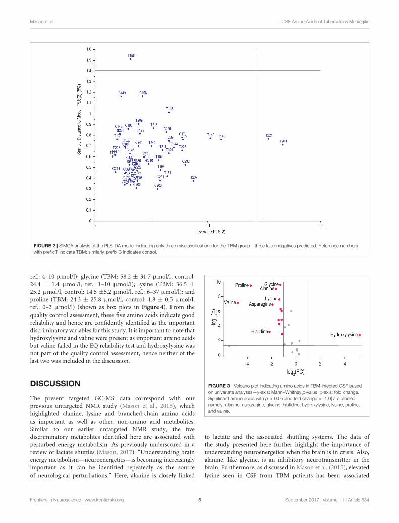
Identification of the ImportantDiscriminatory Amino AcidsFigure 1 illustrates the application of unsupervised PCA andsupervised partial least squares discriminant analysis (PLS-DA).Based on these analyses, the two groups can be differentiatedbased purely on amino acids, but not completely separated.Within the PLS-DA correlation loadings plot there are 11amino acids (circled on the figure)—identified as being the mostimportant discriminatory metabolites based upon leave-one-outcross-validation (R2 = 63.9%, Q2 = 55.3%). These 11 aminoacids are: alanine, alpha-aminobutyric acid, asparagine, glycine,hydroxylysine, lysine, ornithine, proline, serine, threonine, andvaline. Soft independent modeling of class analogy (SIMCA)analysis was performed (Figure 2) based on the PLS-DA model,indicating three misclassifications for the TBM group. Hence,
based on the prediction value of our PLS-DA model, three TBMcases were misclassified as control cases. The strength of thismodel is that there are no false positive classifications.
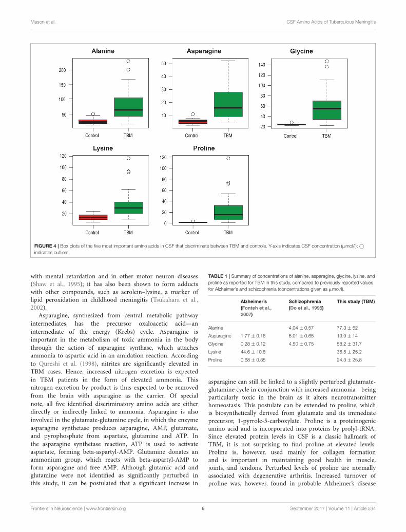
Univariate statistical analyses were also performed usingMann–Whitney p-values and fold changes. The results of theseunivariate analyses are illustrated in the volcano plot (Figure 3).Amino acids with significant values (Mann–Whitney p < 0.05and fold change> 1.0) are indicated, namely, alanine, asparagine,glycine, histidine, lysine, proline, hydroxylysine, and valine.
Based on the combination of both multivariate and univariateanalyses, therefore, themost important, common, discriminatoryamino acids of the control and TBM cases are: alanine (TBM: 77.3± 52 µmol/l, control: 25 ± 9.5 µmol/l, ref.1: 11–53.5 µmol/l);asparagine (TBM: 19.9 ± 14 µmol/l, control: 5.3 ± 1.7 µmol/l,
1Reference ranges given for children (0–10 years) obtained from the Human
Metabolome Database, based upon values reported values from the University of
British Columbia, B.C.’s Children’s Hospital Biochemical Genetics Lab.
Frontiers in Neuroscience | www.frontiersin.org 4 September 2017 | Volume 11 | Article 534
Mason et al. CSF Amino Acids of Tuberculous Meningitis
FIGURE 2 | SIMCA analysis of the PLS-DA model indicating only three misclassifications for the TBM group—three false negatives predicted. Reference numbers
with prefix T indicate TBM; similarly, prefix C indicates control.
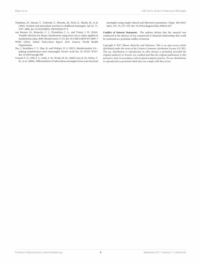
ref.: 4–10 µmol/l); glycine (TBM: 58.2 ± 31.7 µmol/l, control:24.4 ± 1.4 µmol/l, ref.: 1–10 µmol/l); lysine (TBM: 36.5 ±
25.2 µmol/l, control: 14.5 ±5.2 µmol/l, ref.: 6–37 µmol/l); andproline (TBM: 24.3 ± 25.8 µmol/l, control: 1.8 ± 0.5 µmol/l,ref.: 0–3 µmol/l) (shown as box plots in Figure 4). From thequality control assessment, these five amino acids indicate goodreliability and hence are confidently identified as the importantdiscriminatory variables for this study. It is important to note thathydroxylysine and valine were present as important amino acidsbut valine failed in the EQ reliability test and hydroxylysine wasnot part of the quality control assessment, hence neither of thelast two was included in the discussion.
DISCUSSION
The present targeted GC-MS data correspond with ourprevious untargeted NMR study (Mason et al., 2015), whichhighlighted alanine, lysine and branched-chain amino acidsas important as well as other, non-amino acid metabolites.Similar to our earlier untargeted NMR study, the fivediscriminatory metabolites identified here are associated withperturbed energy metabolism. As previously underscored in areview of lactate shuttles (Mason, 2017): “Understanding brainenergy metabolism—neuroenergetics—is becoming increasinglyimportant as it can be identified repeatedly as the sourceof neurological perturbations.” Here, alanine is closely linked
FIGURE 3 | Volcano plot indicating amino acids in TBM-infected CSF based
on univariate analyses—y-axis: Mann–Whitney p-value, x-axis: fold change.
Significant amino acids with p < 0.05 and fold change > |1.0| are labeled;
namely: alanine, asparagine, glycine, histidine, hydroxylysine, lysine, proline,
and valine.
to lactate and the associated shuttling systems. The data ofthe study presented here further highlight the importance ofunderstanding neuroenergetics when the brain is in crisis. Also,alanine, like glycine, is an inhibitory neurotransmitter in thebrain. Furthermore, as discussed in Mason et al. (2015), elevatedlysine seen in CSF from TBM patients has been associated
Frontiers in Neuroscience | www.frontiersin.org 5 September 2017 | Volume 11 | Article 534
Mason et al. CSF Amino Acids of Tuberculous Meningitis
FIGURE 4 | Box plots of the five most important amino acids in CSF that discriminate between TBM and controls. Y-axis indicates CSF concentration (µmol/l); ©
indicates outliers.
with mental retardation and in other motor neuron diseases(Shaw et al., 1995); it has also been shown to form adductswith other compounds, such as acrolein–lysine, a marker oflipid peroxidation in childhood meningitis (Tsukahara et al.,2002).
Asparagine, synthesized from central metabolic pathway
intermediates, has the precursor oxaloacetic acid—an
intermediate of the energy (Krebs) cycle. Asparagine isimportant in the metabolism of toxic ammonia in the body
through the action of asparagine synthase, which attachesammonia to aspartic acid in an amidation reaction. According
to Qureshi et al. (1998), nitrites are significantly elevated inTBM cases. Hence, increased nitrogen excretion is expected
in TBM patients in the form of elevated ammonia. Thisnitrogen excretion by-product is thus expected to be removed
from the brain with asparagine as the carrier. Of special
note, all five identified discriminatory amino acids are eitherdirectly or indirectly linked to ammonia. Asparagine is also
involved in the glutamate-glutamine cycle, in which the enzyme
asparagine synthetase produces asparagine, AMP, glutamate,and pyrophosphate from aspartate, glutamine and ATP. In
the asparagine synthetase reaction, ATP is used to activateaspartate, forming beta-aspartyl-AMP. Glutamine donates anammonium group, which reacts with beta-aspartyl-AMP toform asparagine and free AMP. Although glutamic acid andglutamine were not identified as significantly perturbed inthis study, it can be postulated that a significant increase in
TABLE 1 | Summary of concentrations of alanine, asparagine, glycine, lysine, and
proline as reported for TBM in this study, compared to previously reported values
for Alzheimer’s and schizophrenia (concentrations given as µmol/l).
Alzheimer’s
(Fonteh et al.,
2007)
Schizophrenia
(Do et al., 1995)
This study (TBM)
Alanine 4.04 ± 0.57 77.3 ± 52
Asparagine 1.77 ± 0.16 6.01 ± 0.65 19.9 ± 14
Glycine 0.28 ± 0.12 4.50 ± 0.75 58.2 ± 31.7
Lysine 44.6 ± 10.8 36.5 ± 25.2
Proline 0.68 ± 0.35 24.3 ± 25.8
asparagine can still be linked to a slightly perturbed glutamate-glutamine cycle in conjunction with increased ammonia—beingparticularly toxic in the brain as it alters neurotransmitterhomeostasis. This postulate can be extended to proline, whichis biosynthetically derived from glutamate and its immediateprecursor, 1-pyrrole-5-carboxylate. Proline is a proteinogenicamino acid and is incorporated into proteins by prolyl-tRNA.Since elevated protein levels in CSF is a classic hallmark ofTBM, it is not surprising to find proline at elevated levels.Proline is, however, used mainly for collagen formationand is important in maintaining good health in muscle,joints, and tendons. Perturbed levels of proline are normallyassociated with degenerative arthritis. Increased turnover ofproline was, however, found in probable Alzheimer’s disease
Frontiers in Neuroscience | www.frontiersin.org 6 September 2017 | Volume 11 | Article 534

Mason et al. CSF Amino Acids of Tuberculous Meningitis
cases (Fonteh et al., 2007) without arthritis, possibly reflectingincreased brain degeneration.
In a study on the change in CSF amino acid profiles followingan acute tonic-clonic seizure, Rainesalo et al. (2004) measuredamino acid levels in CSF before and after a seizure. Rainesaloet al. reported an increase in alanine (21.5 ± 1.2 to 23.1 ±
1.4 µmol/l), asparagine (6.2 ± 0.3 to 7.7 ± 0.5 µmol/l), andglycine (5.8 ± 0.5 to 7.4 ± 0.7 µmol/l), and a slight decreasein lysine (17.7 ± 0.9 to 17.1 ± 0.9 µmol/l)—none statisticallysignificant. Understandably, an acute tonic-clonic seizure (fullbody, grand mal, seizure) is a severe physiological response, yetRainesalo et al. reported only slight changes in most amino acids(significant changes in taurine, ornithine, and phenylalanine).Table 1 compares the five discriminatory amino acids identifiedin our study, as being most important in TBM, with those ofstudies of a chronic neuroinflammatory disease, Alzheimer’s, anda psychiatric disorder, schizophrenia. This comparison indicatesthat these particular five amino acids occur at levels 2–10times greater in TBM, further highlighting the severity of thepathophysiological condition of this disease.
CONCLUDING REMARKS
In this study, we used the sensitive method of GC-MS toprofile the amino acids of CSF collected from pediatric patientsclassified as being “definite” or “probable” cases of TBM. Throughstringent quality assessment measures and statistical analyses
(univariate and multivariate) we identified five amino acids inour experimental TBM patients as being reliable and havingstrong discriminatory power. The concentrations of the aminoacidmarkers of TBMhere occur at levels many times greater thanother neuropathological conditions, highlighting the severity ofTBM. Our latest observations justify the need for more detailedstudies on biochemical markers of TBM to be validated in larger-scale, and preferably multi-institutional, follow-up investigationsas amino acid profiling has potential in aiding in earlier diagnosis,and hence crucial earlier treatment of TBM.
AUTHOR CONTRIBUTIONS
All authors listed have made a substantial, direct and intellectualcontribution to the work, and approved it for publication.
ACKNOWLEDGMENTS
Research funding was provided by the Technological InnovationAgency of the Department of Science and Technology of SouthAfrica.
SUPPLEMENTARY MATERIAL
The Supplementary Material for this article can be foundonline at: http://journal.frontiersin.org/article/10.3389/fnins.2017.00534/full#supplementary-material
REFERENCES
Bhigjee, A. I., Padayachee, R., Paruk, H., Hallwirth-Pillay, K. D., Marais,
S., and Connoly, C. (2007). Diagnosis of tuberculous meningitis:
clinical and laboratory parameters. Int. J. Infect. Dis. 11, 248–254.
doi: 10.1016/j.ijid.2006.07.007
Cherian, A., and Thomas, S. V. (2011). Central nervous system tuberculosis. Afr.
Heal. Sci. 11, 116–127.
Corston, R. N., McGale, E. H., Stonier, C., Hutchinson, E. C., and Aber, G.
M. (1981). Cerebrospinal fluid amino acid concentrations in patients with
viral and tuberculous meningitis. J. Neuro. Neurosurg. Psychiatry 44, 791–795.
doi: 10.1136/jnnp.44.9.791
Do, K. Q., Lauer, C. J., Schreiber, W., Zollinger, M., Gutteck-Amsler,
U., Cuenod, M., et al. (1995). γ-Glutamylglutamine and taurine
concentrations are decreased in the cerebrospinal fluid of drug-naive
patients with schizophrenic disorders. J. Neurochem. 65, 2652–2662.
doi: 10.1046/j.1471-4159.1995.65062652.x
Fonteh, A. N., Harrington, R. J., Tsai, A., Liao, P., and Harrington, M. G. (2007).
Free amino acid and dipeptide changes in the body fluids from Alzheimer’s
disease subjects. Amino Acids 32, 213–224. doi: 10.1007/s00726-006-
0409-8
Fowler, B., Burlina, A., Kozich, V., and Vianey-Saban, C. (2008). Quality
of analytical performance in inherited metabolic disorders: the role of
ERNDIM. J. Inherit. Metab. Dis. 31, 680–689. doi: 10.1007/s10545-008-
1025-4
Hristea, A., Olaru, I. D., Baicus, C., Moroti, R., Arama, V., and Ion, M. (2012).
Clinical prediction rule for differentiating tuberculous from viral meningitis.
Int. J. Tuberc. Lung Dis. 16, 793–798. doi: 10.5588/ijtld.11.0687
Marais, S., Thwaites, G., Schoeman, J. F., Török, M. E., Misra, U. K.,
Prasad, K., et al. (2010). Tuberculous meningitis: a uniform case
definition for use in clinical research. Lancet Infect. Dis. 10, 803–812.
doi: 10.1016/S1473-3099(10)70138-9
Mason, S. (2017). Lactate shuttles in neuroenergetics—homeostasis, allostasis and
beyond. Front. Neurosci. 11:43. doi: 10.3389/fnins.2017.00043
Mason, S., Reinecke, C. J., Kulik, W., Van Cruchten, A., Solomons,
R., and van Furth, A. M. T. (2016). Cerebrospinal fluid in
tuberculous meningitis exhibits only the L-enantiomer of
lactic acid. BMC Infect. Dis. 16:251. doi: 10.1186/s12879-016-
1597-9
Mason, S., van Furth, A. M., Mienie, L. J., Engelke, U. F., Wevers, R. A., Solomons,
R., et al. (2015). A hypothetical astrocyte–microglia lactate shuttle derived from
a 1H NMR metabolomics analysis of cerebrospinal fluid from a cohort of
South African children with tuberculousmeningitis.Metabolomics 11, 822–837.
doi: 10.1007/s11306-014-0741-z
Qureshi, G. A., Baig, S. M., Bednar, I., Halawa, A., and Parvez, S. H.
(1998). The neurochemical markers in cerebrospinal fluid to differentiate
between aseptic and tuberculous meningitis. Neurochem. Int. 32, 197–203.
doi: 10.1016/S0197-0186(97)00061-2
Rainesalo, S., Keränen, T., Palmio, J., Peltola, J., Oja, S. S., and Saransaari,
P. (2004). Plasma and cerebrospinal fluid amino acids in epileptic
patients. Neurochem. Res. 29, 319–324. doi: 10.1023/B:NERE.0000010461.
34920.0c
Rock, R. B., Hu, S., Gekker, G., Sheng, W. S., May, B., Kapur, V., et al. (2005).
Mycobacterium tuberculosis-induced cytokine and chemokine expression by
human microglia and astrocytes: effects of dexamethasone. J. Infect. Dis. 192,
2054–2058. doi: 10.1086/498165
Shaw, P. J., Forrest, V., Ince, P. G., Richardson, J. P., and Wastell, H. J.
(1995). CSF and plasma amino acid levels in motor neuron disease: elevation
of CSF glutamate in a subset of patients. Neurodegeneration 4, 209–216.
doi: 10.1006/neur.1995.0026
Solomons, R. S., Visser, D. H., Donald, P. R., Marais, B. J., Schoeman, J. F., and
van Furth, A. M. (2015). The diagnostic value of cerebrospinal fluid chemistry
results in childhood tuberculous meningitis. Childs. Nerv. Syst. 31, 1335–1340.
doi: 10.1007/s00381-015-2745-z
Frontiers in Neuroscience | www.frontiersin.org 7 September 2017 | Volume 11 | Article 534
Mason et al. CSF Amino Acids of Tuberculous Meningitis
Tsukahara, H., Haruta, T., Todoroki, Y., Hiraoka, M., Noiri, E., Maeda, M., et al.
(2002). Oxidant and antioxidant activities in childhood meningitis. Life Sci. 71,
2797–2806. doi: 10.1016/S0024-3205(02)02137-9
van Reenen, M., Reinecke, C. J., Westerhuis, J. A., and Venter, J. H. (2016).
Variable selection for binary classification using error rate p-values applied to
metabolomics data. BMCBioinformatics 17:33. doi: 10.1186/s12859-015-0867-7
WHO (2016). Global Tuberculosis Report 2016. Geneva: World Health
Organization.
Xia, J., Sinelnikov, I. V., Han, B., and Wishart, D. S. (2015). MetaboAnalyst 3.0—
making metabolomics more meaningful. Nucleic Acids Res. 43, W251–W257.
doi: 10.1093/nar/gkv380
Youssef, F. G., Afifi, S. A., Azab, A. M., Wasfy, M. M., Abdel-Aziz, K. M., Parker, T.
M., et al. (2006). Differentiation of tuberculous meningitis from acute bacterial
meningitis using simple clinical and laboratory parameters. Diagn. Microbiol.
Infect. Dis. 55, 275–278. doi: 10.1016/j.diagmicrobio.2006.01.027
Conflict of Interest Statement: The authors declare that the research was
conducted in the absence of any commercial or financial relationships that could
be construed as a potential conflict of interest.
Copyright © 2017 Mason, Reinecke and Solomons. This is an open-access article
distributed under the terms of the Creative Commons Attribution License (CC BY).
The use, distribution or reproduction in other forums is permitted, provided the
original author(s) or licensor are credited and that the original publication in this
journal is cited, in accordance with accepted academic practice. No use, distribution
or reproduction is permitted which does not comply with these terms.
Frontiers in Neuroscience | www.frontiersin.org 8 September 2017 | Volume 11 | Article 534


![Development and Validation of Methods for Impurity ... · 1.2. Amino Acid Analyzer The European Pharmacopoeia utilizes amino acid analysis (AAA) [12] for impurity profiling of AAs](https://img.pdfslide.us/doc/110x75/60620be79ec8427fe1067f59/development-and-validation-of-methods-for-impurity-12-amino-acid-analyzer.jpg)