Embed Size (px)
Citation preview
CEREBRAL VASCULAR ACCIDENTS
Pediatric Critical Care MedicineEmory University
Children’s Healthcare of Atlanta
2
Objectives• Epidemiology• Risk factors• Catergories• Treatments
3
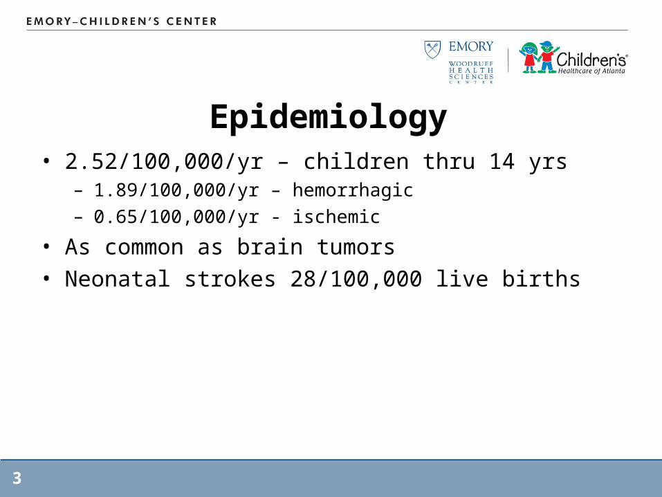
Epidemiology• 2.52/100,000/yr – children thru 14 yrs
– 1.89/100,000/yr – hemorrhagic– 0.65/100,000/yr - ischemic
• As common as brain tumors• Neonatal strokes 28/100,000 live births
4
Epidemiology• Increased awareness & reporting• Improved imagings• Better survival of underlying diseases
5
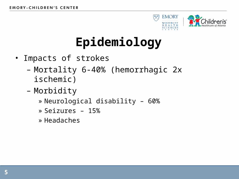
Epidemiology• Impacts of strokes
– Mortality 6-40% (hemorrhagic 2x ischemic)– Morbidity
» Neurological disability – 60%» Seizures – 15%» Headaches
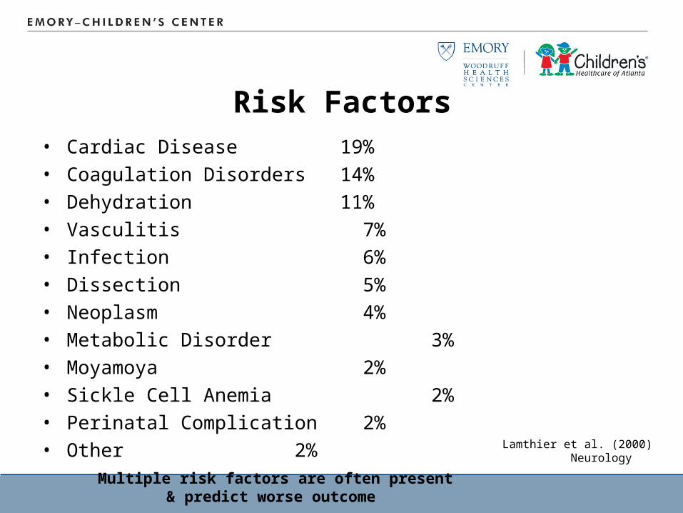
Risk Factors• Cardiac Disease 19%• Coagulation Disorders 14%• Dehydration 11%• Vasculitis 7%• Infection 6%• Dissection 5%• Neoplasm 4%• Metabolic Disorder 3%• Moyamoya 2%• Sickle Cell Anemia 2%• Perinatal Complication 2%• Other 2%
Multiple risk factors are often present& predict worse outcome
Lamthier et al. (2000)Neurology
7
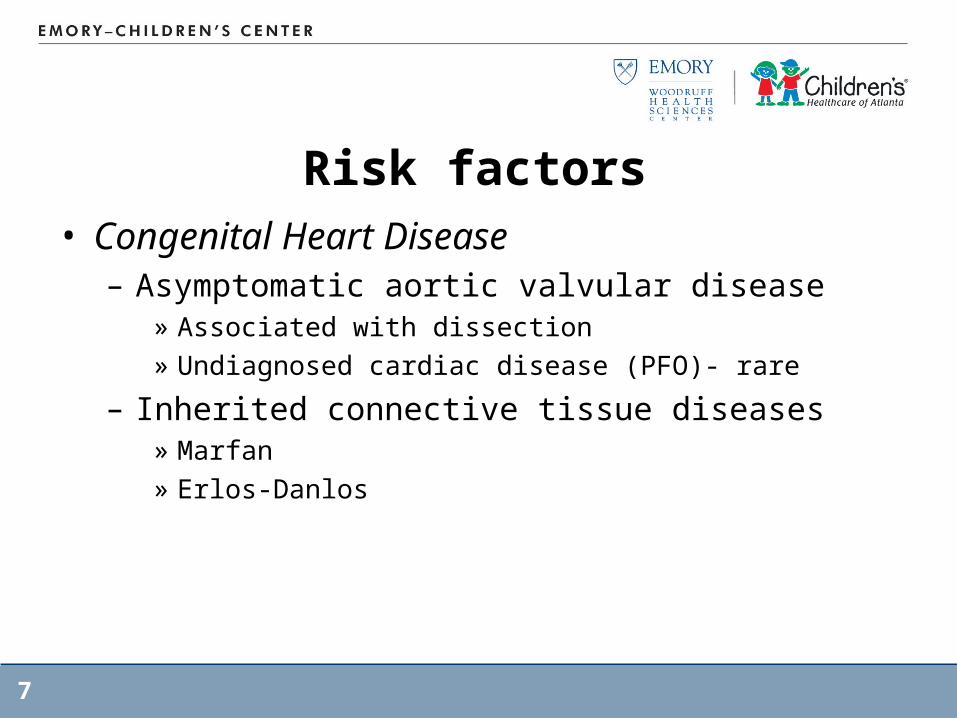
Risk factors• Congenital Heart Disease
– Asymptomatic aortic valvular disease » Associated with dissection » Undiagnosed cardiac disease (PFO)- rare
– Inherited connective tissue diseases» Marfan» Erlos-Danlos
8
Risk factors• Coagulation disorders
– Factor V Leiden » common in Caucasian» most common cause of activated Protein C resistance
– Prothrombin 20210 mutation» Neonatal & childhood CSVT
• Infection, Inflammation, Immune Deficiency– 1/3 cases associated with infection (esp vacicella
within the previous year)– High WBC (in association with SCD) increase recurrence– Inflammation: harmful effects on the endothelium
9
Risk factors• Sickle Cell Disease
– 25 % with stroke by the age 45– Ischemic stroke predominantly in childhood
» Hemorrhagic with steroid and Hypertension» Sinovenous thrombosis, posterior
leukoencephalopathy, watershed ischemia» Silent infarcts
– Hemorrhagic (ICH or SAH) in adult secondary to aneurysm
– High WBC associated with infection can precipitate CVD – indicate chronic infection
10
Risk factors• Anemias
– Hemolytic anemias: thalassemia, hereditary spherocytosis & paroxysmal nocturnal hemoglobinuria
• Metabolic disorders– Homocysteinemia: predispose to vessel abn.– Lipid abnormality:
» Elevation in Cholesterol (9%), TG (31%), Lipoprotein (22%)
» Apolipoprotein abnormality
11
Risk factors• Vascular abnormality
– Vascular adhesion» Adhesion of WBC, RBC, platelets causing endothelial
damage
• Hypertension– Highest risk in young adult & elderly– Largely ignored in pediatric population– ½ strokes with SBP>90th percentile– Abnormality of angiotensinogen gene: 4X increase
in risk of strokes in SCD
12
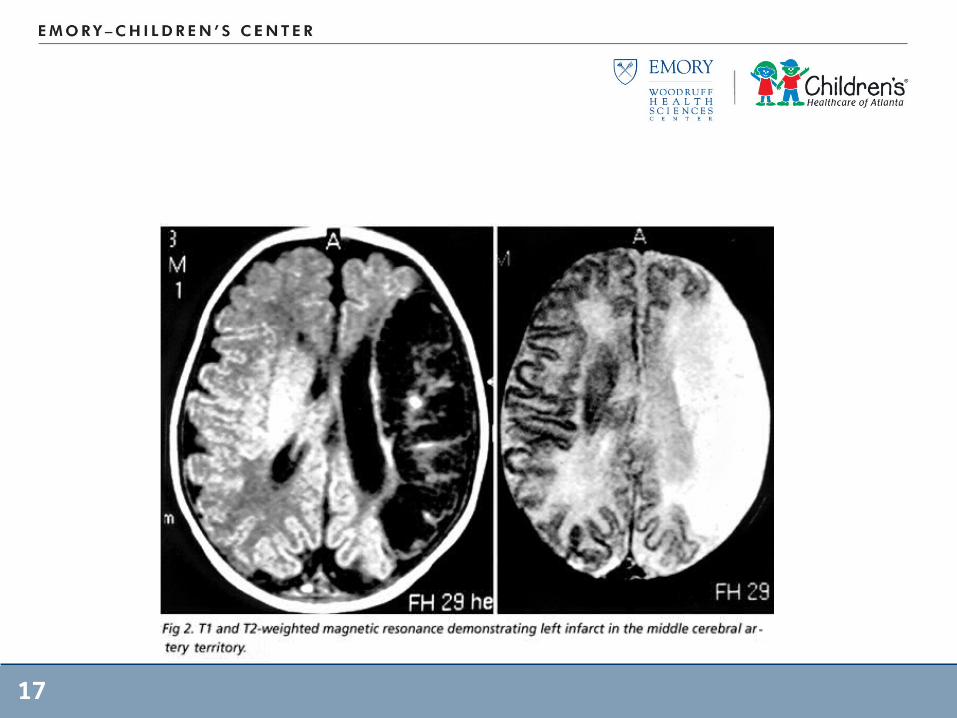
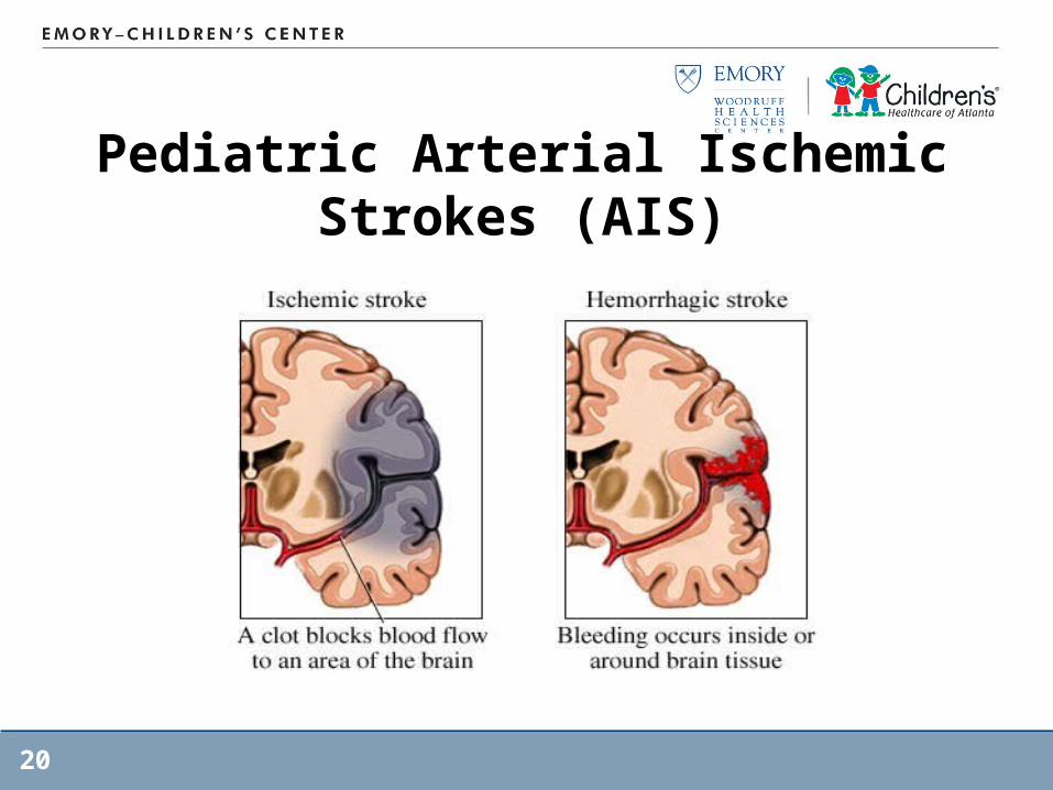
Pediatric Arterial Ischemic Strokes (AIS)
• Primary Hemiparesis: new focal deficits• Ataxic gait• Chorea• Vertigo • Speech and visual disturbance• Headaches with neurological deficits
13
Pediatric Arterial Ischemic Strokes (AIS)
• Median presentation of 5.6 hrs after AIS symptoms onset– ½ presented within the first 6 hrs– ½ presented >24 hrs
• Main factor in delayed presentation: was the failure of parents to recognize that a child was having neurologic symptoms
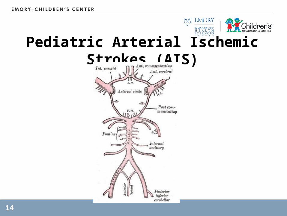
Pediatric Arterial Ischemic Strokes (AIS)
14
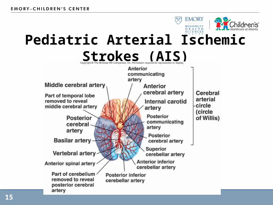
Pediatric Arterial Ischemic Strokes (AIS)
15
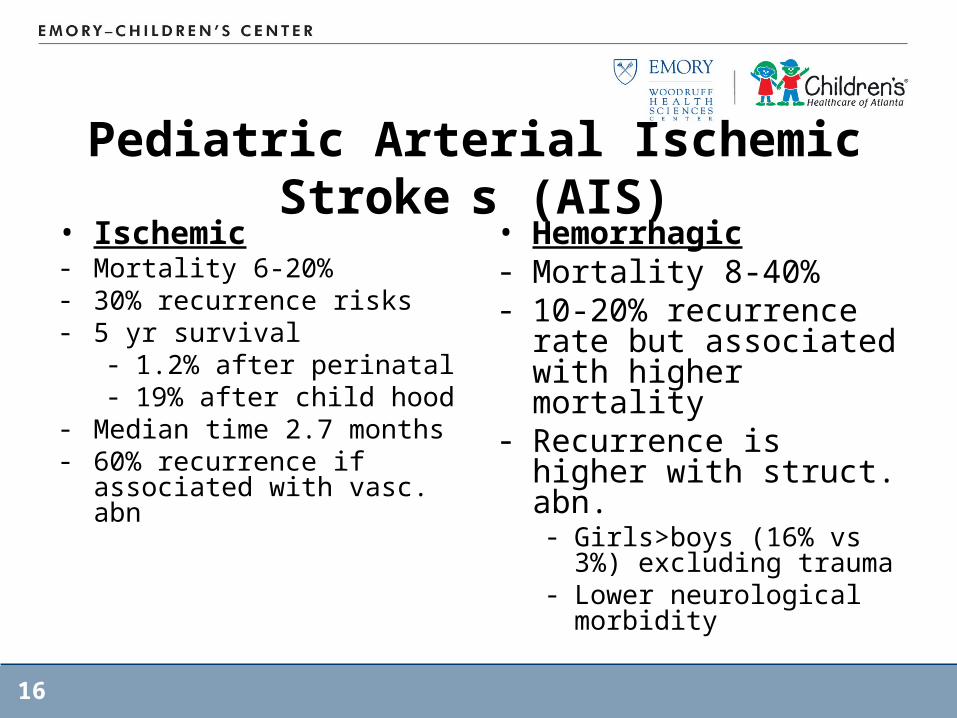
Pediatric Arterial Ischemic Stroke s (AIS)
• Ischemic- Mortality 6-20%- 30% recurrence risks- 5 yr survival
- 1.2% after perinatal- 19% after child hood
- Median time 2.7 months- 60% recurrence if
associated with vasc. abn
• Hemorrhagic- Mortality 8-40%- 10-20% recurrence
rate but associated with higher mortality
- Recurrence is higher with struct. abn.- Girls>boys (16% vs 3%)
excluding trauma- Lower neurological
morbidity
16
17
18
19
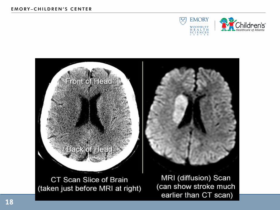
Pediatric Arterial Ischemic Strokes (AIS)
20
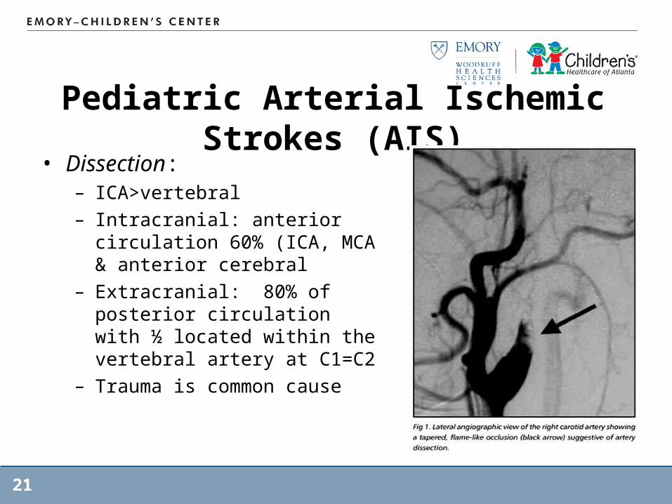
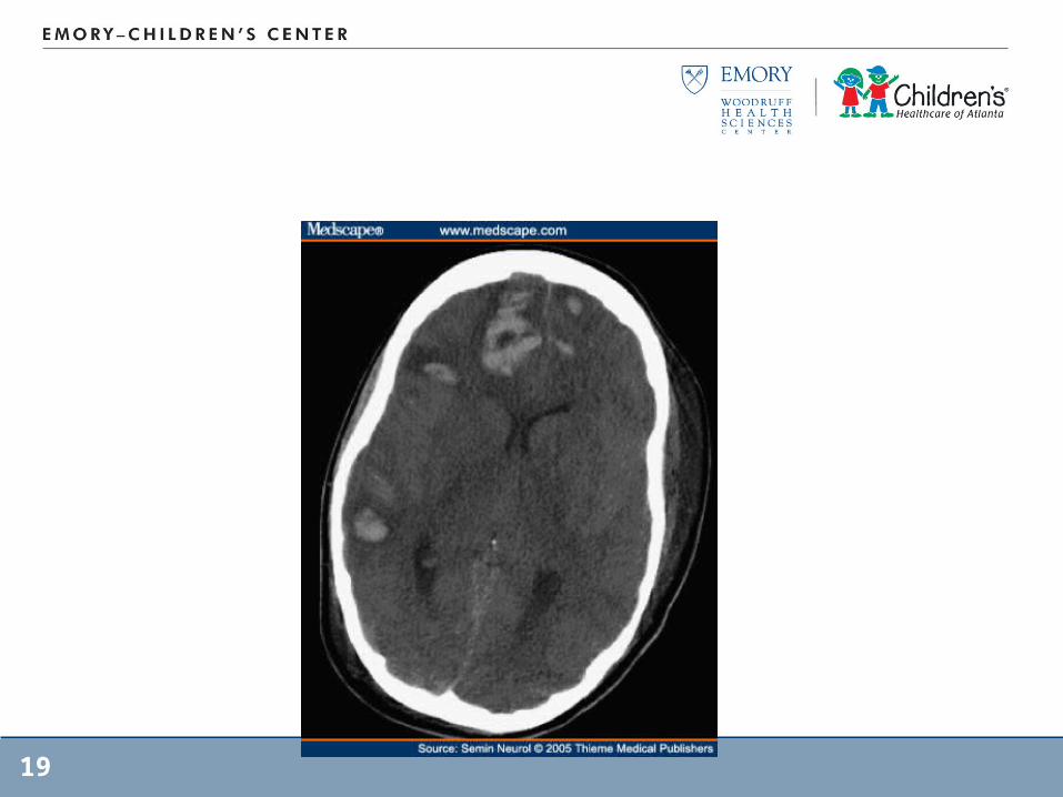
Pediatric Arterial Ischemic Strokes (AIS)
• Dissection:– ICA>vertebral – Intracranial: anterior
circulation 60% (ICA, MCA & anterior cerebral
– Extracranial: 80% of posterior circulation with ½ located within the vertebral artery at C1=C2
– Trauma is common cause
21
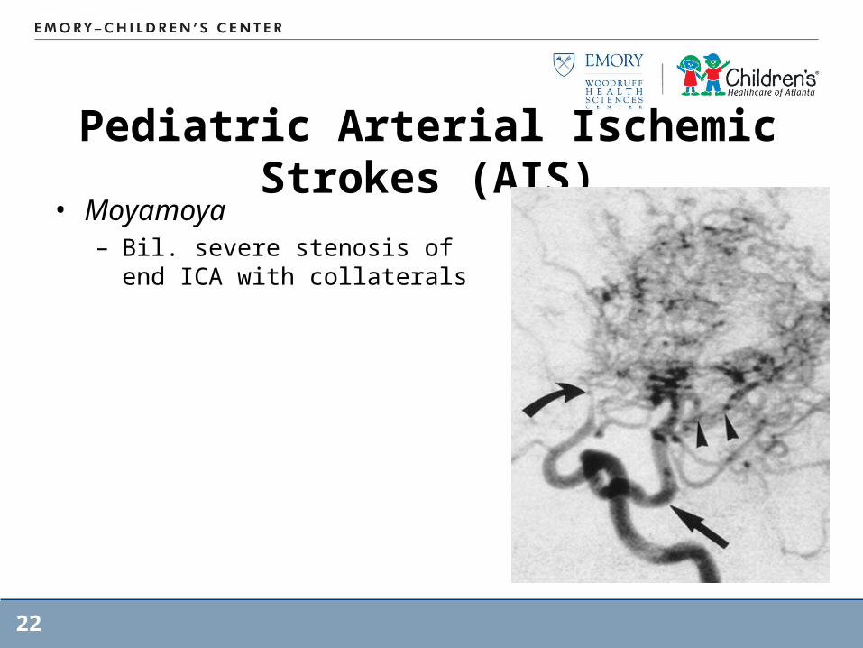
Pediatric Arterial Ischemic Strokes (AIS)
• Moyamoya– Bil. severe stenosis of end ICA
with collaterals
22
23
Pediatric Arterial Ischemic Strokes (AIS)
• Transient Cerebral arteriopathy– Inflammatory response to infection (varicell, Borrelia or
tonsilitis– Multi-focal lesions– Most cases stabilize but some progress to recurrent
strokes
24
Pediatric Arterial Ischemic Strokes (AIS)
• Sickle Cell Disease– Narrowing of distal ICA & proximal MCA, anterior
cerebral arteries– Gradual progression to occlusion– Endothelial proliferation– Silent infarcts occur in MCA territory or in border zones– High recurrence rate– 25% with CVD by age 45
» Ischemic in children» Hemorrhagic in adults
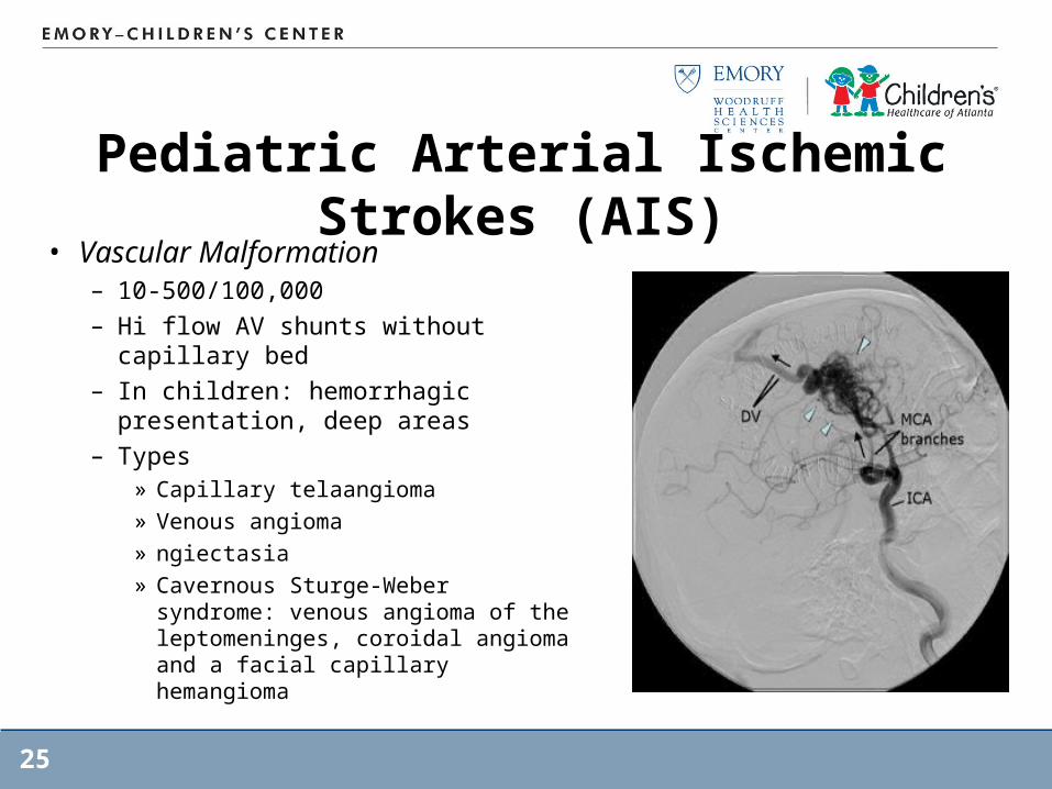
Pediatric Arterial Ischemic Strokes (AIS)
• Vascular Malformation– 10-500/100,000– Hi flow AV shunts without capillary
bed– In children: hemorrhagic
presentation, deep areas– Types
» Capillary telaangioma» Venous angioma» ngiectasia» Cavernous Sturge-Weber
syndrome: venous angioma of the leptomeninges, coroidal angioma and a facial capillary hemangioma
25
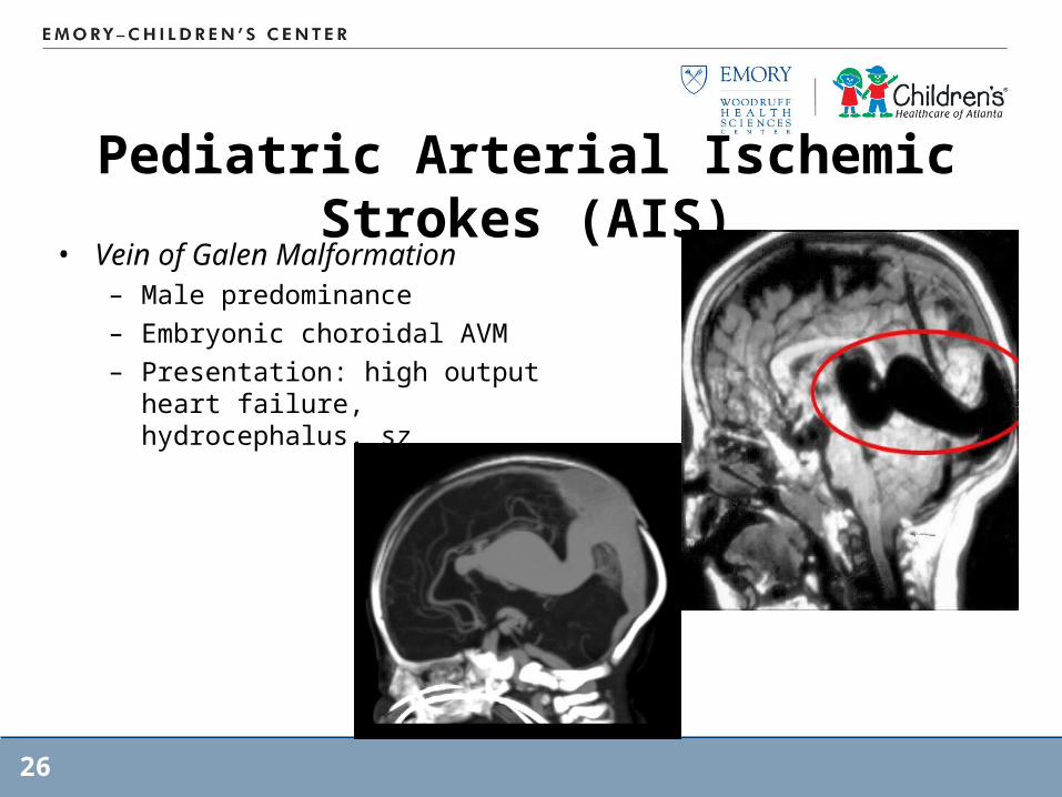
Pediatric Arterial Ischemic Strokes (AIS)
• Vein of Galen Malformation– Male predominance– Embryonic choroidal AVM– Presentation: high output heart
failure, hydrocephalus, sz
26
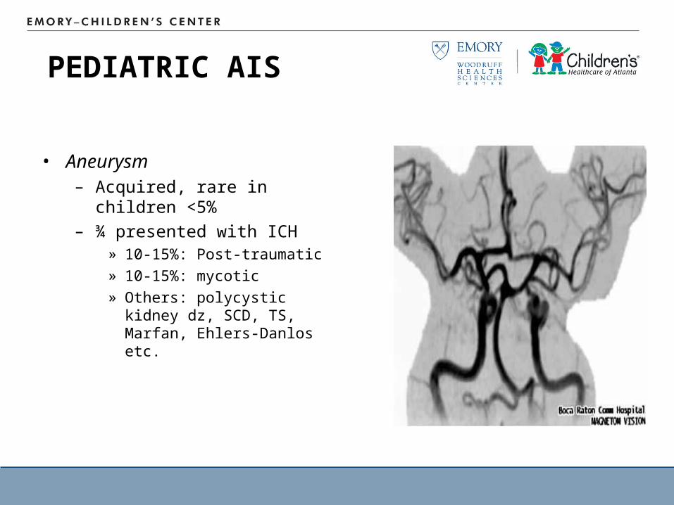
PEDIATRIC AIS
• Aneurysm– Acquired, rare in children
<5%– ¾ presented with ICH
» 10-15%: Post-traumatic» 10-15%: mycotic» Others: polycystic kidney
dz, SCD, TS, Marfan, Ehlers-Danlos etc.
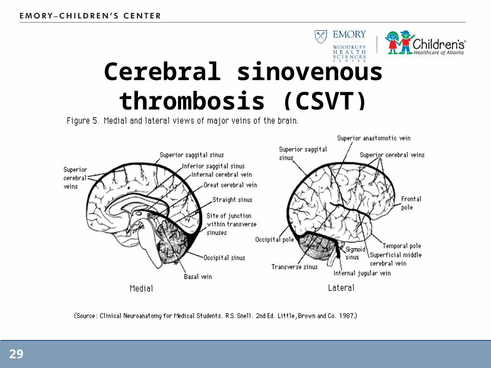
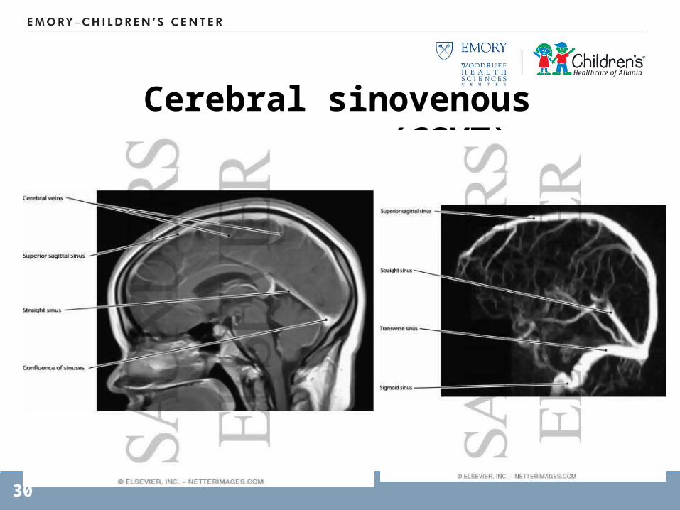
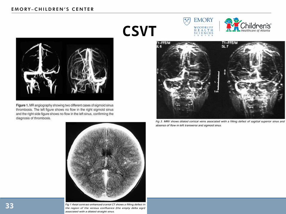
Cerebral sinovenous thrombosis (CSVT)
• Superficial & Deep– Superficial: cortical veins superior sagittal sinus
right lateral sinus– Deep: inferior sagittal sinus & paired internal cerebral
veins, join to form v. of Galen & straight sinus
• Flow is highly responsive to changes in MAP which can cause reversal of flow
• Relative low thrombomodulin prothrombotic
28
Cerebral sinovenous thrombosis (CSVT)
29
Cerebral sinovenous thrombosis (CSVT)
30
CSVT• Pathophysiology
– Mechanical: birth trauma– Trauma, sepsis, underlying disease (malignancy,
systemic inflamation)– Septic foci: inner ear, mastoid or air sinuses– Dehydration, anemia, coagulation disorders
• Venous Infarction: venous HTN by outflow obstruction
• Intracranial Hypertension: disruption of CSF absorption
31
CSVT• Signs/symptoms
– Severe HA associated with vomiting, sleepiness, double vision
– Visual disturbances– Severe dizziness or unsteadiness– Sz activity
32
CSVT
33
Other Strokes Mimics• Posterior Circulation Arterial Strokes
– Posterior infarction of cerebellum, brain stem– Boys>girls– Trauma, subluxation of cervical spines causing arterial
dissection
• Reversible Posterior Leukoencephalopathy– Sx: sz, AMS, disorder of consciousness, visual abn., HA– Predominant post. White matter abn.– Clinical condition: HTN encephalopathy, eclampsia, AC in SCD,
immunosuppression– Acute hypotension (poor cardiac fxn/anemia)– Rapid resolution: vasogenic cerebral edema prob secondary to
autoregulation & endothelial injury
• Acute Disseminated Encephalomyelitis
34
Other Strokes Mimics• Metabolic Strokes: Diabetes, inborn errors of
metabolism– Vascular injury
» Homocysteine: direct endothelial injury» Fabry: lysosomal storage with accumulation and deposition
of glycosphingolipid in blood vessels endothelial cells» Menkes: deficiency in copper; obliteration of intracranial
vasculature
– Non-vascular injury: diabetes, organic acidemias, Urea cycle defects etc.
» MILAS (mitochondrial dz with LA and stroke like sx): lacking of energy supply with generation of oxygen free radicals
» Others: accumulation of toxic substances
35
Diagnosis – w/o SCD• First 24 hours
– Angiogram– MRA– Blood cultures if febrile– Toxicology screen
• 24-72 hours– Echo with bubble study– Limited initial pro-thrombotic evaluation– Lupus anticoagulants, antiphospholipid abs, lipid profile,
lipoprotein A, Homocysteine, gene mutation– Systemic inflammatory disease evaluation: ANA, ESR, CRP, UA
Rollins N, Dowling M, Booth T, Purdy P, AJNR, 2000substances
36
Diagnosis – w/o SCD• After acute setting
– Further prothrombotic evaluation» Protein C & S» Antithrombin» Factor VIII» Confirmation of early abnormal tests
Rollins N, Dowling M, Booth T, Purdy P, AJNR, 2000substances
37
Ischemic Stroke Treatments• General management
– Normo-thermia– Normal oxygen saturation– Cerebral protection with the presence of increase ICP
• Specific management– Early neurosurgery consult as indicated
38
Stroke Treatments• Anticoagulation
– Commonly use in: » AIS: Heparin or LMWH for 5-7 days until cardioembolic stroke
and dissection are excluded» CSVT: 3-6 months of therapy reduced risk of recurrent
systemic or cerebral thrombosis» High risk of embolism with underlying disease» Dissection: 3-6 months with extracranial dissection.» Known prothrombotic abnormalities
– With cardiac embolism: controversial– Balance of risk with precipitate hemorrhage vs recurrence
embolic event (lower risk in children for progression to hemorrhage)
39
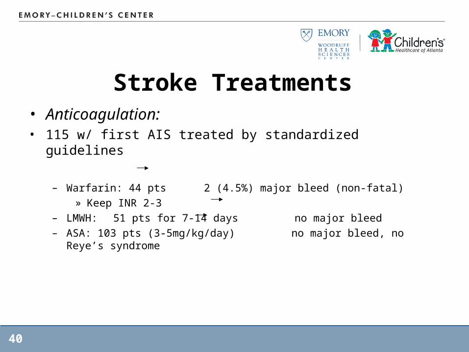
Stroke Treatments• Anticoagulation: • 115 w/ first AIS treated by standardized guidelines
– Warfarin: 44 pts 2 (4.5%) major bleed (non-fatal)» Keep INR 2-3
– LMWH: 51 pts for 7-14 days no major bleed– ASA: 103 pts (3-5mg/kg/day) no major bleed, no Reye’s syndrome
40
Stroke Treatments• Aspirin therapy
– Efficacy and dose are unknown– Usual dose 5mg/lg/day– Long term prophylaxis dose may be lower– No report case of Rye’s syndrome
» One case in adult when the pt increased ASA dosage with flu like sx
41
Stroke Treatments• Thrombolytic agents: urokinase & streptokinase
– No evidence to support efficacy- 203 (pooled literature) for non-cerebral thrombotic complication
» 80% thrombus cleared» 54% minor bleeding (no transfusion needed)» 1 pt with intra-cranial hemorrhage
– Toronto: 29 pts treated with tPA (0.5mg/kg)» 79% - clot was dissolved» ¼ of the pts had bleeding required transfusion
– No good data regarding outcomes, therefore treatment is controversial
42
Stroke Treatments - SCD• ½ will have another stroke• Urgent exchange transfusion (HbS <30% or HgB
10=12.5)• Top off transfusion if exchange transfusion is delayed
or severe anemia• Chronic exchange transfusion
– Keep HgS <50%– Relapse if stop even with period of symptoms free– Risk: iron overload treated with chelation– Use of hydroxyurea to prevent stroke
» Induction of HbF» Generation of Nitric Oxide
43
Intracranial Hemorrhage• Risk factors
– AV malformation 25%– Hematologic anomalies 10-13%– Brain tumors– Cavernous Hemangiomas– Vasculopathy– Vasculitis– Infection– Illicit drug uses
44
Intracranial Hemorrhage• Non traumatic SAH mostly caused by aneurysms• 10% are secondary to CSVT
– Controversial in anti-coag of CSVT with hemorrhage
• 25% mortality with 42% significant disability• No standard management and treatm
45
Treatments: ICH• Treat ICP, cerebral protection• Reverse coagulopathy
– Recombinant activated Factor VII within the first 4 hrs: limited growth of hematoma, reduced mortality, improved functional outcomes
• Treat space occupying lesion• Treat associated vasospasm (SAH) with Triple H
therapy• Supportive treatment
46
Treatments: SAH• Vasospasm associated with 20-30% of aneurysmal SAH• Related to spasmogenic substances generated during
lysis of subarachnoid blood clots• Present no earlier than day 3, peak day 7-8• Triple-H therapy
– Moderate hemodilution– Hypertension– Hypervolumia
• Nimodipine: selective cerebral vessel Ca channel blocker, start within 4 days– Decrease morbidity and mortality– Potential for systemic effect causing severe hypotension
47