Embed Size (px)
Citation preview
Behavioural Neurology 24 (2011) 117–122 117DOI 10.3233/BEN-2011-0283IOS Press
Short Communication
Cerebral perfusion in chronic stroke:Implications for lesion-symptom mappingand functional MRI
Jessica D. Richardsona,∗, Julie M. Bakera, Paul S. Morganb, Chris Rordenc, L. Bonilhad andJulius FridrikssonaaDepartment of Communication Sciences and Disorders, University of South Carolina, Columbia, SC, USAbDepartment of Radiology and Radiological Science, MedicalUniversity of South Carolina, Columbia, SC, USAcGeorgia State/Georgia Tech Center for Advanced Brain Imaging, GA, USAdDepartment of Neurosciences, Medical University of South Carolina, Columbia, SC, USA
Abstract. Lesion-symptom mapping studies are based upon the assumption that behavioral impairments are directly related tostructural brain damage. Given what is known about the relationship between perfusion deficits and impairment in acute stroke,attributing specific behavioral impairments to localized brain damage leaves much room for speculation, as impairments couldalso reflect abnormal neurovascular function in brain regions that appear structurally intact on traditional CT and MRIscans.Compared to acute stroke, the understanding of cerebral perfusion in chronic stroke is far less clear. Utilizing arterial spin labeling(ASL) MRI, we examined perfusion in 17 patients with chronicleft hemisphere stroke. The results revealed a decrease in lefthemisphere perfusion, primarily in peri-infarct tissue. There was also a strong relationship between increased infarct size anddecreased perfusion. These findings have implications for lesion-symptom mapping studies as well as research that relies onfunctional MRI to study chronic stroke.
Keywords: Aphasia, arterial spin labeling (ASL), hypoperfusion, ischemic stroke, peri-infarct
1. Introduction
Lesion symptom mapping studies attempt to inferbrain function by associating the behavioral deficits ob-served with the location of injury observed using brainimaging. Much has been learned about brain-behaviorrelationships from lesion-symptom mapping studies.However, a crucial assumption of such studies is thatbehavioral impairments reflect frank tissue damage andthat neurophysiology is normal in the remaining brainareas that appear structurally intact. Although impairedcerebral perfusion is commonly reported in the acute
∗Corresponding author: Jessica D. Richardson, Ph.D., Depart-ment of Communication Sciences & Disorders, University of SouthCarolina, Columbia, SC 29208, USA. Tel.: +1 803 777 5049; Fax:803 777 4750; E-mail: [email protected].
and sub-acute phases of stroke (see [3,7,11,13,22]), ahandful of studies have reported abnormal perfusion inchronic stroke [1,5,12,19]. It is generally believed thathypoperfusion resolves over time, in accordance withearly behavioral recovery patterns [3] and if present,is probably functionally insignificant [13,21]. How-ever, several single case studies of chronic stroke re-port hypoperfusion in the hemisphere that incurred thedamage [12,13,19] and carefully relate it to functionaldeficits [13,19], complementing an earlier study report-ing the “occasional phenomenon” of hypoperfusion inchronic stroke [1].
Based on the findings reported above, it seems thatattributing specific behavioral impairments to localizedbrain damage in chronic stroke leaves much room forspeculation, as the impairments could also reflect ab-normal function in structurally intact brain regions. If
ISSN 0953-4180/11/$27.50 2011 – IOS Press and the authors. All rights reserved
118 J.D. Richardson et al. / Perfusion in chronic stroke
compromised cerebral perfusion is common in chron-ic stroke, it may need to be taken into account whenexamining lesion-impairment relationships. Unfortu-nately, research focused on cerebral perfusion in chron-ic stroke is limited, and little is known about perfusiondeficits in remaining brain areas or how far such deficitsmight extend beyond the infarct. To shed light on thisissue, the present study provides preliminary informa-tion regarding the relationship between cerebral perfu-sion, time post onset (TPO) of stroke, and infarct sizein chronic patients with cortical stroke.
2. Materials and methods
2.1. Patients
The patients examined in this research were a subsetof a larger group consecutively enrolled for an ongoingaphasia treatment study. The 17 participants (10 fe-males; age range= 41–81 years;M = 61.82 years)had each incurred a single cortical ischemic stroke ofthe left hemisphere at least 4 months prior to study in-clusion (M = 48.47 months, range= 4–246 months).The presumed etiology of stroke was defined by in-terviewing patients regarding their stroke risk factors.Two patients had been diagnosed with atrial fibrillationat the time of the stroke, and two patients were diag-nosed with symptomatic extracranial anterior circula-tion stenosis. The other patients were classified as hav-ing suffered cryptogenic or small vessel strokes. Thestudy was approved by the University of South Caroli-na’s Institutional Review Board, and informed consentwas obtained from all patients.
2.2. Imaging
All MRI data were collected using a 3T Trio sys-tem (Siemens Medical, Erlangen, Germany) with a 12-element head coil. Cerebral perfusion was estimat-ed using a Pulsed Arterial Spin Labeling (PASL) se-quence [25]. PASL imaging utilized a single shot gra-dient echo planar imaging (EPI) sequence with the fol-lowing parameters: parallel imaging (GRAPPA) fac-tor = 2, no fat suppression, matrix size= 64× 64 mm,pixel size = 3.5 × 3.5 mm, 16 axial slices (5 mmthick with 6 mm center-center slice separation), TR= 4000 ms, TE= 12 ms, and bandwidth= 3 kHz/pixel.The selective inversion slab was 120 mm thick; the in-ferior saturation slab was 100 mm thick and directlybelow the imaging slices. The post inversion delay was
700 ms and the post labeling delay was 1000 ms. Thir-ty repeats of selective/non-selective image pairs wereacquired. An estimate of the equilibrium magnetiza-tion (M0) was obtained by a further acquisition of tworepeats of the same sequence (but TR= 10 s and postinversion delay= 5 s). Images were corrected forhead motion and mean images of the selective and non-selective images constructed. Perfusion images wereprocessed using in-house software (www.nottingham.ac.uk/∼njzwww/paul/software/perf.html), and valueswere calculated in units of ml/min/100g tissue, as de-scribed previously [25].
To improve coregistration of images and infarct de-marcation, all participants were scanned with high-resolution T2 and T1 MRI sequences described previ-ously [10]. Structural images were prepared for da-ta analysis using software designed and supported bythe Oxford Centre for Functional MRI of the Brain(FMRIB) – FMRIB’s Software Library (FSL) version4.1 [24]. Cropped and skull-stripped structural MRIimages were normalized to standard space, employinginfarct weighting for improved accuracy. Normalizedimages were resliced to 2 mm isotropic. Each partici-pant’s perfusion images were then coregistered to theirown spatially normalized structural images.
Infarcts were demarcated on axial slices of the nor-malized T2 MRI using MRIcron [23], with the high-resolution native T2 MRI used to guide demarcation.To create the peri-infarct mask, the infarct was dilat-ed 8 mm, and the original infarct was subtracted fromthe diameter of the dilated infarct. Also subtractedwas the peri-infarct area between 0 to 3 mm beyondthe infarct’s rim (partial-volume boundary) in order toaccount for partial lesion volume. This was overlaidon a probabilistic gray matter mask, producing the lefthemisphere peri-infarct gray matter (LHp) mask. Wealso created a mask for the remaining left hemisphere(LHr) by beginning with the probabilistic gray mattermask for the left hemisphere and removing all regionsthat were part of the LHp, infarcted tissue, or infarct-LHp partial-volume boundary. The masks were flippedto obtain the right hemisphere peri-infarct gray homo-logue (RHp) and right hemisphere remaining gray ho-mologue (RHr) masks (see Fig. 1). These four volumesof interest (VOIs) were overlaid on the perfusion im-ages; mean CBF values were obtained for each utilizingMRIcron. In addition, relative values were obtained byexpressing each VOI value as a percentage of its mirrorVOI (i.e., perfusion ratio).
2.3. Data analyses
Statistical analyses were performed using SPSS (Sta-
J.D. Richardson et al. / Perfusion in chronic stroke 119
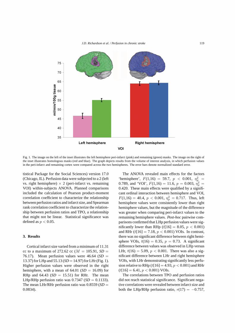
Fig. 1. The image on the left of the inset illustrates the lefthemisphere peri-infarct (pink) and remaining (green) masks. The image on the right ofthe inset illustrates homologous masks (red and blue). The graph depicts results from the volume of interest analysis, in which perfusion valuesin the peri-infarct and remaining cortex were compared across the two hemispheres. The error bars denote normalized standard error.
tistical Package for the Social Sciences) version 17.0(Chicago, IL). Perfusion data were subjected to a 2 (leftvs. right hemisphere)× 2 (peri-infarct vs. remainingVOI) within-subjects ANOVA. Planned comparisonsincluded the calculation of Pearson product-momentcorrelation coefficient to characterize the relationshipbetween perfusion ratios and infarct size,and Spearmanrank correlation coefficient to characterize the relation-ship between perfusion ratios and TPO, a relationshipthat might not be linear. Statistical significance wasdefined asp < 0.05.
3. Results
Cortical infarct size varied from a minimum of 11.31cc to a maximum of 272.62 cc (M = 105.91,SD =
76.17). Mean perfusion values were 46.64 (SD =
13.37) for LHp and 55.13 (SD= 14.97) for LHr (Fig. 1).Higher perfusion values were observed in the righthemisphere, with a mean of 64.01 (SD = 16.09) forRHp and 64.43 (SD = 15.51) for RHr. The meanLHp/RHp perfusion ratio was 0.7347 (SD= 0.1133).The mean LHr/RHr perfusion ratio was 0.8559 (SD=
0.0834).
The ANOVA revealed main effects for the factors‘hemisphere’,F (1,16) = 59.7, p < 0.001, η2
p=
0.789, and ‘VOI’,F (1,16)= 11.6,p = 0.003,η2p
=
0.420. These main effects were qualified by a signifi-cant ordinal interaction between hemisphere and VOI,F (1,16)= 40.4, p < 0.001,η2
p= 0.717. Thus, left
hemisphere values were consistently lower than righthemisphere values, but the magnitude of the differencewas greater when comparing peri-infarct values to theremaining hemisphere values.Post-hocpairwise com-parisons confirmed that LHp perfusion values were sig-nificantly lower than RHp (t[16] = 8.05,p < 0.001)and RHr (t[16] = 7.18,p < 0.001) VOIs. In contrast,there was no significant difference between right hemi-sphere VOIs,t(16) = 0.35, p = 0.73. A significantdifference between values was observed in LHp versusLHr, t(16) = 5.09,p < 0.001. There was also a sig-nificant difference between LHr and right hemisphereVOIs, with LHr demonstrating significantly less perfu-sion relative to RHp (t[16] = 4.93,p < 0.001) and RHr(t[16] = 6.41,p < 0.001) VOIs.
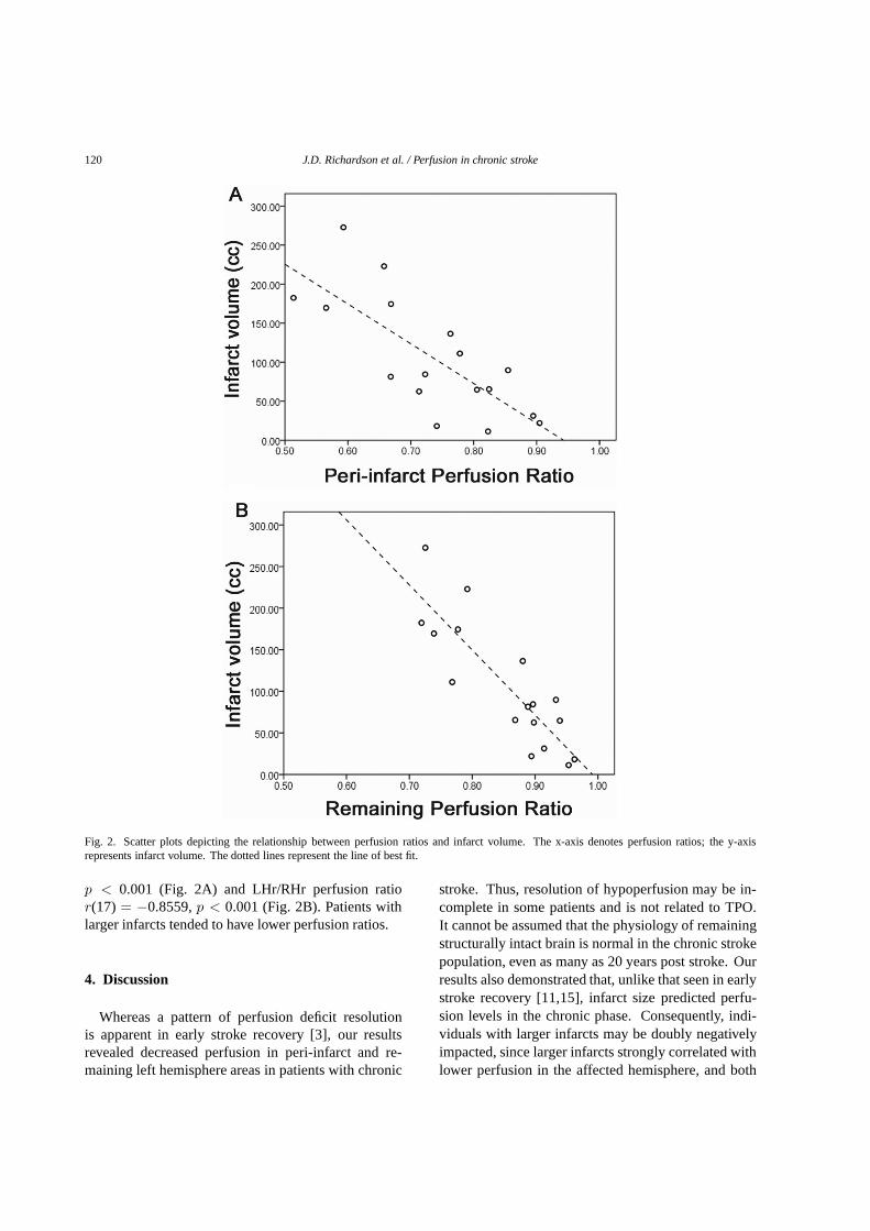
The correlations between TPO and perfusion ratiosdid not reach statistical significance. Significant nega-tive correlations were revealed between infarct size andboth the LHp/RHp perfusion ratio,r(17) = −0.757,
120 J.D. Richardson et al. / Perfusion in chronic stroke
Fig. 2. Scatter plots depicting the relationship between perfusion ratios and infarct volume. The x-axis denotes perfusion ratios; the y-axisrepresents infarct volume. The dotted lines represent the line of best fit.
p < 0.001 (Fig. 2A) and LHr/RHr perfusion ratior(17) = −0.8559,p < 0.001 (Fig. 2B). Patients withlarger infarcts tended to have lower perfusion ratios.
4. Discussion
Whereas a pattern of perfusion deficit resolutionis apparent in early stroke recovery [3], our resultsrevealed decreased perfusion in peri-infarct and re-maining left hemisphere areas in patients with chronic
stroke. Thus, resolution of hypoperfusion may be in-complete in some patients and is not related to TPO.It cannot be assumed that the physiology of remainingstructurally intact brain is normal in the chronic strokepopulation, even as many as 20 years post stroke. Ourresults also demonstrated that, unlike that seen in earlystroke recovery [11,15], infarct size predicted perfu-sion levels in the chronic phase. Consequently, indi-viduals with larger infarcts may be doubly negativelyimpacted, since larger infarcts strongly correlated withlower perfusion in the affected hemisphere, and both

J.D. Richardson et al. / Perfusion in chronic stroke 121
phenomena are possibly related to poorer clinical sta-tus [13,16,22]. Importantly, the potential contributionsof peri-infarct tissue to recoverymight be compromisedby hypoperfusion as there is evidence emphasizing theimportance of peri-infarct function for optimal clinicaloutcomes [10,20].
Much remains to be learned about the relationshipbetween hypoperfusion and function, as there is afore-mentioned evidence of functional deficits clearly at-tributed to hypoperfusion that contrasts with evidenceof patients with hypoperfusion and infarct to prima-ry language areas who do not demonstrate functionaldeficits [21]. Information about perfusion deficits andits effects on behaviors may need to be incorporatedinto future lesion-impairment studies so that a greaterdegree of accuracy can be achieved when character-izing brain-behavior relationships. It is possible thatcerebral perfusion deficits might underlie some of thevariability observed in chronic stroke outcomes, and ifso, should be accounted for in future studies that relatebrain function to stroke recovery.
These results confirm the need for further changesto functional MRI (fMRI) research design in chronicstroke patients since it has been demonstrated that thispopulation often presents with a sustained decrease inthe blood oxygen level dependent (BOLD) signal [5,12]. The majority of stroke recovery studies, howev-er, utilize canonical fMRI protocols that may not ac-count for stroke related changes in blood flow. Basedon the present results, future fMRI studies involvingchronic stroke patients may be improved by accountingfor possible perfusion abnormalities [5]. Furthermore,it is largely unknown how the status of the vessel pa-tency may impact perfusion. It remains to be definedhow intra- and extra-cranial stenosis affect perfusion inchronic stroke patients, particularly when other factorssuch as collateral perfusion [9] may play a significantrole.
Our patients constitute a heterogeneous group thatis representative of the variety of ischemic stroke eti-ologies. The mechanism of sustained hypoperfusionin our patients is unknown, and because at least asubset of our population demonstrate hypoperfusionin the absence of stenosis, mechanisms not specif-ic to occlusion should be explored. Stroke-inducedchanges in neurovascular function, such as cerebrovas-cular reactivity (CVR), might better explain our re-sults. The co-occurrence of hypoperfusion and re-duced CVR has been reported [4,6], and reduced CVRhas been reported in peri-infarct regions in both acuteand chronic recovery [4,6,8,18], and within chronic
recovery in individuals with and without chronic oc-clusion [8]. Another potential explanation could arisefrom a supply-demand situation in which the neurons’metabolic needs, and thus perfusion, are reduced be-cause they are not functioning normally within the cor-tical network. Regardless of the mechanism, it seemshypoperfusion is more common in chronic stroke thanpreviously described. Successful reperfusion treat-ments have been demonstrated for early recovery; forinstance, Hillis and colleagues have shown how reper-fusion and subsequent improvement in visual neglect orspeech-languageabilities can be manipulated by restor-ing blood flow to hypoperfused areas via pharmacolog-ical intervention or surgical procedures [13–15]. Wespeculate that the chronic patient population might ben-efit from similar research seeking to improve perfusionin the affected hemisphere, perhaps via intensive treat-ment [17] or brain stimulation [2], emphasizing theneed for further research regarding the mechanism ofchronic hypoperfusion.
The peri-infarct mask in the present study was on-ly extended 8 mm beyond the infarct, insinuating thatpatients might not demonstrate hypoperfusion beyondthis region. However, it is possible that at least somepatients demonstrate significant perfusion deficits fur-ther than 8 mm beyond the infarct. Encouragingly,neu-rons within hypoperfused areas had not been absorbedinto the ischemic core in these patients and may be vi-able for future treatments that can capitalize on theirrecruitment. Exploration into the many topics present-ed in this discussion is currently underway. With theemergence and continued adoption of noninvasive per-fusion imaging, it is our hope that cerebral perfusion inchronic stroke and its relationship to functional statuswill be more thoroughly characterized.
Acknowledgements
This work was supported by the following grants:DC008355 (PI: JF) and DC009571 (PIs: JF & CR).
We would like to thank: Jiongjiong Wang, PhD, Ra-diology, University of Pennsylvania, Philadelphia, PA19104, USA for providing the ASL sequence; Brian MDale, PhD, Siemens Medical, Morrisville, NC 27560,USA for supporting the ASL sequence under the Mas-ter Research Agreement between Siemens Medical andthe University of South Carolina; and our patients fortheir willingness to participate in our research.
122 J.D. Richardson et al. / Perfusion in chronic stroke
Disclosure/conflict of interest
Paul S. Morgan entered into a Master ResearchAgreement with Siemens Medical that supports the useof the ASL sequence at the University of South Caroli-na.
References
[1] L.M. Auer, G. Pfurtscheller, S. Abobaker and E. Ott, K.-J.Marguc and H. Lechner, Penumbra around chronic cerebralinfarction?Neurological Research10 (1988), 246–251.
[2] J. M. Baker, C. Rorden and J. Fridriksson, Using transcranialdirect-current stimulation to treat stroke patients with aphasia,Stroke41 (2010), 1229–1236.
[3] C. Beaulieu, A. de Crespigny, D.C. Tong, M.E. Moseley,G.W. Albers and M.P. Marks, Longitudinal magnetic reso-nance imaging study of perfusion and diffusion in stroke: Evo-lution of lesion volume and correlation with clinical outcome,Annals of Neurology46 (1999), 568–578.
[4] R.P.H. Bokkers, M.J.P. van Osch, H.B. van der Worp, G.J. deBorst, W.P.T.M. Mali and J. Hendrikse, Symptomatic carotidartery stenosis: Impairment of cerebral autoregulation mea-sured at the brain tissue level with arterial spin-labelingMRimaging,Radiology256(2010), 201–208.
[5] B. Bonakdarpour, T.B. Parrish and C.K. Thompson, Hemo-dynamic response function in patients with stroke-inducedaphasia: Implications for fMRI data analysis,NeuroImage36(2007), 322–331.
[6] L.M. Carusone, J. Srinivasan, D.R. Gitelman, M.M. Mesulamand T.B. Parrish, Hemodynamic response changes in cere-brovascular disease: Implications for functional MR imaging,American Journal of Neuroradiology23 (2002), 1222–1228.
[7] J.A. Chalela, D.C. Alsop, J.B. Gonzalez-Atavales, J.A.Mald-jian, S.E. Kasner and J.A. Detre, Magnetic resonance perfu-sion imaging in acute ischemic stroke using continuous arterialspin labeling,Stroke31 (2000), 680–687.
[8] C.C. Chang, H. Kanno, I. Yamamoto and N. Kuwana,Cerebrovascular reactivity to acetazolamide in alert patientswith cerebral infarction: Usefulness of first-pass radionu-clide angiography using 99m Tc-HMPAO in monitoring cere-bral haemodynamics,Nuclear Medicine Communications22(2001), 1119–1122.
[9] C.P. Derdeyn, T.O. Videen, S.M. Fritsch, D.A. Carpenter, R.L.Grubb and W.J. Powers, Compensatory mechanisms for chron-ic cerebral hypoperfusion in patients with carotid occlusion,Stroke30 (1999), 1019–1024.
[10] J. Fridriksson, L. Bonilha, J.M. Baker, D. Moser and C. Ror-
den, Activity in preserved left hemisphere regions predictsanomia severity in aphasia,Cerebral Cortex20 (2010), 1013–1019.
[11] J. Fridriksson, A.L. Holland, B.M. Coull, E. Plante, T.P.Trouard and P. Beeson, Aphasia severity: Association withcerebral perfusion and diffusion,Aphasiology16(2002), 859–871.
[12] J. Fridriksson, C. Rorden, P.S. Morgan, K.L. Morrow andG.C. Baylis, Measuring the hemodynamic response in chronichypoperfusion,Neurocase12 (2006), 146–150.
[13] A.E. Hillis, Magnetic resonance perfusion imaging in the studyof language,Brain and Language102(2007), 165–175.
[14] A.E. Hillis, J.T. Kleinman, M. Newhart et al., Restoring cere-bral blood flow reveals neural regions critical for naming,TheJournal of Neuroscience26 (2006), 8069–8073.
[15] A.E. Hillis, M. Newhart, J. Heidler, P.B. Barker, E.H. Her-skovits and M. Degaonkar, Anatomy of spatial attention: In-sights from perfusion imaging and hemispatial neglect in acutestroke,The Journal of Neuroscience25 (2005), 3161–3167.
[16] A. Kertesz, Lesion size and location in recovery from aphasia,Journal of Neurolinguistics3 (1988), 49–61.
[17] M. Kononen, J.R. Kuikka, M. Husso-Saastamoinen et al.,Increased perfusion in motor areas after constraint-inducedmovement therapy in chronic stroke: A single-photon emis-sion computerized tomography study,Journal of CerebralBlood Flow & Metabolism25 (2005), 1668–1674.
[18] A. Krainik, M. Hund-Georgiadis, S. Zysset and Y. von Cra-mon, Regional impairment of cerebrovascular reactivity andBOLD signal in adults after stroke,Stroke36 (2005), 1146–1152.
[19] T. Love, D. Swinney, E. Wong and R. Buxton, Perfusion imag-ing and stroke: A more sensitive measure of the brain basesof cognitive deficits,Aphasiology16 (2002), 873–883.
[20] M. Meinzer, T. Flaisch, C. Breitenstein, C. Wienbruch,T.Elbert and B. Rockstroh, Functional re-recruitment of dys-functional brain areas predicts language recovery in chronicaphasia,NeuroImage39 (2008), 2038–2046.
[21] E. Ochfeld, M. Newhart, J. Molitoris et al., Ischemia inBrocaarea is associated with Broca aphasia more reliably in acutethan in chronic stroke,Stroke41 (2010), 325–330.
[22] G. Rodriguez, F. Nobili, F. De Carli et al., Regional cerebralblood flow in chronic stroke patients,Stroke24(1993), 94–99.
[23] C. Rorden, J. Fridriksson and H.O. Karnath, An evaluationof traditional and novel tools for lesion behavior mapping,NeuroImage44 (2009), 1355–1362.
[24] S.M. Smith, M. Jenkinson, M.W. Woolrich et al., Advances infunctional and structural MR image analysis and implementa-tion as FSL,NeuroImage23 (2004), S208–S219.
[25] J. Wang, D.J. Licht, G.-H. Jahng et al., Pediatric perfusionimaging using Pulsed Arterial Spin Labeling,Journal of Mag-netic Resonance Imaging18 (2003), 404–413.

Submit your manuscripts athttp://www.hindawi.com
Stem CellsInternational
Hindawi Publishing Corporationhttp://www.hindawi.com Volume 2014
Hindawi Publishing Corporationhttp://www.hindawi.com Volume 2014
MEDIATORSINFLAMMATION
of
Hindawi Publishing Corporationhttp://www.hindawi.com Volume 2014
Behavioural Neurology
EndocrinologyInternational Journal of
Hindawi Publishing Corporationhttp://www.hindawi.com Volume 2014
Hindawi Publishing Corporationhttp://www.hindawi.com Volume 2014
Disease Markers
Hindawi Publishing Corporationhttp://www.hindawi.com Volume 2014
BioMed Research International
OncologyJournal of
Hindawi Publishing Corporationhttp://www.hindawi.com Volume 2014
Hindawi Publishing Corporationhttp://www.hindawi.com Volume 2014
Oxidative Medicine and Cellular Longevity
Hindawi Publishing Corporationhttp://www.hindawi.com Volume 2014
PPAR Research
The Scientific World JournalHindawi Publishing Corporation http://www.hindawi.com Volume 2014
Immunology ResearchHindawi Publishing Corporationhttp://www.hindawi.com Volume 2014
Journal of
ObesityJournal of
Hindawi Publishing Corporationhttp://www.hindawi.com Volume 2014
Hindawi Publishing Corporationhttp://www.hindawi.com Volume 2014
Computational and Mathematical Methods in Medicine
OphthalmologyJournal of
Hindawi Publishing Corporationhttp://www.hindawi.com Volume 2014
Diabetes ResearchJournal of
Hindawi Publishing Corporationhttp://www.hindawi.com Volume 2014
Hindawi Publishing Corporationhttp://www.hindawi.com Volume 2014
Research and TreatmentAIDS
Hindawi Publishing Corporationhttp://www.hindawi.com Volume 2014
Gastroenterology Research and Practice
Hindawi Publishing Corporationhttp://www.hindawi.com Volume 2014
Parkinson’s Disease
Evidence-Based Complementary and Alternative Medicine
Volume 2014Hindawi Publishing Corporationhttp://www.hindawi.com