Embed Size (px)
Citation preview
Cerebral Histamine H1 Receptor BindingPotential Measured with PET Under a TestDose of Olopatadine, an Antihistamine, IsReduced After Repeated Administration ofOlopatadine
Michio Senda1, Nobuo Kubo2, Kazuhiko Adachi3, Yasuhiko Ikari1,4, Keiichi Matsumoto1, Keiji Shimizu1,and Hideyuki Tominaga1
1Division of Molecular Imaging, Institute of Biomedical Research and Innovation, Kobe, Japan; 2Department ofOtorhinolaryngology, Kansai Medical University, Osaka, Japan; 3Department of Mechanical Engineering, Kobe University,Kobe, Japan; and 4Micron, Inc., Kobe, Japan
Some antihistamine drugs that are used for rhinitis and pollinosishave a sedative effect as they enter the brain and block the H1
receptor, potentially causing serious accidents. Receptor occu-pancy has been measured with PET under single-dose adminis-tration in humans to classify antihistamines as more sedating oras less sedating (or nonsedating). In this study, the effect of re-peated administration of olopatadine, an antihistamine, on thecerebral H1 receptor was measured with PET. Methods: A totalof 17 young men with rhinitis underwent dynamic brain PET with11C-doxepin at baseline, under an initial single dose of 5 mg ofolopatadine (acute scan), and under another 5-mg dose after re-peated administration of olopatadine at 10 mg/d for 4 wk (chronicscan). The H1 receptor binding potential was estimated usingLogan graphical analysis with cerebellum as reference region in-put. Results: The acute scan showed a slight decrease in H1 re-ceptor binding potential across the cerebral cortex (by 15% inthe frontal cortex), but the chronic scan showed a marked de-crease (by 45% from the acute scan in the frontal cortex). Behav-ioral data before and after the PET scans did not reveal anysedative effect. Conclusion: The results may be interpreted aseither intracerebral accumulation of olopatadine or H1 receptordownregulation due to repeated administration. The studyshows feasibility and potential value for PET in evaluating thepharmacologic effect of a drug not only after a single dose butalso after repeated administration.
Key Words: 11C-doxepin; histamine H1 receptor; olopatadine;PET; receptor occupancy
J Nucl Med 2009; 50:887–892DOI: 10.2967/jnumed.108.058537
Antihistamines are widely used as a medication forcommon allergic disorders such as seasonal pollinosis,chronic rhinitis, and urticaria. Some antihistamine drugshave a sedative side effect as they enter the brain and blockhistamine H1 receptor, potentially causing traffic accidentsand other serious events, but the sedative effect is difficult toevaluate because of a large variation in neuropsychologicalmeasures and subjective symptoms. Measurement of cere-bral histamine H1 receptor occupancy using PET with 11C-doxepin under a single administration of antihistamines hasbeen effective in evaluating the sedative effect, allowing thedrugs to be classified as more sedating or as less sedating (ornonsedating) (1,2). Olopatadine is a widely used antihista-mine with high antiallergy efficacy and has been reported tobe less sedating on the basis of PET measurements andneuropsychological tests (3). A mild sedative effect has beenpointed out for olopatadine despite higher efficacy thanfexofenadine, which is a nonsedating antihistamine (4).
Recently, the sedative effect of olopatadine has beenshown in neuropsychological tests to wear off after repeatedadministration (5). Investigators administered eitherolopatadine (10 mg/d) or fexofenadine (120 mg/d) in adouble-blind manner for more than 6 wk to 42 subjects of15–64 y old with seasonal pollinosis and found that thosewho took olopatadine complained of mild sleepiness at 2 wk,with the sleepiness score being significantly higher than thatfor subjects who took fexofenadine (P , 0.05). However, thesedative effect wore off and the sleepiness scale decreased tothe level of fexofenadine after 4 and 7 wk of repeatedadministration. Although the underlying mechanism of thewear-off phenomenon is not clear, such tolerance has beenobserved for other antihistamines, including diphenhydra-mine (6), mequitazine, and dexchlorpheniramine (7).
Received Sep. 25, 2008; revision accepted Feb. 27, 2009.For correspondence or reprints contact: Michio Senda, Division of
Molecular Imaging, Institute of Biomedical Research and Innovation, 2-2Minatojima-Minamimachi, Chuo-ku, Kobe 650-0047, Japan.
E-mail: [email protected] ª 2009 by the Society of Nuclear Medicine, Inc.
ANTIHISTAMINE EFFECT ON CEREBRAL H1 RECEPTOR • Senda et al. 887
by on May 20, 2020. For personal use only. jnm.snmjournals.org Downloaded from
The H1 receptor can be imaged and its binding potential(BP) evaluated with PET, but the technique has not beenapplied to the effect of repeated dosing with antihistamine,possibly because of the limited availability of the PETtechnique and the difficulty in controlling the medicationfor a certain period from the viewpoint of ethics andcompliance. The purpose of the present study was todemonstrate the feasibility of measuring the H1 receptorBP and receptor occupancy using PET with 11C-doxepinafter a 4-wk repeated administration of olopatadine and,thereby, to explore the possible mechanism of the wear-offphenomenon. Contrary to our initial expectation, we founda striking reduction in the H1 receptor BP, as reported here.
MATERIALS AND METHODS
SubjectsThe study protocol and the informed consent documents were
approved by the Ethics Committee of the Institute of BiomedicalResearch and Innovation, and written informed consent wasobtained from every subject.
A total of 17 young Japanese men (age 20–24) with perennialnasal allergy participated in the study. To be included, the subjectshad to be male and 20–29 y old, with clinically moderate allergicrhinitis based on the guideline of the Japanese Society of Aller-gology, a IgE titer score of 2 or higher level for house-dust mites, ahistory of antihistamine treatment but no current treatment withantihistamine medication, and a visual acuity of more than 0.1.The exclusion criteria were contraindications for MRI, organicbrain disease or morphologic brain anomaly, and liver or renaldysfunction.
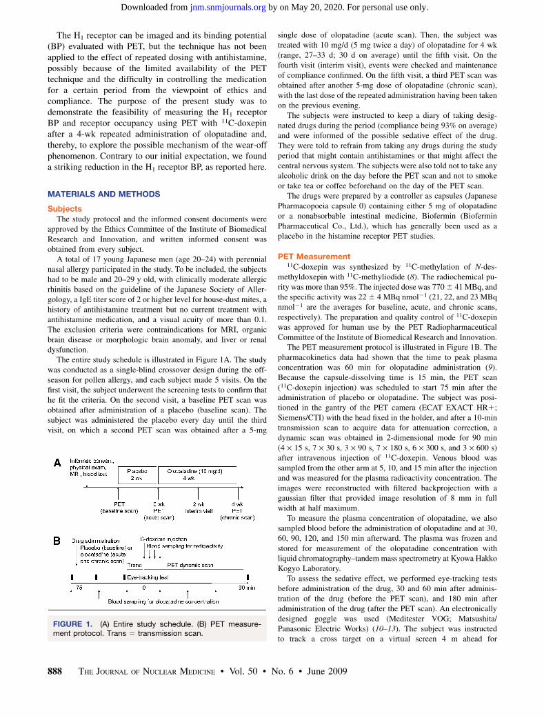
The entire study schedule is illustrated in Figure 1A. The studywas conducted as a single-blind crossover design during the off-season for pollen allergy, and each subject made 5 visits. On thefirst visit, the subject underwent the screening tests to confirm thathe fit the criteria. On the second visit, a baseline PET scan wasobtained after administration of a placebo (baseline scan). Thesubject was administered the placebo every day until the thirdvisit, on which a second PET scan was obtained after a 5-mg
single dose of olopatadine (acute scan). Then, the subject wastreated with 10 mg/d (5 mg twice a day) of olopatadine for 4 wk(range, 27–33 d; 30 d on average) until the fifth visit. On thefourth visit (interim visit), events were checked and maintenanceof compliance confirmed. On the fifth visit, a third PET scan wasobtained after another 5-mg dose of olopatadine (chronic scan),with the last dose of the repeated administration having been takenon the previous evening.
The subjects were instructed to keep a diary of taking desig-nated drugs during the period (compliance being 93% on average)and were informed of the possible sedative effect of the drug.They were told to refrain from taking any drugs during the studyperiod that might contain antihistamines or that might affect thecentral nervous system. The subjects were also told not to take anyalcoholic drink on the day before the PET scan and not to smokeor take tea or coffee beforehand on the day of the PET scan.
The drugs were prepared by a controller as capsules (JapanesePharmacopoeia capsule 0) containing either 5 mg of olopatadineor a nonabsorbable intestinal medicine, Biofermin (BioferminPharmaceutical Co., Ltd.), which has generally been used as aplacebo in the histamine receptor PET studies.
PET Measurement11C-doxepin was synthesized by 11C-methylation of N-des-
methyldoxepin with 11C-methyliodide (8). The radiochemical pu-rity was more than 95%. The injected dose was 770 6 41 MBq, andthe specific activity was 22 6 4 MBq nmol21 (21, 22, and 23 MBqnmol21 are the averages for baseline, acute, and chronic scans,respectively). The preparation and quality control of 11C-doxepinwas approved for human use by the PET RadiopharmaceuticalCommittee of the Institute of Biomedical Research and Innovation.
The PET measurement protocol is illustrated in Figure 1B. Thepharmacokinetics data had shown that the time to peak plasmaconcentration was 60 min for olopatadine administration (9).Because the capsule-dissolving time is 15 min, the PET scan(11C-doxepin injection) was scheduled to start 75 min after theadministration of placebo or olopatadine. The subject was posi-tioned in the gantry of the PET camera (ECAT EXACT HR1;Siemens/CTI) with the head fixed in the holder, and after a 10-mintransmission scan to acquire data for attenuation correction, adynamic scan was obtained in 2-dimensional mode for 90 min(4 · 15 s, 7 · 30 s, 3 · 90 s, 7 · 180 s, 6 · 300 s, and 3 · 600 s)after intravenous injection of 11C-doxepin. Venous blood wassampled from the other arm at 5, 10, and 15 min after the injectionand was measured for the plasma radioactivity concentration. Theimages were reconstructed with filtered backprojection with agaussian filter that provided image resolution of 8 mm in fullwidth at half maximum.
To measure the plasma concentration of olopatadine, we alsosampled blood before the administration of olopatadine and at 30,60, 90, 120, and 150 min afterward. The plasma was frozen andstored for measurement of the olopatadine concentration withliquid chromatography–tandem mass spectrometry at Kyowa HakkoKogyo Laboratory.
To assess the sedative effect, we performed eye-tracking testsbefore administration of the drug, 30 and 60 min after adminis-tration of the drug (before the PET scan), and 180 min afteradministration of the drug (after the PET scan). An electronicallydesigned goggle was used (Meditester VOG; Matsushita/Panasonic Electric Works) (10–13). The subject was instructedto track a cross target on a virtual screen 4 m ahead for
FIGURE 1. (A) Entire study schedule. (B) PET measure-ment protocol. Trans 5 transmission scan.
888 THE JOURNAL OF NUCLEAR MEDICINE • Vol. 50 • No. 6 • June 2009
by on May 20, 2020. For personal use only. jnm.snmjournals.org Downloaded from
measurement of saccadic latency. The subject was also asked howsleepy he felt, on a 4-step scale, after he took the drug.
Data AnalysisQuantitative analysis was performed on several representative
cortical regions to estimate the BP of 11C-doxepin for each scan.Sets of circular regions of interest 1 cm in diameter were placedalong the cortical rims over the dorsolateral prefrontal cortex(DLPFC), frontobasal cortex, temporal cortex, parietal cortex,occipital cortex, and anterior cingulate cortex, as well as in thethalamus and the cerebellum on the baseline scan images of eachsubject to obtain the time–activity curves for each region. Theacute and chronic images were coregistered to the baseline imagesof each subject to obtain the time–activity curves for the sameregions of interest as on the baseline images. The distributionvolume ratio was computed from the time–activity curve for eachregion using Logan graphical analysis with reference region input(14). Cerebellum was used as the reference region because ofnegligible H1 receptor (15–17). The cerebellar efflux constant wasset to be the average of healthy subjects in a previous study (0.022min21) (18). BP (as BPND, where ND 5 nondisplaceable) wascomputed as distribution volume ratio – 1, and receptor occupancywas computed as 1 – (BPND at acute scan/BPND at chronic scan).When the computed receptor occupancy was negative, zero wasassigned. For each region, BPND was compared between the scansusing 2-way ANOVA with Fisher’s least significant difference(19). Statistical significance was tested with Bonferroni adjust-ment by a factor of 7 (7 regions).
We also used an alternative approach that was in line withprevious investigations. The standard input function of 11C-doxepin from previously obtained normal data was calibratedwith the 3-point plasma radioactivity data for each scan of thepresent study. This calibrated input function was applied as theinput function after metabolite correction using the unmetabolized11C-doxepin fraction data of the population (20). The distributionvolume of 11C-doxepin was obtained for each region using Logangraphical analysis with calibrated standard input (21). BP (asBPND) for each region was computed as (distribution volume inthe target region/distribution volume in the cerebellum) 21, andreceptor occupancy was computed in the same way.
RESULTS
In 3 of the 17 subjects, the plasma concentration ofolopatadine rose (arbitrarily above 20 ng mL21) toward thevery end of the PET scan or did not rise at all by 150 min afterthe drug had been administered in the acute or chronic scan or
both. These subjects were thought to have a drug absorptionproblem or unusual pharmacokinetic behavior and wereexcluded from the data analysis. For the remaining 14subjects, the average and SD of the plasma concentrationof olopatadine at 0, 30, 60, 90, 120, and 150 min afteradministration was 0 6 0, 21 6 25, 62 6 28, 46 6 21, 36 6 9,and 33 6 11 ng mL21, respectively, for the acute scan and 7 6
8, 28 6 24, 69 6 29, 52 6 13, 41 6 9, and 35 6 8 ng mL21,respectively, for the chronic scan. The area under the curvewas 57 6 16 and 64 6 14 ng mL21 h (P 5 0.13 by paired ttest) for the duration of the acute and chronic scans, respec-tively.
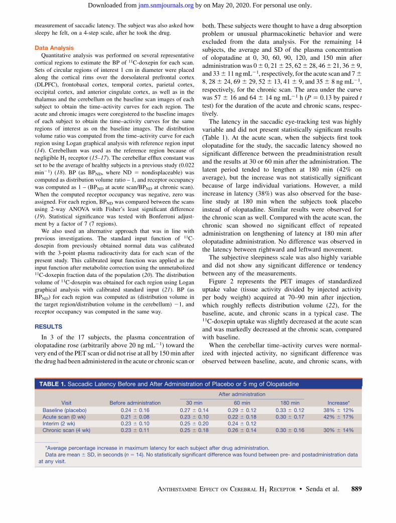
The latency in the saccadic eye-tracking test was highlyvariable and did not present statistically significant results(Table 1). At the acute scan, when the subjects first tookolopatadine for the study, the saccadic latency showed nosignificant difference between the preadministration resultand the results at 30 or 60 min after the administration. Thelatent period tended to lengthen at 180 min (42% onaverage), but the increase was not statistically significantbecause of large individual variations. However, a mildincrease in latency (38%) was also observed for the base-line study at 180 min when the subjects took placeboinstead of olopatadine. Similar results were observed forthe chronic scan as well. Compared with the acute scan, thechronic scan showed no significant effect of repeatedadministration on lengthening of latency at 180 min afterolopatadine administration. No difference was observed inthe latency between rightward and leftward movement.
The subjective sleepiness scale was also highly variableand did not show any significant difference or tendencybetween any of the measurements.
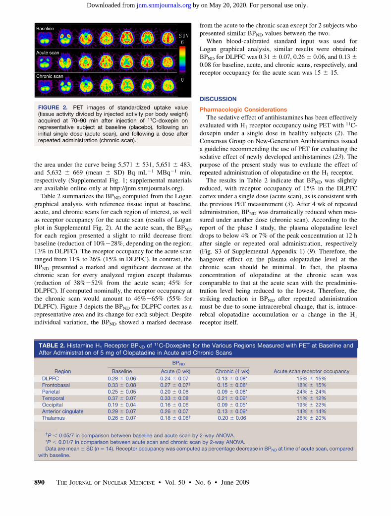
Figure 2 represents the PET images of standardizeduptake value (tissue activity divided by injected activityper body weight) acquired at 70–90 min after injection,which roughly reflects distribution volume (22), for thebaseline, acute, and chronic scans in a typical case. The11C-doxepin uptake was slightly decreased at the acute scanand was markedly decreased at the chronic scan, comparedwith baseline.
When the cerebellar time–activity curves were normal-ized with injected activity, no significant difference wasobserved between baseline, acute, and chronic scans, with
TABLE 1. Saccadic Latency Before and After Administration of Placebo or 5 mg of Olopatadine
After administration
Visit Before administration 30 min 60 min 180 min Increase*
Baseline (placebo) 0.24 6 0.16 0.27 6 0.14 0.29 6 0.12 0.33 6 0.12 38% 6 12%Acute scan (0 wk) 0.21 6 0.08 0.23 6 0.10 0.22 6 0.18 0.30 6 0.17 42% 6 17%
Interim (2 wk) 0.23 6 0.10 0.25 6 0.20 0.24 6 0.12
Chronic scan (4 wk) 0.23 6 0.11 0.25 6 0.18 0.26 6 0.14 0.30 6 0.16 30% 6 14%
*Average percentage increase in maximum latency for each subject after drug administration.
Data are mean 6 SD, in seconds (n 5 14). No statistically significant difference was found between pre- and postadministration data
at any visit.
ANTIHISTAMINE EFFECT ON CEREBRAL H1 RECEPTOR • Senda et al. 889
by on May 20, 2020. For personal use only. jnm.snmjournals.org Downloaded from
the area under the curve being 5,571 6 531, 5,651 6 483,and 5,632 6 669 (mean 6 SD) Bq mL21 MBq21 min,respectively (Supplemental Fig. 1; supplemental materialsare available online only at http://jnm.snmjournals.org).
Table 2 summarizes the BPND computed from the Logangraphical analysis with reference tissue input at baseline,acute, and chronic scans for each region of interest, as wellas receptor occupancy for the acute scan (results of Loganplot in Supplemental Fig. 2). At the acute scan, the BPND
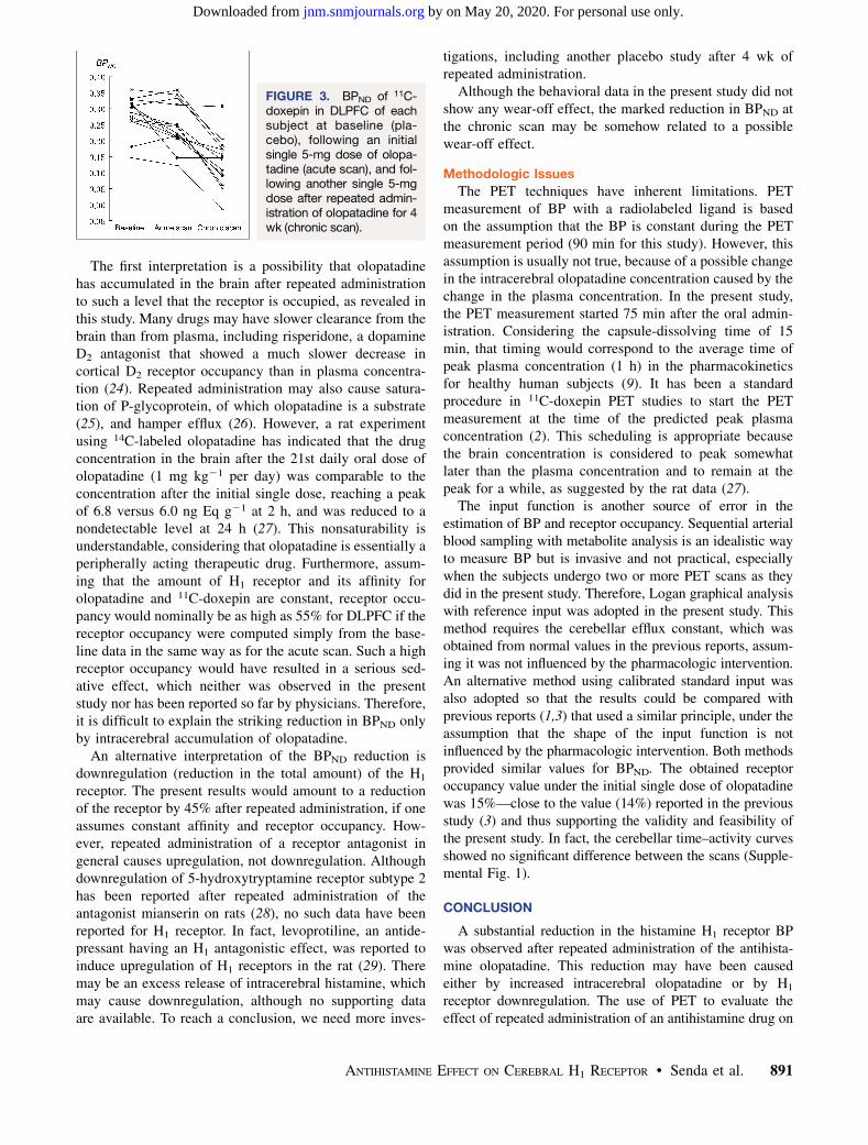
for each region presented a slight to mild decrease frombaseline (reduction of 10%228%, depending on the region;13% in DLPFC). The receptor occupancy for the acute scanranged from 11% to 26% (15% in DLPFC). In contrast, theBPND presented a marked and significant decrease at thechronic scan for every analyzed region except thalamus(reduction of 38%252% from the acute scan; 45% forDLPFC). If computed nominally, the receptor occupancy atthe chronic scan would amount to 46%265% (55% forDLPFC). Figure 3 depicts the BPND for DLPFC cortex as arepresentative area and its change for each subject. Despiteindividual variation, the BPND showed a marked decrease
from the acute to the chronic scan except for 2 subjects whopresented similar BPND values between the two.
When blood-calibrated standard input was used forLogan graphical analysis, similar results were obtained:BPND for DLPFC was 0.31 6 0.07, 0.26 6 0.06, and 0.13 6
0.08 for baseline, acute, and chronic scans, respectively, andreceptor occupancy for the acute scan was 15 6 15.
DISCUSSION
Pharmacologic Considerations
The sedative effect of antihistamines has been effectivelyevaluated with H1 receptor occupancy using PET with 11C-doxepin under a single dose in healthy subjects (2). TheConsensus Group on New-Generation Antihistamines issueda guideline recommending the use of PET for evaluating thesedative effect of newly developed antihistamines (23). Thepurpose of the present study was to evaluate the effect ofrepeated administration of olopatadine on the H1 receptor.
The results in Table 2 indicate that BPND was slightlyreduced, with receptor occupancy of 15% in the DLPFCcortex under a single dose (acute scan), as is consistent withthe previous PET measurement (3). After 4 wk of repeatedadministration, BPND was dramatically reduced when mea-sured under another dose (chronic scan). According to thereport of the phase I study, the plasma olopatadine leveldrops to below 4% or 7% of the peak concentration at 12 hafter single or repeated oral administration, respectively(Fig. S3 of Supplemental Appendix 1) (9). Therefore, thehangover effect on the plasma olopatadine level at thechronic scan should be minimal. In fact, the plasmaconcentration of olopatadine at the chronic scan wascomparable to that at the acute scan with the preadminis-tration level being reduced to the lowest. Therefore, thestriking reduction in BPND after repeated administrationmust be due to some intracerebral change, that is, intrace-rebral olopatadine accumulation or a change in the H1
receptor itself.
FIGURE 2. PET images of standardized uptake value(tissue activity divided by injected activity per body weight)acquired at 70–90 min after injection of 11C-doxepin onrepresentative subject at baseline (placebo), following aninitial single dose (acute scan), and following a dose afterrepeated administration (chronic scan).
TABLE 2. Histamine H1 Receptor BPND of 11C-Doxepine for the Various Regions Measured with PET at Baseline andAfter Administration of 5 mg of Olopatadine in Acute and Chronic Scans
BPND
Region Baseline Acute (0 wk) Chronic (4 wk) Acute scan receptor occupancy
DLPFC 0.28 6 0.06 0.24 6 0.07 0.13 6 0.08* 15% 6 15%
Frontobasal 0.33 6 0.08 0.27 6 0.07y 0.15 6 0.08* 18% 6 15%
Parietal 0.25 6 0.05 0.20 6 0.08 0.09 6 0.08* 24% 6 24%Temporal 0.37 6 0.07 0.33 6 0.08 0.21 6 0.09* 11% 6 12%
Occipital 0.19 6 0.04 0.16 6 0.06 0.09 6 0.05* 19% 6 22%
Anterior cingulate 0.29 6 0.07 0.26 6 0.07 0.13 6 0.09* 14% 6 14%
Thalamus 0.26 6 0.07 0.18 6 0.06y 0.20 6 0.06 26% 6 20%
yP , 0.05/7 in comparison between baseline and acute scan by 2-way ANOVA.
*P , 0.01/7 in comparison between acute scan and chronic scan by 2-way ANOVA.Data are mean 6 SD (n 5 14). Receptor occupancy was computed as percentage decrease in BPND at time of acute scan, compared
with baseline.
890 THE JOURNAL OF NUCLEAR MEDICINE • Vol. 50 • No. 6 • June 2009
by on May 20, 2020. For personal use only. jnm.snmjournals.org Downloaded from
The first interpretation is a possibility that olopatadinehas accumulated in the brain after repeated administrationto such a level that the receptor is occupied, as revealed inthis study. Many drugs may have slower clearance from thebrain than from plasma, including risperidone, a dopamineD2 antagonist that showed a much slower decrease incortical D2 receptor occupancy than in plasma concentra-tion (24). Repeated administration may also cause satura-tion of P-glycoprotein, of which olopatadine is a substrate(25), and hamper efflux (26). However, a rat experimentusing 14C-labeled olopatadine has indicated that the drugconcentration in the brain after the 21st daily oral dose ofolopatadine (1 mg kg21 per day) was comparable to theconcentration after the initial single dose, reaching a peakof 6.8 versus 6.0 ng Eq g21 at 2 h, and was reduced to anondetectable level at 24 h (27). This nonsaturability isunderstandable, considering that olopatadine is essentially aperipherally acting therapeutic drug. Furthermore, assum-ing that the amount of H1 receptor and its affinity forolopatadine and 11C-doxepin are constant, receptor occu-pancy would nominally be as high as 55% for DLPFC if thereceptor occupancy were computed simply from the base-line data in the same way as for the acute scan. Such a highreceptor occupancy would have resulted in a serious sed-ative effect, which neither was observed in the presentstudy nor has been reported so far by physicians. Therefore,it is difficult to explain the striking reduction in BPND onlyby intracerebral accumulation of olopatadine.
An alternative interpretation of the BPND reduction isdownregulation (reduction in the total amount) of the H1
receptor. The present results would amount to a reductionof the receptor by 45% after repeated administration, if oneassumes constant affinity and receptor occupancy. How-ever, repeated administration of a receptor antagonist ingeneral causes upregulation, not downregulation. Althoughdownregulation of 5-hydroxytryptamine receptor subtype 2has been reported after repeated administration of theantagonist mianserin on rats (28), no such data have beenreported for H1 receptor. In fact, levoprotiline, an antide-pressant having an H1 antagonistic effect, was reported toinduce upregulation of H1 receptors in the rat (29). Theremay be an excess release of intracerebral histamine, whichmay cause downregulation, although no supporting dataare available. To reach a conclusion, we need more inves-
tigations, including another placebo study after 4 wk ofrepeated administration.
Although the behavioral data in the present study did notshow any wear-off effect, the marked reduction in BPND atthe chronic scan may be somehow related to a possiblewear-off effect.
Methodologic Issues
The PET techniques have inherent limitations. PETmeasurement of BP with a radiolabeled ligand is basedon the assumption that the BP is constant during the PETmeasurement period (90 min for this study). However, thisassumption is usually not true, because of a possible changein the intracerebral olopatadine concentration caused by thechange in the plasma concentration. In the present study,the PET measurement started 75 min after the oral admin-istration. Considering the capsule-dissolving time of 15min, that timing would correspond to the average time ofpeak plasma concentration (1 h) in the pharmacokineticsfor healthy human subjects (9). It has been a standardprocedure in 11C-doxepin PET studies to start the PETmeasurement at the time of the predicted peak plasmaconcentration (2). This scheduling is appropriate becausethe brain concentration is considered to peak somewhatlater than the plasma concentration and to remain at thepeak for a while, as suggested by the rat data (27).
The input function is another source of error in theestimation of BP and receptor occupancy. Sequential arterialblood sampling with metabolite analysis is an idealistic wayto measure BP but is invasive and not practical, especiallywhen the subjects undergo two or more PET scans as theydid in the present study. Therefore, Logan graphical analysiswith reference input was adopted in the present study. Thismethod requires the cerebellar efflux constant, which wasobtained from normal values in the previous reports, assum-ing it was not influenced by the pharmacologic intervention.An alternative method using calibrated standard input wasalso adopted so that the results could be compared withprevious reports (1,3) that used a similar principle, under theassumption that the shape of the input function is notinfluenced by the pharmacologic intervention. Both methodsprovided similar values for BPND. The obtained receptoroccupancy value under the initial single dose of olopatadinewas 15%—close to the value (14%) reported in the previousstudy (3) and thus supporting the validity and feasibility ofthe present study. In fact, the cerebellar time–activity curvesshowed no significant difference between the scans (Supple-mental Fig. 1).
CONCLUSION
A substantial reduction in the histamine H1 receptor BPwas observed after repeated administration of the antihista-mine olopatadine. This reduction may have been causedeither by increased intracerebral olopatadine or by H1
receptor downregulation. The use of PET to evaluate theeffect of repeated administration of an antihistamine drug on
FIGURE 3. BPND of 11C-doxepin in DLPFC of eachsubject at baseline (pla-cebo), following an initialsingle 5-mg dose of olopa-tadine (acute scan), and fol-lowing another single 5-mgdose after repeated admin-istration of olopatadine for 4wk (chronic scan).
ANTIHISTAMINE EFFECT ON CEREBRAL H1 RECEPTOR • Senda et al. 891
by on May 20, 2020. For personal use only. jnm.snmjournals.org Downloaded from
the H1 receptor is possible and plausible and may be usefulfor developing antihistamine drugs that are less sedating.
ACKNOWLEDGMENTS
This study was financially supported in part by Grant-in-Aid in Scientific Research B 16390350 of the Japan Societyfor Promotion of Science and by Kyowa Hakko Kogyo Co.Ltd., which is the manufacturer of olopatadine (Allelock).We thank Miho Suzuki and Shusaku Tazawa for acquiringthe data, and Jun-ichi Nagase and Masaru Takaishi forcoordinating the study.
REFERENCES
1. Tashiro M, Sakurada Y, Iwabuchi K, et al. Central effects of fexofenadine and
cetirizine: measurement of psychomotor performance, subjective sleepiness, and
brain histamine H1-receptor occupancy using 11C-doxepin positron emission
tomography. J Clin Pharmacol. 2004;44:890–900.
2. Yanai K, Tashiro M. The physiological and pathophysiological roles of neuronal
histamine: an insight from human positron emission tomography studies.
Pharmacol Ther. 2007;113:1–15.
3. Tashiro M, Mochizuki H, Sakurada Y, et al. Brain histamine H1 receptor
occupancy of orally administered antihistamines measured by positron emission
tomography with 11C-doxepin in a placebo-controlled crossover study design
in healthy subjects: a comparison of olopatadine and ketotifen. Br J Clin
Pharmacol. 2006;61:16–26.
4. Kamei H, Noda Y, Ishikawa K, et al. Comparative study of acute effects of single
doses of fexofenadine, olopatadine, d-chlorpheniramine and placebo on psycho-
motor function in healthy volunteers. Hum Psychopharmacol. 2003;18:611–618.
5. Kubo N, Asako M, Hamada S. A single-blinded randomized clinical study of
olopatadine hydrochloride versus fexofenadine hydrochloride with Japanese
ceder/cypress pollinosis. Allergy in Pract. 2005;25:1039–1051.
6. Richardson GS, Roehrs TA, Rosenthal L, Koshorek G, Roth T. Tolerance to
daytime sedative effects of H1 antihistamines. J Clin Psychopharmacol. 2002;
22:511–515.
7. Theunissen EL, Vermeeren A, Ramaekers JG. Repeated-dose effects of
mequitazine, cetirizine and dexchlorpheniramine on driving and psychomotor
performance. Br J Clin Pharmacol. 2006;61:79–86.
8. Iwata R, Pascali C, Bogni A, Yanai K, Kato M, Ido T. A combined loop-SPE
method for the automated preparation of [11C]doxepin. J Labelled Comp
Radiopharm. 2002;45:271–280.
9. Tsunoo M, Momomura S, Masuo M, et al. Phase I clinical study on KW-4679,
an antiallergic drug. Clin Rep. 1995;29:4129–4147.
10. Ferrara M, De Gennaro MFL, Bertini M. Voluntary oculomotor performance
upon awakening after total sleep deprivation. Sleep. 2000;23:801–811.
11. Bocca ML, Denise P. Total sleep deprivation effect on disengagement of spatial
attention as assessed by saccadic eye movements. Clin Neurophysiol. 2006;117:
894–899.
12. Abel LA, Douglas J. Effects of age on latency and error generation in internally
mediated saccades. Neurobiol Aging. 2007;28:627–637.
13. Chan F, Armstrong IT, Pari G, Riopelle RJ, Munoz DP. Deficits in saccadic eye-
movement control in Parkinson’s disease. Neuropsychologia. 2005;43:784–796.
14. Logan J, Fowler JS, Volkow ND, Wang GJ, Ding YS, Alexoff DL. Distribution
volume ratios without blood sampling from graphical analysis of PET data.
J Cereb Blood Flow Metab. 1996;16:834–840.
15. Chang RS, Tran VT, Snyder SH. Heterogeneity of histamine H1-receptors:
species variations in [3H]mepyramine binding of brain membranes. J Neuro-
chem. 1979;32:1653–1663.
16. Yanai K, Watanabe T, Yokoyama H, et al. Mapping of histamine H1 receptors
in the human brain using [11C]pyrilamine and positron emission tomography.
J Neurochem. 1992;59:128–136.
17. Yanai K, Watanabe T, Yokoyama H, et al. Histamine H1 receptors in human brain
visualized in vivo by [11C]doxepin and positron emission tomography. Neurosci
Lett. 1992;137:145–148.
18. Suzuki A, Tashiro M, Kimura Y, et al. Use of reference tissue models for
quantification of histamine H1 receptors in human brain by using positron
emission tomography and [11C]doxepin. Ann Nucl Med. 2005;19:425–433.
19. Hsu JC. Multiple Comparisons, Theory and Methods. London, U.K.: Chapman
and Hall; 1996.
20. Mochizuki H, Kimura Y, Ishii K, et al. Quantitative measurement of histamine
H1 receptors in human brains by PET and [11C]doxepin. Nucl Med Biol.
2004;31:165–171.
21. Logan J, Fowler JS, Volkow ND, et al. Graphical analysis of reversible radioligand
binding from time-activity measurements applied to [N-11C-methyl]-(2)-cocaine
PET studies in human subjects. J Cereb Blood Flow Metab. 1990;10:740–747.
22. Mochizuki H, Kimura Y, Ishii K, et al. Simplified PET measurement for
evaluating histamine H1 receptors in human brains using [11C]doxepin. Nucl
Med Biol. 2004;31:1005–1011.
23. Holgate ST, Canonica GW, Simons FE, et al. Consensus Group on New-
Generation Antihistamines (CONGA): present status and recommendations. Clin
Exp Allergy. 2003;33:1305–1324.
24. Takano A, Suhara T, Ikoma Y, et al. Estimation of the time-course of dopamine
D2 receptor occupancy in living human brain from plasma pharmacokinetics of
antipsychotics. Int J Neuropsychopharmacol. 2004;7:19–26.
25. Mimura N, Nagata Y, Kuwabara T, Kubo N, Fuse E. P-glycoprotein limits the
brain penetration of olopatadine hydrochloride, H1-receptor antagonist. Drug
Metab Pharmacokinet. 2008;23:106–114.
26. Talbot PS, Narandran R, Hwang D-R, et al. Modulation of brain delivery of the
mu opiate receptor agonist loperamide by P-glycoprotein inhibition: a PET study
using [11C]carfentanil in the baboon. J Cereb Blood Flow Metab. 2003;23(suppl
1):686.
27. Ohishi T, Nishiie H, Fuse E, Kobayashi H, Kobayashi S. Disposition of KW-
4679 (2), absorption, distribution, excretion of 14C-KW-4679 after multiple oral
administrations in rats and effect of KW-4679 on drug metabolizing enzyme in
rats. Drug Metab Pharmacokinet. 1995;10:669–682.
28. Conn PJ, Sanders-Bush E. Regulation of serotonin-stimulated phosphoinositide
hydrolysis: relation to the serotonin 5-HT-2 binding site. J Neurosci. 1986;
6:3669–3675.
29. Noguchi S, Inukai T. Repeated treatment with levoprotiline, a novel antidepres-
sant, up-regulates histamine H1 receptors and phosphoinositide hydrolysis
response in vivo. Jpn J Pharmacol. 1992;59:31–35.
892 THE JOURNAL OF NUCLEAR MEDICINE • Vol. 50 • No. 6 • June 2009
by on May 20, 2020. For personal use only. jnm.snmjournals.org Downloaded from
Doi: 10.2967/jnumed.108.058537Published online: May 14, 2009.
2009;50:887-892.J Nucl Med. TominagaMichio Senda, Nobuo Kubo, Kazuhiko Adachi, Yasuhiko Ikari, Keiichi Matsumoto, Keiji Shimizu and Hideyuki Olopatadine
ofDose of Olopatadine, an Antihistamine, Is Reduced After Repeated Administration Receptor Binding Potential Measured with PET Under a Test1Cerebral Histamine H
http://jnm.snmjournals.org/content/50/6/887This article and updated information are available at:
http://jnm.snmjournals.org/site/subscriptions/online.xhtml
Information about subscriptions to JNM can be found at:
http://jnm.snmjournals.org/site/misc/permission.xhtmlInformation about reproducing figures, tables, or other portions of this article can be found online at:
(Print ISSN: 0161-5505, Online ISSN: 2159-662X)1850 Samuel Morse Drive, Reston, VA 20190.SNMMI | Society of Nuclear Medicine and Molecular Imaging
is published monthly.The Journal of Nuclear Medicine
© Copyright 2009 SNMMI; all rights reserved.
by on May 20, 2020. For personal use only. jnm.snmjournals.org Downloaded from