Embed Size (px)
DESCRIPTION
Cerebral Circulation
Citation preview
Dr. Chhaya Saraf
Learning Issues
Arrangement of blood vesselsUnique features of cerebral circulationRegulation of cerebral circulationRole of intracranial tensionBlood Brain Barrier
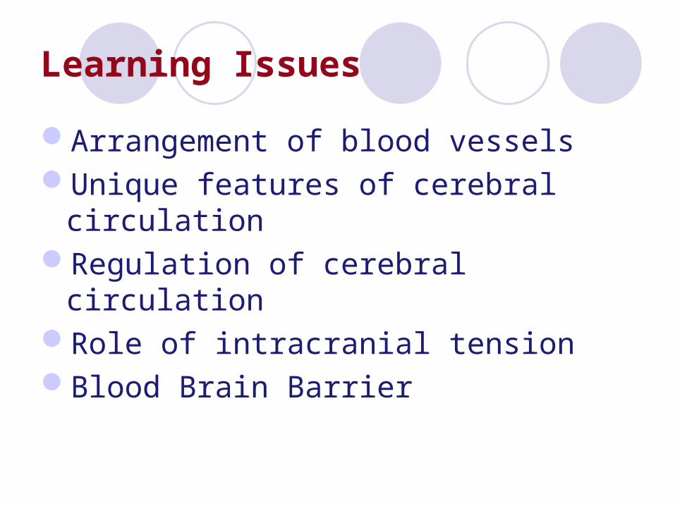
Circle of Willis
Enclosed within rigid structure (cranium).Total volume of blood, CSF, and brain tissue is constant.

•Cerebral veins – unique features (i) no valves, (ii) thin-walled, little smooth muscles, (iii) distribution does not correspond to distribution of arteries.•Drained mainly into internal jugular vein
Cerebral Blood FlowNormally 50-60ml/100gm of brain tissue 750 – 900 ml /min (15% of CO)
No lymphatics
In choroid plexus endothelium shows the gaps but epithelium has tight junctions
Cerebral capillaries have tight junctions, very little vesicular transport, surrounded by end feet of Astrocytes which help in tightening the junctions
Innervation Of Cerebral Blood Vessels
Sympathetic vasoconstrictor nerves from cervical ganglia
Cholinergic neuronsSensory nerves ( sensitive to pain)
Cerebral blood vessels are sensitive to pain though brain substance has no pain fibers

Cerebral circulation• Characteristic features:
–Monro - Kellie Doctrine– contained within rigid structure constant –
inflow/outflow imbalances pressure• Cushing’s Reflex
– Absolute requirement for adequate flow• tissue least tolerant to ischemia
–5 sec ischemia loss of consciousness• glucose-dependent • no contribution to total PR regulation
Measurement & Regional Changes Of Blood FlowPositron Emission Tomography - PETMagnetic Resonance Imaging - MRIInjecting radioactive xenon in carotid artery,
blood flow can be measured by scintillating detectors over the surface of cerebral cortex
By Fick’s PrincipleCerebral blood flow changes 100 to 150% within
sec depending upon local activity of neurons e.g. Reading – visual cortexSpeech – Wernicke’s area
Cerebral Microcirculation
4 times more capillaries in gray matter than white matter
Relatively poor permeability due to tight junctions
Less leaky as supported by glial feet (Astrocytes), prevents overstretching during high pressures
In hypertensives arterioles are usually thickened & constricted preventing the transmission of high pressure
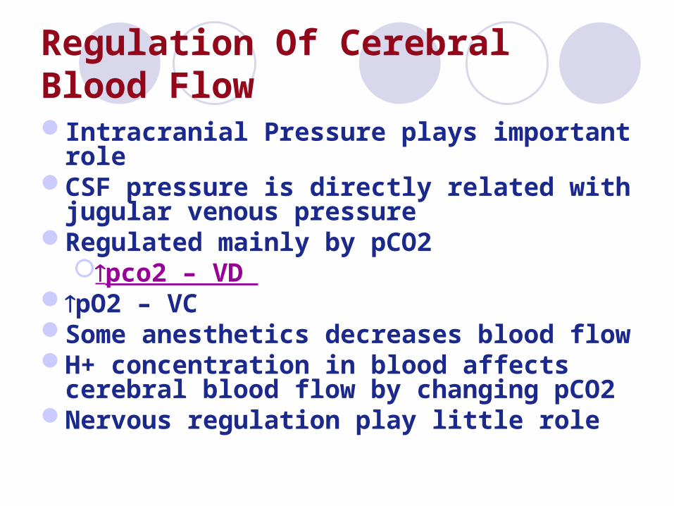
Regulation Of Cerebral Blood FlowIntracranial Pressure plays important roleCSF pressure is directly related with jugular
venous pressureRegulated mainly by pCO2
pco2 – VD pO2 – VC Some anesthetics decreases blood flowH+ concentration in blood affects cerebral
blood flow by changing pCO2Nervous regulation play little role
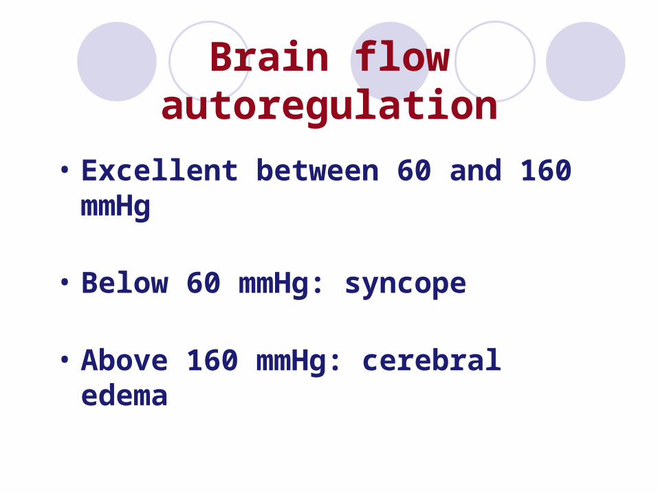
Brain flow autoregulation
• Excellent between 60 and 160 mmHg
• Below 60 mmHg: syncope
• Above 160 mmHg: cerebral edema
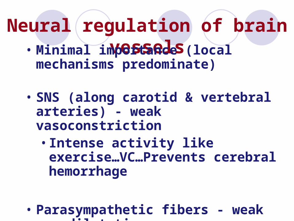
Neural regulation of brain vessels• Minimal importance (local mechanisms
predominate)
• SNS (along carotid & vertebral arteries) - weak vasoconstriction• Intense activity like exercise…VC…Prevents
cerebral hemorrhage
• Parasympathetic fibers - weak vasodilatation
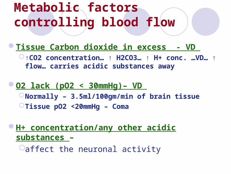
Metabolic factors controlling blood flow
Tissue Carbon dioxide in excess - VD ↑CO2 concentration… ↑ H2CO3… ↑ H+ conc. …VD… ↑
flow… carries acidic substances away
O2 lack (pO2 < 30mmHg)– VD Normally – 3.5ml/100gm/min of brain tissueTissue pO2 <20mmHg – Coma
H+ concentration/any other acidic substances – affect the neuronal activity
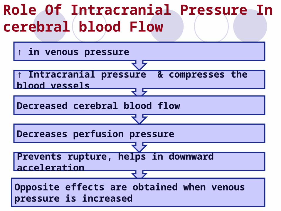
Role Of Intracranial Pressure In cerebral blood Flow
↑ in venous pressure
↑ Intracranial pressure & compresses the blood vessels
Decreased cerebral blood flow
Decreases perfusion pressure
Prevents rupture, helps in downward acceleration
Opposite effects are obtained when venous pressure is increased
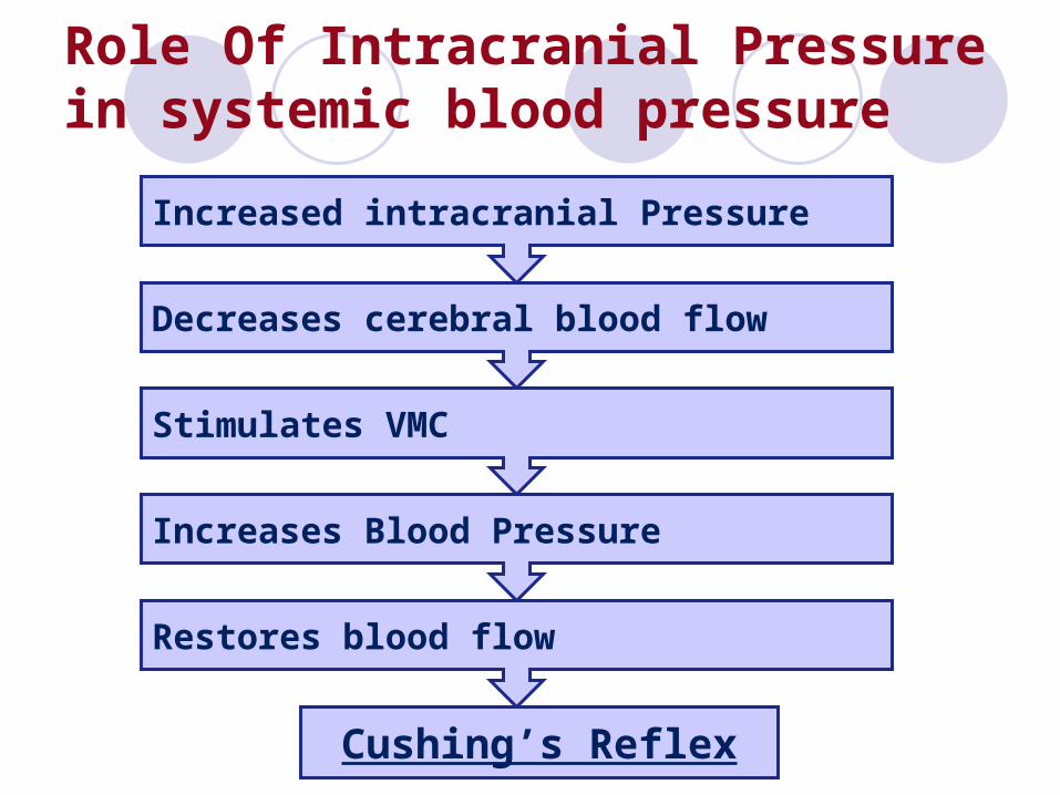
Role Of Intracranial Pressure in systemic blood pressure
Increased intracranial Pressure
Decreases cerebral blood flow
Stimulates VMC
Increases Blood Pressure
Restores blood flow
Cushing’s Reflex
Cerebral Stroke
Due to atherosclerosis…block in some small arteries…local cerebral ischemia
Hypertensives are more prone to cerebral hemorrhage
Mostly in middle cerebral artery – affects midportion of brain
Posterior cerebral artery will affect the occipital lobe
Midbrain involvement – sensory & motor abnormalities
Blood Brain Barrier
Present in choroid plexus as well as capillary levelNot present in some parts of hypothalamus,
posterior pituitary gland, adjacent median eminence & Area Postrema Important for homeostasis
Not well developed at birthFactors responsible:-
Tight junctions leading to poor permeabilityGlial cells (Astrocytes) are interposed between
neurons & capillaries
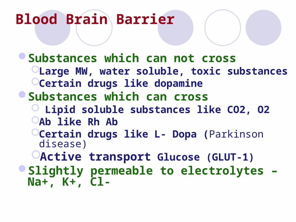
Substances which can not crossLarge MW, water soluble, toxic substances Certain drugs like dopamine
Substances which can cross Lipid soluble substances like CO2, O2Ab like Rh AbCertain drugs like L- Dopa (Parkinson disease)Active transport Glucose (GLUT-1)
Slightly permeable to electrolytes – Na+, K+, Cl-
Blood Brain Barrier
Study materialGuyton – 11th edition, chap. 61Ganong – 21st edition, chap. 32
Thanks


![CEREBRAL CIRCULATION AND CEREBROSPINAL FLUID [CSF]](https://img.pdfslide.us/doc/110x75/56814ee4550346895dbc77ad/cerebral-circulation-and-cerebrospinal-fluid-csf.jpg)