Embed Size (px)
Citation preview

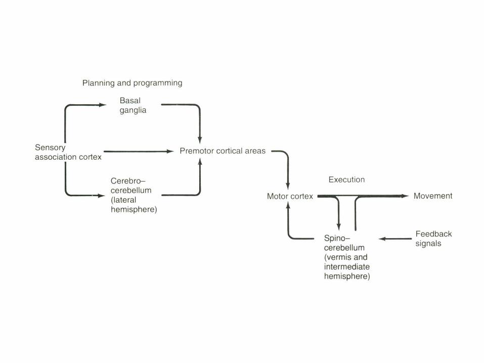
CEREBELLUM
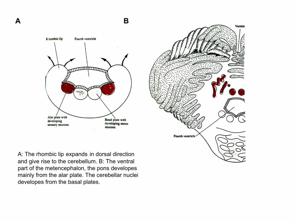
A B
A: The rhombic lip expands in dorsal direction and give rise to the cerebellum. B: The ventral part of the metencephalon, the pons developesmainly from the alar plate. The cerebellar nuclei developes from the basal plates.
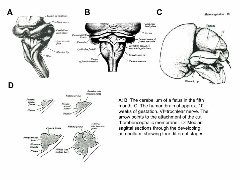
A B C
DA: B: The cerebellum of a fetus in the fifth month. C: The human brain at approx. 10 weeks of gestation. VI=trochlear nerve. The arrow points to the attachment of the cut rhombencephalic membrane. D: Median sagittal sections through the developing cerebellum, showing four different stages.
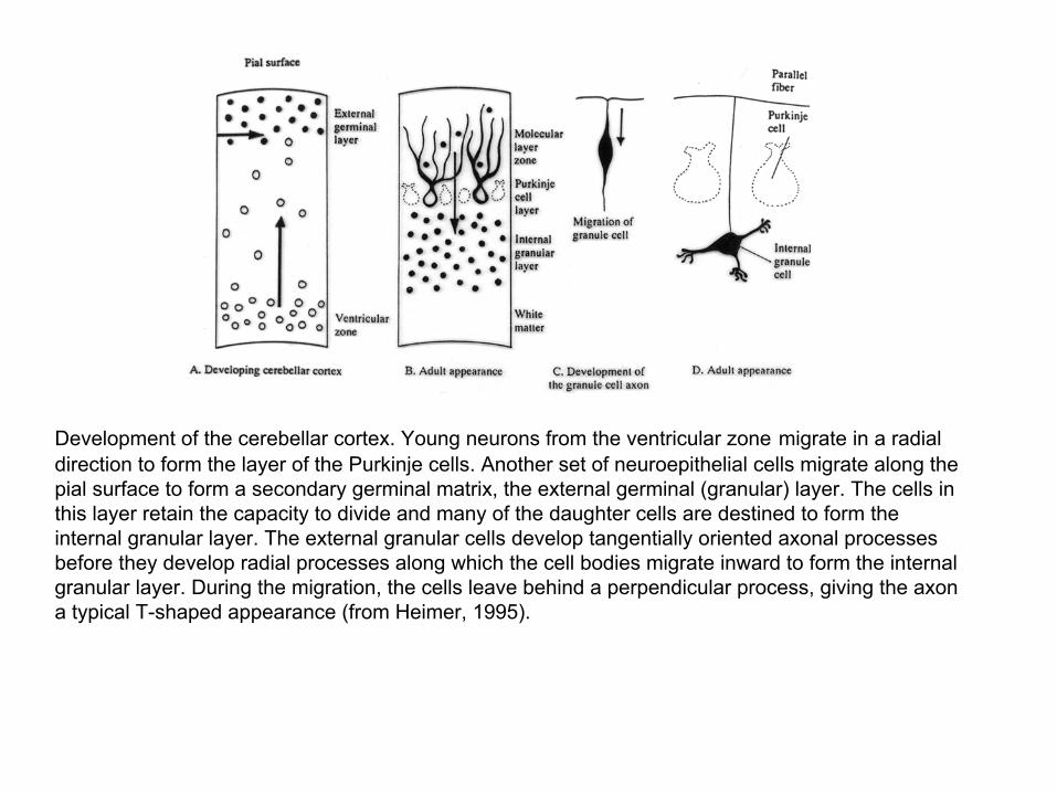
Development of the cerebellar cortex. Young neurons from the ventricular zone migrate in a radial direction to form the layer of the Purkinje cells. Another set of neuroepithelial cells migrate along the pial surface to form a secondary germinal matrix, the external germinal (granular) layer. The cells in this layer retain the capacity to divide and many of the daughter cells are destined to form the internal granular layer. The external granular cells develop tangentially oriented axonal processes before they develop radial processes along which the cell bodies migrate inward to form the internal granular layer. During the migration, the cells leave behind a perpendicular process, giving the axon a typical T-shaped appearance (from Heimer, 1995).
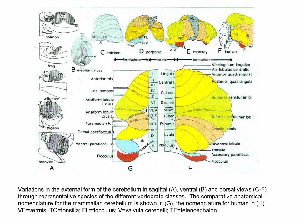
Variations in the external form of the cerebellum in sagittal (A), ventral (B) and dorsal views (C-F) through representative species of the different vertebrate classes. The comparative anatomical nomenclature for the mammalian cerebellum is shown in (G), the nomenclature for human in (H). VE=vermis; TO=tonsilla; FL=flocculus; V=valvula cerebelli; TE=telencephalon.
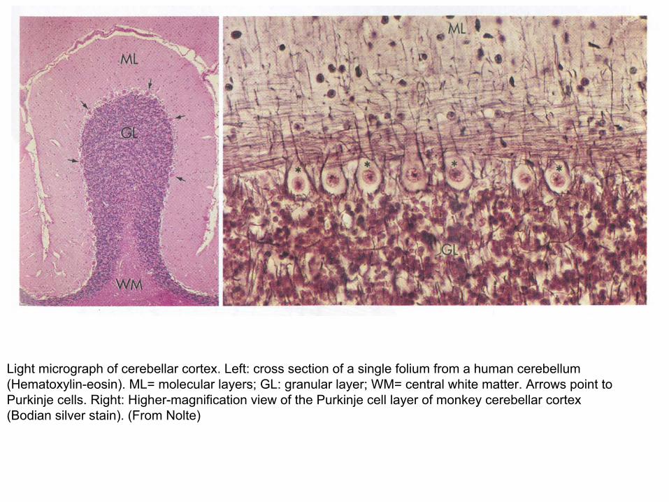
Light micrograph of cerebellar cortex. Left: cross section of a single folium from a human cerebellum (Hematoxylin-eosin). ML= molecular layers; GL: granular layer; WM= central white matter. Arrows point to Purkinje cells. Right: Higher-magnification view of the Purkinje cell layer of monkey cerebellar cortex (Bodian silver stain). (From Nolte)
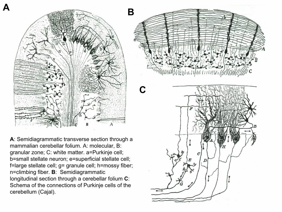
A: Semidiagrammatic transverse section through a mammalian cerebellar folium. A: molecular, B: granular zone; C: white matter. a=Purkinje cell; b=small stellate neuron; e=superficial stellate cell; f=large stellate cell; g= granule cell; h=mossy fiber; n=climbing fiber. B: Semidiagrammaticlongitudinal section through a cerebellar folium C: Schema of the connections of Purkinje cells of the cerebellum (Cajal).
A B
C
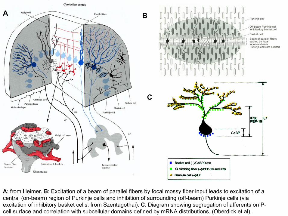
A: from Heimer. B: Excitation of a beam of parallel fibers by focal mossy fiber input leads to excitation of a central (on-beam) region of Purkinje cells and inhibition of surrounding (off-beam) Purkinje cells (via excitation of inhibitory basket cells, from Szentagothai). C: Diagram showing segregation of afferents on P-cell surface and correlation with subcellular domains defined by mRNA distributions. (Oberdick et al).
A B
C
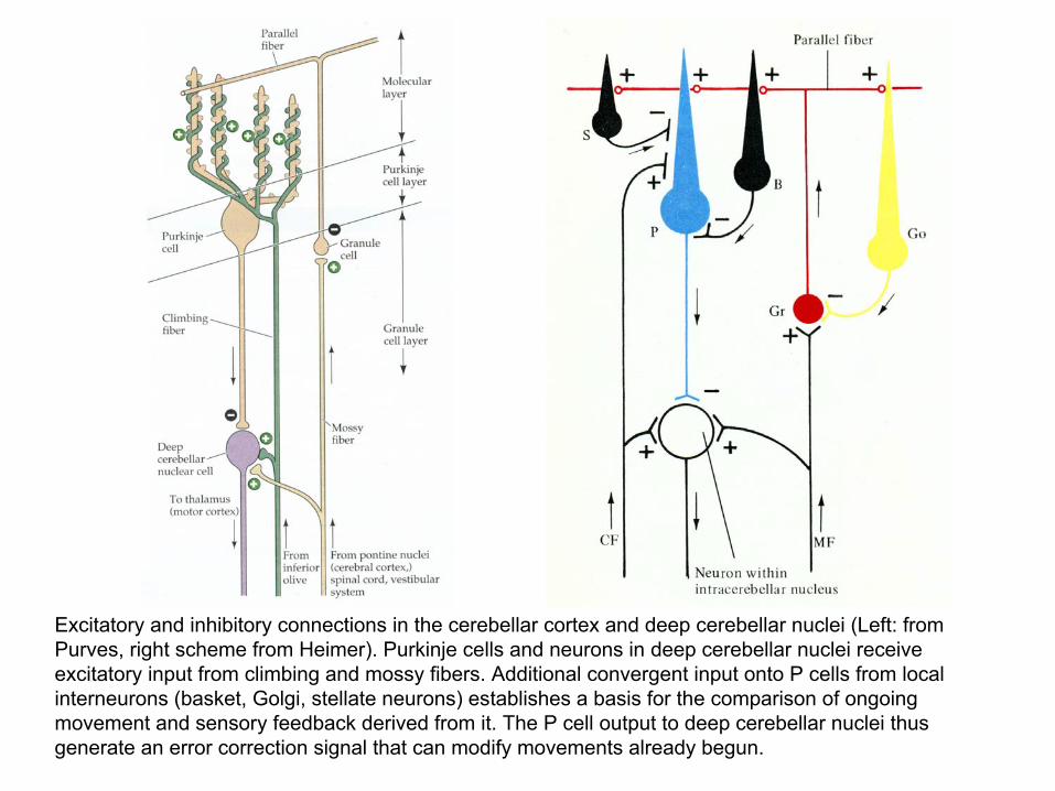
Excitatory and inhibitory connections in the cerebellar cortex and deep cerebellar nuclei (Left: from Purves, right scheme from Heimer). Purkinje cells and neurons in deep cerebellar nuclei receive excitatory input from climbing and mossy fibers. Additional convergent input onto P cells from local interneurons (basket, Golgi, stellate neurons) establishes a basis for the comparison of ongoing movement and sensory feedback derived from it. The P cell output to deep cerebellar nuclei thus generate an error correction signal that can modify movements already begun.
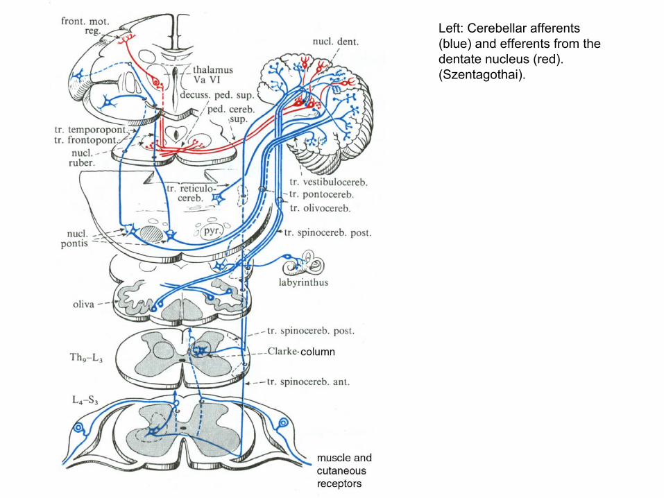
Left: Cerebellar afferents (blue) and efferents from the dentate nucleus (red). (Szentagothai).
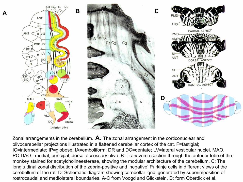
Zonal arrangements in the cerebellum. A: The zonal arrangement in the corticonuclear and olivocerebellar projections illustrated in a flattened cerebellar cortex of the cat. F=fastigial; IC=intermediate; IP=globose; IA=emboliform; DR and DC=dentate; LV=lateral vestibular nuclei. MAO, PO,DAO= medial, principal, dorsal accessory olive. B: Transverse section through the anterior lobe of the monkey stained for acetylcholineesterase, showing the modular architecture of the cerebellum. C: The longitudinal zonal distribution of the zebrin-positive and ‘negative’ Purkinje cells in different views of thecerebellum of the rat. D: Schematic diagram showing cerebellar ‘grid’ generated by superimposition of rostrocaudal and mediolateral boundaries. A-C from Voogd and Glickstein, D: form Oberdick et al.
A B C
D
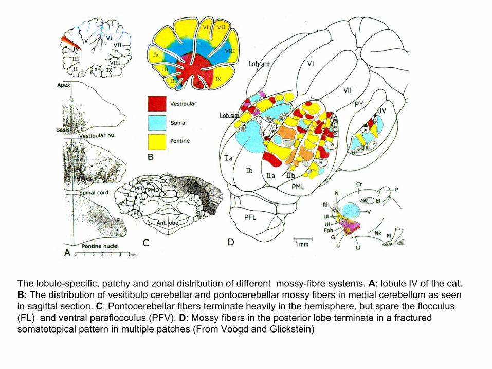
The lobule-specific, patchy and zonal distribution of different mossy-fibre systems. A: lobule IV of the cat. B: The distribution of vesitibulo cerebellar and pontocerebellar mossy fibers in medial cerebellum as seen in sagittal section. C: Pontocerebellar fibers terminate heavily in the hemisphere, but spare the flocculus(FL) and ventral paraflocculus (PFV). D: Mossy fibers in the posterior lobe terminate in a fractured somatotopical pattern in multiple patches (From Voogd and Glickstein)
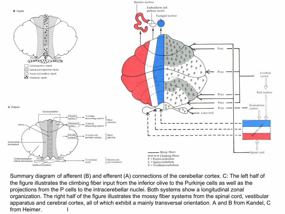
Summary diagram of afferent (B) and efferent (A) connections of the cerebellar cortex. C: The left half of the figure illustrates the climbing fiber input from the inferior olive to the Purkinje cells as well as the projections from the P cells to the intracerebellar nuclei. Both systems show a longitudinal zonal organization. The right half of the figure illustrates the mossy fiber systems from the spinal cord, vestibular apparatus and cerebral cortex, all of which exhibit a mainly transversal orientation. A and B from Kandel, C from Heimer. l
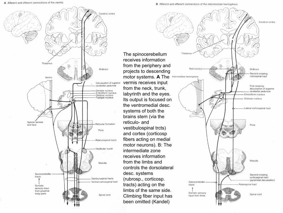
The spinocerebellumreceives information from the periphery and projects to descending motor systems. A The vermis receives input from the neck, trunk, labyrinth and the eyes. Its output is focused on the ventromedial desc. systems of both the brains stem (via the reticulo- and vestibulospinal trcts) and cortex (corticospfibers acting on medial motor neurons). B: The intermediate zone receives information from the limbs and controls the dorsolateraldesc. systems (rubrosp., corticosp. tracts) acting on the limbs of the same side. Climbing fiber input has been omitted (Kandel)

Left figure: The cerebrocerebellum (the lateral zone of each hemisphere) receives cortical input via the pontinenuclei (color inset from Nolte) and influences the motor and premotor cortices via the VA nucleus of the thalamus (Kandel). Right: Diagram illustrating the distribution within the basal pons of the rhesus monkey of projections derived from various cortical areas using the same color code. Cortical areas shown in yellow are not currently thought to have pontine projections. There is a complex mosaic of terminations in the pons, with each cerebral areas having preferential sites of pontine terminations (Schmahmann, Nolte).

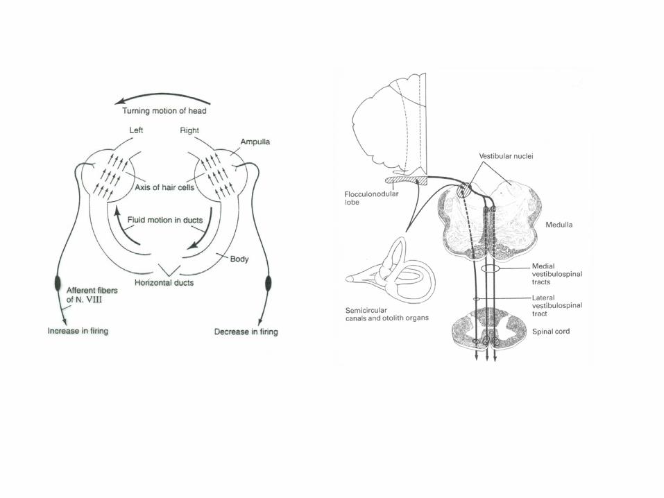
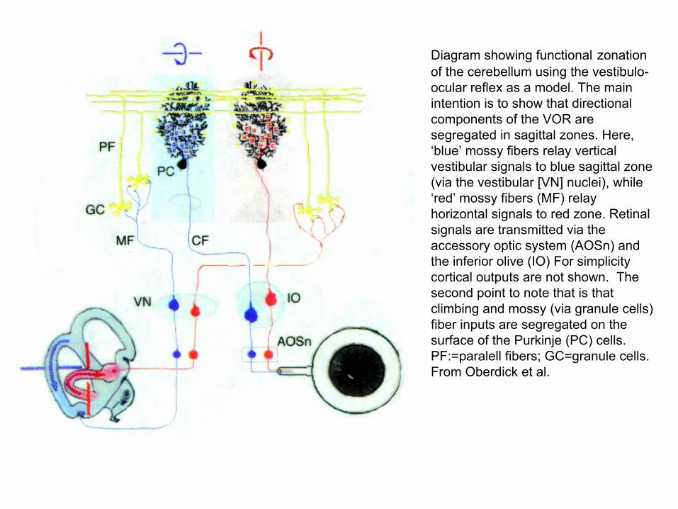
Diagram showing functional zonationof the cerebellum using the vestibulo-ocular reflex as a model. The main intention is to show that directional components of the VOR are segregated in sagittal zones. Here, ‘blue’ mossy fibers relay vertical vestibular signals to blue sagittal zone (via the vestibular [VN] nuclei), while ‘red’ mossy fibers (MF) relay horizontal signals to red zone. Retinal signals are transmitted via the accessory optic system (AOSn) and the inferior olive (IO) For simplicity cortical outputs are not shown. The second point to note that is that climbing and mossy (via granule cells) fiber inputs are segregated on the surface of the Purkinje (PC) cells. PF:=paralell fibers; GC=granule cells. From Oberdick et al.
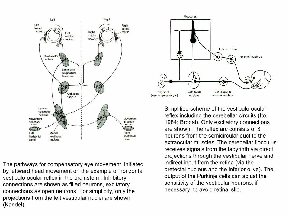
The pathways for compensatory eye movement initiated by leftward head movement on the example of horizontal vestibulo-ocular reflex in the brainstem . Inhibitory connections are shown as filled neurons, excitatory connections as open neurons. For simplicity, only the projections from the left vestibular nuclei are shown (Kandel).
Simplified scheme of the vestibulo-ocular reflex including the cerebellar circuits (Ito, 1984; Brodal). Only excitatory connections are shown. The reflex arc consists of 3 neurons from the semicircular duct to the extraocular muscles. The cerebellar flocculusreceives signals from the labyrinth via direct projections through the vestibular nerve and indirect input from the retina (via the pretectal nucleus and the inferior olive). The output of the Purkinje cells can adjust the sensitivity of the vestibular neurons, if necessary, to avoid retinal slip.
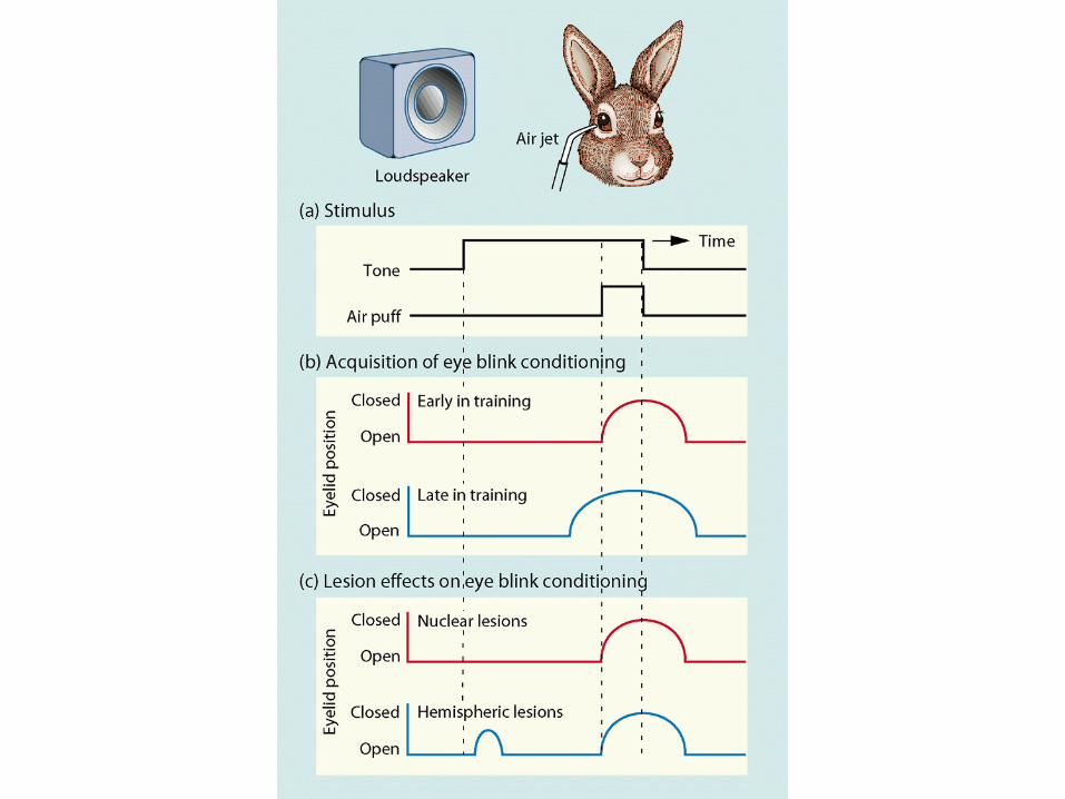
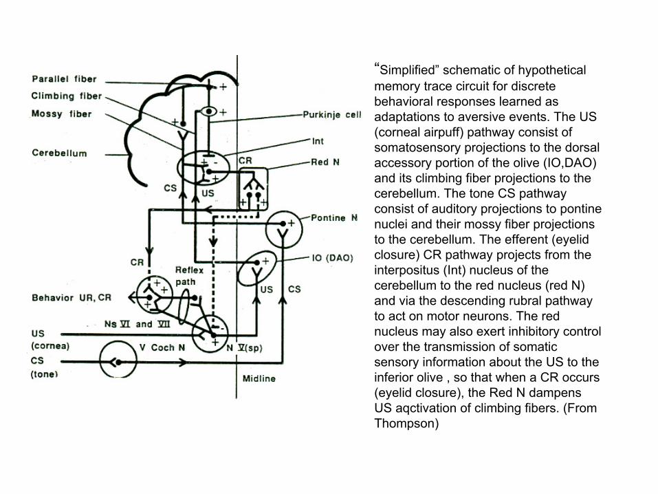
“Simplified” schematic of hypothetical memory trace circuit for discrete behavioral responses learned as adaptations to aversive events. The US (corneal airpuff) pathway consist of somatosensory projections to the dorsal accessory portion of the olive (IO,DAO) and its climbing fiber projections to the cerebellum. The tone CS pathway consist of auditory projections to pontinenuclei and their mossy fiber projections to the cerebellum. The efferent (eyelid closure) CR pathway projects from the interpositus (Int) nucleus of the cerebellum to the red nucleus (red N) and via the descending rubral pathway to act on motor neurons. The red nucleus may also exert inhibitory control over the transmission of somatic sensory information about the US to the inferior olive , so that when a CR occurs (eyelid closure), the Red N dampens US aqctivation of climbing fibers. (From Thompson)

Pathways mediating the unconditioned and conditioned blink reflex after Holstege.
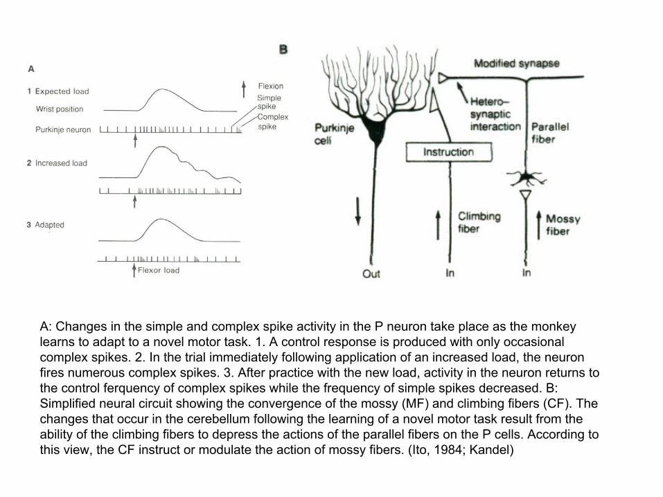
A: Changes in the simple and complex spike activity in the P neuron take place as the monkey learns to adapt to a novel motor task. 1. A control response is produced with only occasional complex spikes. 2. In the trial immediately following application of an increased load, the neuron fires numerous complex spikes. 3. After practice with the new load, activity in the neuron returns to the control ferquency of complex spikes while the frequency of simple spikes decreased. B: Simplified neural circuit showing the convergence of the mossy (MF) and climbing fibers (CF). The changes that occur in the cerebellum following the learning of a novel motor task result from the ability of the climbing fibers to depress the actions of the parallel fibers on the P cells. According to this view, the CF instruct or modulate the action of mossy fibers. (Ito, 1984; Kandel)
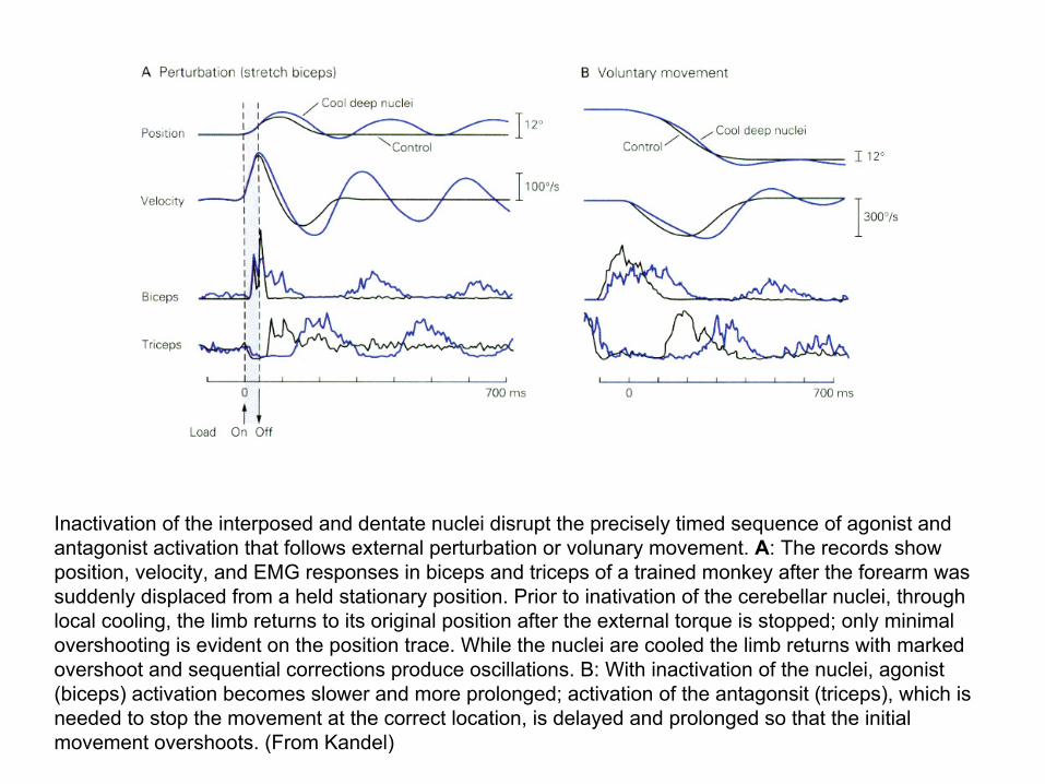
Inactivation of the interposed and dentate nuclei disrupt the precisely timed sequence of agonist and antagonist activation that follows external perturbation or volunary movement. A: The records show position, velocity, and EMG responses in biceps and triceps of a trained monkey after the forearm was suddenly displaced from a held stationary position. Prior to inativation of the cerebellar nuclei, through local cooling, the limb returns to its original position after the external torque is stopped; only minimal overshooting is evident on the position trace. While the nuclei are cooled the limb returns with marked overshoot and sequential corrections produce oscillations. B: With inactivation of the nuclei, agonist (biceps) activation becomes slower and more prolonged; activation of the antagonsit (triceps), which is needed to stop the movement at the correct location, is delayed and prolonged so that the initial movement overshoots. (From Kandel)
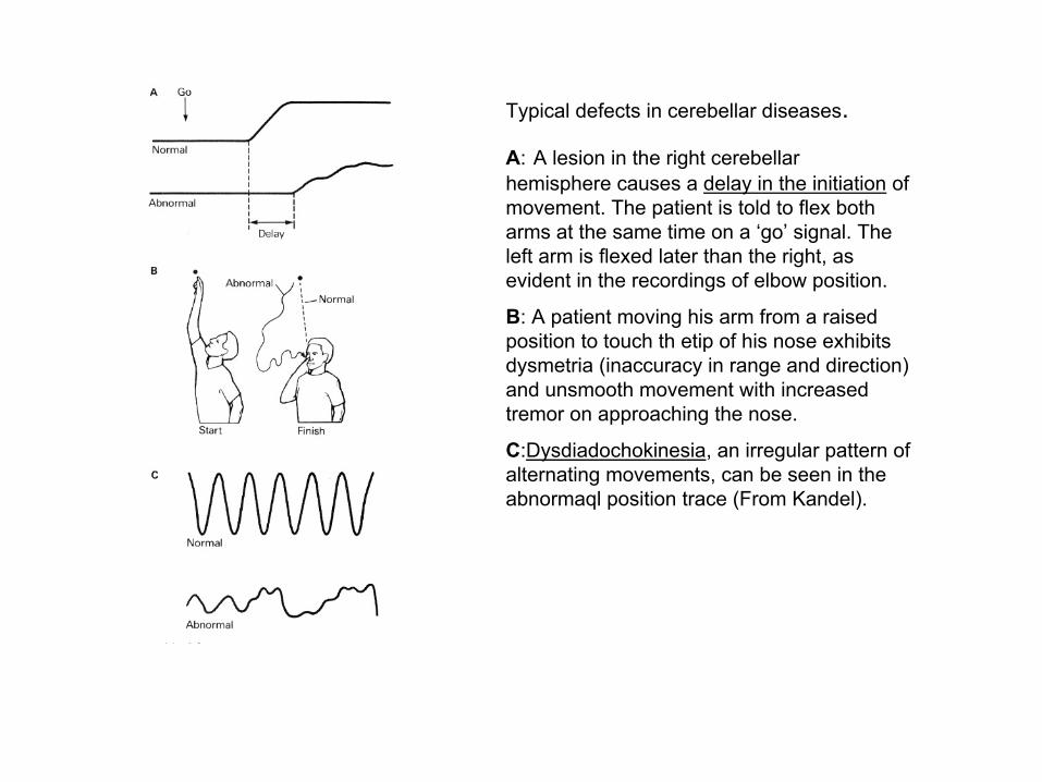
Typical defects in cerebellar diseases.
A: A lesion in the right cerebellarhemisphere causes a delay in the initiation of movement. The patient is told to flex both arms at the same time on a ‘go’ signal. The left arm is flexed later than the right, as evident in the recordings of elbow position.
B: A patient moving his arm from a raised position to touch th etip of his nose exhibits dysmetria (inaccuracy in range and direction) and unsmooth movement with increased tremor on approaching the nose.
C:Dysdiadochokinesia, an irregular pattern of alternating movements, can be seen in the abnormaql position trace (From Kandel).
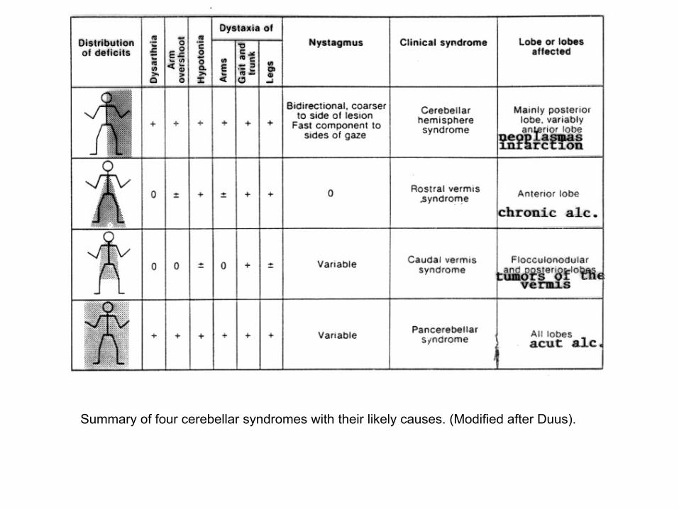
Summary of four cerebellar syndromes with their likely causes. (Modified after Duus).
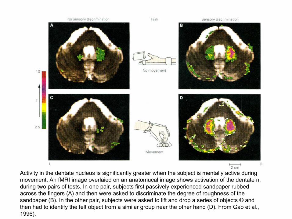
Activity in the dentate nucleus is significantly greater when the subject is mentally active during movement. An fMRI image overlaied on an anatomucal image shows activation of the dentate n. during two pairs of tests. In one pair, subjects first passively experienced sandpaper rubbed across the fingers (A) and then were asked to discriminate the degree of roughness of the sandpaper (B). In the other pair, subjects were asked to lift and drop a series of objects © and then had to identify the felt object from a similar group near the other hand (D). From Gao et al., 1996).