Embed Size (px)
Citation preview
Evidence review
Endovascular treatment of varicose veins CEP09017
August 2009
Contents 2
CEP09017: August 2009
Summary ................................................................................................ 3
Introduction ............................................................................................. 4
Methods ................................................................................................ 11
Evidence review .................................................................................... 19
Conclusions .......................................................................................... 27
Acknowledgements ............................................................................... 28
Glossary ................................................................................................ 30
References............................................................................................ 33
Appendix 1: Products and suppliers ...................................................... 37
Appendix 2: Summary of selected controlled trials ................................ 42
Appendix 3: Summary of economic evidence ........................................ 51
Appendix 4: Recording expert opinion ................................................... 53
Author and report information ................................................................ 58
Summary 3
CEP09017: August 2009
The product Endovascular ablation techniques are a minimally invasive alternative to traditional ligation and stripping of the great saphenous vein. These techniques employ devices designed to destroy the internal lining of the vein and close it off by heating, using laser or radio-frequency energy, or chemical destruction (sclerotherapy).
Field of use Varicose veins cause major socio-economic and health problems and varicose vein procedures consumed approximately £40 million of the NHS budget in 2005-6 [1]. Varicose veins are not an acute life-threatening condition but do have an impact on quality of life and can involve significant expenditure, in particular in the provision of nursing care for venous ulcers.
National guidance NICE recommends that both radio-frequency ablation (RFA) and endovenous laser ablation (EVLA) may be used with normal arrangements for clinical governance, consent and audit [2;3]. Recent guidance on foam sclerotherapy (FSC) has been more cautious and requires clinicians undertaking the procedure to make special arrangements for audit [4-6].
Evidence reviewed Eighteen publications comparing the endovascular techniques with surgery were reviewed [7-24] and the expert opinion of consultants with relevant experience was sought. This report includes consideration of clinical factors and issues relating to cost.
CEP’s verdict All of the endovascular techniques have the potential to improve both patient comfort post procedure and recovery rates. The evidence relating to long term efficacy is limited. There is also potential to reduce procedure costs by changing from a surgical procedure carried out under general anaesthetic in the operating theatre to an endovascular procedure carried out under local anaesthetic in an outpatient clinic setting. Further good quality randomised controlled trials are required to clarify the long term efficacy of the endovascular techniques.
Introduction 4
CEP09017: August 2009
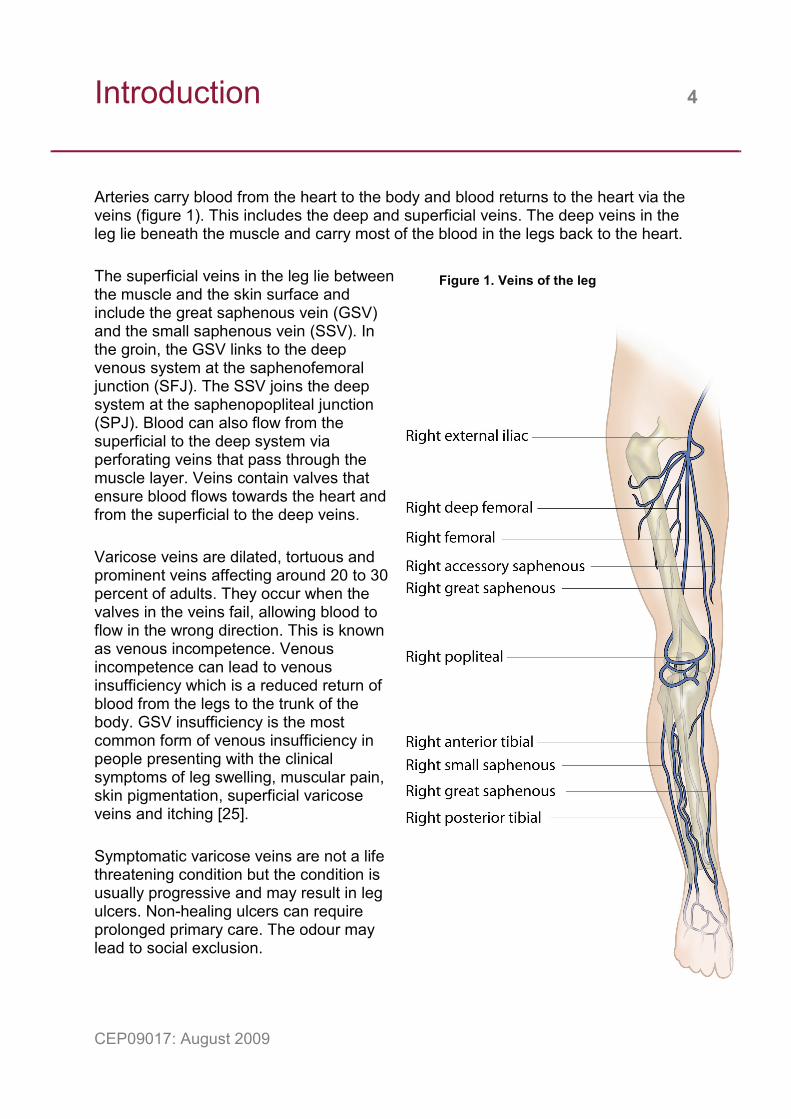
Arteries carry blood from the heart to the body and blood returns to the heart via the veins (figure 1). This includes the deep and superficial veins. The deep veins in the leg lie beneath the muscle and carry most of the blood in the legs back to the heart.
The superficial veins in the leg lie between the muscle and the skin surface and include the great saphenous vein (GSV) and the small saphenous vein (SSV). In the groin, the GSV links to the deep venous system at the saphenofemoral junction (SFJ). The SSV joins the deep system at the saphenopopliteal junction (SPJ). Blood can also flow from the superficial to the deep system via perforating veins that pass through the muscle layer. Veins contain valves that ensure blood flows towards the heart and from the superficial to the deep veins.
Varicose veins are dilated, tortuous and prominent veins affecting around 20 to 30 percent of adults. They occur when the valves in the veins fail, allowing blood to flow in the wrong direction. This is known as venous incompetence. Venous incompetence can lead to venous insufficiency which is a reduced return of blood from the legs to the trunk of the body. GSV insufficiency is the most common form of venous insufficiency in people presenting with the clinical symptoms of leg swelling, muscular pain, skin pigmentation, superficial varicose veins and itching [25].
Symptomatic varicose veins are not a life threatening condition but the condition is usually progressive and may result in leg ulcers. Non-healing ulcers can require prolonged primary care. The odour may lead to social exclusion.
Figure 1. Veins of the leg
Introduction 5
CEP09017: August 2009
Recurrence of varicose veins may be due to inadequate initial treatment or may occur following satisfactory treatment in progressive cases because of:
• enlargement of secondary veins and branches which take on the function of the destroyed GSV
• the new growth and development of multiple tiny vein branches that provide new connections between the deep and superficial veins.
Occasionally the GSV is a duplicate system and both branches need to be treated. Such duplication should be identified by colour duplex ultrasound prior to the procedure.
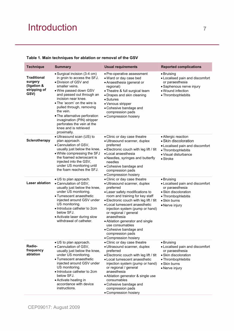
Surgery is the traditional treatment for more severe cases and in the UK this usually comprises ligation of the SFJ and stripping of the GSV from the groin to knee. This may be supplemented by phlebectomy comprising surgical removal of visible superficial varicosities. Over the last decade several minimally invasive endovascular varicose vein treatments have been developed as alternatives to surgery. The main techniques for ablation or removal of the GSV are summarised in table 1. Advances in ultrasound technology and the wider availability of colour duplex ultrasound imaging have contributed to the adoption of these techniques.
Colour duplex ultrasound is an imaging technique that combines information on blood flow direction and velocity displayed as colour, overlaid on the conventional black and white ultrasound image. Colour duplex ultrasound is used to identify the incompetent venous segments and guide catheter insertion into the GSV, usually close to the knee. Colour duplex scanning and catheter insertion are facilitated by the patient lying on a tilting table with their legs below the level of the heart to increase vein diameter. Prior to the start of the endovascular treatment, the table is tilted until the leg is raised above the level of the heart (Trendelenburg position) to partially empty the vein of blood and reduce its diameter. During EVLA and RFA ultrasound monitoring is used to guide the catheter tip along the vein and ensure that it is positioned just below the SFJ prior to starting ablation. Ultrasound is used in FSC to guide puncture of non-visible varicose veins and to monitor foam distribution.
For treatments using EVLA or RFA a local tumescent anaesthesia is used. The tumescent liquid is infused around the outside of the vein, usually using a tumescent pump. The local tumescent anaesthetic is intended to:
• empty the vein of blood by external compression
• act as a heat shield, protecting tissue surrounding the vein from thermal damage
• act as an analgesic, reducing pain and discomfort during the treatment.
Introduction 6
CEP09017: August 2009
Treatment of residual varicosities following RFA and EVLA may be carried out during a single treatment episode or at a later date using foam or minor surgical phlebectomy under local anaesthetic.
The endovenous procedures may all be carried out under local anaesthetic, and if they are adopted it is possible to move from treatment in a sterile operating theatre to ambulant outpatient treatment.
Foam sclerotherapy Liquid sclerotherapy (LSC) has been in widespread use for many years[26] and more recently ultrasound-guided FSC for varicose veins has been developed as a variation of LSC. Sclerosant is a chemical detergent which causes irritation and inflammation of the inside wall of the vein, causing the vein to collapse and close. Increasing the volume of the sclerosant by producing a foam increases the contact time of the chemical with the walls of larger veins and prevents dilution of the sclerosant by blood. The air bubbles in the foam improve visibility by ultrasound. As the sclerosant initiates a tight spasm in the vein, the foam is dispersed along the vein. The sclerotherapy agents used are sodium tetradecyl sulphate (STS) marketed as Fibrovein in the UK and polidocanol. Varisolve, a foamed version of polidicanol, is produced as a ready to use canister. FSC should only use pharmaceuticals licensed for sclerotherapy of varicose veins. Only STS and ethanolamine oleate are currently licensed in the UK for sclerotherapy, although the latter does not appear to be in widespread use. Mixing liquid sclerosant with air to make foam constitutes unlicensed use [5]. According to the British National Formulary ‘Unlicensed use of medicines becomes necessary if the clinical need cannot be met by licensed medicines; such use should be supported by appropriate evidence and experience’ [27]. Foam must be made immediately before injection, usually using the Tessari method [28].
Introduction 7
CEP09017: August 2009
Table 1. Main techniques for ablation or removal of the GSV
Technique Summary Usual requirements Reported complications
Traditional surgery (ligation & stripping of GSV)
• Surgical incision (3-4 cm) in groin to access the SFJ.
• Division of GSV and smaller veins.
• Wire passed down GSV and passed out through an incision near knee.
• The ‘acorn’ on the wire is pulled through, removing the vein. • The alternative perforation
invagination (PIN) stripper perforates the vein at the knee and is retrieved proximally.
• Pre-operative assessment • Ward or day case bed • Anaesthesia (general or
regional) • Theatre & full surgical team • Drapes and skin cleaning • Sutures • Venous stripper • Cohesive bandage and
compression pads • Compression hosiery
• Bruising • Localised pain and discomfort
or paraesthesia • Saphenous nerve injury • Wound infection • Thrombophlebitis
Sclerotherapy
• Ultrasound scan (US) to plan approach.
• Cannulation of GSV, usually just below the knee.
• While compressing the SFJ the foamed scleroscant is injected into the GSV, under US monitoring until the foam reaches the SFJ.
• Clinic or day case theatre • Ultrasound scanner, duplex
preferred • Electronic couch with leg lift / tilt • Local anaesthesia • Needles, syringes and butterfly
needles • Cohesive bandage and
compression pads • Compression hosiery
• Allergic reaction • Skin discoloration • Localised pain and discomfort • Thrombophlebitis • Visual disturbance • Stroke
Laser ablation
• US to plan approach. • Cannulation of GSV,
usually just below the knee, under US monitoring.
• Tumescent anaesthetic injected around GSV under US monitoring.
• Introduce catheter to 2cm below SFJ.
• Activate laser during slow withdrawal of catheter.
• Clinic or day case theatre • Ultrasound scanner, duplex
preferred • Laser safety modifications to
room and training for key staff • Electronic couch with leg lift / tilt • Local tumescent anaesthetic
injection system (pump or hand) or regional / general anaesthesia
• Ablation generator and single use consumables
• Cohesive bandage and compression pads
• Compression hosiery
• Bruising • Localised pain and discomfort
or paraesthesia • Skin discoloration • Thrombophlebitis • Skin burns • Nerve injury
Radio-frequency ablation
• US to plan approach. • Cannulation of GSV,
usually just below the knee, under US monitoring.
• Tumescent anaesthetic injected around GSV under US monitoring.
• Introduce catheter to 2cm below SFJ.
• Activate heating in accordance with device instructions.
• Clinic or day case theatre • Ultrasound scanner, duplex
preferred • Electronic couch with leg lift / tilt • Local tumescent anaesthetic
injection system (pump or hand) or regional / general anaesthesia
• Ablation generator & single use consumables
• Cohesive bandage and compression pads
• Compression hosiery
• Bruising • Localised pain and discomfort
or paraesthesia • Skin discoloration • Thrombophlebitis • Skin burns • Nerve injury
Introduction 8
CEP09017: August 2009
Laser ablation EVLA treatment is designed to cause thermal damage to the vein lining. During laser activation light energy is transmitted through an optical fibre to the tip, usually producing a fine beam within the vein.
Lasers used for EVLA may be either diodes or solid state Nd:YAG lasers (eg Fotona XP-2 from Pierson Surgical). The active material in a diode laser is a small semi-conductor chip. Diode lasers have the advantage of small size and may be desktop or trolley mounted or stored in a cupboard. They are available in a number of wavelengths to target different absorbing molecules, produce less heat than Nd:YAG and require minimal maintenance for reliable operation. The active material in the Nd:YAG laser is a solid rod of artificial garnet and because the laser generates heat a cooling system is incorporated in the device. The laser is floor standing and will require suitable storage space. Nd:YAG and diode lasers may be used for other cosmetic, aesthetic or dermatological applications and cost effectiveness will increase if they have multiple uses.
Diode lasers producing infrared wavelengths are common (eg Delta from Angiodynamics and Varilase from Pyramed at 810nm, SDL Medical at 806.5 nm or Biolitec ELVeS from Promed at 810 and 980nm) as these match an energy absorption peak in both oxyhaemoglobin and deoxyhaemoglobin. At these wavelengths laser energy is preferentially absorbed by blood causing rapid heating and coagulation. Heat also damages the vein wall and the vein shrinks.
Laser energy at longer wavelengths (eg Biolitec ELVeS painless from Promed at 1470nm) is preferentially absorbed by water, so both the water within blood and the vein wall lining directly absorb the laser energy. The manufacturers claim that the 1470 nm diode laser results in less post-operative pain, bruising and swelling, but there are currently no published randomised controlled clinical trials to support this claim.
Full details of laser accessories including catheters, fibres and introducer kits are available from suppliers (appendix 1).
The use of lasers may require minor modifications to the room (eg fitting window blinds), appointment of one of the team as laser protection supervisor, written safety procedures and safety training for staff[29].
Radiofrequency ablation RFA uses electrical energy to heat the vein wall. Several different RFA devices are available and although they are constructed differently they all work on the same
Introduction 9
CEP09017: August 2009
principle. The RF generators are all compact and portable. This technique also requires the injection of tumescent anaesthesia to minimise pain and reduce the risk of skin burns during the procedure.
There are currently three devices used in the UK for RFA.
• The VNUS Closure Plus catheter uses multiple electrodes that fan out radially as the catheter sheath is retracted, each touching the vein wall so the RF current passes through the blood to a return electrode positioned centrally in the vein. Treatment progress is monitored using a thermocouple within the catheter.
• The VNUS Closure Fast catheter uses RF energy to achieve temperatures between 95°C and 120°C in a seven centimetre long heating element. The temperature is monitored by a thermocouple within the catheter tip.
• The Celon RFITT catheter comprises two concentric bipolar electrodes, one at the tip and the other several millimetres down the catheter shaft. An electrical current at 470 kHz is conducted through the adjacent blood and vein wall. The generator monitors electrical impedance, which rises as blood and the vein wall heat up to 85°C [30]. A continuous audible signal indicates that ablation is complete.
Full details of RFA accessories including catheter kits are available from suppliers (appendix 1).
Training Surgery is the long established method for the removal of varicose veins by which other methods of treatment are compared. It is currently the only method with formal training requirements. Varicose vein surgery is a core activity of the vascular surgery syllabus and venous disease is a speciality in the final stages of training [31]. This is monitored on a competency basis using logbooks, reviews and RITAs (Record/Review of In house Training Assessments) which are soon to be replaced with ARCP (Annual Review of Competent Progression).
The VEIN study [32] also recommended that for traditional surgery, procedures should be added to a central database to monitor outcome and performance.
At this time, there is no structured training for any of the endovascular varicose vein treatments. Some of these can be performed by doctors of different specialities such as dermatologists and radiologists in addition to surgeons. Where the endovascular practitioner is not trained in vascular ultrasound, this element can be carried out by a specialist vascular ultrasound practitioner.
Introduction 10
CEP09017: August 2009
The VEIN study [32] also recommended the following for endovascular practitioners:
• colour duplex ultrasound imaging skills
• competence in venous cannulation under ultrasound guidance
• complete training in the theoretical aspects of the technique
• training for the specific equipment used. Most manufacturers provide training as part of a package when purchasing their products
• training and mentoring by an experienced vascular specialist in the new type of treatment until competency has been demonstrated
• for EVLA, MHRA recommends a ‘Core of Knowledge’ training course in laser safety for laser operators, their assistants and laser protection supervisors [29]. In the UK, a number of organisations run Core of Knowledge courses on laser safety.
National guidance NICE recommends that both RF ablation [2] and laser ablation [3] may be used with normal arrangements for clinical governance, consent and audit. Recent guidance on FSC, published by NICE in May 2007, is more cautious and requires clinicians undertaking FSC to make special arrangements for audit [5]. These are:
• inform the clinical governance lead in the NHS trust
• fully inform the patients of the potential side effects associated with the procedure and of the alternative treatment options
• audit and review clinical outcomes of all patients having FSC treatment [4;5].
The guidance explains that ‘current evidence on ultrasound-guided foam shows that it is efficacious in the short term’ but that ‘evidence of long-term efficacy is limited’. NICE has therefore developed an audit tool [6] to monitor long term outcomes.
Patient reported outcome measures From April 2009 patients undergoing varicose vein surgery (and other treatments) will be asked to complete a patient reported outcome measures (PROMs) questionnaire. PROMs are intended to provide a direct measure of the success of medical interventions, as judged by patients, to enable clinical teams to benchmark their performance and research the success of different treatment options [33]. The results will be available to the public on the NHS choices website. Results for varicose vein procedures will be available in early 2010 on the DH website.
Methods 11
CEP09017: August 2009
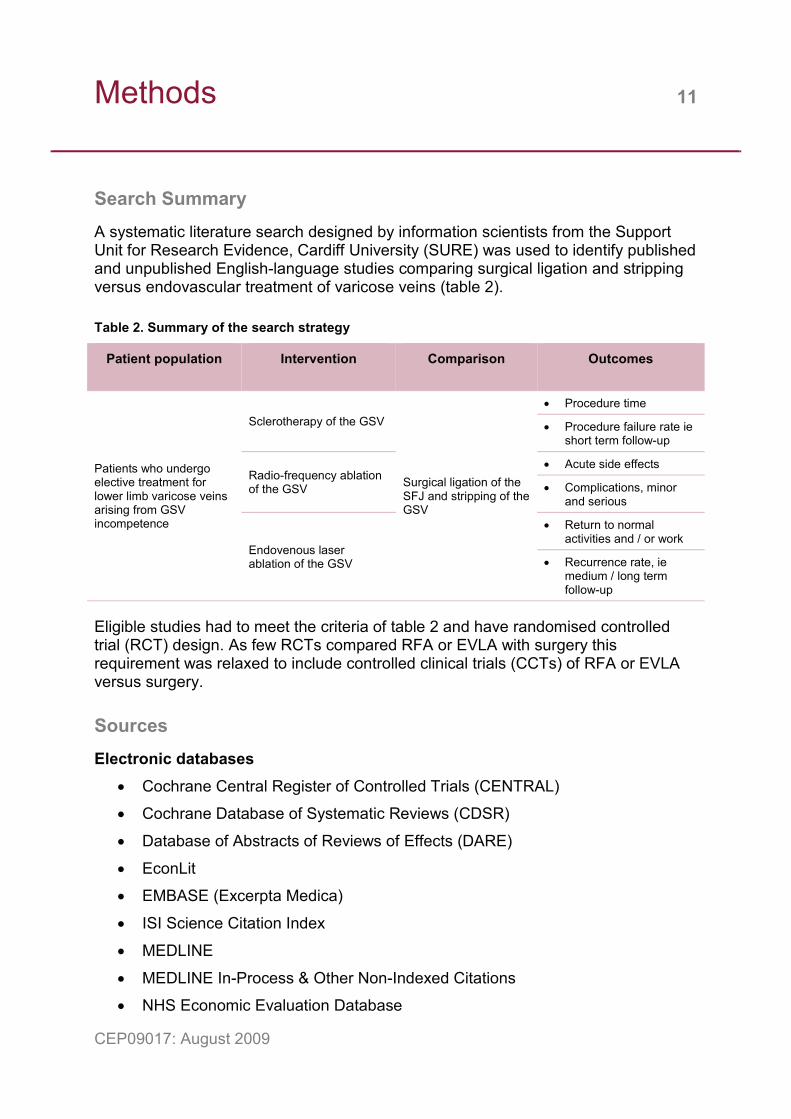
Search Summary A systematic literature search designed by information scientists from the Support Unit for Research Evidence, Cardiff University (SURE) was used to identify published and unpublished English-language studies comparing surgical ligation and stripping versus endovascular treatment of varicose veins (table 2).
Table 2. Summary of the search strategy
Patient population Intervention Comparison Outcomes
Patients who undergo elective treatment for lower limb varicose veins arising from GSV incompetence
Sclerotherapy of the GSV
Surgical ligation of the SFJ and stripping of the GSV
• Procedure time
• Procedure failure rate ie short term follow-up
Radio-frequency ablation of the GSV
• Acute side effects
• Complications, minor and serious
Endovenous laser ablation of the GSV
• Return to normal activities and / or work
• Recurrence rate, ie medium / long term follow-up
Eligible studies had to meet the criteria of table 2 and have randomised controlled trial (RCT) design. As few RCTs compared RFA or EVLA with surgery this requirement was relaxed to include controlled clinical trials (CCTs) of RFA or EVLA versus surgery.
Sources Electronic databases
• Cochrane Central Register of Controlled Trials (CENTRAL)
• Cochrane Database of Systematic Reviews (CDSR)
• Database of Abstracts of Reviews of Effects (DARE)
• EconLit
• EMBASE (Excerpta Medica)
• ISI Science Citation Index
• MEDLINE
• MEDLINE In-Process & Other Non-Indexed Citations
• NHS Economic Evaluation Database
Methods 12
CEP09017: August 2009
• SCOPUS
• Google Scholar
• VascularWeb Conference proceedings
• Vascular and Endovascular Controversies Update 4-7 April 2008
• 30th Charing Cross International Symposium, 12-15 April 2008
• VEITH Symposium
• International Congress of Endovascular Interventions
• Web of Knowledge
• ZETOC Trials and research registers
• CenterWatch
• CinicalTrials
• Current Controlled Trials metaRegister
• International Standard Randomised Controlled Trial Number Register
• WHO International Clinical Trials Registry
• National Research Register – to Sept 2007
• UK Clinical Research Network
• Health Services Research Projects in Progress (HSRProj) Websites
• NICE (guidelines and technology appraisals)
• NIHR Health Technology Assessment Programme
• NLM – Health Services/Technology Assessment
• New Zealand Health Technology Assessment
• Health Services/Technology Assessment Text (HSTAT)
• The West Midlands Health Technology Assessment Collaboration (WMHTAC)
• SHPIC (Scottish Health Purchasing Information Centre)
• SIGN
• Guidelines in Practice
• Canadian Agency for Drugs and Technologies in Heath (CADTH)
Methods 13
CEP09017: August 2009
• Department of Health
• Medicines and Healthcare products Regulatory Agency (MHRA)
• NHS National Patient Safety Agency (NPSA)
• European Society for Vascular Surgery
• Vascular Society of Great Britain and Ireland
• Society for Vascular Surgery (US)
• International Journal of Technology Assessment in Health Care (2008 only)
The economic review used the following databases:
• NIHR Health Technology Assessment Programme
• Database of Abstracts of Reviews of Effects (DARE)
• NHS Economic Evaluation Database
• PubMed
An additional search was conducted, searching national guidelines and medical technology agencies ECRI and HAYS. The reference lists of relevant papers were scanned for further studies.
Search terms 1 varicose vein:.mp. 2 exp varicose vein/ 3 exp saphenous vein/ 4 saphenous vein:.mp. 5 varicos:.tw. 6 ((varicose or saphenous) adj4 vein:)).tw. 7 (saphenous adj3 reflux).tw. 8 venous insufficiency.tw. 9 GSV or GSV.tw. 10 exp sclerotherapy/ 11 exp sclerosing solutions/ 12 exp sodium tetradecyl sulfate/ 13 ultrasonography, interventional/ 14 Catheter Ablation/ae, ec, mt [Adverse Effects, Economics, Methods]
Methods 14
CEP09017: August 2009
15 (sclerothap: or sclerosing).mp. or sclerosant:.tw. 16 ((foam or microfoam or injection or ultrasound-guid: or liquid) adj4 sclerotherap:)).tw. 17 radiofrequency powered segmental thermal ablation.tw. 18 echo-guided compression sclerotherap:.tw. 19 ((endoluminal or endovenous) adj4 (obliteration or laser or therap:)).tw. 20 (EVLT or ELVS or EVLA or RFA or RFITT).tw. 21 (RSTA or TIPP or STRIP-PHLEB).tw. 22 phlebectomy.mp. 23 stab avulsion.tw. 24 (venous adj sclerotherap:).tw. 25 radiofrequency.mp. 26 VNUS closure:.tw. 27 starclose.tw. 28 vascular closure.tw. 29 closurefast.tw. 30 varisolve.tw. 31 trans?illuminat:.tw. 32 tessari.tw. 33 (sotradecol or polidocanol).mp. or sclerovein.tw. 34 ethanolamine oleate.tw. 35 sodium tetradecyl sulfate.mp. 36 or/1-9 37 or/10-35 38 36 and 37 39 limit 38 to english language
Economic search terms were: 1 varicose veins 2 Economics (MeSH) 3 #1 and #2 4 #1 and #2 not ulcer
Methods 15
CEP09017: August 2009
5 cost effective and #1 6 cost effective and #1 not ulcer 7 QALY or utility and #1 not ulcer 8 cost and #1 not ulcer 9 ablation and #1 and #2 not ulcer 10 stripping and #1 and #2 not ulcer 11 endovenous and #1 and #2 not ulcer 12 sclerotherapy and #1 and #2 not ulcer 13 vnus and #1 and #2 not ulcer The reference lists of relevant systematic reviews were also scanned for potentially relevant studies.
Inclusion and exclusion criteria The search identified 3202 references, reduced to 921 after the removal of duplicates and clearly irrelevant articles. The criteria below were applied to the titles and abstracts, resulting in 173 articles. All articles were published between 1996 and 2008, with no restriction on country of origin
Inclusion criteria: 1. Intervention study comparing standard treatment with one / more minimally
invasive techniques (radiofrequency ablation, eg VNUS closure, endovenous laser treatment, sclerotherapy, transilluminate powered phlebectomy) OR comparing one / more minimally invasive techniques against no treatment OR comparing two / more minimally invasive techniques for the treatment of varicose veins or venous ulcers
2. Adults (>15 years) with confirmed symptomatic varicose veins OR great saphenous vein incompetence OR chronic venus insufficiency/disease OR venus ulcers
3. Prospective study designs with a control group: For all interventions: (i) Randomised controlled trial for all interventions (ii) cost-effectiveness study or economic evaluation iii) systematic review or meta-analysis to be unpicked for RCTs. To fill evidence gaps (identified for laser and radiofrequency treatments) the following were considered: (iv) controlled trial without randomisation
Methods 16
CEP09017: August 2009
(v) controlled before and after study 4. Observational / qualitative data only when it is collected as part of a controlled
study 5. Individual and multi-centre study designs 6. Study outcomes to include vein occlusion, quality of life (QOL), time to return to
routine activities / work, symptoms and adverse symptoms related to the procedure, cosmetic appearance, recurrent varicose veins and venous flare formation, overall patient satisfaction, cost-effectiveness
Exclusion criteria: 1. Treatment technique / method not commercially available in the United Kingdom,
eg hyperheated steam 2. Studies including patients with superficial, thread, facial or spider veins or
varicose hand veins 3. Studies evaluating drug therapies or complimentary medicine only 4. Studies that are only available as conference proceedings
173 full-text articles resulted from these criteria and were read by information scientists. They identified 41 published randomised controlled trials (RCTs), 5 ongoing RCTs and 4 published controlled clinical trials (CCTs). Two specialist registrars in surgery and a consultant clinical scientist independently reviewed the 45 published full-text articles to ensure that each directly compared the surgical technique traditionally used in the UK (ligation and stripping) with one of the three endovascular techniques described. Collective discussion, resulted in the selection of 18 published papers [7-24] for critical analysis (tables 6-8, appendix 2).
For economic review, evaluations of costs and outcomes of varicose vein interventions were included. There was no restriction on country of origin, but only English language papers were included. Papers that did not include a cost or resource comparison of traditional surgery versus endovascular treatments were excluded. Thread veins, spider veins and facial veins were outside the scope of this review. 60 published papers were identified as potentially relevant for review. Eight were selected for critical analysis using the Drummond checklist [34] and this was then further refined to five [9;10;15;19;20].
Methods 17
CEP09017: August 2009
Critical analysis Two specialist registrars in surgery and a consultant clinical scientist independently critically reviewed the 18 studies identified for clinical evidence [7-24] using a checklist [35] with arbitration by a consultant vascular surgeon. An evidence level was applied to each study using the classification scheme described [36]. Results from each study were extracted according to the outcomes specified in table 2. The studies are summarised in tables 6-8, appendix 2 and described in the clinical issues section.
Critical analysis of the economic papers is summarised in table 9, appendix 3 and described in the Economic issues section.
Limitations Recent advances in technology, surgical and endosurgical practice mean that studies reporting disease recurrence over a medium to long term follow-up period (2-10 years) were conducted using devices or techniques that have since been modified and are no longer current practice.
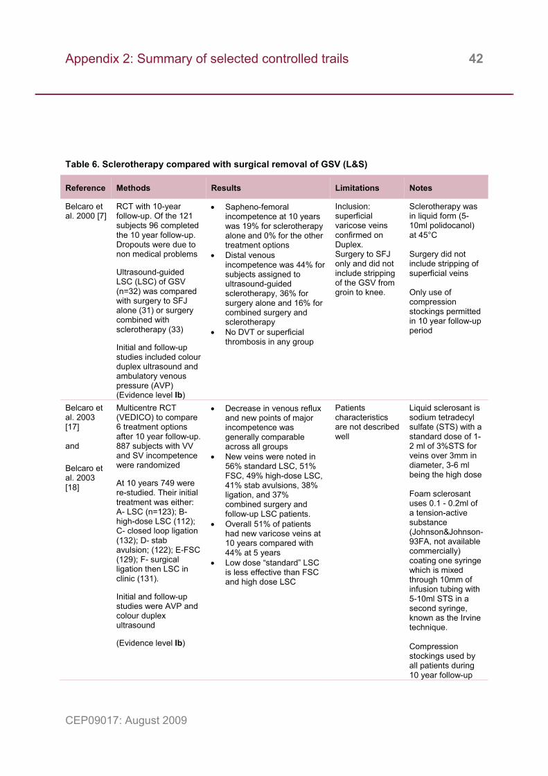
For sclerotherapy, only one study reports clinical outcome of a foamed sclerotherapy product currently licensed for use in the UK. The author of references 7, 17 and 18, Belcaro, has been erased from the register by the General Medical Council (GMC) for misconduct. The GMC are unable to give guidance as to the impact of this on the credibility of the studies. The journals have not retracted the papers concerned.
Laser wavelengths used in the studies reviewed were 810 nm in four studies and 980 nm in two studies. A variety of techniques for fibre withdrawal and laser settings were used, including continuous wave or laser pulsed at 1 or 1.5 seconds. For RFA, all of the studies used the VNUS Closure Plus system.
There is no single agreed classification system for the severity of varicose veins [20]. Across clinical trials there is likely to be variation in classification of severity of cases, which may misrepresent patient groups. This may also occur due to the exclusion of patients not suitable for randomisation.
The VEIN review [32] considered larger numbers of studies by relaxing the standard of evidence required.
Economic evidence confirmed that studies investigating the economic implications of varicose vein treatments are scarce. In most cases the studies did not take place in the UK and practices were not directly comparable with those in the UK. General anaesthesia is used for all procedures in many of the studies. In the UK,
Methods 18
CEP09017: August 2009
endovascular procedures are often performed in a clinic setting using local anaesthesia. This can make a large difference to costs. Alternative health and social care systems in other countries also lead to differences in payments for treatment and the economic impact of time off work.
The evidence is supplemented by a summary of typical procedures gathered during a survey of consultants (table 10, appendix 4).
Survey A questionnaire survey seeking cost related information was completed by six UK surgeons in September 2008 (table 11, appendix 4). Each respondent was a consultant vascular surgeon with established expertise in two or more treatment options. The surgeons work separately in NHS hospitals distributed across the UK and completed the questionnaire independently, based on their own experience.
To assist questionnaire design a study author observed expert surgeons performing each procedure. None of these surgeons contributed data to the survey.
The responses were collated and summarised to achieve a “typical procedure” resource use for each of the four treatment methods (table 3). Differences in costs between trusts are expected due to local variation in:
• details of the procedure
• existing equipment
• choice of supplier
• quantity supplied.
These variations may be very significant and therefore numerical values are not given.
Table 4, appendix 1, gives list prices for some of the main capital items and associated consumables from different suppliers. Prices will vary depending on negotiation between purchaser and supplier.
Evidence review 19
CEP09017: August 2009
Clinical issues Foam sclerotherapy The FSC procedure is generally well tolerated by patients. Less pain and a higher quality of life is reported in the initial post-treatment period following sclerotherapy compared with surgery [19]. Transient neurological symptoms resolving within 24 hours have been reported [21]. These problems were not observed when the sclerotherapy technique was modified and the dose of sclerosant reduced [21]. Common adverse events reported are skin discolouration and limb pain [19;21].
There is a gradual deterioration in venous competence after all treatments for varicose veins. Most of the sclerotherapy studies [17-19;21] reveal that the recurrence of venous incompetence and symptoms is significantly higher for sclerotherapy than for surgery. There were more recurrences 3 months after FSC using Fibrovein (13 percent) than following surgery (7 percent) [19]. This clinical outcome is considerably better than for Varisolve (32 percent and 13 percent, respectively) [21], a product which is not licensed in the UK. In a controlled trial of LSC and surgery both had similar satisfactory outcomes at one year [20], but the study power and follow-up were insufficient to compare recurrence rates. The authors note the increased recurrence rate for sclerotherapy reported in other trials and suggest that a proportion of patients identified as having recurrences because of careful trial follow up may not have presented with recurrence outside the trial environment.
In some cases the relatively poorer long term outcome for FSC compared with surgery may not be of prime importance. Significant benefits accrue from avoiding the risks and side effects associated with surgery for high risk patients and this can be achieved using a simple FSC technique [17;18].
The VEIN review [32] found that FSC is far more effective than LSC. FSC can be carried out successfully on almost any patient with clinically significant venous disease, no matter how elderly, frail, obese or ill they are. The VEIN report concludes that ultrasound guided FSC is a safe and effective method of treating varicose veins but the relative advantages or disadvantages of this treatment in the longer term are yet to be published.
Endovenous laser ablation EVLA results in fewer complications when compared with surgery [8-10;22-24]. Post-operative assessment by colour duplex ultrasound, clinical assessment and quality of life questionnaires reveal similar outcomes for EVLA and surgery [8;22;24]. The majority of the studies reported similar post-operative pain scores for EVLA and surgery and the differences in reported time to return to work or daily activities post procedure were not consistent for the two groups. Wound infections only occurred in
Evidence review 20
CEP09017: August 2009
the surgery groups. Bruising was less frequent following EVLA compared with surgery and skin burns and nerve damage were rare complications of EVLA.
The VEIN study concluded that rates of successful treatment, quality of life, cosmesis and patient satisfaction are similar for surgery and EVLA [32]. Generally EVLA is suitable only for straight veins but experienced practitioners can treat more tortuous veins. Laser wavelength determines laser penetration and absorption but the VEIN study concludes that there is no evidence that wavelength affects clinical outcome [32].
Radio-frequency ablation The VNUS Closure Plus system was used in all of the studies reviewed. There was no consistent difference in procedure time between surgery and RFA [11;12;15;16]. Differences in post-procedure pain, bruising and return to work or normal activities consistently favoured RFA compared with surgery. Skin burns and nerve damage were rare complications of RFA. Clinical and duplex ultrasound assessment of efficacy outcomes showed similar results for RFA and surgery [11;12;14;16].
The VEIN study concludes that RFA is established as an acceptable and efficacious treatment for varicose veins [32].
Summary FSC is the fastest technique and is associated with a high level of patient acceptability, particularly in cases where the cosmetic outcome is not considered a high priority. Unfortunately there is a higher recurrence rate of varicose vein symptoms in the long term varying from 13 percent to 33 percent for FSC compared with from 7 percent to 14 percent for surgery, depending on the specific outcome measure used [7;17-21]. FSC is a very useful technique where surgery is not a good option. FSC can have an additional useful role as a less painful and equally effective alternative to phlebectomy of the visible varicose veins. This may be performed either during the main treatment procedure or at a follow-up clinic.
EVLA and RFA give largely similar long term recurrence rates when compared with surgery. EVLA and RFA have better immediate outcomes (less pain, earlier return to work) than surgery providing tumescence is used. EVLA and RFA also result in reduced post-operative infection rates and recovery times when compared with surgery. Both of these techniques have the potential to provide a clinically effective replacement for traditional surgery.
Economic issues There are very few robust economic papers available comparing endovascular treatments with surgery. Most of the studies were undertaken primarily for clinical
Evidence review 21
CEP09017: August 2009
data with a brief cost comparison. Many of the trials were not based in the UK and make assumptions that are not appropriate for NHS settings. Both surgery and endovascular treatments were often in day-theatre under general anaesthetic. This leads to cost results that do not represent typical UK practice, where endovascular treatment is usually performed under local anaesthetic in a clinic setting. In the UK, surgical ligation and stripping uses a full theatre team and general anaesthetic.
Current economic evidence from the UK on cost effectiveness of these treatments is not sufficient to draw definitive conclusions. Longer term data for recurrence rates and up-to-date direct and indirect costs are needed to assess the efficacy and cost-effectiveness of new treatments.
These papers are discussed in more detail in table 9, appendix 3.
Sclerotherapy compared with surgery Two papers compared costs of sclerotherapy with surgery. Both were based on trials in the UK. One trial used FSC for GSV, but with an additional ligation of the SFJ [19]. The other [20] formed part of the UK REACTIV trial used LSC and is included due to the extent of the economic analysis provided. This analysis is limited by a small sample group, but illustrates the factors that can be considered over and above the simple procedure cost. Similar factors would be appropriate for consideration in all of the endovascular techniques.
Both trials used general anaesthetic for surgery and local anaesthesia in a clinic setting for sclerotherapy.
Overall procedure cost was considerably less at £672.97 for FSC versus £1120.64 for surgery [19]. The lower cost came from:
• shorter procedure time (therefore lower staff costs)
• lower anaesthetic cost (and no requirement for an anaesthetist)
• reduced recovery time in theatre and on the ward
• reduced capital and overhead cost.
FSC incurred costs for ultrasound and more expensive stockings but remained less expensive than surgery.
Clinical results from this study [19] showed that the median time to return to work was lower in the FSC group, at 2 days versus 8 days in the surgical group. The Aberdeen Varicose Veins Questionnaire (AVVQ) score was reduced at 3 months, by 46% in the FSC group and by 40% in the surgery group.
Evidence review 22
CEP09017: August 2009
The study comparing LSC with surgery [20] also showed that the LSC procedure was cheaper than a surgical procedure, including post treatment GP follow up. This study reported few differences between surgery and LSC with respect to changes in symptoms or patient satisfaction at 1 year. The only significant difference in total NHS cost over 2 years came from the initial treatment (p<0.05). The total NHS costs over 2 years were £701.28 for surgery compared with £210.94 for sclerotherapy.
The study also considered patient benefits, complication rates, repeat treatments and other factors. These were used to calculate expected costs and benefits for 10 years after treatment and then find the incremental cost effectiveness ratio (ICER). ICER is the incremental cost of one treatment over another, divided by the incremental benefit in effectiveness. Using this measure to compare both techniques to conservative treatment, surgery has a lower ICER (2083 £/QALY) than LSC (3388 £/QALY). The study concludes that although LSC is cost effective, a blend of surgical and conservative treatment could give greater benefit than LSC. It must be emphasised that this deals with LSC not FSC and that this analysis of 77 randomised patients was too small to draw firm conclusions.
Laser treatment compared with surgery Two non-UK papers of poor applicability to the UK compared EVLA with traditional surgery [9;10].
A cost comparison analysis alongside an RCT [9] found little difference between EVLA and surgical treatment when comparing complications, symptomatic relief, time to resume normal activities, time to return to work and costs. All treatments were performed as office-based procedures using local anaesthetic. Procedure time was not measured. The authors considered that the study may have failed to demonstrate a generalisable difference in the time taken to return to work between the groups because of the Danish system of fully paid sick leave.
The other cost consequences analysis [10] showed that EVLA led to improved outcomes, fewer complications and shorter sick leave in comparison to surgical treatment. All procedures took place in theatre using general anaesthetic. The cost difference between procedures was attributable to the cost of a vein stripper required for surgery versus that of a catheter and generator for EVLA. Savings in the total cost were made by a faster return to work, although the study only recruited employed patients which may overestimate the saving.
In both studies, consistency of treatment was ensured by the same surgeons performing both treatments [9;10]. The laser procedure was performed under guided colour duplex ultrasound and tumescent anaesthesia. Both found that the endovascular procedure was more expensive than surgery, but that when time to return to work costs were considered surgery was a more expensive option. For both
Evidence review 23
CEP09017: August 2009
studies, there are significant limitations in applying these cost conclusions to the UK. Both studies have fixed prices for varicose vein treatment regardless of technique. The differences reported in total procedure price come from instruments (ie laser catheter and generator), and, in one [9], additional duplex imaging. Neither study considered the cost of room alterations or staff training for laser safety.
Literature comparing laser treatment with surgery is limited and no cost effectiveness studies in the UK were identified. Neither of the included studies provided clear results and no robust conclusion could be drawn.
RFA compared with surgery Only one paper compared RFA with traditional surgical treatment - a short term (50 day follow-up) cost-consequences analysis alongside an RCT from a single centre in Finland [15]. The sample was small; 33 patients were randomised to either RFA or traditional surgery. All procedures were carried out in theatre using general anaesthetic so the study design could not evaluate the potential saving from clinic-based RFA. The author concluded that RFA was more costly than surgery. RFA costs included:
• catheter
• RFA generator
• use of an ultrasound machine
• longer procedure time than surgery (this is not found in all other studies)
• use of a radiologist in addition to a surgeon (not normal in UK)
• procedure takes place in theatre using general anaesthetic (not normal in the UK once practice established).
The study found 11.6 lost working days following surgery and 4.5 following RFA. When the indirect cost of productivity loss was included, RFA was found to be a more cost effective treatment. A sensitivity analysis considered the numbers of patients not in employment and the number of operations per year. Where 60% of patients are employed, the study found RFA total costs to be lower than surgical total costs when 43 or more procedures were done per year. The study is not fully applicable to the UK and did not provide clear results. No robust conclusion could be drawn.
Expert opinion Cost savings from endovascular techniques are identified in the survey as dependent on a change being made from treatment in theatres to treatment in an office or clinic. This reduces staff and equipment requirements and frees theatres for other procedures. There is also normally a reduction in procedure time and in pre- and
Evidence review 24
CEP09017: August 2009
post-operative stay. These factors have to be weighed against the availability of a suitable office location and the costs of equipping it.
EVLA and RFA also require capital costs for laser or radiofrequency generators and have high consumable costs.
Residual varicosities may be removed following EVLA or RFA using foam or phlebectomy. This may be during the same session as the main treatment, or in an additional outpatient visit. Additional visits will increase costs slightly.FSC has the greatest potential for cost savings (looking only at the costs of a procedure, not a full economic analysis) since it can be done in an office location, requires relatively little capital investment and few consumables.
Endovascular treatment can bring cost savings to the patient, and the wider economy, with a faster return to work. A full economic analysis of each method is not covered in the scope of this report, however recovery times, complications and repeat procedures should be considered when deciding which procedure to use.
Traditional vascular surgery is carried out in theatres, usually under a general anaesthetic and requires a high level of staffing, as detailed in table 3. Equipment in theatres is also costly. Typical list prices for major equipment are: operating table £30,000 [37], anaesthetic machine £22,000 [38] and electrosurgery unit £12,053 [39]. This equipment will already be in place in any theatre, however a cost for maintenance and replacement should be included in calculations. This cost would normally be shared with other services that use theatres. There is also an opportunity cost which arises because a theatre that is required for varicose vein surgery is not available for other surgical procedures.
In addition to direct procedure costs for surgery, there are staffing and location costs for recovery and a short stay on a day ward. In some cases, patients may stay overnight.
Although a colour duplex ultrasound machine is not required during surgery, it is used for pre-operative assessment by many surgeons, requiring a clinic visit ideally on the same day as the procedure to mark the incompetent veins on the leg. Other pre-operative assessments may also be required, such as laboratory tests and ECG.
All of the endovascular techniques discussed in this report can be done in a clinic setting, although it is quite common to use theatres until the procedure is established. Equipment requirements include:
• a tilting table that allows the Trendelenburg position
Evidence review 25
CEP09017: August 2009
• use of a colour duplex ultrasound machine (it may be possible to utilise an existing machine)
• general equipment such as storage cupboards and equipment trolleys
• portable examination light
• an area for reception of patients and for a brief stay post procedure. For FSC there must be facilities to cope with anaphylaxis, eg a resuscitation trolley. There are no other capital costs required for this procedure and consumables have a relatively low cost.
For either EVLA or RFA an appropriate generator and a tumescent pump are required. Additionally a catheter kit is needed which typically incorporates an entry needle, guide wire, introducer sheath and laser or RFA catheter. Procedure packs are also sold by most generator suppliers containing the other consumables required for the procedure, in some cases including the tumescent drugs.
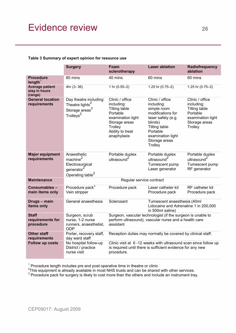
Table 3 lists equipment and procedure times taken from the expert opinion survey. There are strong preferences for different techniques between individuals and this may be reflected in the answers given.
Evidence review 26
CEP09017: August 2009
Table 3 Summary of expert opinion for resource use
1 Procedure length includes pre and post operative time in theatre or clinic 2This equipment is already available in most NHS trusts and can be shared with other services. 3 Procedure pack for surgery is likely to cost more than the others and include an instrument tray.
Surgery Foam sclerotherapy
Laser ablation Radiofrequency ablation
Procedure length1
80 mins 40 mins 60 mins 60 mins
Average patient stay in hours (range)
4hr (3- 36) 1 hr (0.55–2) 1.25 hr (0.75–2) 1.25 hr (0.75–2)
General location requirements
Day theatre including: Theatre lights2 Storage areas2 Trolleys2
Clinic / office including: Tilting table Portable examination light Storage areas Trolley Ability to treat anaphylaxis
Clinic / office including: simple room modifications for laser safety (e.g. blinds) Tilting table Portable examination light Storage areas Trolley
Clinic / office including: Tilting table Portable examination light Storage areas Trolley
Major equipment requirements
Anaesthetic machine2 Electrosurgical generator2 Operating table2
Portable duplex ultrasound2
Portable duplex ultrasound2 Tumescent pump Laser generator
Portable duplex ultrasound2 Tumescent pump RF generator
Maintenance Regular service contract
Consumables – main items only
Procedure pack3 Vein stripper
Procedure pack Laser catheter kit Procedure pack
RF catheter kit Procedure pack
Drugs – main items only
General anaesthesia
Sclerosant
Tumescent anaesthesia (40ml Lidocaine and Adrenaline 1 in 200,000 in 500ml saline)
Staff requirements for procedure
Surgeon, scrub nurse, 1-2 nurse runners, anaesthetist, ODP
Surgeon, vascular technologist (if the surgeon is unable to perform ultrasound), vascular nurse and a health care assistant
Other staff requirements
Porter, recovery staff, day ward staff
Reception duties may normally be covered by clinical staff.
Follow up costs No hospital follow-up District / practice nurse visit
Clinic visit at 6 -12 weeks with ultrasound scan since follow up is required until there is sufficient evidence for any new procedure.
Conclusions 27
CEP09017: August 2009
Varicose veins cause major socio-economic and health problems. In the UK varicose vein procedures consumed approximately £40 million in 2005-6 [1]. Varicose veins are not an acute life-threatening condition but do have an impact on quality of life and can involve significant expenditure, in particular in the provision of nursing care for venous ulcers. Treatment of varicose veins gives a benefit to patients and both surgery and LSC are well below the NICE threshold of £30,000 per QALY [20]. This is also likely to be true for FSC, RFA and EVLA.
Surgery is the most common treatment within the NHS at present. It is very effective in removing varicose veins and has a high long term success rate. Surgery in the UK is performed using general anaesthetic in theatre. Patients may experience significant bruising, post-procedure pain, and complications such as groin infections. Return to work is frequently reported as between one and two weeks, or even more, resulting in a high social and economic cost to society. Endovascular techniques offer an alternative to surgery and NICE has concluded that they are sufficiently safe and effective for use in the NHS. NICE recommends audit procedures and patient information for FSC.
All the endovascular techniques considered can be performed without a general anaesthetic. This frees theatres for other surgical procedures and leads to a, faster recovery, lower staff costs, reduced equipment needs and reduced ward time pre and post procedure. They also give a quicker return to normal activities.
FSC is the cheapest procedure and requires no special equipment (tables 3, 4). It can be used on almost any patient, including those not suited to general anaesthesia and those with tortuous veins. Its effects do not last as long as other treatments, meaning repeat procedures may be necessary. This reduces the overall cost effectiveness of FSC. It is less painful immediately after the procedure than surgery and allows a faster return to normal activities, but may not be as cosmetically pleasing as some other procedures. There are various side effects associated with FSC, some of which are serious, although rare.
Although EVLA and RFA are both effective in the short term, there is a lack of evidence for long term results. They require capital investment and expensive consumables. There is a lack of robust UK evidence on cost implications. Patients may have a faster return to normal activities resulting in a cost saving to the wider economy. RFA studies show a reduction in post-procedure pain and bruising.
There is significant potential to improve both patient comfort post procedure and recovery rates with all of the endovascular techniques considered. There is also potential to reduce procedure costs, depending on individual circumstances. There is a lack of evidence for long term efficacy. FSC has poorer long term effectiveness than surgery, while EVLA and RFA may be similar to surgery in this regard.
Acknowledgements 28
CEP09017: August 2009
We should like to thank the following for their contribution to this evidence review.
Ian Bayliff, Buyer, NHS Supply Chain
Professor Andrew Bradbury1, Consultant Vascular Surgeon, Heart of England NHS Foundation Trust
Philip Coleridge-Smith2, Consultant Surgeon, University College London Hospitals NHS Foundation Trust
Jonothan Earnshaw3, Consultant General and Vascular Surgeon, Gloucestershire Hospitals NHS Foundation Trust
Paul Edwards, Consultant Surgeon, Countess of Chester Hospital NHS Foundation Trust
Peggy Edwards, Patient Safety Manager, National Patient Safety Agency
Michael J Gough1, Leeds Vascular Institute, Leeds Teaching Hospitals NHS Trust
Susan Hill, Consultant Vascular Surgeon, Cardiff and Vale NHS Trust
Mel King4, Senior Medical Device Specialist, MHRA
Hilary Kitcher, Support Unit for Research Evidence (SURE), Cardiff University
Tim Lees1, Consultant Vascular Surgeon, Newcastle Upon Tyne Hospitals NHS Foundation Trust
Ian Loftus, Consultant Vascular Surgeon, St George’s Healthcare NHS Trust
Shane MacSweeney, Consultant Vascular Surgeon, Nottingham University Hospitals NHS Trust
Media Resources, School of Medicine, Cardiff University
Maxine Priest5, Education Special Interest Group Lead, AFPP
Aisling Roberts, Chair, Society of Vascular Nurses
Lesley Sander, Support Unit for Research Evidence (SURE), Cardiff University
Tracey Wall6, Consultant Anaesthetist, Abertawe Bro Morgannwg University NHS Trust
David West7, Medical Director, Veincentre
Acknowledgements 29
CEP09017: August 2009
Ken Woodburn, Consultant Vascular Surgeon, Royal Cornwall Hospitals NHS Trust
Elaine Young, President, Society for Vascular Technology
Cardiff and Vale NHS Trust
Imperial College Healthcare NHS Trust
Manufacturers and suppliers of listed products
1 Nominated by the Vascular Society 2 Nominated by the British Association of Sclerotherapists 3 Nominated by the Venous Forum 4 Nominated by the Medicines and Healthcare products Regulatory Agency 5 Nominated by the Association for Perioperative Practice 6 Nominated by the Vascular Anaesthesia Society 7 Nominated by the British Society of Interventional Radiology
Glossary 30
CEP09017: August 2009
Aberdeen Varicose Veins Questionnaire (AVVQ) is a validated specific quality of life questionnaires for lower limb venous disease [40]
Ablation Destruction of unwanted tissue.
Ambulatory venous pressure (AVP) The blood pressure in the veins of the leg measured on weight bearing.
Anaphylaxis A life threatening hypersensitive allergic reaction.
Cannulation Insertion of a flexible tube into a blood vessel
Colour duplex ultrasound An imaging technique that combines information on blood flow direction and speed displayed as colour, overlaid on the conventional black and white ultrasound image.
Deep vein thrombosis (DVT) A disorder involving a thrombus (blood clot) in one of the deep veins of the leg. A deep vein thrombosis is potentially life threatening.
EQ-5D Health outcome measure based on five dimensions of health : Mobility, self-care, usual activities, pain/discomfort and anxiety/depression.
Endovascular Relating to a procedure in which a catheter is inserted into a blood vessel for treatment of vascular disease.
Endovenous laser ablation (EVLA) A technique designed to destroy the internal lining of varicose veins by heating using laser energy.
Foam sclerotherapy (FSC) The use of sclerosing chemicals constituted as a foam to treat varicosities. The agent produces inflammation and later fibrosis and obliteration of the lumen.
Great saphenous vein (GSV) Also known as the long saphenous vein, the GSV is a truncal vein which runs from
Glossary 31
CEP09017: August 2009
the ankle to the groin where it drains into the femoral vein. It is the longest vein in the body and, usually being from one to two centimetres deep in the thigh and lies more superficially near the knee and in the lower leg and primarily drains blood from superficial veins in the leg.
HRQoL Health-related quality of life
ICER Incremental Cost Effectiveness Ratio. Ratio of change in costs to change in effects of one intervention compared with an alternative.
Incompetent vein A vein that does not function adequately because a faulty valve allows blood to flow away from the heart in the reverse direction from normal.
Ligation The procedure of tying off a blood vessel with a suture.
Liquid sclerotherapy (LSC) The use of sclerosing chemical in liquid form to treat varicosities. The agent produces inflammation and later fibrosis and obliteration of the lumen.
Non-steroidal anti-inflammatory drug (NSAID) One of a group of drugs having antipyretic, analgesic and anti-inflammatory effects eg aspirin, ibuprofen.
Quality of life (QoL) A measure of a person’s ability to cope successfully with the full range of challenges encountered in the real world.
QALY Quality Adjusted Life Years. Outcome measure that captures both quality and quantity elements in a single measure.
Radio-frequency ablation (RFA) A technique designed to destroy the internal lining of varicose veins by heating, using radio-frequency (RF) energy.
Reflux An abnormal backwards flow of blood.
Glossary 32
CEP09017: August 2009
Saphenofemoral junction (SFJ) The junction between the great saphenous vein and the femoral vein at the groin.
Small saphenous vein (SSV) A superficial vein originating at the ankle and terminating at the popliteal vein behind the knee.
Sodium tetradecyl sulfate (STS) A synthetic surface-active substance used in sclerotherapy. It is a clear solution with the properties of soap that is non-viscous, has a low surface tension and is readily miscible with blood.
Tessari method A method for producing a foam from a liquid sclerosent using two syringes and a 3-way tap. The solution is quickly passed from one syringe to the other twenty times via the tap. After the first ten passages the tap is narrowed. The procedure results in a foam of high quality and consistency.
Trendelenburg position A position in which the head is low and the body and legs are on an inclined plane.
Tumescent anaesthesia Administration of a local infiltration anaesthetic, lidocaine, through the use of large volumes of fluid. In varicose vein procedures administration is through several injections. The anaesthetic infiltrates around the vein, along the entire length to be treated.
Varicose veins (VV) Tortuous dilated veins with incompetent valves.
Venous insufficiency An abnormal circulatory condition consisting of reduced return of venous blood from the legs to the trunk of the body.
Visual analogue scale (VAS) A measurement scale for a characteristic that ranges across a continuum of values and cannot easily be measured. A VAS is usually a horizontal line with a label at each end eg ‘no pain’ and ‘the worst pain ever’ at the other end. The patient marks the scale according to how they judge the severity of their pain.
References 33
CEP09017: August 2009
(1) House of Commons Hansard written answers for 12 Mar 2007
(2) National Institute for Health and Clinical Excellence. Interventional procedure overview of radiofrequency ablation of varicose veins (VNUS closure). 2003. Available at: http://www.nice.org.uk/ip132overview
(3) National Institute for Health and Clinical Excellence. Endovenous laser treatment of the long saphenous vein: Guidance. 2004. Available at: http://www.nice.org.uk/Guidance/IPG52/Guidance/pdf/English
(4) National Institute for Health and Clinical Excellence. Systematic review of the safety and efficacy of foam sclerotherapy for venous disease of the lower limbs. 2006. Available at: http://www.nice.org.uk/guidance/index.jsp?action=download&o=31296
(5) National Institute for Health and Clinical Excellence. IPG217 Ultrasound guided foam sclerotherapy for varicose veins: Guidance. 2007. Available at: http://www.nice.org.uk/IPG217
(6) National Institute for Health and Clinical Excellence. IPG217 Ultrasound-guided foam sclerotherapy for varicose veins: Audit criteria. 2007. Available at: http://www.nice.org.uk/IPG217AuditCriteria
(7) Belcaro GN. Endovascular sclerotherapy, surgery, and surgery plus sclerotherapy in superficial venous incompetence: A randomized, 10-year follow-up trial - Final results. Angiology 2000 Jul;51(7):529-34.
(8) Mekako AI, Hatfield J, Bryce J, Lee D, McCollum PT, Chetter I. A nonrandomised controlled trial of endovenous laser therapy and surgery in the treatment of varicose veins. Annals of Vascular Surgery 2006;20(4):451-7.
(9) Rasmussen LH, Bjoern L, Lawaetz M, Blemings A, Lawaetz B, Eklof B. Randomized trial comparing endovenous laser ablation of the great saphenous vein with high ligation and stripping in patients with varicose veins: Short-term results. Journal of Vascular Surgery 2007;46(2):308-15.
(10) Vuylsteke M, V. Endovenous laser obliteration for the treatment of primary varicose veins. Phlebology 2006 Jun;21(2):Jun:80-7
(11) Hinchliffe RJ, Ubhi J, Beech A, Ellison J, Braithwaite BD, Hinchliffe RJ, et al. A prospective randomised controlled trial of VNUS closure versus surgery for the treatment of recurrent long saphenous varicose veins.[see comment]. European Journal of Vascular & Endovascular Surgery 2006 Feb;31(2):212-8.
(12) Lurie FC. Prospective randomized study of endovenous radiofrequency obliteration (Closure procedure) versus ligation and stripping in a selected
References 34
CEP09017: August 2009
patient population (EVOLVeS Study). Journal of Vascular Surgery 2003 Aug;38(2).
(13) Lurie F, Creton D, Eklof B, Kabnick LS, Kistner RL, Pichot O, et al. Prospective randomised study of endovenous radiofrequency obliteration (closure) versus ligation and vein stripping (EVOLVeS): Two-year follow-up. European Journal of Vascular and Endovascular Surgery 2005;29(1):67-73.
(14) Perala JR. Radiofrequency endovenous obliteration versus stripping of the long saphenous vein in the management of primary varicose veins: 3-Year outcome of a randomized study. Annals of Vascular Surgery 2005 Sep;19(5):669-72
(15) Rautio T, Ohinmaa A, Perala J, Ohtonen P, Heikkinen T, Wiik H, et al. Endovenous obliteration versus conventional stripping operation in the treatment of primary varicose veins: a randomized controlled trial with comparison of the costs. Journal of Vascular Surgery 2002 May;35(5):958-65.
(16) Stotter LS, I. Comparative outcomes of radiofrequency endoluminal ablation, invagination stripping, and cryostripping in the treatment of great saphenous vein insufficiency. Phlebology 2006 Jun;21(2):Jun:60-4
(17) Belcaro GC. Foam-sclerotherapy, surgery, sclerotherapy, and combined treatment for varicose veins: A 10-year, prospective, randomized, controlled, trial (VEDICO* trial). Angiology 2003 May;54(3):307-15.
(18) Belcaro GC. Treatments for varicose veins: Surgery, sclerotherapy, foamsclerotherapy and combined (surgery+sclerotherapy) options. A 10-year, prospective, randomised, controlled, follow-up study. The VEDICO* trial and EST (European Sclerotherapy Trial). Angeiologie 2003 Jan;55(1):29-36.
(19) Bountouroglou DG, Azzam M, Kakkos SK, Pathmarajah M, Young P, Geroulakos G, et al. Ultrasound-guided foam sclerotherapy combined with sapheno-femoral ligation compared to surgical treatment of varicose veins: early results of a randomised controlled trial. European Journal of Vascular & Endovascular Surgery 2006 Jan;31(1):93-100.
(20) Michaels JA, Campbell WB, Brazier JE, Macintyre JB, Palfreyman SJ, Ratcliffe J, et al. Randomised clinical trial, observational study and assessment of cost-effectiveness of the treatment of varicose veins (REACTIV trial). [Review] [220 refs]. Health Technology Assessment (Winchester, England) 2006 Apr 20;10(13):1-196.
(21) Wright DG. Varisolve polidocanol microfoam compared with surgery or sclerotherapy in the management of varicose veins in the presence of trunk
References 35
CEP09017: August 2009
vein incompetence: European randomized controlled trial. Phlebology 2006 Dec;21(4):180-90.
(22) Darwood RJ, Theivacumar N, Dellagrammaticas D, Mavor AI, Gough MJ, Darwood RJ, et al. Randomized clinical trial comparing endovenous laser ablation with surgery for the treatment of primary great saphenous varicose veins. British Journal of Surgery 2008 Mar;95(3):294-301.
(23) de Medeiros CA LG. Comparison of endovenous treatment with an 810 nm laser versus conventional stripping of the great saphenous vein in patients with primary varicose veins. Dermatologic surgery : official publication for American Society for Dermatologic Surgeryl 2005 Dec;31(12):1685-94.
(24) Kalteis MB, I. High ligation combined with stripping and endovenous laser ablation of the great saphenous vein: Early results of a randomized controlled study. Journal of Vascular Surgery 2008 Apr;47(4):822-9
(25) Bradbury A, Evans C, Allan P, Lee A, Vaughan Ruckley C, Fowkes FGR. What are the symptoms of varicose veins? Edinburgh vein study cross sectional population survey. British Medical Journal 1999;6(318):353-6.
(26) Caggiati A. Historical Background. In: Labropoulos N, Stansby G, editors. Venous and Lymphatic Diseases.New York: Taylor and Francis; 2006. p. 1-7.
(27) British Medical Association and the Royal Pharmaceutical Society of Great Britain. British National Formulary No. 57. 2009.
(28) Cavezzi A. A new sclerosing foam in the treatment of varicose veins: Tessari method. Minerva Cardioangiol 2000;48((Suppl)1: 248).
(29) Medicines and Healthcare Regulatory Agency. Device Bulletin DB2008(03): Guidance on the safe use of lasers, intense light source systems and LEDs in medical, surgical, dental and aesthetic practices. 2008. Available at: http://www.mhra.gov.uk/Publications/Safetyguidance/DeviceBulletins/CON014775
(30) Medical Devices Agency. MDA 01022 Bipolar Electrosurgery Review. 2001. Available at: www.pasa.nhs.uk/cep
(31) ISCP. Intercollegiate Surgical Curriculum Programme. 2009.
(32) Berridge D, Lees T, Michaels J, Davies A, Earnshaw J, VEnous INtervention (VEIN) Project. 2009. Summary:http://www.roysocmed.ac.uk/academ/downloads/veg102_apr09.pdf
(33) Department of Health. News Distribution Service for Government and the Public Sector. News Item: NHS ready for PROM date. 2009 Feb 6. Available
References 36
CEP09017: August 2009
at:http://nds.coi.gov.uk/content/detail.aspx?NewsAreaId=2&ReleaseID=392024&SubjectId=16&AdvancedSearch=true
(34) Drummond MF, Sculpher MJ, Torrance GW, O'Brian BJ, Stoddart GL. Critical assessment of economic evaluation. Methods for the economic evaluation of health care programmes. Third ed. New York: Oxford University Press; 2005. p. 27-53.
(35) Oxman AD. Checklists for review articles. 10-9-1994.
(36) Hadorn DC, Baker D, Hodges JS, Hicks N, Hadorn DC, Baker D, et al. Rating the quality of evidence for clinical practice guidelines. Journal of Clinical Epidemiology 1996 Jul;49(7):749-54.
(37) NHS Purchasing and Supply Agency's Centre for Evidence based Purchasing (CEP). CEP Buyers' guide: operating tables (draft CEP report circulated to CEDAR; due for publication in 2009). 2009.
(38) Cardiff and Vale NHS Trust. Personal communication from the Department of Clinical Engineering, Cardiff and Vale NHS Trust to CEDAR regarding the Fabius GS anaesthetic machine (Draeger Medical). 27-1-2009.
(39) Valleylab. Personal communication from Valleylab (part of Covidien) to CEDAR, regarding the Force FX electrosurgery unit. 2-4-2009.
(40) Garratt AM, Macdonald LM, Ruta DA, Russell IT, Buckingham JK, Krukowski ZH. Towards measurement of outcome for patients with varicose veins. Qual Health Care 1993;2(1):5-10.
Appendix 1: Products and suppliers 37
CEP09017: August 2009
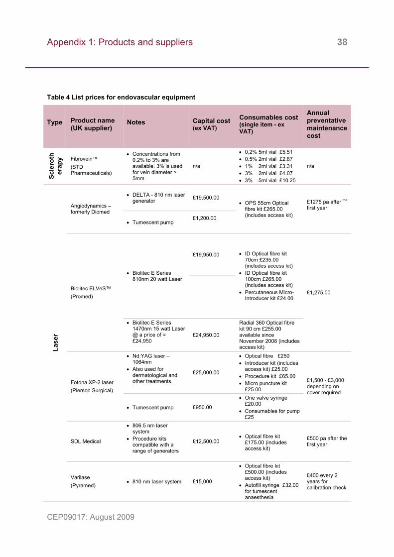
Endovascular ablation products Table 4 gives list prices for equipment used in endovascular ablation techniques. Supplier details are shown on table 5. All prices are exclusive of VAT. The list is correct at the time of publication however new products and suppliers may become available and the products listed may be superseded in time.
Prices are usually negotiable with potential savings of up to 50% on list price depending on factors including the type of purchaser (eg NHS) and quantity to be supplied. Capital equipment may be available for rental, or occasionally as a free loan if there is an agreement to purchase sufficient consumable items. The consumable items are a significant cost and must be considered when choosing a laser or RFA generator. It may be possible to use EVLA catheters from one supplier with a different supplier’s generator to achieve cost savings. Purchasers must consider which is the most effective solution according to local needs.
Laser treatment requires that staff wear protective glasses, and patients have a protective eye covering. The type of glasses required depends on both the wavelength and type of laser. Typical costs are from £125 to £300 per pair. Glasses should be CE marked and conform to BS EN 207.
RFA and EVLA require an access kit containing disposables to give access to the vein, typically including a needle, guidewire and introducer sheath. These may be included in the main kit, a procedure pack or purchased separately. This is listed in table 4 for each supplier.
Appendix 1: Products and suppliers 38
CEP09017: August 2009
Table 4 List prices for endovascular equipment
Type Product name (UK supplier)
Notes Capital cost (ex VAT)
Consumables cost (single item - ex VAT)
Annual preventative maintenance cost
Scle
roth
erap
y Fibrovein™ (STD Pharmaceuticals)
• Concentrations from 0.2% to 3% are available. 3% is used for vein diameter > 5mm
n/a
• 0,2% 5ml vial £5.51 • 0.5% 2ml vial £2.87 • 1% 2ml vial £3.31 • 3% 2ml vial £4.07 • 3% 5ml vial £10.25
n/a
Lase
r
Angiodynamics – formerly Diomed
• DELTA - 810 nm laser generator £19,500.00
• OPS 55cm Optical fibre kit £265.00 (includes access kit)
£1275 pa after the
first year
• Tumescent pump £1,200.00
Biolitec ELVeS™ (Promed)
• Biolitec E Series 810nm 20 watt Laser
£19,950.00 • ID Optical fibre kit 70cm £235.00 (includes access kit)
• ID Optical fibre kit 100cm £265.00 (includes access kit)
• Percutaneous Micro-Introducer kit £24.00
£1,275.00
• Biolitec E Series 1470nm 15 watt Laser @ a price of = £24,950
£24,950.00
Radial 360 Optical fibre kit 90 cm £255.00 available since November 2008 (includes access kit)
Fotona XP-2 laser (Pierson Surgical)
• Nd:YAG laser – 1064nm
• Also used for dermatological and other treatments.
£25,000.00
• Optical fibre £250 • Introducer kit (includes
access kit) £25.00 • Procedure kit £65.00 • Micro puncture kit
£25.00
£1,500 - £3,000 depending on cover required
• Tumescent pump £950.00
• One valve syringe £20.00
• Consumables for pump £25
SDL Medical
• 806.5 nm laser system
• Procedure kits compatible with a range of generators
£12,500.00
• Optical fibre kit
£175.00 (includes access kit)
£500 pa after the first year
Varilase (Pyramed)
• 810 nm laser system £15,000
• Optical fibre kit £500.00 (includes access kit)
• Autofill syringe £32.00 for tumescent anaesthesia
£400 every 2 years for calibration check

Appendix 1: Products and suppliers 39
CEP09017: August 2009
Type Product name (UK supplier)
Notes Capital cost (ex VAT)
Consumables cost (single item - ex VAT)
Annual preventative maintenance cost
Rad
io-fr
eque
ncy
Celon Lab Precision (Olympus Medical)
• Celon lab precision generator & procurve catheter launch 2007
• Procurve catheter also used for perforator surgery
• Loan generator available through loyalty scheme
• UK distributers at complete Aloha Ultrasound range
• Tumescent pump
£10,600.00
• Pro Curve K10003442 catheter £330.00
• Intravenous Sheath Kit £100.00 (includes access kit)
• 4D Access kit (for tortuous veins) £115.00
• Intravenous Guidewire 150cm £30.00
• Mobile Trolley £2,100.00
£960 pa after the first year
VNUS Closure (VNUS Medical Technologies UK)
• Closure Fast catheter launched in 2006
• Closure Plus catheter cited in critical analysis studies
• RFF Stylet catheter is for treating perforators
• Loan generator available by negotiation
£9,995.00
• Closure Fast catheter £519.00
• VNUS RFF Stylet™ £210.00
• Procedure kit including (includes access kit, tumescent pump consumables and other consumables) £59.00
3 years maintenance included in purchase price
• Tumescent pump available as free loan with sufficient consumable purchase.
£695.00
Appendix 1: Products and suppliers 40
CEP09017: August 2009
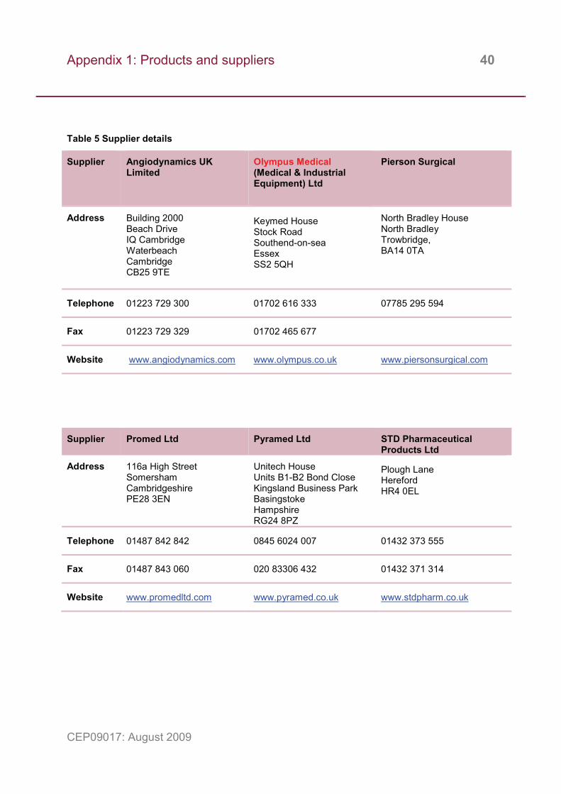
Table 5 Supplier details
Supplier Angiodynamics UK Limited
Olympus Medical (Medical & Industrial Equipment) Ltd
Pierson Surgical
Address Building 2000 Beach Drive IQ Cambridge Waterbeach Cambridge CB25 9TE
Keymed House Stock Road Southend-on-sea Essex SS2 5QH
North Bradley House North Bradley Trowbridge, BA14 0TA
Telephone 01223 729 300 01702 616 333 07785 295 594
Fax 01223 729 329 01702 465 677
Website www.angiodynamics.com www.olympus.co.uk www.piersonsurgical.com
Supplier Promed Ltd Pyramed Ltd STD Pharmaceutical Products Ltd
Address 116a High Street Somersham Cambridgeshire PE28 3EN
Unitech House Units B1-B2 Bond Close Kingsland Business Park Basingstoke Hampshire RG24 8PZ
Plough Lane Hereford HR4 0EL
Telephone 01487 842 842 0845 6024 007 01432 373 555
Fax 01487 843 060 020 83306 432 01432 371 314
Website www.promedltd.com www.pyramed.co.uk www.stdpharm.co.uk
Appendix 1: Products and suppliers 41
CEP09017: August 2009
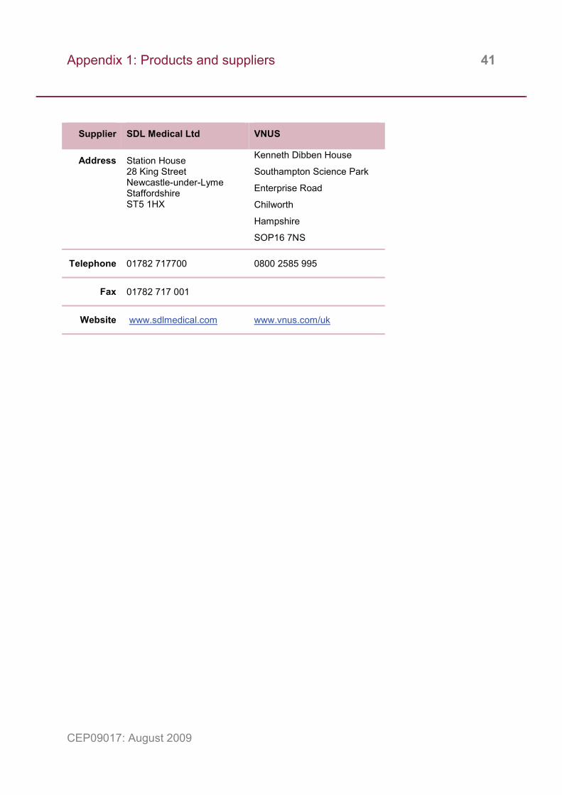
Supplier SDL Medical Ltd VNUS
Address Station House 28 King Street Newcastle-under-Lyme Staffordshire ST5 1HX
Kenneth Dibben House
Southampton Science Park
Enterprise Road
Chilworth
Hampshire
SOP16 7NS
Telephone 01782 717700 0800 2585 995
Fax 01782 717 001
Website www.sdlmedical.com www.vnus.com/uk
Appendix 2: Summary of selected controlled trails 42
CEP09017: August 2009
Table 6. Sclerotherapy compared with surgical removal of GSV (L&S)
Reference Methods Results Limitations Notes
Belcaro et al. 2000 [7]
RCT with 10-year follow-up. Of the 121 subjects 96 completed the 10 year follow-up. Dropouts were due to non medical problems Ultrasound-guided LSC (LSC) of GSV (n=32) was compared with surgery to SFJ alone (31) or surgery combined with sclerotherapy (33) Initial and follow-up studies included colour duplex ultrasound and ambulatory venous pressure (AVP) (Evidence level Ib)
• Sapheno-femoral incompetence at 10 years was 19% for sclerotherapy alone and 0% for the other treatment options
• Distal venous incompetence was 44% for subjects assigned to ultrasound-guided sclerotherapy, 36% for surgery alone and 16% for combined surgery and sclerotherapy
• No DVT or superficial thrombosis in any group
Inclusion: superficial varicose veins confirmed on Duplex. Surgery to SFJ only and did not include stripping of the GSV from groin to knee.
Sclerotherapy was in liquid form (5-10ml polidocanol) at 45°C Surgery did not include stripping of superficial veins Only use of compression stockings permitted in 10 year follow-up period
Belcaro et al. 2003 [17] and Belcaro et al. 2003 [18]
Multicentre RCT (VEDICO) to compare 6 treatment options after 10 year follow-up. 887 subjects with VV and SV incompetence were randomized At 10 years 749 were re-studied. Their initial treatment was either: A- LSC (n=123); B- high-dose LSC (112); C- closed loop ligation (132); D- stab avulsion; (122); E-FSC (129); F- surgical ligation then LSC in clinic (131). Initial and follow-up studies were AVP and colour duplex ultrasound (Evidence level Ib)
• Decrease in venous reflux and new points of major incompetence was generally comparable across all groups
• New veins were noted in 56% standard LSC, 51% FSC, 49% high-dose LSC, 41% stab avulsions, 38% ligation, and 37% combined surgery and follow-up LSC patients.
• Overall 51% of patients had new varicose veins at 10 years compared with 44% at 5 years
• Low dose “standard” LSC is less effective than FSC and high dose LSC
Patients characteristics are not described well
Liquid sclerosant is sodium tetradecyl sulfate (STS) with a standard dose of 1-2 ml of 3%STS for veins over 3mm in diameter, 3-6 ml being the high dose Foam sclerosant uses 0.1 - 0.2ml of a tension-active substance (Johnson&Johnson-93FA, not available commercially) coating one syringe which is mixed through 10mm of infusion tubing with 5-10ml STS in a second syringe, known as the Irvine technique. Compression stockings used by all patients during 10 year follow-up
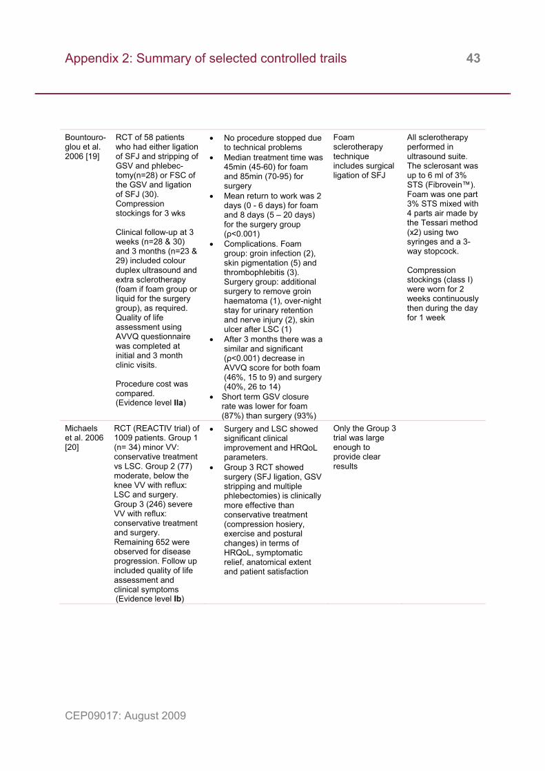
Appendix 2: Summary of selected controlled trails 43
CEP09017: August 2009
Bountouro-glou et al. 2006 [19]
RCT of 58 patients who had either ligation of SFJ and stripping of GSV and phlebec-tomy(n=28) or FSC of the GSV and ligation of SFJ (30). Compression stockings for 3 wks Clinical follow-up at 3 weeks (n=28 & 30) and 3 months (n=23 & 29) included colour duplex ultrasound and extra sclerotherapy (foam if foam group or liquid for the surgery group), as required. Quality of life assessment using AVVQ questionnaire was completed at initial and 3 month clinic visits. Procedure cost was compared. (Evidence level IIa)
• No procedure stopped due to technical problems
• Median treatment time was 45min (45-60) for foam and 85min (70-95) for surgery
• Mean return to work was 2 days (0 - 6 days) for foam and 8 days (5 – 20 days) for the surgery group (ρ<0.001)
• Complications. Foam group: groin infection (2), skin pigmentation (5) and thrombophlebitis (3). Surgery group: additional surgery to remove groin haematoma (1), over-night stay for urinary retention and nerve injury (2), skin ulcer after LSC (1)
• After 3 months there was a similar and significant (ρ<0.001) decrease in AVVQ score for both foam (46%, 15 to 9) and surgery (40%, 26 to 14)
• Short term GSV closure rate was lower for foam (87%) than surgery (93%)
Foam sclerotherapy technique includes surgical ligation of SFJ
All sclerotherapy performed in ultrasound suite. The sclerosant was up to 6 ml of 3% STS (Fibrovein™). Foam was one part 3% STS mixed with 4 parts air made by the Tessari method (x2) using two syringes and a 3-way stopcock. Compression stockings (class I) were worn for 2 weeks continuously then during the day for 1 week
Michaels et al. 2006 [20]
RCT (REACTIV trial) of 1009 patients. Group 1 (n= 34) minor VV: conservative treatment vs LSC. Group 2 (77) moderate, below the knee VV with reflux: LSC and surgery. Group 3 (246) severe VV with reflux: conservative treatment and surgery. Remaining 652 were observed for disease progression. Follow up included quality of life assessment and clinical symptoms (Evidence level Ib)
• Surgery and LSC showed significant clinical improvement and HRQoL parameters.
• Group 3 RCT showed surgery (SFJ ligation, GSV stripping and multiple phlebectomies) is clinically more effective than conservative treatment (compression hosiery, exercise and postural changes) in terms of HRQoL, symptomatic relief, anatomical extent and patient satisfaction
Only the Group 3 trial was large enough to provide clear results
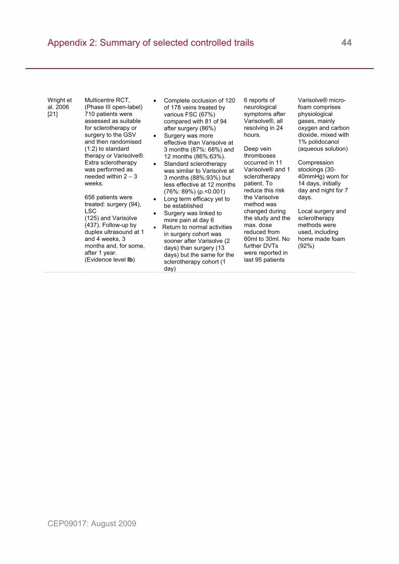
Appendix 2: Summary of selected controlled trails 44
CEP09017: August 2009
Wright et al. 2006 [21]
Multicentre RCT, (Phase III open-label) 710 patients were assessed as suitable for sclerotherapy or surgery to the GSV and then randomised (1:2) to standard therapy or Varisolve®. Extra sclerotherapy was performed as needed within 2 – 3 weeks. 656 patients were treated: surgery (94), LSC (125) and Varisolve (437). Follow-up by duplex ultrasound at 1 and 4 weeks, 3 months and, for some, after 1 year. (Evidence level Ib)
• Complete occlusion of 120 of 178 veins treated by various FSC (67%) compared with 81 of 94 after surgery (86%)
• Surgery was more effective than Varisolve at 3 months (87%: 68%) and 12 months (86%:63%).
• Standard sclerotherapy was similar to Varisolve at 3 months (88%:93%) but less effective at 12 months (76%: 89%) (ρ.<0.001)
• Long term efficacy yet to be established
• Surgery was linked to more pain at day 6
• Return to normal activities in surgery cohort was sooner after Varisolve (2 days) than surgery (13 days) but the same for the sclerotherapy cohort (1 day)
6 reports of neurological symptoms after Varisolve®, all resolving in 24 hours. Deep vein thromboses occurred in 11 Varisolve® and 1 sclerotherapy patient. To reduce this risk the Varisolve method was changed during the study and the max. dose reduced from 60ml to 30ml. No further DVTs were reported in last 95 patients
Varisolve® micro-foam comprises physiological gases, mainly oxygen and carbon dioxide, mixed with 1% polidocanol (aqueous solution) Compression stockings (30-40mmHg) worn for 14 days, initially day and night for 7 days. Local surgery and sclerotherapy methods were used, including home made foam (92%)
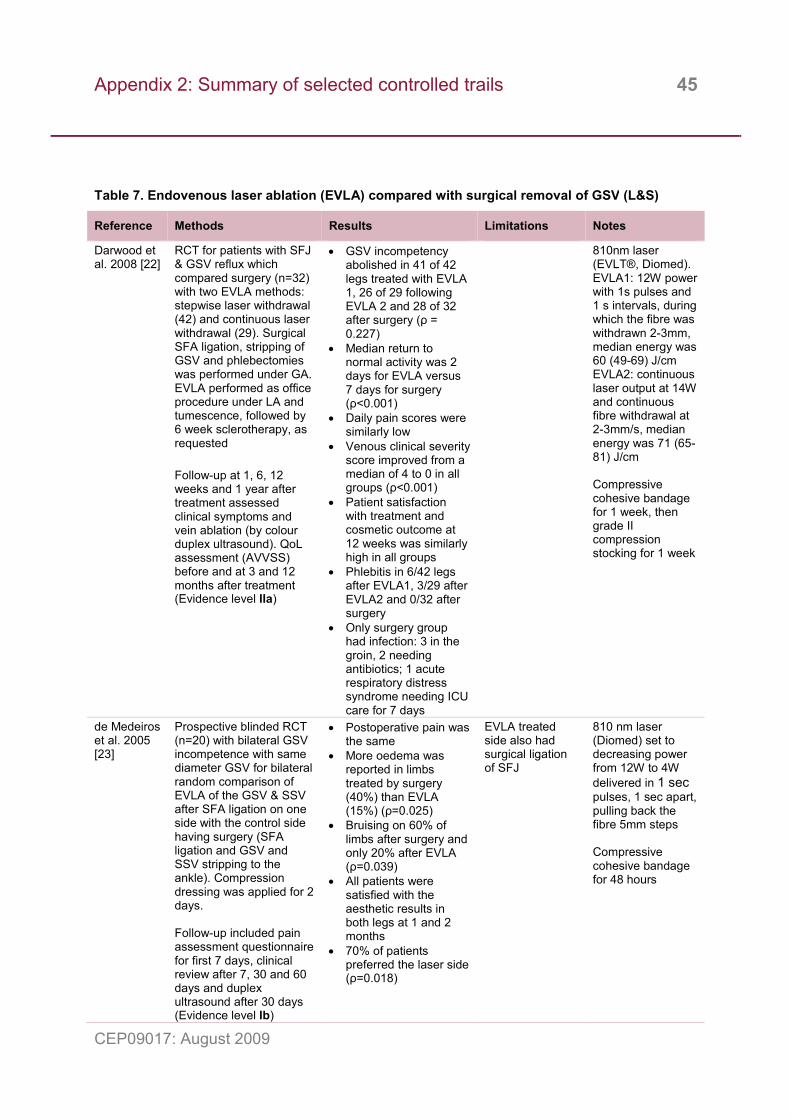
Appendix 2: Summary of selected controlled trails 45
CEP09017: August 2009
Table 7. Endovenous laser ablation (EVLA) compared with surgical removal of GSV (L&S)
Reference Methods Results Limitations Notes
Darwood et al. 2008 [22]
RCT for patients with SFJ & GSV reflux which compared surgery (n=32) with two EVLA methods: stepwise laser withdrawal (42) and continuous laser withdrawal (29). Surgical SFA ligation, stripping of GSV and phlebectomies was performed under GA. EVLA performed as office procedure under LA and tumescence, followed by 6 week sclerotherapy, as requested Follow-up at 1, 6, 12 weeks and 1 year after treatment assessed clinical symptoms and vein ablation (by colour duplex ultrasound). QoL assessment (AVVSS) before and at 3 and 12 months after treatment (Evidence level IIa)
• GSV incompetency abolished in 41 of 42 legs treated with EVLA 1, 26 of 29 following EVLA 2 and 28 of 32 after surgery (ρ = 0.227)
• Median return to normal activity was 2 days for EVLA versus 7 days for surgery (ρ<0.001)
• Daily pain scores were similarly low
• Venous clinical severity score improved from a median of 4 to 0 in all groups (ρ<0.001)
• Patient satisfaction with treatment and cosmetic outcome at 12 weeks was similarly high in all groups
• Phlebitis in 6/42 legs after EVLA1, 3/29 after EVLA2 and 0/32 after surgery
• Only surgery group had infection: 3 in the groin, 2 needing antibiotics; 1 acute respiratory distress syndrome needing ICU care for 7 days
810nm laser (EVLT®, Diomed). EVLA1: 12W power with 1s pulses and 1 s intervals, during which the fibre was withdrawn 2-3mm, median energy was 60 (49-69) J/cm EVLA2: continuous laser output at 14W and continuous fibre withdrawal at 2-3mm/s, median energy was 71 (65-81) J/cm Compressive cohesive bandage for 1 week, then grade II compression stocking for 1 week
de Medeiros et al. 2005 [23]
Prospective blinded RCT (n=20) with bilateral GSV incompetence with same diameter GSV for bilateral random comparison of EVLA of the GSV & SSV after SFA ligation on one side with the control side having surgery (SFA ligation and GSV and SSV stripping to the ankle). Compression dressing was applied for 2 days. Follow-up included pain assessment questionnaire for first 7 days, clinical review after 7, 30 and 60 days and duplex ultrasound after 30 days (Evidence level Ib)
• Postoperative pain was the same
• More oedema was reported in limbs treated by surgery (40%) than EVLA (15%) (ρ=0.025)
• Bruising on 60% of limbs after surgery and only 20% after EVLA (ρ=0.039)
• All patients were satisfied with the aesthetic results in both legs at 1 and 2 months
• 70% of patients preferred the laser side (ρ=0.018)
EVLA treated side also had surgical ligation of SFJ
810 nm laser (Diomed) set to decreasing power from 12W to 4W delivered in 1 sec pulses, 1 sec apart, pulling back the fibre 5mm steps Compressive cohesive bandage for 48 hours
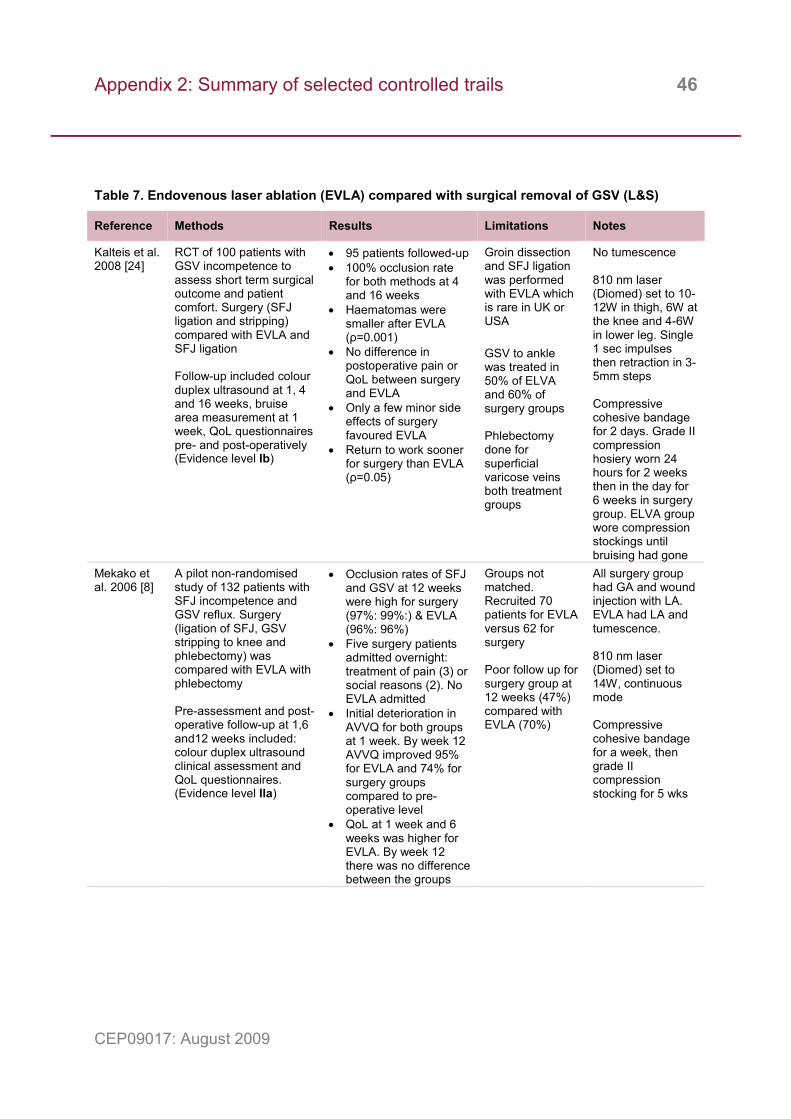
Appendix 2: Summary of selected controlled trails 46
CEP09017: August 2009
Table 7. Endovenous laser ablation (EVLA) compared with surgical removal of GSV (L&S)
Reference Methods Results Limitations Notes
Kalteis et al. 2008 [24]
RCT of 100 patients with GSV incompetence to assess short term surgical outcome and patient comfort. Surgery (SFJ ligation and stripping) compared with EVLA and SFJ ligation Follow-up included colour duplex ultrasound at 1, 4 and 16 weeks, bruise area measurement at 1 week, QoL questionnaires pre- and post-operatively (Evidence level Ib)
• 95 patients followed-up • 100% occlusion rate
for both methods at 4 and 16 weeks
• Haematomas were smaller after EVLA (ρ=0.001)
• No difference in postoperative pain or QoL between surgery and EVLA
• Only a few minor side effects of surgery favoured EVLA
• Return to work sooner for surgery than EVLA (ρ=0.05)
Groin dissection and SFJ ligation was performed with EVLA which is rare in UK or USA GSV to ankle was treated in 50% of ELVA and 60% of surgery groups Phlebectomy done for superficial varicose veins both treatment groups
No tumescence 810 nm laser (Diomed) set to 10-12W in thigh, 6W at the knee and 4-6W in lower leg. Single 1 sec impulses then retraction in 3-5mm steps Compressive cohesive bandage for 2 days. Grade II compression hosiery worn 24 hours for 2 weeks then in the day for 6 weeks in surgery group. ELVA group wore compression stockings until bruising had gone
Mekako et al. 2006 [8]
A pilot non-randomised study of 132 patients with SFJ incompetence and GSV reflux. Surgery (ligation of SFJ, GSV stripping to knee and phlebectomy) was compared with EVLA with phlebectomy Pre-assessment and post-operative follow-up at 1,6 and12 weeks included: colour duplex ultrasound clinical assessment and QoL questionnaires. (Evidence level IIa)
• Occlusion rates of SFJ and GSV at 12 weeks were high for surgery (97%: 99%:) & EVLA (96%: 96%)
• Five surgery patients admitted overnight: treatment of pain (3) or social reasons (2). No EVLA admitted
• Initial deterioration in AVVQ for both groups at 1 week. By week 12 AVVQ improved 95% for EVLA and 74% for surgery groups compared to pre-operative level
• QoL at 1 week and 6 weeks was higher for EVLA. By week 12 there was no difference between the groups
Groups not matched. Recruited 70 patients for EVLA versus 62 for surgery Poor follow up for surgery group at 12 weeks (47%) compared with EVLA (70%)
All surgery group had GA and wound injection with LA. EVLA had LA and tumescence. 810 nm laser (Diomed) set to 14W, continuous mode Compressive cohesive bandage for a week, then grade II compression stocking for 5 wks
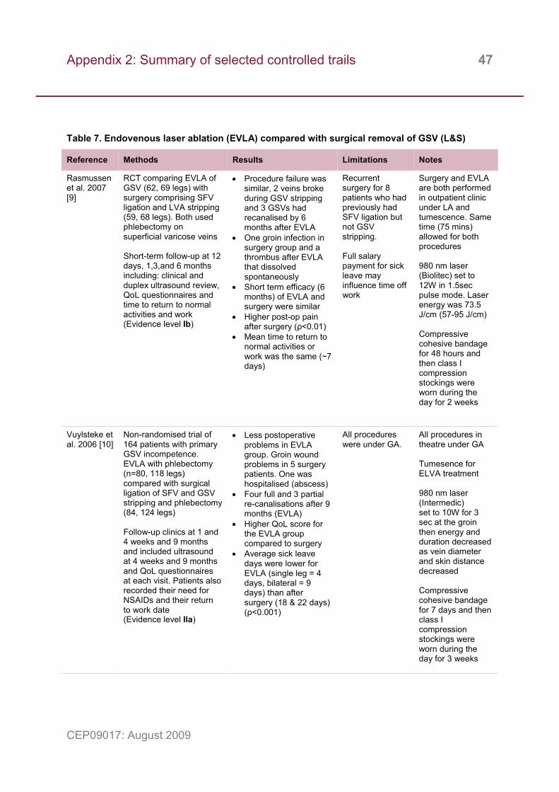
Appendix 2: Summary of selected controlled trails 47
CEP09017: August 2009
Table 7. Endovenous laser ablation (EVLA) compared with surgical removal of GSV (L&S)
Reference Methods Results Limitations Notes
Rasmussen et al. 2007 [9]
RCT comparing EVLA of GSV (62, 69 legs) with surgery comprising SFV ligation and LVA stripping (59, 68 legs). Both used phlebectomy on superficial varicose veins Short-term follow-up at 12 days, 1,3,and 6 months including: clinical and duplex ultrasound review, QoL questionnaires and time to return to normal activities and work (Evidence level Ib)
• Procedure failure was similar, 2 veins broke during GSV stripping and 3 GSVs had recanalised by 6 months after EVLA
• One groin infection in surgery group and a thrombus after EVLA that dissolved spontaneously
• Short term efficacy (6 months) of EVLA and surgery were similar
• Higher post-op pain after surgery (ρ<0.01)
• Mean time to return to normal activities or work was the same (~7 days)
Recurrent surgery for 8 patients who had previously had SFV ligation but not GSV stripping. Full salary payment for sick leave may influence time off work
Surgery and EVLA are both performed in outpatient clinic under LA and tumescence. Same time (75 mins) allowed for both procedures 980 nm laser (Biolitec) set to 12W in 1.5sec pulse mode. Laser energy was 73.5 J/cm (57-95 J/cm) Compressive cohesive bandage for 48 hours and then class I compression stockings were worn during the day for 2 weeks
Vuylsteke et al. 2006 [10]
Non-randomised trial of 164 patients with primary GSV incompetence. EVLA with phlebectomy (n=80, 118 legs) compared with surgical ligation of SFV and GSV stripping and phlebectomy (84, 124 legs) Follow-up clinics at 1 and 4 weeks and 9 months and included ultrasound at 4 weeks and 9 months and QoL questionnaires at each visit. Patients also recorded their need for NSAIDs and their return to work date (Evidence level IIa)
• Less postoperative problems in EVLA group. Groin wound problems in 5 surgery patients. One was hospitalised (abscess)
• Four full and 3 partial re-canalisations after 9 months (EVLA)
• Higher QoL score for the EVLA group compared to surgery
• Average sick leave days were lower for EVLA (single leg = 4 days, bilateral = 9 days) than after surgery (18 & 22 days) (ρ<0.001)
All procedures were under GA.
All procedures in theatre under GA Tumesence for ELVA treatment 980 nm laser (Intermedic) set to 10W for 3 sec at the groin then energy and duration decreased as vein diameter and skin distance decreased Compressive cohesive bandage for 7 days and then class I compression stockings were worn during the day for 3 weeks
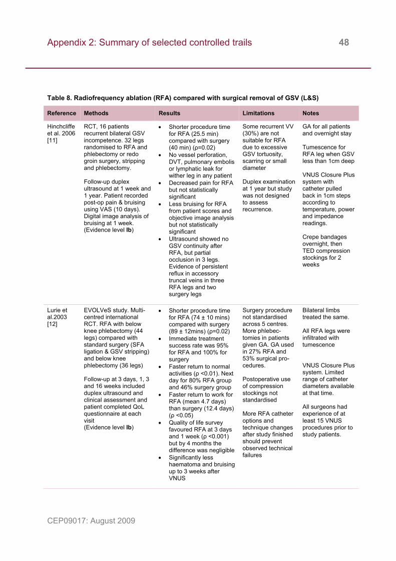
Appendix 2: Summary of selected controlled trails 48
CEP09017: August 2009
Table 8. Radiofrequency ablation (RFA) compared with surgical removal of GSV (L&S)
Reference Methods Results Limitations Notes
Hinchcliffe et al. 2006 [11]
RCT, 16 patients recurrent bilateral GSV incompetence. 32 legs randomised to RFA and phlebectomy or redo groin surgery, stripping and phlebectomy. Follow-up duplex ultrasound at 1 week and 1 year. Patient recorded post-op pain & bruising using VAS (10 days). Digital image analysis of bruising at 1 week. (Evidence level Ib)
• Shorter procedure time for RFA (25.5 min) compared with surgery (40 min) (ρ=0.02)
• No vessel perforation, DVT, pulmonary embolis or lymphatic leak for wither leg in any patient
• Decreased pain for RFA but not statistically significant
• Less bruising for RFA from patient scores and objective image analysis but not statistically significant
• Ultrasound showed no GSV continuity after RFA, but partial occlusion in 3 legs. Evidence of persistent reflux in accessory truncal veins in three RFA legs and two surgery legs
Some recurrent VV (30%) are not suitable for RFA due to excessive GSV tortuosity, scarring or small diameter Duplex examination at 1 year but study was not designed to assess recurrence.
GA for all patients and overnight stay Tumescence for RFA leg when GSV less than 1cm deep VNUS Closure Plus system with catheter pulled back in 1cm steps according to temperature, power and impedance readings. Crepe bandages overnight, then TED compression stockings for 2 weeks
Lurie et al.2003 [12]
EVOLVeS study. Multi-centred international RCT. RFA with below knee phlebectomy (44 legs) compared with standard surgery (SFA ligation & GSV stripping) and below knee phlebectomy (36 legs) Follow-up at 3 days, 1, 3 and 16 weeks included duplex ultrasound and clinical assessment and patient completed QoL questionnaire at each visit (Evidence level Ib)
• Shorter procedure time for RFA (74 ± 10 mins) compared with surgery (89 ± 12mins) (ρ=0.02)
• Immediate treatment success rate was 95% for RFA and 100% for surgery
• Faster return to normal activities (ρ <0.01). Next day for 80% RFA group and 46% surgery group
• Faster return to work for RFA (mean 4.7 days) than surgery (12.4 days) (ρ <0.05)
• Quality of life survey favoured RFA at 3 days and 1 week (ρ <0.001) but by 4 months the difference was negligible
• Significantly less haematoma and bruising up to 3 weeks after VNUS
Surgery procedure not standardised across 5 centres. More phlebec-tomies in patients given GA. GA used in 27% RFA and 53% surgical pro-cedures. Postoperative use of compression stockings not standardised More RFA catheter options and technique changes after study finished should prevent observed technical failures
Bilateral limbs treated the same. All RFA legs were infiltrated with tumescence VNUS Closure Plus system. Limited range of catheter diameters available at that time. All surgeons had experience of at least 15 VNUS procedures prior to study patients.
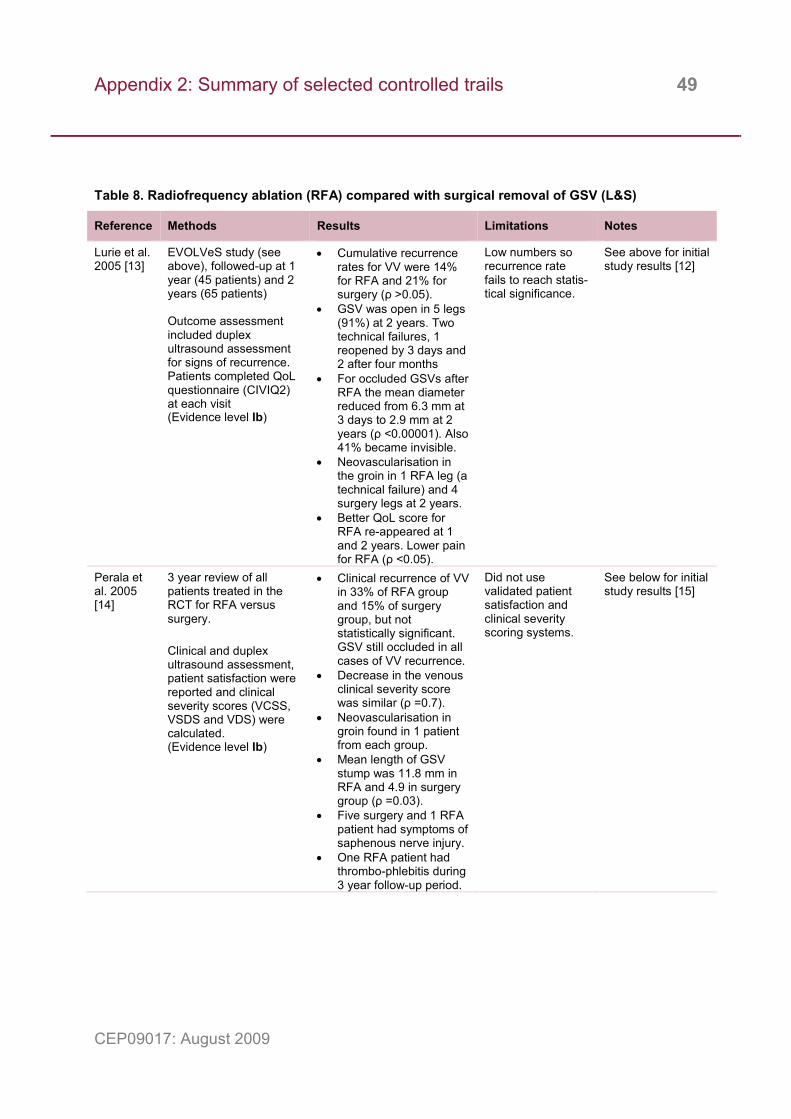
Appendix 2: Summary of selected controlled trails 49
CEP09017: August 2009
Table 8. Radiofrequency ablation (RFA) compared with surgical removal of GSV (L&S)
Reference Methods Results Limitations Notes
Lurie et al. 2005 [13]
EVOLVeS study (see above), followed-up at 1 year (45 patients) and 2 years (65 patients) Outcome assessment included duplex ultrasound assessment for signs of recurrence. Patients completed QoL questionnaire (CIVIQ2) at each visit (Evidence level Ib)
• Cumulative recurrence rates for VV were 14% for RFA and 21% for surgery (ρ >0.05).
• GSV was open in 5 legs (91%) at 2 years. Two technical failures, 1 reopened by 3 days and 2 after four months
• For occluded GSVs after RFA the mean diameter reduced from 6.3 mm at 3 days to 2.9 mm at 2 years (ρ <0.00001). Also 41% became invisible.
• Neovascularisation in the groin in 1 RFA leg (a technical failure) and 4 surgery legs at 2 years.
• Better QoL score for RFA re-appeared at 1 and 2 years. Lower pain for RFA (ρ <0.05).
Low numbers so recurrence rate fails to reach statis-tical significance.
See above for initial study results [12]
Perala et al. 2005 [14]
3 year review of all patients treated in the RCT for RFA versus surgery. Clinical and duplex ultrasound assessment, patient satisfaction were reported and clinical severity scores (VCSS, VSDS and VDS) were calculated. (Evidence level Ib)
• Clinical recurrence of VV in 33% of RFA group and 15% of surgery group, but not statistically significant. GSV still occluded in all cases of VV recurrence.
• Decrease in the venous clinical severity score was similar (ρ =0.7).
• Neovascularisation in groin found in 1 patient from each group.
• Mean length of GSV stump was 11.8 mm in RFA and 4.9 in surgery group (ρ =0.03).
• Five surgery and 1 RFA patient had symptoms of saphenous nerve injury.
• One RFA patient had thrombo-phlebitis during 3 year follow-up period.
Did not use validated patient satisfaction and clinical severity scoring systems.
See below for initial study results [15]
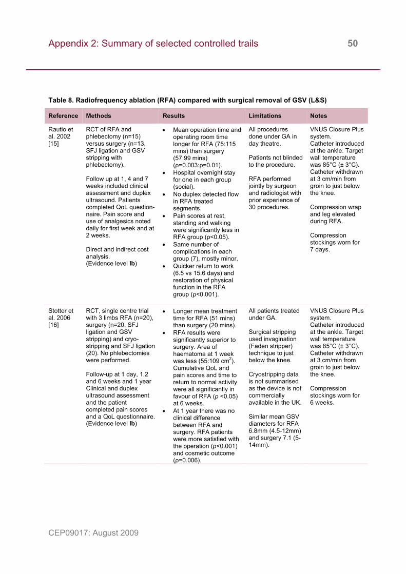
Appendix 2: Summary of selected controlled trails 50
CEP09017: August 2009
Table 8. Radiofrequency ablation (RFA) compared with surgical removal of GSV (L&S)
Reference Methods Results Limitations Notes
Rautio et al. 2002 [15]
RCT of RFA and phlebectomy (n=15) versus surgery (n=13, SFJ ligation and GSV stripping with phlebectomy). Follow up at 1, 4 and 7 weeks included clinical assessment and duplex ultrasound. Patients completed QoL question-naire. Pain score and use of analgesics noted daily for first week and at 2 weeks. Direct and indirect cost analysis. (Evidence level Ib)
• Mean operation time and operating room time longer for RFA (75:115 mins) than surgery (57:99 mins) (ρ=0.003:ρ=0.01).
• Hospital overnight stay for one in each group (social).
• No duplex detected flow in RFA treated segments.
• Pain scores at rest, standing and walking were significantly less in RFA group (ρ<0.05).
• Same number of complications in each group (7), mostly minor.
• Quicker return to work (6.5 vs 15.6 days) and restoration of physical function in the RFA group (ρ<0.001).
All procedures done under GA in day theatre. Patients not blinded to the procedure. RFA performed jointly by surgeon and radiologist with prior experience of 30 procedures.
VNUS Closure Plus system. Catheter introduced at the ankle. Target wall temperature was 85°C (± 3°C). Catheter withdrawn at 3 cm/min from groin to just below the knee. Compression wrap and leg elevated during RFA. Compression stockings worn for 7 days.
Stotter et al. 2006 [16]
RCT, single centre trial with 3 limbs RFA (n=20), surgery (n=20, SFJ ligation and GSV stripping) and cryo-stripping and SFJ ligation (20). No phlebectomies were performed. Follow-up at 1 day, 1,2 and 6 weeks and 1 year Clinical and duplex ultrasound assessment and the patient completed pain scores and a QoL questionnaire. (Evidence level Ib)
• Longer mean treatment time for RFA (51 mins) than surgery (20 mins).
• RFA results were significantly superior to surgery. Area of haematoma at 1 week was less (55:109 cm2). Cumulative QoL and pain scores and time to return to normal activity were all significantly in favour of RFA (ρ <0.05) at 6 weeks.
• At 1 year there was no clinical difference between RFA and surgery. RFA patients were more satisfied with the operation (ρ<0.001) and cosmetic outcome (ρ=0.006).
All patients treated under GA. Surgical stripping used invagination (Faden stripper) technique to just below the knee. Cryostripping data is not summarised as the device is not commercially available in the UK. Similar mean GSV diameters for RFA 6.8mm (4.5-12mm) and surgery 7.1 (5-14mm).
VNUS Closure Plus system. Catheter introduced at the ankle. Target wall temperature was 85°C (± 3°C). Catheter withdrawn at 3 cm/min from groin to just below the knee. Compression stockings worn for 6 weeks.
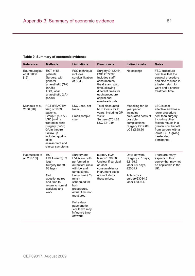
Appendix 3: Summary of economic evidence 51
CEP09017: August 2009
Table 9. Summary of economic evidence
Reference Methods Limitations Direct costs Indirect costs Notes
Bountouroglou et al. 2006 [19]
RCT of 58 patients. Surgery, with general anaesthetic (GA) (n=28) FSC, local anaesthetic (LA) (n=30).
FSC technique includes surgical ligation of SFJ.
Surgery £1120.64 FSC £672.97 Includes staff, consumables, theatre and ward time, allowing different times for each procedure, capital and overhead costs.
No costings FSC procedure cost less that the surgical procedure and also resulted in a faster return to work and a shorter treatment time.
Michaels et al. 2006 [20]
RCT (REACTIV trial) of 1009 patients. Group 2 (n-=77) LSC (n=41), treated in clinic Surgery (n=36) GA in theatre Follow up included quality of life assessment and clinical symptoms
LSC used, not foam. Small sample size.
Total discounted NHS Costs for 2 years, including GP visits: Surgery £701.28 LSC £210.94
Modelling for 10 year period including calculated costs of possible complications: Surgery £919.80 LCS £628.60
LSC is cost effective and has a lower procedure cost than surgery. Including other factors results in a greater cost benefit from surgery with a lower ICER, giving it extended dominance.
Rasmussen et al. 2007 [9]
RCT EVLA (n=62, 69 legs) Surgery (n=59, 68 legs). QoL questionnaires and time to return to normal activities and work.
Surgery and EVLA are both performed in outpatient clinic with LA and tumescence. Same time (75 mins) scheduled for both procedures, actual time not measured. Full salary payment for sick leave may influence time off work.
surgery €924 laser €1390.66 Unclear if surgical or laser consumables or instrument costs are included in these prices.
Days off work: Surgery 7.7 days, €2159.5 laser 6.9 days, €2005.7 Total costs: surgery€3084.5 laser €3396.4
There are many aspects of this survey that may not be applicable in the UK.
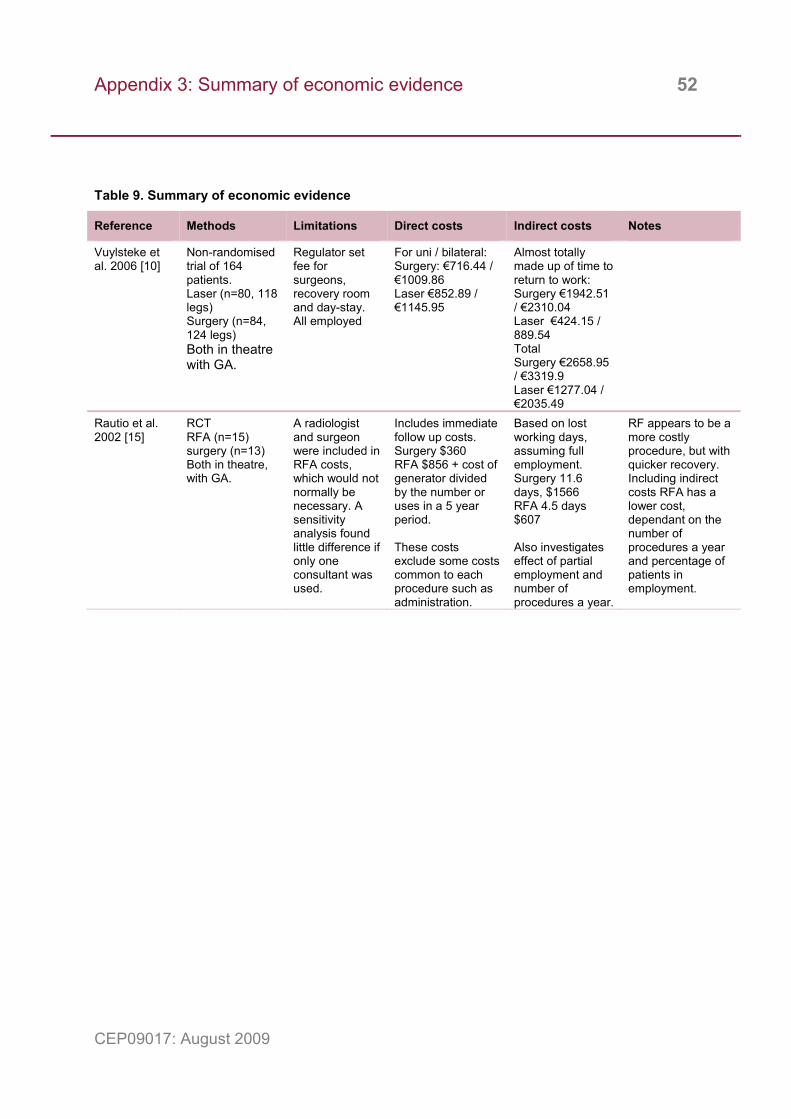
Appendix 3: Summary of economic evidence 52
CEP09017: August 2009
Table 9. Summary of economic evidence
Reference Methods Limitations Direct costs Indirect costs Notes
Vuylsteke et al. 2006 [10]
Non-randomised trial of 164 patients. Laser (n=80, 118 legs) Surgery (n=84, 124 legs) Both in theatre with GA.
Regulator set fee for surgeons, recovery room and day-stay. All employed
For uni / bilateral: Surgery: €716.44 / €1009.86 Laser €852.89 / €1145.95
Almost totally made up of time to return to work: Surgery €1942.51 / €2310.04 Laser €424.15 / 889.54 Total Surgery €2658.95 / €3319.9 Laser €1277.04 / €2035.49
Rautio et al. 2002 [15]
RCT RFA (n=15) surgery (n=13) Both in theatre, with GA.
A radiologist and surgeon were included in RFA costs, which would not normally be necessary. A sensitivity analysis found little difference if only one consultant was used.
Includes immediate follow up costs. Surgery $360 RFA $856 + cost of generator divided by the number or uses in a 5 year period. These costs exclude some costs common to each procedure such as administration.
Based on lost working days, assuming full employment. Surgery 11.6 days, $1566 RFA 4.5 days $607 Also investigates effect of partial employment and number of procedures a year.
RF appears to be a more costly procedure, but with quicker recovery. Including indirect costs RFA has a lower cost, dependant on the number of procedures a year and percentage of patients in employment.
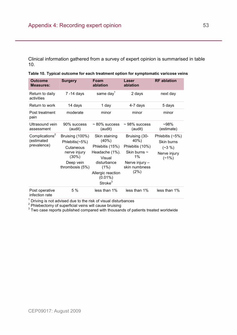
Appendix 4: Recording expert opinion 53
CEP09017: August 2009
Clinical information gathered from a survey of expert opinion is summarised in table 10.
Table 10. Typical outcome for each treatment option for symptomatic varicose veins
Outcome Measures:
Surgery Foam ablation
Laser ablation
RF ablation
Return to daily activities
7 -14 days same day1 2 days next day
Return to work 14 days 1 day 4-7 days 5 days
Post treatment pain
moderate minor minor minor
Ultrasound vein assessment
90% success (audit)
~ 80% success (audit)
~ 98% success (audit)
~98% (estimate)
Complications2 (estimated prevalence)
Bruising (100%) Phlebitis(~5%)
Cutaneous nerve injury
(30%) Deep vein
thrombosis (5%)
Skin staining (40%)
Phlebitis (15%) Headache (1%).
Visual disturbance
(1%) Allergic reaction
(0.01%) Stroke3
Bruising (30-40%)
Phlebitis (10%) Skin burns ~
1% Nerve injury – skin numbness
(2%)
Phlebitis (~5%) Skin burns
(~3 %) Nerve injury
(~1%)
Post operative infection rate
5 % less than 1% less than 1% less than 1%
1 Driving is not advised due to the risk of visual disturbances 2 Phlebectomy of superficial veins will cause bruising 3 Two case reports published compared with thousands of patients treated worldwide
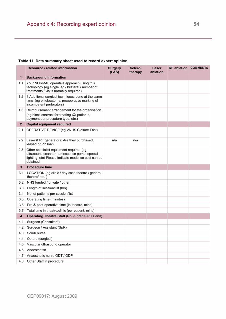
Appendix 4: Recording expert opinion 54
CEP09017: August 2009
Table 11. Data summary sheet used to record expert opinion
Resource / related information Surgery (L&S)
Sclero-therapy
Laser ablation
RF ablation COMMENTS
1 Background information 1.1 Your NORMAL operative approach using this
technology (eg single leg / bilateral / number of treatments / visits normally required)
1.2 ? Additional surgical techniques done at the same time (eg phlebectomy, preoperative marking of incompetent perforators)
1.3 Reimbursement arrangement for the organisation (eg block contract for treating XX patients, payment per procedure type, etc.)
2 Capital equipment required 2.1 OPERATIVE DEVICE (eg VNUS Closure Fast)
2.2 Laser & RF generators: Are they purchased, leased or on loan
n/a n/a
2.3 Other specialist equipment required (eg ultrasound scanner, tumescence pump, special lighting, etc) Please indicate model so cost can be obtained
3 Procedure time 3.1 LOCATION (eg clinic / day case theatre / general
theatre/ etc. )
3.2 NHS funded / private / other 3.3 Length of session/list (hrs) 3.4 No. of patients per session/list 3.5 Operating time (minutes) 3.6 Pre & post-operative time (in theatre, mins) 3.7 Total time in theatre/clinic (per patient, mins) 4 Operating Theatre Staff (No. & grade/AfC Band)
4.1 Surgeon (Consultant) 4.2 Surgeon / Assistant (SpR) 4.3 Scrub nurse 4.4 Others (surgical) 4.5 Vascular ultrasound operator 4.6 Anaesthetist 4.7 Anaesthetic nurse ODT / ODP 4.8 Other Staff in procedure
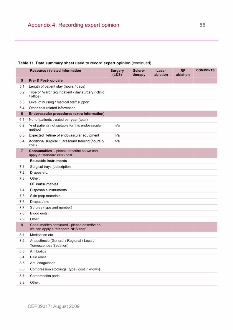
Appendix 4: Recording expert opinion 55
CEP09017: August 2009
Table 11. Data summary sheet used to record expert opinion (continued)
Resource / related information Surgery (L&S)
Sclero-therapy
Laser ablation
RF ablation
COMMENTS
5 Pre- & Post- op care 5.1 Length of patient stay (hours / days) 5.2 Type of “ward” (eg inpatient / day surgery / clinic
/ office)
5.3 Level of nursing / medical staff support 5.4 Other cost related information 6 Endovascular procedures (extra information)
6.1 No. of patients treated per year (total) 6.2 % of patients not suitable for this endovascular
method n/a
6.3 Expected lifetime of endovascular equipment n/a 6.4 Additional surgical / ultrasound training (hours &
cost) n/a
7 Consumables - please describe so we can apply a “standard NHS cost”
Reusable instruments 7.1 Surgical trays (description 7.2 Drapes etc. 7.3 Other:
OT consumables 7.4 Disposable instruments 7.5 Skin prep materials 7.6 Drapes / etc 7.7 Sutures (type and number) 7.8 Blood units 7.9 Other 8 Consumables continued - please describe so
we can apply a “standard NHS cost”
8.1 Medication etc. 8.2 Anaesthesia (General / Regional / Local /
Tumescence / Sedation)
8.3 Antibiotics 8.4 Pain relief 8.5 Anti-coagulation
8.6 Compression stockings (type / cost if known)
8.7 Compression pads
8.8 Other:
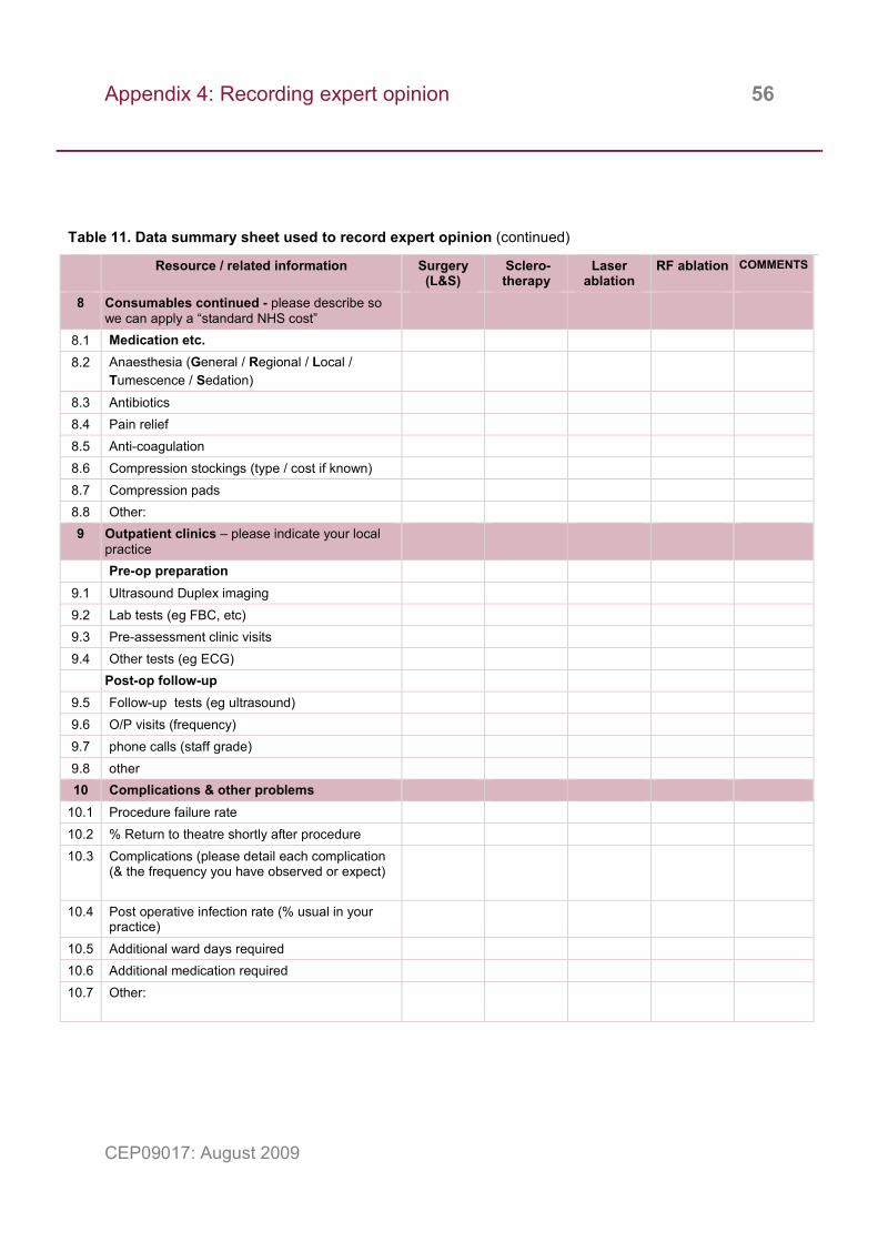
Appendix 4: Recording expert opinion 56
CEP09017: August 2009
Table 11. Data summary sheet used to record expert opinion (continued)
Resource / related information Surgery (L&S)
Sclero-therapy
Laser ablation
RF ablation COMMENTS
8 Consumables continued - please describe so we can apply a “standard NHS cost”
8.1 Medication etc. 8.2 Anaesthesia (General / Regional / Local /
Tumescence / Sedation)
8.3 Antibiotics 8.4 Pain relief 8.5 Anti-coagulation 8.6 Compression stockings (type / cost if known) 8.7 Compression pads 8.8 Other: 9 Outpatient clinics – please indicate your local
practice
Pre-op preparation 9.1 Ultrasound Duplex imaging 9.2 Lab tests (eg FBC, etc) 9.3 Pre-assessment clinic visits 9.4 Other tests (eg ECG)
Post-op follow-up 9.5 Follow-up tests (eg ultrasound) 9.6 O/P visits (frequency) 9.7 phone calls (staff grade) 9.8 other 10 Complications & other problems
10.1 Procedure failure rate 10.2 % Return to theatre shortly after procedure 10.3 Complications (please detail each complication
(& the frequency you have observed or expect)
10.4 Post operative infection rate (% usual in your practice)
10.5 Additional ward days required 10.6 Additional medication required 10.7 Other:
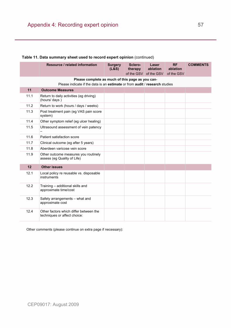
Appendix 4: Recording expert opinion 57
CEP09017: August 2009
Table 11. Data summary sheet used to record expert opinion (continued)
Resource / related information Surgery (L&S)
Sclero-therapy
of the GSV
Laser ablation
of the GSV
RF ablation
of the GSV
COMMENTS
Please complete as much of this page as you can- Please indicate if the data is an estimate or from audit / research studies
11 Outcome Measures 11.1 Return to daily activities (eg driving)
(hours/ days )
11.2 Return to work (hours / days / weeks) 11.3 Post treatment pain (eg VAS pain score
system)
11.4 Other symptom relief (eg ulcer healing) 11.5 Ultrasound assessment of vein patency
–
11.6 Patient satisfaction score 11.7 Clinical outcome (eg after 5 years) 11.8 Aberdeen varicose vein score 11.9 Other outcome measures you routinely
assess (eg Quality of Life)
12 Other issues
12.1 Local policy re reusable vs. disposable instruments
12.2 Training – additional skills and approximate time/cost
12.3 Safety arrangements – what and approximate cost
12.4 Other factors which differ between the techniques or affect choice:
Other comments (please continue on extra page if necessary):
Author and report information 58
CEP09017: August 2009
Evidence review: Endovascular treatment for varicose veins Grace Carolan Rees, Diane C Crawford, Megan Dale, Joelle Williams, Kevin Conway, Harsheet Sethi, David McLain, Ruth Turley, Rani Bhogal Clinical Engineering Device Assessment and Reporting (CEDAR) Cardiff Medicentre Cardiff CF14 4UJ Tel: 029 2068 2120 Fax: 029 2075 0239 mark FAO CEDAR Email: [email protected] For more information on CEDAR and our earlier reports visit www.cedar.wales.nhs.uk About CEP The Centre for Evidence-based Purchasing (CEP) is part of the Policy and Innovation Directorate of the NHS Purchasing and Supply Agency. We underpin purchasing decisions by providing objective evidence to support the uptake of useful, safe and innovative products and related procedures in health and social care.
We are here to help you make informed purchasing decisions by gathering evidence globally to support the use of innovative technologies, assess value and cost effectiveness of products, and develop nationally agreed protocols.
Sign up to our email alert service All our publications since 2002 are available in full colour to download from our website. To sign up to our email alert service and receive new publications straight to your mailbox contact:
Centre for Evidence-based Purchasing Room 152C Skipton House 80 London Road SE1 6HL Tel: 020 7972 6080 Fax: 020 7975 5795 Email: [email protected] www.pasa.nhs.uk/cep © Crown Copyright 2009