Embed Size (px)
Citation preview
7/29/2019 Cep Halos Prins
http://slidepdf.com/reader/full/cep-halos-prins 1/6
Sensitive Chemiluminescence Determination of ThirteenCephalosporin Antibiotics with Luminol–Copper(II) Reaction
JIANXIU DU* and HONG LI Key Laboratory of Analytical Chemistry for Life Science of Shaanxi Province, School of Chemistry and Materials Science, Shaanxi Normal
University, Xi’an 710062, China (J.D.); and Weinan Vocational and Technical College, Weinan 714000, China (H.L.)
A new chemiluminescence reaction, the luminol–Cu2þ reaction, was
investigated for the determination of thirteen (13) cephalosporin
antibiotics, namely cefalexin, cefadroxil, cefradine, cefazolin sodium,
cefaclor, cefuroxime axetil, cefotaxime sodium, cefoperazone sodium,
ceftriaxone sodium, ceftazidime, cefetamet pivoxil hydrochloride, cefix-
ime, and cefpodoxime. It was found that, without adding any special
oxidant, strong chemiluminescent (CL) signal could be produced from the
reaction of the alkaline luminol with the above-mentioned antibiotics in
the presence of Cu2þ. The experimental conditions for the reaction were
carefully optimized with flow-injection mode. The detection limits are 0.3
ng/mL cefalexin, 3 ng/mL cefadroxil, 0.3 ng/mL cefradine, 0.02 lg/mL
cefazolin sodium, 0.8 ng/mL cefaclor, 0.02 lg/mL cefuroxime axetil, 5 ng/
mL cefotaxime sodium, 0.02 lg/mL cefoperazone sodium, 0.8 ng/mL
ceftriaxone sodium, 1 ng/mL ceftazidime, 0.08 ng/mL cefetamet pivoxil
hydrochloride, 0.8 ng/mL cefixime, and 2 ng/mL cefpodoxime. The
proposed method was validated by direct application to commercial
formulations and spiked milk samples containing cefradine. A possible
reaction mechanism is also discussed.
Index Headings: Cephalosporin antibiotics; Chemiluminescence; Flow
injection.
INTRODUCTION
Cephalosporin antibiotics, referred to as the b-lactamantibiotics, are the most frequently prescribed class of antibiotics against infections. They have significant activityagainst both gram-positive and gram-negative bacteria.1
Cephalosporin antibiotics are grouped into ‘‘generations’’ basedon their spectrum of antimicrobial activity. The therapeuticimportance of these antibiotics requires the development of sensitive, rapid analytical methods for industrial quality controland clinical monitoring.
A wide variety of analytical methods have been reported for the determination of cephalosporin antibiotics in pharmaceu-tical preparations and in biological fluids, including spectro-photometry,2–4 fluorimetry,5,6 electrochemical methods,7,8
liquid chromatography,9–11 and capillary electrophoresis.12,13
Recently, a comprehensive review of the analysis of cephalo-sporin antibiotics was presented by El-Shaboury.14 Thechemiluminescence (CL) method with the advantages of simplicity, rapidity, and sensitivity has also been explored for
the analysis of cephalosporin antibiotics.15–28 The involved CLsystems and the analytical merits of the reported CL methodsfor the determination of cephalosporin antibiotics are summa-rized in Table I.
Luminol (5-amino-2,3-dihydrophalazine-1,4-dione), one of the best-known and widely used liquid-phase CL agents, isoxidized to produce CL in the presence of an oxidant in analkaline solution. Many familiar oxidants, such as H2O2,
K3Fe(CN)6, KIO4, and KMnO4, have been employed for thispurpose. These CL reactions are the basis for the determinationof a variety of both inorganic and organic species, which act asa catalyst, an enhancer, or an inhibitor during the reaction.Unfortunately, these determinations suffer from high back-ground signal because strong CL emission usually accompa-nies the reaction between luminol and the oxidant. This, tosome extent, became a hindrance in improving the sensitivityof detection.
We here found that CL was directly produced by the reactionof cephalosporin antibiotics, including cefalexin, cefadroxil,cefradine, cefazolin sodium, cefaclor, cefuroxime axetil,
cefotaxime sodium, cefoperazone sodium, ceftriaxone sodium,ceftazidime, cefetamet pivoxil hydrochloride, cefixime, andcefpodoxime, with luminol in an alkaline condition without adding any special oxidants. In the presence of Cu2þ the CLsignal was significantly enhanced. This reaction system hasbeen established as a flow-injection analysis for the determi-nation of these antibiotics. The experimental conditions for thereaction are optimized, and the possible reaction mechanism isdiscussed. A direct application to commercial drug formula-tions and spiked milk samples is presented.
EXPERIMENTAL
Apparatus. Chemiluminescent measurements were carried
out on an IFFM-D type flow-injection CL analyzer (Xi’anRemex Analysis Instrument Co., Ltd, China). PTFE tubing(0.8-mm inner diameter) was used as the connection material inthe flow system. CL spectra were scanned on a 970CRTfluorescence spectrophotometer (Shanghai Analysis Instrument Plant) with the excitation source shutter closed. Absorptionspectra were taken on a TU-1901 ultraviolet–visible (UV-Vis)spectrophotometer (Beijing Currency Instrumental Ltd, China).pH measurements were made with a pHS-3C(A) pH meter (Shanghai Dapu Instruments Co., Ltd, China).
Chemicals. All chemicals were of analytical grade; doublydistilled deionized water was used to prepare the solutions. Thestandards of cephalosporin antibiotics were obtained from theNational Institute for the Control of Pharmaceutical and
Biological Products (Beijing, China). Luminol was synthesizedby the Institute of Analytical Science of Shaanxi NormalUniversity (Xi’an, China). CuSO4Á5H2O and other reagentswere purchased from Xi’an Chemical Reagent Factory (Xi’an,China).
Stock solutions of cephalosporin antibiotics (50.0 lg/mL)were prepared by dissolving 0.0500 g of each standard inwater. These stock solutions were stored at 4 8C i n a refrigerator and protected from light. Working solutions of cephalosporin antibiotics were freshly prepared by diluting thecorresponding stock solution with water. Luminol solution (5.0310À4 mol/L) was prepared by diluting the appropriate amount
Received 2 March 2010; accepted 2 July 2010.* Author to whom correspondence should be sent. E-mail: jxdu@snnu.
edu.cn.
1154 Volume 64, Number 10, 2010 APPLIED SPECTROSCOPY0003-7028/10/6410-1154$2.00/0
Ó 2010 Society for Applied Spectroscopy
7/29/2019 Cep Halos Prins
http://slidepdf.com/reader/full/cep-halos-prins 2/6
of 1.03 10À2 mol/L luminol stock solution (prepared in 1.03
10À2 mol/L NaOH solution) with 0.1 mol/L carbonate buffer
solution to provide a final pH of 12.5. A 5.0 310À5 mol/L Cu2þ
solution was prepared by dissolving the appropriate amount of
CuSO4Á5H2O in water.
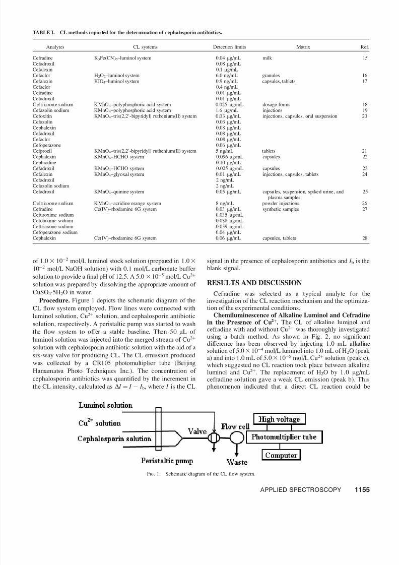
Procedure. Figure 1 depicts the schematic diagram of the
CL flow system employed. Flow lines were connected with
luminol solution, Cu2þ solution, and cephalosporin antibiotic
solution, respectively. A peristaltic pump was started to wash
the flow system to offer a stable baseline. Then 50 lL of
luminol solution was injected into the merged stream of Cu2þ
solution with cephalosporin antibiotic solution with the aid of a
six-way valve for producing CL. The CL emission produced
was collected by a CR105 photomultiplier tube (Beijing
Hamamatsu Photo Techniques Inc.). The concentration of
cephalosporin antibiotics was quantified by the increment in
the CL intensity, calculated as D I ¼ I À I b, where I is the CL
signal in the presence of cephalosporin antibiotics and I b is theblank signal.
RESULTS AND DISCUSSION
Cefradine was selected as a typical analyte for theinvestigation of the CL reaction mechanism and the optimiza-tion of the experimental conditions.
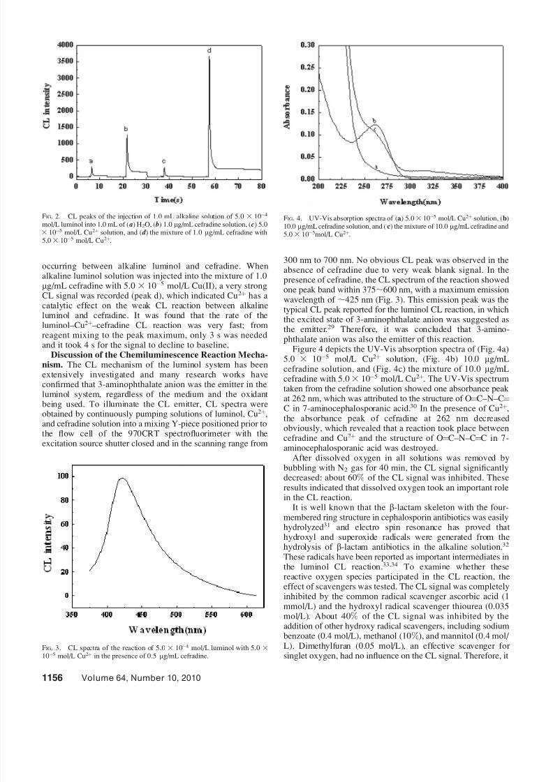
Chemiluminescence of Alkaline Luminol and Cefradinein the Presence of Cu2þ. The CL of alkaline luminol andcefradine with and without Cu2þ was thoroughly investigatedusing a batch method. As shown in Fig. 2, no significant difference has been observed by injecting 1.0 mL alkalinesolution of 5.0310À4 mol/L luminol into 1.0 mL of H2O (peaka) and into 1.0 mL of 5.03 10À5 mol/L Cu2þ solution (peak c),which suggested no CL reaction took place between alkalineluminol and Cu2þ. The replacement of H2O by 1.0 lg/mLcefradine solution gave a weak CL emission (peak b). Thisphenomenon indicated that a direct CL reaction could be
TABLE I. CL methods reported for the determination of cephalosporin antibiotics.
Analytes CL systems Detection limits Matrix Ref.
Cefradine K3Fe(CN)6 –luminol system 0.04 lg/mL milk 15Cefadroxil 0.08 lg/mLCefalexin 0.1 lg/mLCefaclor H2O2 –luminol system 6.0 ng/mL granules 16Cefalexin KIO4 –luminol system 0.9 ng/mL capsules, tablets 17Cefaclor 0.4 ng/mLCefradine 0.01 lg/mL
Cefadroxil 0.01 lg/mLCeftriaxone sodium KMnO4 –polyphosphoric acid system 0.025 lg/mL dosage forms 18Cefazolin sodium KMnO4 –polyphosphoric acid system 1.6 lg/mL injections 19Cefoxitin KMnO4 –tris(2,20-bipyridyl) ruthenium(II) system 0.03 lg/mL injections, capsules, oral suspension 20Cefazolin 0.03 lg/mLCephalexin 0.08 lg/mLCefadroxil 0.08 lg/mLCefaclor 0.08 lg/mLCefoperazone 0.06 lg/mLCefprozil KMnO4 –tris(2,20-bipyridyl) ruthenium(II) system 5 ng/mL tablets 21Cephalexin KMnO4 –HCHO system 0.096 lg/mL capsules 22Cephradine 0.10 lg/mLCefadroxil KMnO4 –HCHO system 0.025 lg/mL capsules 23Cefalexin KMnO4 –glyoxal system 0.01 lg/mL injections, capsules, tablets 24Cefadroxil 2 ng/mLCefazolin sodium 2 ng/mLCefadroxil KMnO4 –quinine system 0.05 lg/mL capsules, suspension, spiked urine, and
plasma samples
25
Ceftriaxone sodium KMnO4 –acridine orange system 8 ng/mL powder injections 26Cefradine Ce(IV)–rhodamine 6G system 0.03 lg/mL synthetic samples 27Cefuroxime sodium 0.035 lg/mLCefotaxime sodium 0.038 lg/mLCeftriaxone sodium 0.039 lg/mLCefoperazone sodium 0.04 lg/mLCephalexin Ce(IV)–rhodamine 6G system 0.06 lg/mL capsules, tablets 28
FIG. 1. Schematic diagram of the CL flow system.
APPLIED SPECTROSCOPY 1155
7/29/2019 Cep Halos Prins
http://slidepdf.com/reader/full/cep-halos-prins 3/6
occurring between alkaline luminol and cefradine. When
alkaline luminol solution was injected into the mixture of 1.0lg/mL cefradine with 5.0 3 10À5 mol/L Cu(II), a very strongCL signal was recorded (peak d), which indicated Cu2þ has a catalytic effect on the weak CL reaction between alkalineluminol and cefradine. It was found that the rate of theluminol–Cu2þ –cefradine CL reaction was very fast; fromreagent mixing to the peak maximum, only 3 s was neededand it took 4 s for the signal to decline to baseline.
Discussion of the Chemiluminescence Reaction Mecha-nism. The CL mechanism of the luminol system has beenextensively investigated and many research works haveconfirmed that 3-aminophthalate anion was the emitter in theluminol system, regardless of the medium and the oxidant being used. To illuminate the CL emitter, CL spectra were
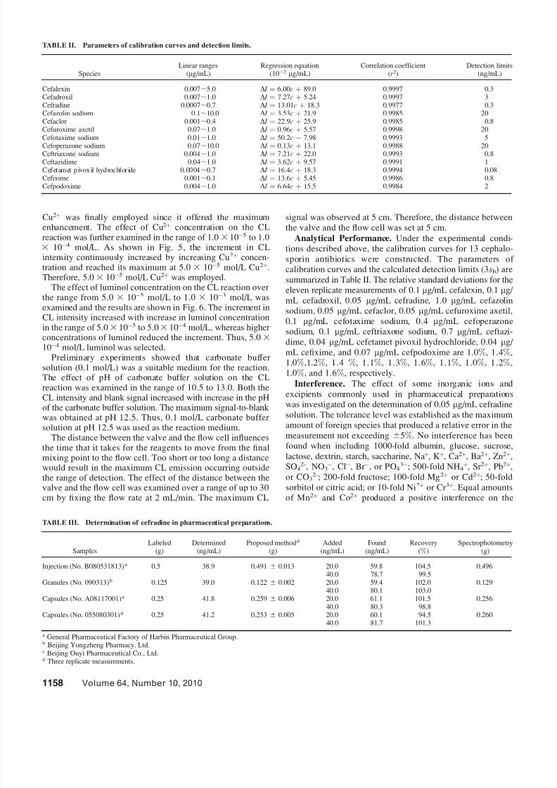
obtained by continuously pumping solutions of luminol, Cu2þ,and cefradine solution into a mixing Y-piece positioned prior tothe flow cell of the 970CRT spectrofluorimeter with theexcitation source shutter closed and in the scanning range from
300 nm to 700 nm. No obvious CL peak was observed in the
absence of cefradine due to very weak blank signal. In thepresence of cefradine, the CL spectrum of the reaction showedone peak band within 375;600 nm, with a maximum emissionwavelength of ;425 nm (Fig. 3). This emission peak was thetypical CL peak reported for the luminol CL reaction, in whichthe excited state of 3-aminophthalate anion was suggested asthe emitter.29 Therefore, it was concluded that 3-amino-phthalate anion was also the emitter of this reaction.
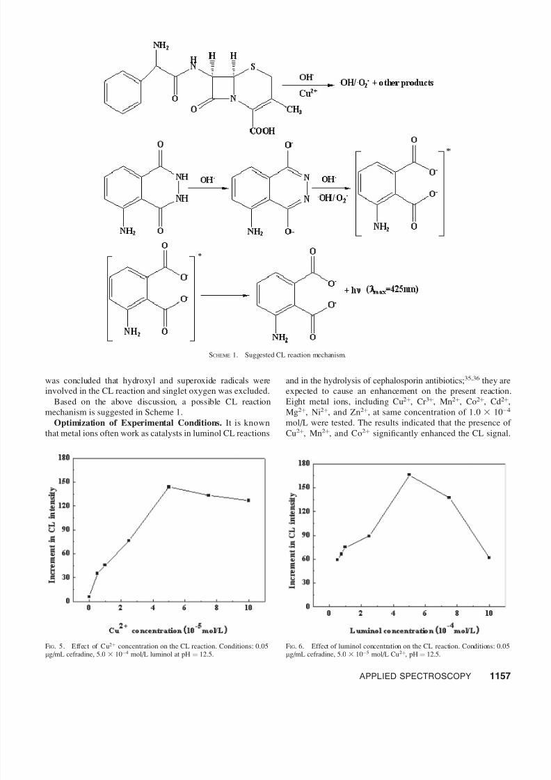
Figure 4 depicts the UV-Vis absorption spectra of (Fig. 4a)5.0 3 10À5 mol/L Cu2þ solution, (Fig. 4b) 10.0 lg/mLcefradine solution, and (Fig. 4c) the mixture of 10.0 lg/mLcefradine with 5.0 3 10À5 mol/L Cu2þ. The UV-Vis spectrumtaken from the cefradine solution showed one absorbance peakat 262 nm, which was attributed to the structure of O¼C–N–C¼
C in 7-aminocephalosporanic acid.30
In the presence of Cu2þ
,the absorbance peak of cefradine at 262 nm decreasedobviously, which revealed that a reaction took place betweencefradine and Cu2þ and the structure of O¼C–N–C¼C in 7-aminocephalosporanic acid was destroyed.
After dissolved oxygen in all solutions was removed bybubbling with N2 gas for 40 min, the CL signal significantlydecreased: about 60% of the CL signal was inhibited. Theseresults indicated that dissolved oxygen took an important rolein the CL reaction.
It is well known that the b-lactam skeleton with the four-membered ring structure in cephalosporin antibiotics was easilyhydrolyzed31 and electro spin resonance has proved that hydroxyl and superoxide radicals were generated from the
hydrolysis of b-lactam antibiotics in the alkaline solution.32
These radicals have been reported as important intermediates inthe luminol CL reaction.33,34 To examine whether thesereactive oxygen species participated in the CL reaction, theeffect of scavengers was tested. The CL signal was completelyinhibited by the common radical scavenger ascorbic acid (1mmol/L) and the hydroxyl radical scavenger thiourea (0.035mol/L). About 40% of the CL signal was inhibited by theaddition of other hydroxy radical scavengers, including sodiumbenzoate (0.4 mol/L), methanol (10%), and mannitol (0.4 mol/ L). Dimethylfuran (0.05 mol/L), an effective scavenger for singlet oxygen, had no influence on the CL signal. Therefore, it
FIG. 2. CL peaks of the injection of 1.0 mL alkaline solution of 5.03 10À4
mol/L luminol into 1.0 mL of ( a) H2O, ( b) 1.0 lg/mL cefradine solution, ( c) 5.03 10À5 mol/L Cu2þ solution, and ( d ) the mixture of 1.0 lg/mL cefradine with5.03 10À5 mol/L Cu2þ.
FIG. 3. CL spectra of the reaction of 5.0 3 10À4 mol/L luminol with 5.0 310À5 mol/L Cu2þ in the presence of 0.5 lg/mL cefradine.
FIG. 4. UV-Vis absorption spectra of (a) 5.03 10À5 mol/L Cu2þ solution, (b)10.0 lg/mL cefradine solution, and (c) the mixture of 10.0 lg/mL cefradine and5.03 10À5mol/L Cu2þ.
1156 Volume 64, Number 10, 2010
7/29/2019 Cep Halos Prins
http://slidepdf.com/reader/full/cep-halos-prins 4/6
was concluded that hydroxyl and superoxide radicals wereinvolved in the CL reaction and singlet oxygen was excluded.
Based on the above discussion, a possible CL reaction
mechanism is suggested in Scheme 1.Optimization of Experimental Conditions. It is known
that metal ions often work as catalysts in luminol CL reactions
and in the hydrolysis of cephalosporin antibiotics;35,36 they areexpected to cause an enhancement on the present reaction.Eight metal ions, including Cu2þ, Cr 3þ, Mn2þ, Co2þ, Cd2þ,
Mg2þ, Ni2þ, and Zn2þ, at same concentration of 1.0 3 10À4
mol/L were tested. The results indicated that the presence of Cu2þ, Mn2þ, and Co2þ significantly enhanced the CL signal.
SCHEME 1. Suggested CL reaction mechanism.
FIG. 5. Effect of Cu2þ concentration on the CL reaction. Conditions: 0.05lg/mL cefradine, 5.0 3 10À4 mol/L luminol at pH ¼ 12.5.
FIG. 6. Effect of luminol concentration on the CL reaction. Conditions: 0.05lg/mL cefradine, 5.0 3 10À5 mol/L Cu2þ, pH ¼ 12.5.
APPLIED SPECTROSCOPY 1157
7/29/2019 Cep Halos Prins
http://slidepdf.com/reader/full/cep-halos-prins 5/6
Cu2þ was finally employed since it offered the maximumenhancement. The effect of Cu2þ concentration on the CLreaction was further examined in the range of 1.0 3 10À5 to 1.03 10À4 mol/L. As shown in Fig. 5, the increment in CLintensity continuously increased by increasing Cu2þ concen-tration and reached its maximum at 5.0 3 10À5 mol/L Cu2þ.
Therefore, 5.0 3 10À5 mol/L Cu2þ was employed.The effect of luminol concentration on the CL reaction over
the range from 5.0 3 10À5 mol/L to 1.0 3 10À3 mol/L wasexamined and the results are shown in Fig. 6. The increment inCL intensity increased with increase in luminol concentrationin the range of 5.03 10À5 to 5.03 10À4 mol/L, whereas higher concentrations of luminol reduced the increment. Thus, 5.0 310À4 mol/L luminol was selected.
Preliminary experiments showed that carbonate buffer solution (0.1 mol/L) was a suitable medium for the reaction.The effect of pH of carbonate buffer solution on the CLreaction was examined in the range of 10.5 to 13.0. Both theCL intensity and blank signal increased with increase in the pHof the carbonate buffer solution. The maximum signal-to-blankwas obtained at pH 12.5. Thus, 0.1 mol/L carbonate buffer solution at pH 12.5 was used as the reaction medium.
The distance between the valve and the flow cell influencesthe time that it takes for the reagents to move from the finalmixing point to the flow cell. Too short or too long a distancewould result in the maximum CL emission occurring outsidethe range of detection. The effect of the distance between thevalve and the flow cell was examined over a range of up to 30cm by fixing the flow rate at 2 mL/min. The maximum CL
signal was observed at 5 cm. Therefore, the distance betweenthe valve and the flow cell was set at 5 cm.
Analytical Performance. Under the experimental condi-tions described above, the calibration curves for 13 cephalo-sporin antibiotics were constructed. The parameters of calibration curves and the calculated detection limits (3sb) aresummarized in Table II. The relative standard deviations for theeleven replicate measurements of 0.1 lg/mL cefalexin, 0.1 lg/ mL cefadroxil, 0.05 lg/mL cefradine, 1.0 lg/mL cefazolinsodium, 0.05 lg/mL cefaclor, 0.05 lg/mL cefuroxime axetil,0.1 lg/mL cefotaxime sodium, 0.4 lg/mL cefoperazonesodium, 0.1 lg/mL ceftriaxone sodium, 0.7 lg/mL ceftazi-dime, 0.04 lg/mL cefetamet pivoxil hydrochloride, 0.04 lg/ mL cefixime, and 0.07 lg/mL cefpodoxime are 1.0%, 1.4%,1.0%,1.2%, 1.4 %, 1.1%, 1.3%, 1.6%, 1.1%, 1.0%, 1.2%,1.0%, and 1.6%, respectively.
Interference. The effect of some inorganic ions andexcipients commonly used in pharmaceutical preparationswas investigated on the determination of 0.05 lg/mL cefradine
solution. The tolerance level was established as the maximumamount of foreign species that produced a relative error in themeasurement not exceeding 65%. No interference has beenfound when including 1000-fold albumin, glucose, sucrose,lactose, dextrin, starch, saccharine, Na þ, Kþ, Ca 2þ, Ba 2þ, Zn2þ,SO4
2-, NO3À, ClÀ, Br À, or PO4
3À; 500-fold NH4þ, Sr 2þ, Pb2þ,
or CO32-; 200-fold fructose; 100-fold Mg2þ or Cd2þ; 50-fold
sorbitol or citric acid; or 10-fold Ni2þ or Cr 3þ. Equal amountsof Mn2þ and Co2þ produced a positive interference on the
TABLE III. Determination of cefradine in pharmaceutical preparations.
Samples
Labeled
(g)
Determined
(ng/mL)
Proposed methodd
(g)
Added
(ng/mL)
Found
(ng/mL)
Recovery
(%)
Spectrophotometry
(g)
Injection (No. B080531813)a 0.5 38.9 0.491 6 0.013 20.0 59.8 104.5 0.49640.0 78.7 99.5
Granules (No. 090313)b 0.125 39.0 0.122 6 0.002 20.0 59.4 102.0 0.12940.0 80.1 103.0
Capsules (No. A08117001)a 0.25 41.8 0.259 6 0.006 20.0 61.1 101.5 0.25640.0 80.3 98.8
Capsules (No. 055080301)d 0.25 41.2 0.253 6 0.005 20.0 60.1 94.5 0.26040.0 81.7 101.3
a General Pharmaceutical Factory of Harbin Pharmaceutical Group.b Beijing Yongzheng Pharmacy. Ltd.c Beijing Ouyi Pharmaceutical Co., Ltd.d Three replicate measurements.
TABLE II. Parameters of calibration curves and detection limits.
SpeciesLinear ranges
(lg/mL)Regression equation
(10À2 lg/mL)Correlation coefficient
(r 2)Detection limits
(ng/mL)
Cefalexin 0.007;5.0 D I ¼ 6.00c þ 89.0 0.9997 0.3Cefadroxil 0.007;1.0 D I ¼ 7.27c þ 5.24 0.9997 3Cefradine 0.0007;0.7 D I ¼ 13.01c þ 18.3 0.9977 0.3Cefazolin sodium 0.1;10.0 D I ¼ 3.53c þ 21.9 0.9985 20Cefaclor 0.001;0.4 D I ¼ 22.9c þ 25.9 0.9985 0.8Cefuroxime axetil 0.07;1.0 D I ¼ 0.96c þ 5.57 0.9998 20
Cefotaxime sodium 0.01;1.0 D I ¼ 50.2c À 7.98 0.9993 5Cefoperazone sodium 0.07;10.0 D I ¼ 0.13c þ 13.1 0.9988 20Ceftriaxone sodium 0.004;1.0 D I ¼ 7.21c þ 22.0 0.9993 0.8Ceftazidime 0.04;1.0 D I ¼ 3.62c þ 9.57 0.9991 1Cefetamet pivoxil hydrochloride 0.0004;0.7 D I ¼ 16.4c þ 18.3 0.9994 0.08Cefixime 0.001;0.1 D I ¼ 13.6c þ 5.45 0.9986 0.8Cefpodoxime 0.004;1.0 D I ¼ 6.64c þ 15.5 0.9984 2
1158 Volume 64, Number 10, 2010
7/29/2019 Cep Halos Prins
http://slidepdf.com/reader/full/cep-halos-prins 6/6
signal possibly because they had an action similar to that of Cu2þ.
Determination of Cefradine in Pharmaceutical Prepa-rations. The proposed method was applied to the determina-tion of cefradine in several pharmaceutical preparationspurchased from local drugstores. Five capsules, granules, or injections were accurately weighed to obtained the meanweight. They were finely powdered and homogenized, and a portion of the powder, corresponding to 50 mg of cefradine,was accurately weighed and dissolved in 100 mL of water. Themixture was filtered and the filtrate was appropriately dilutedwith water for sample analysis. The results are shown in TableIII. Statistical analysis of the results by using a t -test showed
that there were no significant differences between the proposedmethod and spectrophotometry37 at a confidence level of 95%.The recovery studies were also carried out on samples to whichknown amounts of cefradine were added. The results are alsolisted in Table III. Recoveries with the range of 94.5% to104.5% were achieved.
Determination of Cefradine in Spiked Milk Samples.Known amounts of cefradine standard solution were added into1.0 mL milk samples, followed by addition of 1.50 mL of 10%aqueous trichloroacetic acid to promote protein precipitation.15
The mixture was vortexed for several minutes and thencentrifuged at 4000 rpm for 10 min. The supernatant wascollected, filtered through a 0.2 lm membrane filter, andevaporated to near dryness to remove excess trichloroacetic
acid. The residual was dissolved into 50 mL of water anddetermined by the proposed method. Table IV shows theresults of the recovery studies of cefradine from spiked milksamples, and recoveries within the range of 92.1% to 102.2%were achieved.
CONCLUSION
A new chemiluminescence method has been suggested for the determination of thirteen cephalosporin antibiotics, basedupon their direct CL reaction with alkaline luminol with Cu2þ
as a catalyst in the absence of extra added oxidant. The absenceof an intentionally added oxidant reduces the backgroundsignal and leads to better sensitivity. The application of themethod to the analysis of some pharmaceutical preparationsand spiked milk samples containing cefradine was evaluated.The proposed method is simple, sensitive, and offers analternative detection method to liquid chromatography andcapillary electrophoresis.
ACKNOWLEDGMENTS
This work was supported by the Fundamental Research Funds for theCentral Universities (Program No. GK200902012) and Excellent PreresearchProjects of Science and Technology of Shaanxi Normal University (ProgramNo. 200902016).
1. Q. D. You, Medicinal Chemistry (Chemical Industry Press, Beijing, 2004).2. G. A. Saleh, S. R. El-Shaboury, F. A. Mohamed, and A. H. Rageh,
Spectrochim. Acta, Part A 73, 946 (2009).3. G. A. Saleh, H. F. Askal, M. F. Radwan, and M. A. Omar, Talanta 54,
1205 (2001).4. I. F. Al-Momani, J. Pharm. Biomed. Anal. 25, 751 (2001).5. M. A. Omar, O. H. Abdelmageed, and T. Z. Attia, Talanta 77, 1394 (2009).6. L. I. Bebawy, K. E. Kelani, and L. A. Fattah, J. Pharm. Biomed. Anal. 32,
1219 (2003).7. E. S. Jamasbi, A. Rouhollahi, S. Shahrokhian, and S. Haghgoo, Talanta 71,
1669 (2007).8. T. M. Reddy, M. Sreedhar, and S. J. Reddy, J. Pharm. Biomed. Anal. 31,
811 (2003).9. Chinese Pharmacopoeia Commission, Chinese Pharmacopoeia (Part II)
(Chemical Industry Press, Beijing, 2005).10. Y. Cai, Y. Q. Cai, S. F. Mou, and Y. Q. Lu, Chin. J. Anal. Chem. 34, 745
(2006).11. V. F. Samanidou, E. A. Hapeshi, and I. N. Papdoyannis, J. Chromatogr., B
788, 147 (2003).12. M. Andrasi, A. Gaspar, and A. Klekner, J. Chromatogr., B 846, 355
(2007).
13. P. Puig, F. Borrull, C. Aguilar, and M. Calull, Chromatographia 65, 501(2007).
14. S. R. El-Shaboury, G. A. Saleh, F. A. Mohamed, and A. H. Rageh, J.Pharm. Biomed. Anal. 45, 1 (2007).
15. W. Liu, Z. J. Zhang, and Z. Q. Liu, Anal. Chim. Acta 592, 187 (2007).16. S. F. Li, X. W. Wei, X. J. Lu, L. Zhang, and W. Y. Liu, Fenxi shiyanshi
25, 9 (2006).17. H. Yao, Y. Tang, Y. H. Li, and Y. Y. Sun, Anal. Lett. 36, 2975 (2003).18. D. Y. Zhang, Y. J. Ma, M. Zhou, L. Li, and H. Chen, Anal. Sci. 22, 183
(2006).19. D. Y. Zhang, Y. J. Ma, M. Zhou, Y. Q. Yang, X. Y. Zhou, and H. Chen,
Fenxi shiyanshi 25, 44 (2006).20. C. Thongpoon, B. Liawruangrath, S. Liawruangrath, R. A. Wheatley, and
A. Townshend, Anal. Chim. Acta 553, 123 (2005).21. N. A. Alarfaj and S. A. Abd El-Razeq, J. Pharm. Biomed. Anal. 41, 1423
(2006).22. J. W. Wang and H. Y. Xiong, Fenxi shiyanshi 23, 9 (2004).
23. C. Thongpoon, B. Liawruangrath, S. Liawruangrath, R. A. Wheatley, andA. Townshend, J. Pharm. Biomed. Anal. 42, 277 (2006).
24. Y. Y. Sun, Y. H. Tang, H. Yao, and X. H. Zheng, Talanta 64, 156 (2004).25. F. A. Aly, N. A. Alarfaffj, and A. A. Alwarthan, Talanta 47, 471 (1998).26. B. Liu, J. Z. Wang, and Y. Man, Fenxi Ceshi Xuebao 22, 45 (2003).27. J. D. Yang, Y. M. Huang, and S. P. Liu, Yaowu Fenxi Zazhi 22, 352
(2002).28. S. L. Fan, C. Lu, J. Wang, and Q. X. Zhou, Fenxi shiyanshi 20, 61 (2001).29. E. H. White and M. M. Bursey, J. Am. Chem. Soc. 86, 941 (1964).30. W. Y. Liu, Pharmaceutical Analysis (People’s Medical Publishing House,
Beijing, 1999), 4th ed.31. R. Broderson, Acta Pharmacol. Toxicol. 3, 345 (1947).32. H. Kubo, M. Saitoh, S. Murase, T. Inomata, Y. Yoshimura, and H.
Nakazawa, Anal. Chim. Acta 389, 89 (1999).33. G. Merenyi, J. Lind, and T. E. Ericksen, J. Biolumin. Chemilumin. 5, 53
(1990).34. K. Faulkner and I. Fridovich, J. Free Radic. Biol. Med. 15, 447 (1993).
35. N. P. Gensmantel, P. Proctor, and M. I. Page, J. Chem. Soc., Perkin Trans.2 11, 1725 (1980).
36. J. V. Uri, P. Actor, L. Philips, and J. A. Weisbach, Experientia 31, 54(1975).
37. M. Wu and X. Lou, Chin. J. Antibiot. 26, 226 (2001).
TABLE IV. Determination of cefradine in spiked milk samples.
SampleAdded
(ng/mL)Found
(ng/mL)R.S.D
(n ¼ 5)Recovery
(%)
No. 1 0.0 0.0No. 2 30.0 28.9 3.0% 96.3No. 3 50.0 51.1 2.9% 102.2No. 4 70.0 64.5 3.6% 92.1
APPLIED SPECTROSCOPY 1159