Embed Size (px)
Citation preview

INTRODUCTION
Over 500 million years ago in the early Cambrian, a group ofanimals evolved a basic morphology that would allow them totake over the world, becoming one of the most populous anddiverse phyla on the planet. This group, the Arthropods,includes over a million species of spiders, mites, ticks,centipedes, millipedes, crustaceans and insects. Theirsegmented body plan consists of a series of repeatedmorphological units, which are grouped into tagmata dedicatedto specific functions. Each class of arthropods has a uniquedivision of body tagmata. For example, while the insects havethree tagmata, the head, thorax and abdomen, myriapods havejust two, the head and trunk (see Fig. 1).
The process of tagmosis, as well as independentdifferentiation of individual segments, has allowed a greatdegree of specialization that can account for the great successof the arthropods. However, until recently, we have hadlittle conception of the mechanism by which such body planchanges were accomplished. To understand the origin of themorphological diversity upon which natural selection acts, itis necessary to understand how the process of embryonicdevelopment evolves. We can infer the evolution of
development by comparing the mechanisms of developmentin different species. The extensive work in Drosophiladevelopmental genetics facilitates this, as it provides somebasis for speculating about the developmental processes ofother arthropods.
The body plan of Drosophila is encoded in part by thepatterned expression of a set of transcription factors called theHox proteins, which divide the embryo into a series of uniquedomains from anterior to posterior, and thereby assign spatialidentity to the segments. The Hox genes are now known to becrucial players in the development of nearly all animals, bothprotostomes and deuterostomes (Manak and Scott, 1994).Furthermore, because the Hox genes coordinate a large suiteof downstream targets that work together to create segmentalidentity, a shift in the expression pattern of a Hox genecan cause major morphological change without necessarilybeing disastrous to the animal. Thus, changes in Hox geneexpression may provide a mechanism of relatively rapidmacroevolutionary change.
Among the arthropods, the expression patterns of the Hoxgenes have been characterized in chelicerates, crustaceans andinsects, with interesting implications for the evolution of theunique morphologies of those groups (see Fig. 10). Although
1225Development 129, 1225-1238 (2002)Printed in Great Britain © The Company of Biologists Limited 2002DEV5988
The diversity of the arthropod body plan has long been afascinating subject of study. A flurry of recent research hasanalyzed Hox gene expression in various arthropod groups,with hopes of gaining insight into the mechanisms thatunderlie their evolution. The Hox genes have been analyzedin insects, crustaceans and chelicerates. However, theexpression patterns of the Hox genes have not yet beencomprehensively analyzed in a myriapod. We present theexpression patterns of the ten Hox genes in a centipede,Lithobius atkinsoni, and compare our results to those fromstudies in other arthropods. We have three major findings.First, we find that Hox gene expression is remarkablydynamic across the arthropods. The expression patterns ofthe Hox genes in the centipede are in many casesintermediate between those of the chelicerates and those ofthe insects and crustaceans, consistent with the proposedintermediate phylogenetic position of the Myriapoda.Second, we found two ‘extra’ Hox genes in the centipede
compared with those in Drosophila. Based on its pattern ofexpression, Hox3 appears to have a typical Hox-like role inthe centipede, suggesting that the novel functions of theHox3 homologs zenand bicoid were adopted somewhere inthe crustacean-insect clade. In the centipede, the expressionof the gene fushi tarazusuggests that it has both a Hox-likerole (as in the mite), as well as a role in segmentation (as ininsects). This suggests that this dramatic change in functionwas achieved via a multifunctional intermediate, acondition maintained in the centipede. Last, we foundthat Hox expression correlates with tagmatic boundaries,consistent with the theory that changes in Hox genes had amajor role in evolution of the arthropod body plan.
Key words: Body plan, Centipede, Chilopoda, Lithobius, Hox, labial,proboscipedia,Hox3,Deformed,Sex combs reduced,fushi tarazu,Antennapedia,Ultrabithorax,abdominal-A,Abdominal-B
SUMMARY
Exploring the myriapod body plan: expression patterns of the ten Hox genes
in a centipede
Cynthia L. Hughes and Thomas C. Kaufman*
Howard Hughes Medical Institute, Department of Biology, Indiana University, Bloomington, IN 47405, USA*Author for correspondence (e-mail: [email protected])
Accepted 12 December 2001

1226
fragments of the Hox genes have been cloned from themyriapods (centipedes and millipedes), the expression patternof most of the Hox genes has not been determined (Cook et al.,2001; Grenier et al., 1997). As recent molecular phylogeniesplace the myriapods outside the insect-crustacean clade, theabsence of Hox gene expression data for the group leaves a gapin the middle of the arthropod tree (Giribet et al., 2001; Hwanget al., 2001; Cook et al., 2001; Boore et al., 1998; Regier andShultz, 1997; Friedrich and Tautz, 1995). Thus, it has beendifficult to infer the full course of the evolution of these genesin the arthropods.
Besides the importance of the myriapods’ phylogeneticposition, they also have an interesting body plan. As noted, themyriapod body is divided into two tagmata, the head and trunk.The long trunk is typically fairly homonomous. That is, thereis little specialization among the many pairs of legs. Moreover,the trunk can vary greatly in length and number of segments,even within a species (Minelli and Bortoletto, 1988). Thisrelatively unspecialized, homonomous trunk is probablysimilar to the body plan of the arthropod ancestor.
There are also interesting differences in body plan within themyriapods. The head may include two, three or four setsof mouthpart appendages (in millipedes and pauropods,symphylans, and centipedes, respectively). In centipedes, thelast pair of ‘mouthparts’ – their notorious poison fangs – isactually a modified pair of legs co-opted from the trunk andare therefore referred to here as maxillipeds.
We present sequence and expression data for the Hox genesin the centipede Lithobius atkinsoni. Having established theHox expression patterns in a myriapod, we now have data thatrepresent all four extant classes of arthropods, and thereby arebetter able to infer the course of Hox evolution within thisfascinating and diverse group.
MATERIALS AND METHODS
Centipede husbandryWild-caught centipedes from North Carolina were supplied throughCarolina Biological Supply. They were identified as Lithobiusatkinsoni, thanks to help from Gerald Summers. Adult animals werehoused in plastic tubs with layers of pine bark wood chips over apoured plaster-of-Paris floor, with vented lids to maintain moderatehumidity. Tubs were sprayed with water every few days, and cricketsor mealworms were provided every few weeks. Intraspecific predationis minimal unless the animals are crowded or starved.
Eggs were collected periodically by rinsing out the wood chips andtubs with water and catching the eggs in a sieve (mesh number 60).Eggs are laid year-round, and are deposited individually in dampcrevices. The mother often coats each egg in a sphere of detritus;however, this is easily recognized and removed without damaging theegg. The clear eggshells allow the embryos to be staged by simpleobservation under a dissecting microscope. Embryos were maintaineduntil the desired stage in watchglasses with moistened, shreddedcoconut fiber, which is sold through pet shops as a substrate forreptiles (‘Bed-a-Beast’).
Embryo preparationThe extended-germband stage embryo can be seen through the eggshellat about a week after egg deposition, at room temperature. Embryoswere fixed for 30-60 minutes in 4% paraformaldehyde. The fixativepermeates the embryo through the eggshell. After fixation, embryoswere dissected from the eggshell and stored in ethanol at –20°C.
CloningRNA was prepared from collections of mixed-stage embryos usingTrizol reagent, following manufacturer’s instructions. Total RNA waspoly-A selected with the Qiagen Oligotex kit. The BoehringerMannheim 5′/3′ RACE Kit and Ambion RLM RACE kits were usedto produce cDNA, and PCR was performed using the Advantage2PCR System (Clontech).
Sets of degenerate primers were used to amplify portions of thevarious Hox genes. The primers were designed based on thesequences of orthologs from other arthropod species; primersequences are available upon request. From the clones of thehomeobox regions, exact primers were designed for 3′ RACE, whichproduced longer clones suitable for making in situ probes. In the caseof the abdominal-Agene, 3′RACE primers were designed based onthe abd-A sequence of a similar centipede (Genbank AccessionNumber, AF362094). A variety of annealing temperatures were testedto optimize PCR amplification. A short set of five initial ramp cycles(with a gradually increasing temperature between the annealing andextension steps), or alternatively, a set of five initial ‘touchdown’cycles (with an extension temperature 5-10°C higher than the maincycles) were each found to improve amplification. The clonedLithobius gene sequences are available through GenBank with thefollowing Accession Numbers: labial, AF435002; proboscipedia,AF435003; Hox3, AF435001; Deformed, AF434997; Sex combsreduced, AF435004; fushi tarazu, AF435000; Antennapedia,AF434996; Ultrabithorax, AF435005; abdominal-A, AF434994; andAbdominal-B, AF434995.
Sequences of orthologs from other species used for alignmentswere retrieved from GenBank. The Accession Numbers are asfollows: Drosophila lab, X13103; Tribolium lab, AF231104;Porcellio lab, AF148935; Lithobius forficatus lab, AF362084;Cupiennius lab, AJ007431; Drosophila pb, AAF54089; Artemia pb,AF363018; Lithobius forficatus pb2, AF362086, pb1, AF362085;Archegozetes pb, AAC35935; Drosophila bcd, P09081; Drosophilazen, P09089; z2, P09090; Tribolium zen, X97819; zen2, AF321227;Schistocerca zen, X92654; Pachymerium Hox3, CAB75744;Cupiennius Hox3, CAA06645; Drosophila Dfd, X05136; TriboliumDfd, U81038; Thermobia Dfd, AF104005; Artemia Dfd, X70078;Pachymerium Dfd, AJ272191; Lithobius forficatus Dfd, AF362087;
C. L. Hughes and T. C. Kaufman
Fig. 1. Arthropod body plans and phylogeny. The four major groupsof extant arthropods are illustrated here, with a tree based on severalrecent molecular phylogenies that group the insects with thecrustacea (Giribet et al., 2001; Hwang et al,. 2001; Cook et al., 2001;Boore et al., 1998; Regier and Shultz, 1997; Friedrich and Tautz,1995). In the tree shown, myriapods are retained within theMandibulata with insects and crustaceans (Giribet et al., 2001).Tagmatic boundaries are indicated by broken lines; names fortagmata of different groups are also indicated. Note that some groupsof arthropods, for example, the crustaceans, include species with avariety of tagmatic plans not illustrated here.

1227Centipede Hox genes
Cupiennius Dfd, AJ007432; Drosophila Scr, X14475; Tribolium Scr,AF227628; Artemia Scr, X70080; Ethmostigmus Scr, AF010178;Lithobius forficatus Scr1, AF362088; Scr2, AF362089; ArchegozetesScr, AF071407; Drosophila ftz, X00854; Tribolium ftz, U14732;Schistocerca ftz, X73982; Lithobius forficatus ftz, AF362090;Archegozetes ftz, AF237818; Drosophila Antp, M20705; SchistocercaAntp, U32943; Porcellio Antp, AF241662; Ethmostigmus Antp,AF010175; Lithobius forficatus Antp1, AF362091; Antp2, AF362092;Cupiennius Antp, AJ007433; Drosophila Ubx, X76210; ManducaUbx, U63300; ArtemiaUbx, X70081; Ethmostigmus Ubx, AF010179;Lithobius forficatus Ubx, AF362093; Cupiennius Ubx1, AJ007434;Ubx2, AJ007435; Junonia abd-A, L41931; Tribolium abd-A,AF017415; Artemia abd-A, X70076; Ethmostigmus abd-A,AF010174; Lithobius forficatus abd-A, AF362094; Cupiennius abd-A, AJ007436; Drosophila Abd-B, A34220; Tribolium Abd-B,AF227923; Schistocerca Abd-B, S33375; Lithobius forficatus Abd-B,AF362095; Cupiennius Abd-B, AJ131397. Sequences were alignedusing the Clustal function of MacVector software.
In situ hybridizationIn situ probes were prepared using the Ambion MEGAscript orMAXIscript kits, with digoxigenin-UTP or biotin-UTP, and weremock-digested in carbonate buffer, then precipitated, resuspended andquantified. The optimal concentration of each probe was establishedempirically, by testing concentrations between about 0.01-1.0 µg/ml.
The centipede in situ hybridization protocol was developed basedon multiple protocols, especially that of O’Neill and Bier (O’Neill andBier, 1994), with some critical added modifications. To make the fixedembryos permeable, it was necessary to start with a 50:50heptane/ethanol soak for 20 minutes, followed by a 1 hour soak inRIPA detergent mix [150 mM NaCl, 1% NP-40, 0.5% SodiumDeoxycholate (DOC), 0.1% SDS, 1mM EDTA, 50mM Tris-HCl, pH8.0]. These were followed by proteinase digestion of 7.5 minutes, apost-fixation for 20 minutes, and then hybridization for up to 48 hoursat 56°C. After probe was removed, a long soak of 24-36 hours inhybridization buffer at 60°C helped to reduce background. Shortwashes in a lower-salt buffer [2×saline sodium citrate (SSC), 50%formamide, 0.1% Tween] also helped to reduce background. Anti-digoxigenen and anti-biotin antibodies conjugated to alkalinephosphatase were used (Roche), with overnight incubations at 4°C.The purplish-blue stain is the result of an NBT + BCIP color reaction.(Interested readers are encouraged to contact the authors for a full,detailed in situ protocol.)
Microscopy and imagesDevelopmental stages of the centipede embryos were recorded usingscanning electron microscopy (Jeol). Results of in situ hybridizationwere analyzed and photographed through a dissecting microscope(Nikon), using a blue filter (Tiffen 80A) to correct the color balanceof the halogen illumination. DAPI-stained embryos and close-ups ofin situ stained embryos were photographed on a transmissionmicroscope (Zeiss). Images were prepared using Adobe Photoshopand Illustrator, with some minor image adjustments.
RESULT
EmbryologyThe extended-germband embryo of Lithobius atkinsoniisillustrated in Fig. 2. The scanning electron micrograph showsthe outer form of the embryo, while the DAPI staining revealsthe nuclei. The identity of each segment is labeled in thediagram. The embryo at this stage lies along the surface of theyolk, just under the chorion, with the ventral side outwards ina crescent-shape. Soon after this stage, the embryo contractsand folds in half ventrally, to form a ‘C’ shape, while the dorsal
membrane expands to enclose the entire yolk mass. Followingthis ventral flexure, the appendages elongate and differentiate,and several weeks later the hatchling emerges as a tinycentipede with eight pairs of legs. Additional leg-bearingsegments are added at each molt during juvenile development,up to a final number of 15.
The observed development of this species of Lithobius isconsistent with that previously described for a similar species(Hertzel, 1984). Lithobiusembryogenesis in general is alsosimilar to that of other centipede families. However, theembryo is not split along the ventral midline as in theScolopendra, as even in early stages of embryogenesis a thinlayer of cells connects the left and right halves of thegermband.
Hox gene sequencesDegenerate PCR was used to acquire short clones of homeoboxregions of the genes. Using these sequences to design exactprimers, we then performed 3′ RACE to acquire longer clonessuitable for making in situ hybridization probes. The sequencesof these clones are shown in Fig. 3, aligned with homologousgenes from other arthropod species. The sequencescorresponding to each in situ probe are marked.
Gene homology was determined by alignment with otherdescribed arthropod Hox genes from GenBank. Sequenceswere retrieved that corresponded to the ten Hox genes: labial,proboscipedia, Hox3/zen, Deformed, Sex combs reduced, fushitarazu, Antennapedia, Ultrabithorax, abdominal-A and
Fig. 2.The centipede extended-germband embryo is illustrated by aschematic diagram (A), a scanning electron micrograph (B) and aDAPI-stained embryo (C). Head segments are labeled in bluelettering: ocular, Oc; antennal, Ant; intercalary, Int; mandibular, Mn;maxillary I, Mx1; and maxillary II, Mx2. The labrum (Lm) probablyrepresents the highly-modified, fused appendages of the intercalarysegment (see Haas et al., 2001a; Haas et al., 2001b). The segmentthat will give rise to the poison fangs, or maxillipeds, is labeled inpurple, as it is a trunk segment that has been co-opted into the head(Mxpd). The leg-bearing trunk segments are labeled in red (L1-L7).(The final L8 segment develops later in embryogenesis than isillustrated here.) The telson is labeled in green (Te). The stomadeumlies just behind the labrum (asterisk); the proctodeum lies to theposterior of the germband (dagger).

1228 C. L. Hughes and T. C. Kaufman
Fig. 3. LithobiusHox gene sequences. The partial sequences of cloned portions of the LithobiusHox genes are aligned with orthologs from afew other arthropod species. Small arrows highlight the centipede sequences (Lithobius). Regions of the homeobox within the clones aremarked above the sequences. The primers used for Lithobiusare marked with boxes, indicating that that region of the sequence is somewhatuncertain. The sequence corresponding to the 5′ end of each in situ probe is marked by a bar. The arrow indicates that the probe sequenceextends further to the 3′end of the transcript. All sequences except those of Lithobius atkinsoniwere acquired from GenBank; for AccessionNumbers, see Materials and Methods.

1229Centipede Hox genes
Abdominal-B. Note that although fushi tarazuand the Hox3homologs zen, z2 and bicoid do not behave like typical Hoxgenes in Drosophila, they appear to have been more typicalHox genes ancestrally (see Discussion). No evidence forduplications of any of the genes was found in Lithobiusatkinsoni; however, we cannot exclude the possibility ofadditional unrecovered Hox genes.
The head genes: lab, pb, Dfd and ScrIn other arthropods, the gene labial (lab) is the most anteriorlyexpressed of the Hox genes. Likewise, in the centipede, lab isexpressed strongly in the labrum and intercalary segment, andweakly in the mandibular segment (Fig. 4A). The labrum is athick structure that could potentially accumulate backgroundstaining as an artifact. However, staining in the labrum is seenconsistently only with the laband pbprobes; therefore, we
interpret this staining as a bona fide region of the expressiondomain for these genes. Interestingly, in both cases the labrumstaining is seen in conjunction with staining in the intercalarysegment. This result is consistent with a recent suggestion thatthe labrum represents the fused appendages of the intercalarysegment (Haas et al., 2001a; Haas et al., 2001b). For thecentipede embryos shown here, it should be noted that theoccasional staining of the antennae is merely backgroundaccumulation. The antennae are cup-like, and in some embryosthey accumulate chromagen with all probes tested, includingnegative control sense probes (not shown).
The gene proboscipedia(pb) is expressed in very different
Fig. 4. The head Hox genes. (A) Two embryos stained for labialareshown, one full-length (left) and one magnified to show details of theexpression pattern (right). Expression of labial is strong in thelabrum (Lm) and the intercalary (Int), with weaker expression in themandibular segment (Mn). (B) Expression of proboscipediais shownin a younger (left) and older stage embryo (right). Staining of pb isstrong in the labrum and intercalary segment, weaker in themandibular segment and mandibular limb-buds, and strong in themaxillary I and II distal limb-buds (Mx1, Mx2). The maxillary IIappendage is much longer than that of maxillary I. The arrowheadpoints out the expression of pb in distal maxillary II. (C) Expressionof Deformedin two embryos shows expression to be across themandibular segment, except for spots in the limb-buds (white arrow),in the segment and limb-buds of maxillary I and in a ring around thelimb-bud of maxillary II (arrowhead). There is also some expressionin the very posterior of the intercalary segment (black arrow). (D)Expression of Sex combs reducedis shown in a younger (left) and anolder (right) embryo. In both stages, strongest expression is seen inthe maxillary II segment and limb-buds, and the limb-buds only ofthe maxilliped segment (Mxpd). Expression near the ventral midlineextends from the maxillary I to the first leg segment (arrowheads).Additional, presumptive neural expression is seen laterally in all thetrunk segments (arrow).
Fig. 5. The trunk Hox genes. (A) Three embryos illustrateexpression of Antennapedia. Strongest expression in is themaxilliped limb-buds and segment (arrows). Weakerexpression extends to the posterior in the youngest embryo(left), but extends only from L1 to L4 in the oldest embryoon the right (bracket). The anterior boundary of expression isin the posterior of the maxillary II segment (arrowhead). (B)Expression of Ultrabithoraxis shown in three embryos.From the youngest stage shown here (left) to the oldest,expression begins in the limb-buds and the posterior region(arrowhead) of the first leg segment (L1), and extendsthrough most of the trunk. In the later stage (right),expression is absent from the last few segments of theposterior. From late extended germband stage (middle) on,expression in the trunk segments takes the form of a rosetteof patches of presumptive neural tissue (arrow). (C) Anearly- (left) and late-stage embryo (right) show expression ofabdominal-A, which is similar to that of Ubx. Expressionextends from the limb-buds of L1, with a ventral boundaryin the posterior of the segment (arrowheads), and extends allthe way along the trunk. Expression of abd-Adoes not fadefrom the posterior in older embryos. (D) Embryos of fourstages show expression of Abdominal-B. In early embryos,expression comes on in the posterior, even in cells still in the growth zone (top left), with especially strong expression circumferential to theproctodeum (bottom left; arrowhead). In extended-germband embryos, expression is strongest in the last few segments (middle), fading fromL8 in the oldest embryos and then limited to the telson (right; Te). Another, weaker domain of expression is seen in segments from extended-germband stage through older embryos (middle and right). This domain extends from the posterior of the L1 segment (arrow) on backwardsthrough segments L2-L7 of the trunk (bracket).

1230
domains in crustaceans versus insects; thus,it was important to analyze the expression ina myriapod. The pb probes reveals a patternof expression that extends over foursegments: intercalary/labrum, mandibular,maxillary I and maxillary II (Fig. 4B). Theexpression is strong in the intercalarysegment and labrum. In the mandibular segment, stainingextends across both the segment and the limb-buds, but is weakand spotty. Expression in maxillary I and II is limited to thedistal appendages. Interestingly, this expression domainresembles a combination of the crustacean and insectexpression patterns (see Discussion).
Expression of Deformed(Dfd) extends from the veryposterior edge of the intercalary segment to the maxillary IIlimb-buds (Fig. 4C). Dfd is expressed across the mandibular
segment and limb-buds, but is excluded from the central regionof the limb-buds. In maxillary I, expression extends across theentire segment and limb-buds. In the maxillary II segment,expression is only seen in the middle region of the appendages.
Sex combs reduced(Scr) is expressed primarily in maxillaryII and maxillipeds (Fig. 4D). In the maxillary II segment,expression is strong in the segment and the limb-buds, but inthe maxillipeds expression is limited to the limb-buds. Twoadditional domains of expression are seen: a medial domainjust outside the ventral midline, which extends from themaxillary I segment to the L1 leg segment; and, more laterally,spots of presumptive neural expression in each of the trunksegments.
The trunk genes: Antp , Ubx, abd-A and Abd-BThe gene Antennapedia(Antp) is expressed most strongly inthe maxilliped limb-buds and segment, but is also weaklyexpressed in the segments and limb-buds of more posterior legs(Fig. 5A). In early stages, the posterior expression fadesgradually along the entire trunk, but in later embryos, theexpression reaches only to L4. The segmental expression hasits anterior boundary in the extreme posterior of the maxillaryII segment.
Expression of the gene Ultrabithorax(Ubx) is shown forextended-germband stages of embryogenesis in Fig. 5B(expression in earlier embryos for Ubxand abd-Ais shownseparately in Fig. 6). In extended-germband embryos, Ubxexpression is strong in the limb-buds of the first leg segment(Fig. 5B; L1), with a distinct boundary along the posterior ofthe L1 segment. This expression pattern of Ubx, with an
C. L. Hughes and T. C. Kaufman
Fig. 6. Ubxand abd-Ain early embryos. Thesame embryos are shown with Ubxor abd-Ainsitu hybridization staining (left) and with DAPI-staining (right) to facilitate identification ofsegments (labeled). (A) Ubxexpression in a veryyoung embryo, which has just formed the L3segment. Expression is visible in the extremeposterior of the L1 segment (arrow), in the L2and L3 segments, and further back inunsegmented tissue of the proliferation zone.Punctate expression is due to staining of nascenttranscripts. (B) Ubxexpression in an embryo thathas formed five pairs of walking legs. The lateralexpression is beginning to extend more anteriorly(arrowhead). (C) Expression of abd-Ain anembryo that has just formed the L6 segment. Theanterior boundary is at the posterior of L1(arrow), even at the limb-bud (arrowhead). (D)Expression of abd-Ain an extended-germbandembryo. Now the expression domain extends intothe L1 limb-buds (arrow). Abbreviations: Int,intercalary; Mn, mandibular; Mx1, maxillary I;Mx2, maxillary II; Mxpd, maxilliped; L1, first leg(etc.); PZ, proliferation zone.
Fig. 7. Expression of Hox3. Three embryos illustrate sequentialstages of Hox3 expression. In young embryos (A,B), expression isstrong throughout the mandibular limb-buds (arrowheads), withsmall patches of expression in part of the intercalary segment(arrows). (Staining of the antennae in A is backgroundaccumulation.) In an older embryo (C), the intercalary expression isgone, and mandibular expression is seen only in the limb-budmesoderm (black arrowhead), and is absent from the ectodermallayer (white arrowhead).

1231Centipede Hox genes
anterior boundary in the first leg segment, is similarto that seen in a Scolopendran centipede (Grenier etal., 1997). In the early extended-germband stage,expression extends through all the segments andlimb-buds of the trunk, but in later embryogenesis,expression fades from the extreme posterior. Inaddition, in later embryos, ventral trunk expressionfades from regions of the segment, leaving rosette-like patches of expression that may be proneural.
The gene abdominal-A(abd-A)is expressed in a pattern verysimilar to that of Ubx(Fig. 5C). Inboth early and late extendedgermband embryos, expressionstarts in the limb-buds andsegment of L1 (again with aboundary in the posterior of thesegment), and extends along thetrunk. Unlike Ubx, however, theexpression of abd-Adoes not fadeaway from the posterior-mostsegments in older embryos.
Abdominal-B(Abd-B) comes onsurprisingly early, in embryos still
Fig. 8. Expression of fushi tarazu. Embryos of eightsuccessive stages are shown to illustrate the dynamicchanges in fushi tarazuexpression during development.In the younger embryos (A-D), one can see strongexpression in the proliferation zone (brackets), withstripes forming in the newest segments (arrowheads). Theolder segments have broad expression across them, fromthe posterior of the maxillary I segment (arrows), acrossmaxillary II (Mx2), on back to the posterior. In olderembryos (E-H), expression has faded from theproliferation zone and from across the trunk segments,leaving the strong expression in the maxillary I and IIsegments (arrows; Mx2), and a presumptive neural patternin each trunk segment (white arrows). In the oldestembryos (G,H), expression has intensified in the limb-buds of the maxilliped (arrowhead), while strongexpression is maintained in the maxillary II segment(Mx2).
Fig. 9. Summary of centipede Hoxexpression. (A) The expressionpatterns of the ten centipede Hoxgenes are illustrated in cartoon formfor an extended-germband embryo.Note that only the expression domainspresumably corresponding to asegment identity function areillustrated here (e.g. for ftz). (B) Thesame expression data is showndiagramatically, for comparison ofdomain boundaries with each otherand with tagmata and appendages ofthe centipede (shown for a newly-hatched larva, with seven full-sizelegs and an eighth not yet full length).Striped patterns indicate weakerexpression.

1232
forming segments (Fig. 5D), with expression in the growthzone and a bright ring of expression around the proctodeum.In later embryos, strongest expression is seen in the last fewsegments, eventually becoming restricted to the telson. Thereis another weak domain of expression of Abd-B along thesegments of the trunk, with an anterior boundary in theposterior of the first leg segment.
Ubx and abd-A in early embryosThe anterior boundary of Ubxand abd-A expression ispresumed to play an important role in determining tagmaticboundaries in crustaceans (Averof and Patel, 1997). Inaddition, there has been some indication of a dynamic shift inthis boundary in a centipede(Akam, 2000). Therefore,we analyzed in more detailthe anterior boundary ofexpression of these genesin early embryos stillundergoing segmentformation (Fig. 6).
We found that for both Ubx and abd-A, expression in earlyembryos is restricted slightly more towards the posterior thanin older embryos. For both genes, the initial expression domainhas its anterior boundary in the second leg segment (L2 in Fig.6A-C). As the embryos age, expression becomes apparent inthe posterior of the first leg segment, and eventually expressionis seen in the limb-buds of the first leg segment (Fig. 6D). Atnone of the stages examined, from embryos with newly formedL3 segments to embryos past germband flexure, did we seeexpression in the maxilliped segment or limb-buds [contrary toa previous report in a similar centipede (Akam, 2000)].Interestingly, in newly formed segments, accumulation of Ubxtranscripts is low in the cytoplasm, but two distinct spots of
C. L. Hughes and T. C. Kaufman
Fig. 10. Shifting Hox domainsacross the arthropods. Theexpression domains of Hoxgenes from studies of variousarthropods are illustrated herein simplified fashion for easeof comparison. Solid barsindicate strong expression,while striped bars indicateweak or transient expression.As this diagram represents thetemporal and spatialcomplexity of each gene as asingle bar, in some cases usinginformation from multiplespecies, it is necessarilyhighly simplified. Therefore,we have included the sourcereferences, listed on the right(1-43), in addition to specialnotes on the expressionpatterns (a-p). For thisinformation see below.Different arthropod speciesoften have differing numbersof segments; the segment-boxes illustrated here arebased on the spidersCupienniusand Achaearanea(Chelicerate); the centipedeLithobius, at hatching(Myriapod); the pillbugPorcellio (Crustacean); andthe firebrat Thermobia(Insect). Question marks forHox3and ftzindicate thatthese genes have not yet beenanalyzed in a crustacean. Inthe insects, Hox3homologsand ftzhave highly divergedfunctions, so these are treatedseparately in Figs 11 and 12.

1233Centipede Hox genes
staining can be seen in each cell, indicative of a high level oftranscription from the two chromosomal copies of the gene(not shown). At the anterior of the Ubxdomain, there is a strictboundary between these Ubx-expressing cells and theirneighbors that lack any detectable Ubx expression.
Expression of Hox3The Hox3gene is expressed in the centipede in a pattern limitedto the intercalary and mandibular segments (Fig. 7). Throughoutembryogenesis there is strong expression in the mandibularlimb-buds, with no expression within the segment. In earlyembryos, this domain fills the developing limb-buds (Fig. 7A),but in older embryos this domain is restricted to the interiormesodermal layer of the mandibles, with no expression in theoverlying ectoderm (Fig. 7C). In addition, young embryos showexpression in two small lateral patches in the anterior of theintercalary segment, just under the antennae (Fig. 7A,B). Thisexpression is absent from older embryos (Fig. 7C).
Expression of fushi tarazuThe expression pattern of the centipede fushi tarazu(ftz) geneis complex, and changes dramatically throughout development(Fig. 8). In the earliest embryos expression is very strong inthe proliferation zone, with stripes apparently emanating offthis area (Fig. 8A,B). There is also expression in the whole ofeach segment up to maxillary II, with a distinct set of bands
just to the posterior of the maxillary I segment (Fig. 8C,D). Atsubsequent stages, the proliferation zone expression becomesweaker and limited to a chevron above the proctodeum. Thesegmental expression gradually fades as well, except for themaxillary I and II expression, which becomes more intense(Fig. 8E). As the broad expression across the trunk segmentsfades, it resolves into a presumably neural pattern of small dotsin a line across the anterior of each segment (Fig. 8F). In theoldest embryos, there is strong expression maintained in themaxillary II segment and a bit of the posterior of maxillary I,accompanied by expression in the limb-buds of the maxillipedsegment. There is also possible weak expression in the limb-buds of more posterior trunk segments (Fig. 8G,H).Presumptive neural expression is still faintly visible in thesegments of the oldest embryos examined (Fig. 8H). Tosummarize, ftz is expressed in the following domains: first, inthe proliferation zone and the segments arising from it; thengradually stronger in the segments of maxillary I and II; laterwith expression in the limb-buds of the maxillipeds; and finallyin the developing nervous system of the trunk.
DISCUSSION
The expression data for the centipede Hox genes issummarized in Fig. 9. The expression of each gene is shown
References(1) Damen et al., 1998; (2) Telford and Thomas, 1998a; (3) Abzhanov et al., 1999; (4) Telford and Thomas, 1998b; (5) Damen and Tautz, 1998;(6) Telford, 2000; (7) Damen and Tautz, 1999; (8) this work; (9) Grenier et al., 1997; (10) Abzhanov and Kaufman, 1999a; (11) Abzhanov andKaufman, 1999b; (12) Abzhanov and Kaufman, 2000b; (13) Abzhanov and Kaufman, 2000a; (14) Averof and Akam, 1995; (15) Averof andPatel, 1997; (16) Peterson et al., 1999; (17) Rogers and Kaufman, 1997; (18) Nie et al., 2001; (19) Diederich et al., 1989; (20) Rogers et al.,2002; (21) Shippy et al., 2000; (22) Pultz et al., 1988; (23) Fleig et al., 1992; (24) Brown et al., 1999; (25) Chadwick and McGinnis, 1987; (26)Kokubo et al., 1997; (27) Rogers et al., 1997; (28) Walldorf et al., 2000; (29) Curtis et al., 2001; (30) Martinez-Arias et al., 1987; (31) Wirz etal., 1986; (32) Hayward et al., 1995; (33) Zheng, 1999; (34) Nagata, 1996; (35) Kelsh et al., 1994; (36) Bennett et al., 1999; (37) White andWilcox, 1985; (38) Tear et al., 1990; (39) Shippy et al., 1998; (40) Nagy et al., 1991; (41) Macias et al., 1990; (42) Kelsh et al., 1993; (43)Delorenzi and Bienz, 1990.NotesaRef. 3 also reports weak staining throughout the opisthosoma.bRef. 2 also reports staining in the opisthosoma; Ref. 3 reports two paralogs of Dfd.cIn early embryos, there is also some opisthosomal staining.d Ref. 1 reports two paralogs of Ubx, and Ubx-2mRNA is expressed slightly more anteriorly than that of Ubx-1or protein.eAdditional small spots of expression in the Op2 segment correspond to the future genital pores.fOnly the ‘Hox’ domain of ftz is illustrated here.gIn early embryos, expression of Antpextends along the entire trunk, but later fades from posterior segments.hStriped bars indicate that translation of Scrtranscript in the Mx2 and T1(Mxpd) segments is delayed until late embryogenesis, where theappearance of Scr protein correlates with transformation of the maxillipeds (in Porcellio); expression is absent from Mx1 in Procambarus(Ref.12).iExpression of Porcellio Antpis shown here; expression in Procambarusbecomes restricted more to the anterior; expression in Artemiaextendsfrom posterior Mx1 to the end of the thorax (T11).jThe anterior border of Ubxvaries in correspondence with the number of maxilliped segments (Ref. 15); in Artemiaexpression extends to theend of the thorax (T11) (Ref. 14).kThe top bar indicates expression of abdAin Porcellioand Procambarus(although Porcelliolack the extension of expression into T7 and T8);the bottom bar indicates the expression of abd-Ain Artemia.lExpression of Abd-Bin Artemiais in genital segments I and II, which lie between the thorax and abdomen; the genital segments are followedby six abdominal segments that are not shown here.mThe typical insect expression in the Mx and Lb segments is indicated here by a solid bar; the striped bar indicates that some insects haveadditional weak expression in the Mn and/or Int segments. Note that Oncopeltuslacks expression of pbin the Mx appendage, a change inexpression that may be correlated to the unique sucking mouthparts of Hemipterans (Ref. 17).nThe striped bar indicates that although in Drosophilaexpression of Scris strong throughout the T1 segment, in other insects expression islimited to a few specific patches in the T1 segment (Ref. 27). Note that there is also expression of Scr in the mesoderm of the legs.oExpression is shown as for Thermobia; in later embryos of Drosophilaexpression of Antpbecomes restricted to the thorax.pExpression shown is based on Thermobiaand Schistocerca; in Drosophila, two Abd-Btranscripts, m and r, have unique functions, and the mdomain extends more anteriorly (Ref. 43).
1234
in two diagrammatic forms. In Fig. 9A, the major expressiondomain of each gene is illustrated in cartoon form on anextended-germband embryo. In Fig. 9B, the extent of eachgene expression domain is illustrated in bar form, below adiagram showing the segments and appendages of a larvalcentipede.
From the intercalary segment to the telson, all segmentsexpress at least one Hox gene (Fig. 9A,B). The expressiondomains of the Hox genes in the centipede follow theircanonical order in the complex, as known from other species(Manak and Scott, 1994). Although the genes obey this ‘ruleof co-linearity’, there is a certain amount of overlap betweenadjacent genes.
The expression of the Hox genes corresponds roughly withthe tagmatic divisions in the centipede (Fig. 9B). Theexpression of the genes lab, pb, Hox3and Dfdis confined tothe head, while the trunk is apparently under the control ofAntp, Ubx, abd-A and Abd-B. Interestingly, the maxillipedsegment has expression of three genes that extend both intothe head (Scrand ftz) and into the trunk (Antp). The maxillipedsegment is thought to be homologous to the first trunk orthoracic segment of other mandibulate arthropods. Theappendages of this segment in the centipede, however, havebeen highly modified. While their leg-like structure is stillevident, they develop to become short and broad fangs,complete with a poison gland. Thus, the first legs of the
C. L. Hughes and T. C. Kaufman
Fig. 11. Evolution of Hox3expression and function. Theexpression domains of Hox3homologs from work in otherspecies are illustrated here in cartoon-form for comparisonto that of Lithobius. In the mite, expression of Hox3 is in atypical Hox-like segmental domain, extending from thepedipalps into the opisthosoma (Telford and Thomas,1998b). A similar Hox-like pattern is seen in spiders aswell (Damen and Tautz, 1998; Abzhanov et al., 1999).Lithobiusexpression is also Hox-like, but is limited to themandibular segment, plus a small anterolateral region ofthe intercalary segment. As indicated by the questionmarks, the expression of Hox3has not yet been analyzedin a crustacean or a basal insect. Within the insects, theHox3ortholog zenis expressed in the extra-embryonictissues of the grasshopper Schistocerca, the beetleTribolium and the fruit fly Drosophila(Falciani et al., 1996; Rushlow and Levine, 1990). The extra-embryonic tissue is located primarily at theposterior pole of the Schistocercaegg, at the anterior and dorsal edge of the Tribolium egg, and along the dorsal surface of the Drosophilaegg.There is also a duplicate copy of the zengene in Drosophilacalled z2, which has a very similar expression pattern (Rushlow and Levine, 1990).In Drosophila, the Hox3ortholog bicoidis maternally loaded into the anterior of the egg (Frohnhöfer and Nüsslein-Volhard, 1986). Thus, threeseparate functions are illustrated for homologs of Hox3 in the arthropods: a Hox-like segmental identity function (Hox3in the mite andcentipede), a function in extra-embryonic tissues (zenin the insects) and a function in early anteroposterior polarity (bicoid in Drosophila).
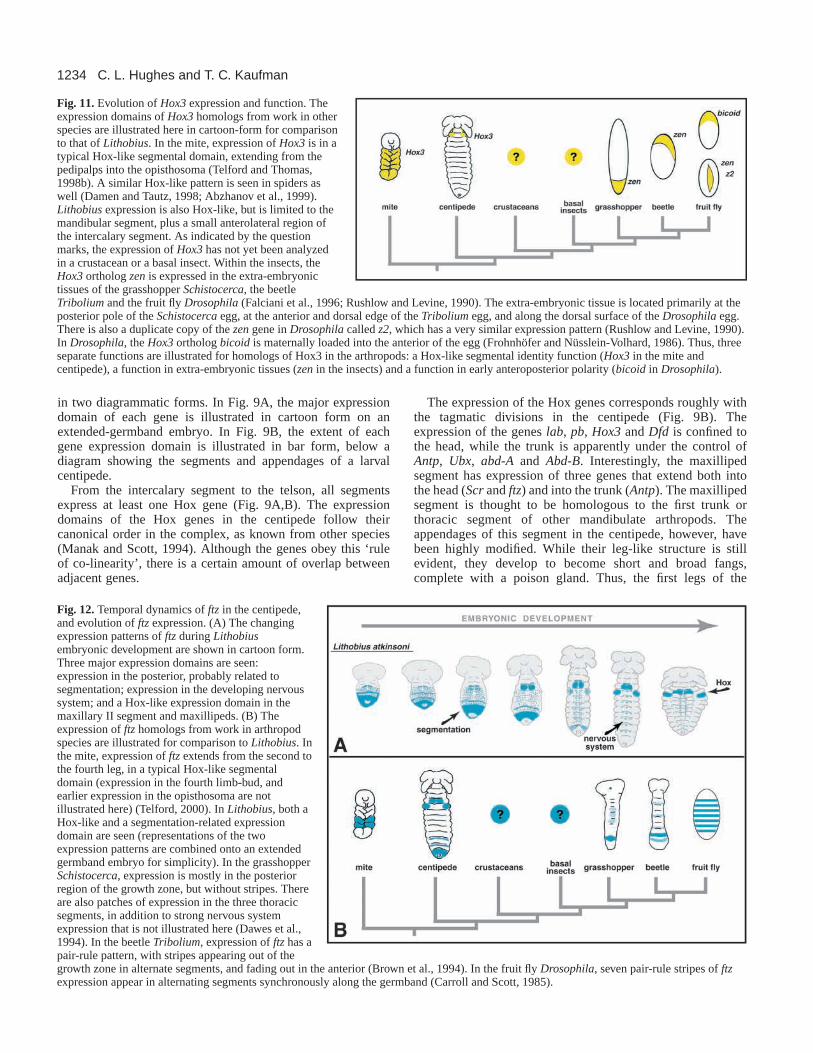
Fig. 12.Temporal dynamics of ftzin the centipede,and evolution of ftz expression. (A) The changingexpression patterns of ftzduring Lithobiusembryonic development are shown in cartoon form.Three major expression domains are seen:expression in the posterior, probably related tosegmentation; expression in the developing nervoussystem; and a Hox-like expression domain in themaxillary II segment and maxillipeds. (B) Theexpression of ftzhomologs from work in arthropodspecies are illustrated for comparison to Lithobius. Inthe mite, expression of ftzextends from the second tothe fourth leg, in a typical Hox-like segmentaldomain (expression in the fourth limb-bud, andearlier expression in the opisthosoma are notillustrated here) (Telford, 2000). In Lithobius, both aHox-like and a segmentation-related expressiondomain are seen (representations of the twoexpression patterns are combined onto an extendedgermband embryo for simplicity). In the grasshopperSchistocerca, expression is mostly in the posteriorregion of the growth zone, but without stripes. Thereare also patches of expression in the three thoracicsegments, in addition to strong nervous systemexpression that is not illustrated here (Dawes et al.,1994). In the beetle Tribolium, expression of ftzhas apair-rule pattern, with stripes appearing out of thegrowth zone in alternate segments, and fading out in the anterior (Brown et al., 1994). In the fruit fly Drosophila, seven pair-rule stripes of ftzexpression appear in alternating segments synchronously along the germband (Carroll and Scott, 1985).

1235Centipede Hox genes
centipede are modified to become more mouthpart-like, andare used for prey capture and manipulation. This mixedhead/trunk identity of the segment seems to be reflected in theHox code found there. While the segment itself has only a‘trunk’ Hox gene (Antp), the appendages have expression ofAntp as well as the ‘head’ genes Scrand ftz, which are alsoexpressed in the maxillary II segment. It remains to bedetermined how these genes contribute to the development ofthe centipede fangs. It would also be interesting to knowwhether the evolution of this novel appendage is correlatedwith a shift in the expression of these genes. Further studiesof Hox expression in other myriapods such as a millipede, orfunctional studies in the centipede, would be very interestingregarding these issues.
Shrinking domains of head Hox genesComparing the expression of the centipede Hox genes withthose of other arthropods reveals significant variability in theobserved patterns (Fig. 10). For example, in the cheliceratehead the Hox expression domains broadly overlap. These samegenes are expressed in much more restricted domains in thehead segments of crustaceans and insects. Interestingly, theexpression domains of these genes in the centipede areintermediate between these two extremes. For example, thegene lab is expressed over five segments in the spider, twosegments in the centipede, and only a single segment in thecrustaceans and insects (see Fig. 10). Likewise, the three-segment expression domain of centipede Dfd is intermediatebetween the four-segment domain in the spider and mite, andthe two-segment domains of the crustaceans and insects. Moststriking is the comparison between expression of pbamong thefour groups. In the spider, pb is expressed over five segments,from the pedipalps through the fourth walking leg. In thecentipede, the expression domain covers four segments, fromthe intercalary to the maxillary II. In the crustaceans, theexpression is restricted to the antennal II segment, which ishomologous to the intercalary segment. In the insects, however,the expression of pbis more posterior, limited mainly to theappendages of the maxillary and labial segments (homologousto the maxillary I and II segments of the centipede). Theseexpression patterns suggest that the centipede may retain someHox expression domains in an intermediate state of theirevolution, from the broad domains of the chelicerates to themore-restricted, less overlapping patterns of the crustaceansand insects. Moreover, the expression domain of pbapparentlybecame differently subdivided in different lineages; towardsthe anterior in the crustaceans, and towards the posterior in theinsects.
The centipede trunk Expression of genes along the centipede trunk is, like themorphology of the trunk, fairly homonomous. Antennapediaextends along the whole trunk in early stages, and later retractsto cover legs one through four (Fig. 5A). It is not clear whetherthis later, more restricted domain imparts any developmentaldifference to these segments, as none is evidentmorphologically. It is intriguing to note that this restriction tothe anteriormost segments of the trunk is reminiscent of asimilar restriction of Antpexpression in the pleon ofmalacostracan crustaceans and the thorax of insects (see Fig.10). Perhaps the domain of Antp expression was restricted
to the anterior portion of the trunk in the myriapod-likemandibulate ancestor, but was only exploited fully in thespecialized differentiation of the crustaceans and insects. In thecentipede, Ubx and abd-A expression patterns are similarlyexpressed along the trunk, although Ubxexpression fades fromthe extreme posterior segments. Expression of Abd-B isstrongest in the telson, but faint expression extends over themid-region of leg segments two to seven. As the genes Ubx,abd-Aand Abd-Bare likely to have similar roles in patterningthe trunks of all mandibulates, we suggest that the myriapodshave developed their unique body plan largely by expandingthe number of segments under the control of the ‘trunk’ genes.This is a similar scenario to that provided by recent findingsthat snakes seem to have created an elongated body byincreasing the numbers of somites under the control of thoracicHox genes (Cohn and Tickle, 1999).
Genes with changing rolesThose familiar with the developmental genetics of Drosophilamay find it odd to refer to zenand fushi tarazuas ‘Hox genes’.In fact, only recently have these been recognized as such. Yetrecent studies indicate that these genes were probably typicalHox genes in the arthropod ancestor, but have undergoneremarkable functional transitions in some arthropod lineages.
The expression of the insect orthologs of Hox3– bicoid, zen,and z2– reflects a remarkably versatile repertoire of functions(see Fig. 11). The gene bicoid encodes an anterior-specifyingmorphogen deposited maternally into the Drosophila egg(Frohnhöfer and Nüsslein-Volhard, 1986). The gene zerknüllt(zen) plays a role in the specification of Drosophila extra-embryonic tissues, whereas z2, the adjacent duplication of zen,has a similar expression pattern but no discernable function(Pultz et al., 1988; Rushlow and Levine, 1990). Whenhomologs were cloned from other insects, it was realized thatthese genes had sequence similarity with both the Hox3genesof vertebrates as well as the zengene of Drosophila; thus, zenis actually a highly derived homolog of Hox3 with a novelfunction (Falciani et al., 1996). More interesting still, whenbicoid and zenhomologs were cloned from another fly, it wasdiscovered that these genes have sequence similarity as well(Stauber et al., 1999). Therefore it is likely that, despite its all-important role in early Drosophiladevelopment, bicoidmayactually be a fairly recent duplication of the zengene that hasdiverged greatly in function. Thus, the Hox3 gene hasapparently been ‘caught in the act’ of changing functiondrastically in evolution – twice! To those interested inunderstanding the mechanisms of gene evolution, such a geneis worthy of much study. Researchers are currently working toclarify the timing of the zento zen+ bicoid duplication anddivergence in the higher insects (Stauber et al., 1999; Stauberet al., 2000).
The results we present are relevant to the earlier functionalchange, from Hox3(with a Hox-like role) to zen(with a rolein extra-embryonic tissues). In spiders and a mite, the Hox3gene has a typical Hox-like expression pattern, with a broaddomain whose anterior boundary is approximately co-linearwith the other Hox genes (Telford and Thomas, 1998b; Damenand Tautz, 1998; Abzhanov et al., 1999). The homologousgenes of the grasshopper Schistocercaand the beetle Triboliumapparently have zen-like roles, with expression in the extra-embryonic serosa (Falciani et al., 1996). The centipede Hox3

1236
gene presented here has a Hox-like expression pattern in thesegments of the embryonic germband, with no hint of an extra-embryonic domain. Thus, we have narrowed the window ofthe change in developmental function from Hox3to zen tosomewhere in the insect-crustacean clade. Further studies oncrustaceans and lower insects may be able to pinpoint moreprecisely the phylogenetic timing of the change, and perhapsshed light on the context and the process by which this rogueHox gene escaped from its role in determining segmentidentity.
With regard to fushi tarazu, we think we may havediscovered just such a process of Hox gene change. Althoughftz has a role in segmentation in Drosophila, ancestrally in thearthropods it seems to have been a more typical Hox gene. Ourresults here suggest that the transition between a Hox-like roleand a role in segmentation may have occurred via anintermediate state in which the gene played multiple roles indevelopment, and that this transition state was maintained inthe centipede lineage.
Among the chelicerates, the sequence and expression of ftzwas analyzed in the mite Archegozetes. The sequence of miteftz revealed its homology to the Lox5gene of annelids, and theexpression pattern is that of a typical Hox gene (see Fig. 12)(Telford, 2000). Yet in Drosophila, ftzis a pair-rule gene, witha striking pattern of seven stripes in alternating segments of theembryo (Carroll and Scott, 1985). In Tribolium, ftz has amodified pair-rule pattern, with stripes that appear out of thegrowth zone in alternate segments (Brown et al., 1994). InSchistocerca, the gene is expressed strongly in the posterior ofthe embryo, with additional expression in the nervous system,and some weak expression in the thorax (Dawes et al., 1994).It is unclear whether or not the expression in the region of theposterior growth zone of the grasshopper is related to a role insegment formation.
Two recent studies have explored the biochemical functionsof Schistocerca, Tribolium and Drosophila Ftz proteins bymisexpressing them in Drosophila(Löhr et al., 2001; Alonsoet al., 2001). Löhr et al. found that the SchistocercaandTribolium Ftz proteins retain some ability to function as a Hoxprotein when misexpressed, whereas the Drosophila proteindoes not. Expression data suggest that neither SchistocercanorTribolium ftz play a Hox-like role in their native context;yet apparently the YPWM-motif and homeodomain presentin each gene can confer homeotic phenotypes and affectHox target genes when misexpressed in the Drosophilaenvironment.
These studies have also explored the ability of misexpressedSchistocercaand TriboliumFtz proteins to mimic the disrupted-segmentation phenotype of misexpressed Drosophila Ftz.Tribolium Ftz could partially mimic this effect, whileSchistocercaFtz could not. Probably owing to the absence ofthe LXXLL motif, the SchistocercaFtz protein has only weakinteraction with DrosophilaFtz-F1, which is a necessary co-factor for the segmentation phenotype in the Drosophilaenvironment. Thus, the acquisition of the LXXLL-motif in theinsects may have led to an integral role for the Ftz-F1interaction for the Drosophilasegmentation process. However,these results do not rule out a role in segmentation for the ftzgenes of other arthropods by an LXXLL-motif independentmechanism.
In fact, our results suggest that a role for ftz in the process
of segmentation may have an ancient origin, and may beconserved across the mandibulate arthropods (myriapods,crustaceans and insects). In early centipede embryos, thepattern of expression in the posterior growth zone plus stripesin new segments (not unlike that of even-skipped; C. L. H. andT. C. K., unpublished) suggests a role in segment formation.But in later embryos, a clear Hox-like domain in the maxillaryII and maxilliped segments emerges. Thus, we suggest thatfushi tarazumade its evolutionary transition from a Hox-likerole to a role in segmentation via an intermediate stage that isretained in the centipede. Based on its combined domains ofexpression, it would appear that ftzmay be able to play multipleroles in the same embryo, one of which was lost in the insects(perhaps owing to redundancy with Scr) (Telford, 2000).Further studies of ftzhomologs in the crustaceans and insectsshould clarify where in arthropod evolution the Hox role waslost.
The results we report suggest that the complex, dynamicexpression domains in the centipede reflect multiple roles forthe centipede ftzgene. The observed expression domains of thisgene in the centipede suggest that major transitions in thefunction of a developmentally important gene may happengradually via a multifunctional intermediate, and notnecessarily only by duplication and divergence of two copiesof a gene.
Further explorationsOur results, compared with others, suggest a dynamic role forthe Hox genes in arthropod evolution. However, many morestudies are needed to test the hypotheses presented here.Comparison of the expression patterns of other species, suchas a millipede, for example, would be informative. Ultimately,we would like to bring functional techniques to bear on thesequestions. Currently, comparisons of development betweendifferent arthropods relies heavily on correlating expressionpattern with inferred and presumed function, but expansion ofknockout and misexpression techniques to more species willallow us to test our models of evolution directly.
We are grateful to Gerald Summers for his help identifying ourcentipede species, to Gary Grumbling and Joseph Duffy for in situhybridization advice, to Rudi Turner for preparing SEM micrographs,and to Paul Z. Liu for critical reading of the manuscript. The authorsalso acknowledge the inspiration of J. L. Cloudsley-Thompson whonoted that ‘Centipedes seem to exert a weird fascination on the morbidappetites of the hysterical and insane.’ C. L. H. thanks the HowardHughes Medical Institute for their financial support through an HHMIPredoctoral Fellowship. T. C. K. is an investigator of the HowardHughes Medical Institute.
REFERENCES
Abzhanov, A. and Kaufman, T. C. (1999a). Homeotic genes and thearthropod head: Expression patterns of the labial, proboscipedia, andDeformedgenes in crustaceans and insects.Proc. Natl. Acad. Sci. USA96,10224-10229.
Abzhanov, A. and Kaufman, T. C.(1999b). Novel regulation of the homeoticgene Scr associated with a crustacean leg-to-maxilliped appendagetransformation.Development126, 1121-1128.
Abzhanov, A. and Kaufman, T. C.(2000a). Crustacean (malacostracan) Hoxgenes and the evolution of the arthropod trunk.Development127, 2239-2249.
Abzhanov, A. and Kaufman, T. C.(2000b). Embryonic expression patterns
C. L. Hughes and T. C. Kaufman

1237Centipede Hox genes
of the Hox genes of the crayfish Procambarus clarkii (Crustacea,Decapoda). Evol. Dev2, 271-283.
Abzhanov, A., Popadic, A. and Kaufman, T. C.(1999). Chelicerate Hoxgenes and the homology of arthropod segments.Evol Dev. 1, 77-89.
Akam, M. (2000). Developmental genetics and the diversity of animal form:Hox genes in arthropods. In The Biology of Biodiversity(ed. M. Kato), pp.195-208. Tokyo, New York: Springer-Verlag.
Alonso, C. R., Maxton-Kuechenmeister, J. and Akam, M.(2001). Evolutionof Ftz protein function in insects.Curr. Biol. 11, 1473-1478.
Averof, M. and Akam, M. (1995). Hox genes and the diversification of insectand crustacean body plans.Nature376, 420-423.
Averof, M. and Patel, N. H. (1997). Crustacean appendage evolutionassociated with changes in Hox gene expression.Nature388, 682-686.
Bennett, R. L., Brown, S. J. and Denell, R. E.(1999). Molecular and geneticanalysis of the Tribolium Ultrabithoraxortholog, Ultrathorax.Dev. GenesEvol. 209, 608-619.
Boore, J. L., Lavrov, D. V. and Brown, W. M. (1998). Gene translocationlinks insects and crustaceans.Nature392, 667-668.
Brown, S., Holtzman, S., Kaufman, T. and Denell, R. (1999).Characterization of the Tribolium Deformedortholog and its ability todirectly regulate Deformedtarget genes in the rescue of a DrosophilaDeformednull mutant.Dev. Genes Evol. 209, 389-398.
Brown, S. J., Hilgenfeld, R. B. and Denell, R. E.(1994). The beetleTribolium castaneumhas a fushi tarazuhomolog expressed in stripes duringsegmentation.Proc. Natl. Acad. Sci. USA 91, 12922-12926.
Carroll, S. B. and Scott, M. P.(1985). Localization of the fushi tarazuproteinduring Drosophilaembryogenesis.Cell 43, 47-58.
Chadwick, R. and McGinnis, W. (1987). Temporal and spatial distributionof transcripts from the Deformedgene of Drosophila.EMBO J. 6, 779-790.
Cohn, M. J. and Tickle, C.(1999). Developmental basis of limblessness andaxial patterning in snakes.Nature399, 474-479.
Cook, C. E., Smith, M. L., Telford, M. J., Bastianello, A. and Akam, M.(2001). Hox genes and the phylogeny of the arthropods.Curr. Biol. 11, 759-763.
Curtis, C. D., Brisson, J. A., DeCamillis, M. A., Shippy, T. D., Brown, S.J. and Denell, R. E.(2001). Molecular characterization of Cephalothorax,the Tribolium ortholog of Sex combs reduced.Genesis30, 12-20.
Damen, W. G. M. and Tautz, D.(1998). A hox class 3 orthologue from thespider Cupiennius saleiis expressed in a Hox-gene-like fashion.Dev. GenesEvol. 208, 586-590.
Damen, W. G. M. and Tautz, D.(1999). Abdominal-Bexpression in a spidersuggests a general role for Abdominal-Bin specifying the genital structure.J. Exp. Zool. 285, 85-91.
Damen, W. G. M., Hausdorf, M., Seyfarth, E.-A. and Tautz, D.(1998). Aconserved mode of head segmentation in arthropods revealed by theexpression pattern of Hox genes in a spider.Proc. Natl. Acad. Sci. USA95,10665-10670.
Dawes, R., Dawson, I., Falciani, F., Tear, G. and Akam, M.(1994). Dax, alocust Hox gene related to fushi-tarazubut showing no pair-rule expression.Development120, 1561-1572.
Delorenzi, M. and Bienz, M. (1990). Expression of Abdominal-Bhomeoproteins in Drosophilaembryos.Development108, 323-330.
Diederich, R. J., Merrill, V. K., Pultz, M. A. and Kaufman, T. C. (1989).Isolation, structure, and expression of labial, a homeotic gene of theAntennapedia Complex involved in Drosophila head development.GenesDev. 3, 399-414.
Falciani, F., Hausdorf, B., Schroder, R., Akam, M., Tautz, D., Denell, R.and Brown, S.(1996). Class 3 Hox genes in insects and the origin of zen.Proc. Natl. Acad. Sci. USA93, 8479-8484.
Fleig, R., Walldorf, U., Gehring, W. J. and Sander, K.(1992). Developmentof the Deformed protein pattern in the embryo of the honeybee ApismelliferaL. (Hymenoptera). Roux’s Arch. Dev. Biol. 201, 235-242.
Friedrich, M. and Tautz, D. (1995). Ribosomal DNA phylogeny of the majorextant arthropod classes and the evolution of myriapods.Nature376, 165-167.
Frohnhöfer, H. G. and Nüsslein-Volhard, C.(1986). Organization of anteriorpattern in the Drosophilaembryo by the maternal gene bicoid. Nature324,120-125.
Giribet, G., Edgecombe, G. and Wheeler, W.(2001). Arthropod phylogenybased on eight molecular loci and morphology.Nature413, 157-161.
Grenier, J. K., Garber, T. L., Warren, R., Whitington, P. M. and Carroll,S. (1997). Evolution of the entire arthropod Hox gene set predated theorigin and radiation of the onychophoran/arthropod clade.Curr. Biol. 7,547-553.
Haas, M. S., Brown, S. J. and Beeman, R. W.(2001a). Homeotic evidencefor the appendicular origin of the labrum in Tribolium castaneum. Dev.Genes Evol. 211, 96-102.
Haas, M. S., Brown, S. J. and Beeman, R. W.(2001b). Pondering theprocephalon: The segmental origin of the labrum.Dev. Genes Evol. 211, 89-95.
Hayward, D. C., Patel, N. H., Rehm, E. J., Goodman, C. S. and Ball, E.E. (1995). Sequence and expression of grasshopper Antennapedia:Comparison to Drosophila.Dev. Biol. 172, 452-465.
Hertzel, G. (1984). Die Segmentation des Keimstreifens von Lithobiusforficatus(L.) (Myriapoda, Chilopoda). Zool. Jb. Anat. 112, 369-386.
Hwang, U., Friedrich, M., Tautz, D., Park, C. and Kim, W. (2001).Mitochondrial protein phylogeny joins myriapods with chelicerates.Nature413, 154-157.
Kelsh, R., Dawson, I. and Akam, M.(1993). An analysis of Abdominal-Bexpression in the locust Schistocercagregaria.Development117, 293-305.
Kelsh, R., Weinzierl, R. O. J., White, R. A. H. and Akam, M.(1994).Homeotic gene expression in the locust Schistocerca: An antibody thatdetects conserved epitopes in Ultrabithorax and abdominal-A proteins.Dev.Genetics15, 19-31.
Kokubo, H., Ueno, K., Amanai, K. and Suzuki, Y.(1997). Involvement ofthe Bombyx Scrgene in development of the embryonic silk gland.Dev. Biol.186, 46-57.
Löhr, U., Yussa, M. and Pick, L. (2001). Drosophila fushi tarazu: a gene onthe border of homeotic function.Curr. Biol. 11, 1403-1412.
Macias, A., Casanova, J. and Morata, G.(1990). Expression and regulationof the abd-Agene of Drosophila. Development110, 1197-1208.
Manak, J. R. and Scott, M. P. (1994). A class act: Conservation ofhomeodomain protein functions.Development120, 61-77.
Martinez-Arias, A., Ingham, P. W., Scott, M. P. and Akam, M. E.(1987).The spatial and temporal deployment of Dfd and Scrtranscripts throughoutdevelopment of Drosophila.Development100, 673-684.
Minelli, A. and Bortoletto, S. (1988). Myriapod metamerism and arthropodsegmentation.Biol. J. Linn. Soc. 33, 323-343.
Nagata, T., Suzuki, Y., Ueno, K., Kokubo, H., Xu, X., Hui, C., Hara, W.and Fukuta, M. (1996). Developmental expression of the BombyxAntennapediahomologue and homeotic changes in the Nc mutant.GenesCells1, 555-568.
Nagy, L. M., Booker, R. and Riddiford, L. M. (1991). Isolation andembryonic expression of an abdominal-A-like gene from the lepidopteran,Manduca sexta. Development112, 119-130.
Nie, W., Stronach, B., Panganiban, G., Shippy, T., Brown, S. and Denell,R. (2001). Molecular characterization of Tclabialand the 3′end of theTribolium homeotic complex.Dev. Genes Evol. 211, 244-251.
O’Neill, J. W. and Bier, E. (1994). Double-label in situhybridizationusing biotin and digoxigenin-tagged RNA probes.BioTechniques17, 870-875.
Peterson, M. D., Rogers, B. T., Popadic, A. and Kaufman, T. C.(1999).The embryonic expression pattern of labial, posterior homeotic complexgenes and the teashirt homologue in an apterygote insect.Dev. Genes Evol.209, 77-90.
Pultz, M. A., Diederich, R. J., Cribbs, D. L. and Kaufman, T. C.(1988).The proboscipedialocus of the Antennapedia complex: a molecular andgenetic analysis.Genes Dev. 2, 901-920.
Regier, J. C. and Shultz, J. W.(1997). Molecular phylogeny of the majorarthropod groups indicates polyphyly of crustaceans and a new hypothesisfor the origin of hexapods.Mol. Biol. Evol. 14, 902-913.
Rogers, B. T. and Kaufman, T. C.(1997). Structure of the insect head inontogeny and phylogeny: A view from Drosophila. Int. Rev. Cyt. 174, 1-84.
Rogers, B. T., Peterson, M. D. and Kaufman, T. C.(1997). Evolution of theinsect body plan as revealed by the Sex combs reducedexpression pattern.Development124, 149-157.
Rogers, B. T., Peterson, M. D. and Kaufman, T. C.(2002). The developmentand evolution of insect mouthparts as revealed by the expression patterns ofgnathocephalic genes.Evol. Dev. (in press).
Rushlow, C. and Levine, M. (1990). Role of the zerknullt gene in dorsal-ventral pattern formation in Drosophila. In Advances in Genetics, Vol. 27.Genetic Regulatory Hierarchies in Development(ed. T. R. F. Wright). SanDiego, CA: Academic Press.
Shippy, T. D., Brown, S. J. and Denell, R. E.(1998). Molecularcharacterization of the Tribolium abdominal-Aortholog and implications forthe products of the Drosophilagene.Dev. Genes Evol. 207, 446-452.
Shippy, T. D., Guo, J., Brown, S. J., Beeman, R. W. and Denell, R. E.

1238
(2000). Analysis of maxillipedia expression pattern and larval cuticularphenotype in wild-type and mutant Tribolium. Genetics155, 721-731.
Stauber, M., Jaeckle, H. and Schmidt-Ott, U. (1999). The anteriordeterminant bicoid of Drosophila is a derived Hox class 3 gene.Proc. Natl.Acad. Sci. USA96, 3786-3789.
Stauber, M., Taubert, H. and Schmidt-Ott, U. (2000). Function of bicoidand hunchback homologs in the basal cyclorrhaphan fly Megaselia(Phoridae). Proc. Natl. Acad. Sci. USA97, 10844-10849.
Tear, G., Akam, M. and Martinez-Arias, A. (1990). Isolation of anabdominal-Agene from the locust Schistocerca gregariaand its expressionduring early embryogenesis.Development110, 915-926.
Telford, M. J. (2000). Evidence for the derivation of the Drosophila fushitarazugene from a Hox gene orthologous to lophotrochozoan Lox5. Curr.Biol. 10, 349-352.
Telford, M. J. and Thomas, R. H.(1998a). Expression of homeobox genes
shows chelicerate arthropods retain their deutocerebral segment.Proc. Natl.Acad. Sci. USA95, 10671-10675.
Telford, M. J. and Thomas, R. H. (1998b). Of mites and zen: expressionstudies in a chelicerate arthropod confirm zenis a divergent Hox gene.Dev.Genes Evol. 208, 591-594.
Walldorf, U., Binner, P. and Fleig, R.(2000). Hox genes in the honey beeApis mellifera.Dev. Genes Evol. 210, 483-492.
White, R. A. H. and Wilcox, M. (1985). Distribution of Ultrabithorax proteinsin Drosophila. EMBO J. 4, 2035-2044.
Wirz, J., Fessler, L. I. and Gehring, W. J. (1986). Localization of theAntennapediaprotein in Drosophilaembryos and imaginal discs.EMBO J.5, 3327-3334.
Zheng, Z. Khoo, A, Fambrough, D., Garza, L. and Booker, R. (1999).Homeotic gene expression in the wild-type and a homeotic mutant of themoth Manduca sexta.Dev. Genes Evol. 209, 460-472.
C. L. Hughes and T. C. Kaufman