Embed Size (px)
DESCRIPTION
Exciting things happen at the intersection of ideas. When knowledge domains collide, fireworks can fly. That’s certainly true in the pathbreaking work of Northwestern University researchers who have harnessed the interdisciplinary power of medicine, chemistry, engineering, and more to discover the “zinc sparks” that occur when egg and sperm meet during conception. (See page 18.) The discovery could transform our understanding of reproduction and fertility. In this issue of CenterPiece, we explore advances in medical science at Northwestern. Whether it is precision medicine’s potential to exploit genomic information to effect “customized” cures, or else biomedical engineering breakthroughs that deliver better diagnostic tests to those who need them most, Northwestern scholars are combining their expertise to improve people’s lives.
Citation preview
Research Scholarship, Collaboration, and Outreach at Northwestern University
SPRING 2015
CENTERPIECE02CIGHT Brings Health Innovation to Africa
14Closing in on Precision Medicine
18Sparks Fly in Quest for a Good Egg
30Biology and Beyond
CREATING NEW KNOWLEDGE
Exciting things happen at the intersection of ideas. When knowledge domains collide, fireworks can fly. That’s certainly
true in the pathbreaking work of Northwestern University researchers who have harnessed the interdisciplinary power of medicine, chemistry, engineering, and more to discover the “zinc sparks” that occur when egg and sperm meet during conception. (See page 18.) The discovery could transform our understanding of reproduction and fertility.
In this issue of CenterPiece, we explore advances in medical science at Northwestern. Whether it is precision medicine’s potential to exploit genomic information to effect “customized” cures, or else biomedical engineering breakthroughs that deliver better diagnostic tests to those who need them most, Northwestern scholars are combining their expertise to improve people’s lives.
Office for Research Jay Walsh
Vice President for Research
Meg McDonald
Assistant Vice President for Research
Matt Golosinski
Director of Research Communications
Roger Anderson
Publications Editor
Jeanine Shimer
Designer
Writers
Roger Anderson Christina Cala Sara Kupper Joan T. Naper Patricia Reese Erin Spain
Designer
Jeanine Shimer
Address all correspondence to:
Matt Golosinski Director of Research Communications Northwestern University Office for Research, 633 Clark Street Evanston, Illinois 60208
This publication is available online at:
research.northwestern.edu/orpfc/
publications/centerpiece
discover.northwestern.edu
©2015 Northwestern University.
All rights reserved.
BIRTH OF INNOVATION
CenterPiece | Spring 2015 1
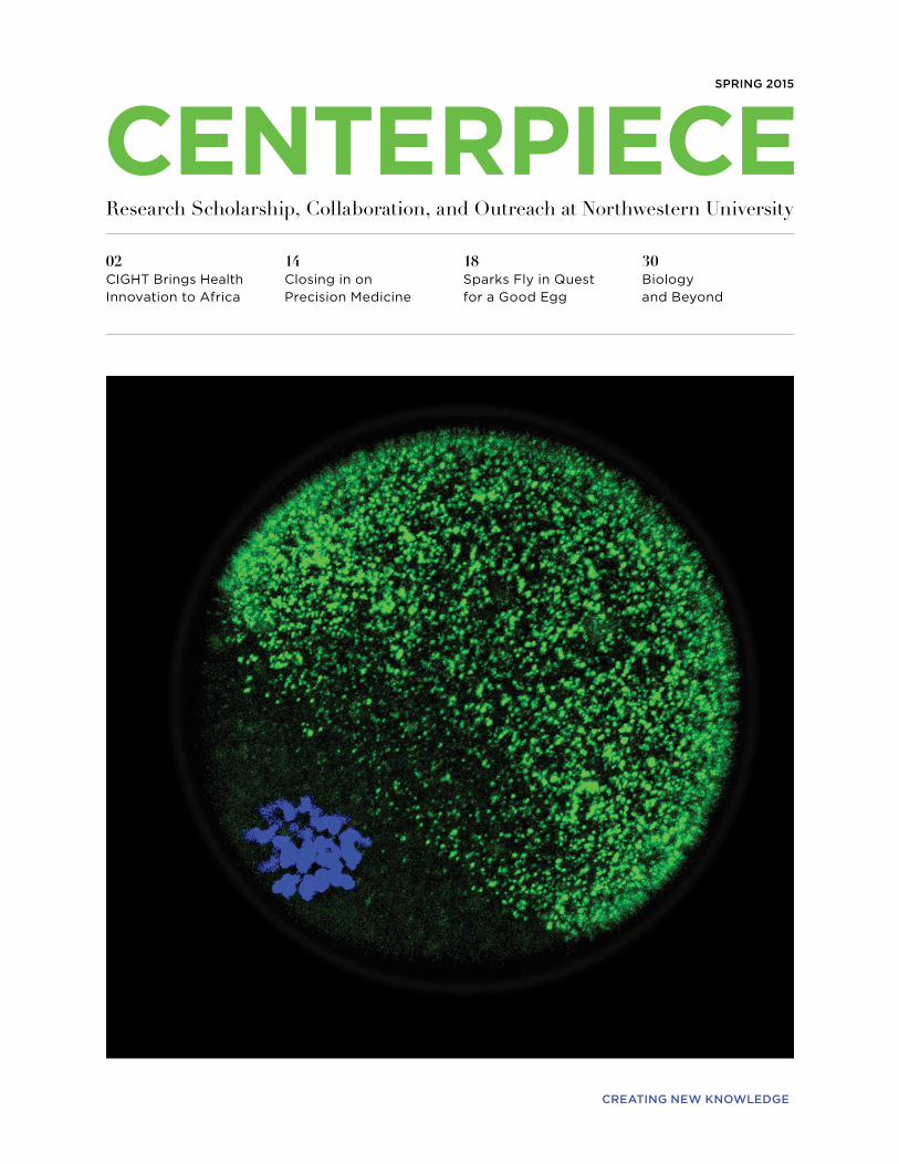
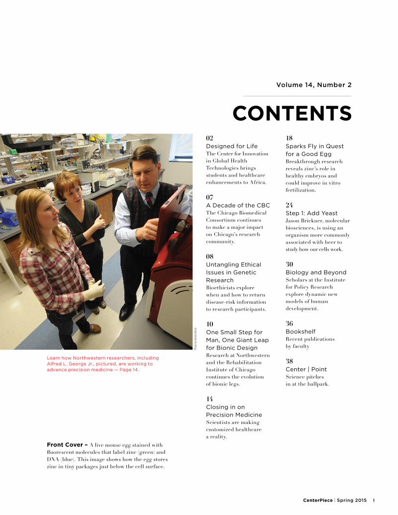
Front Cover – A live mouse egg stained with fluorescent molecules that label zinc (green) and DNA (blue). This image shows how the egg stores zinc in tiny packages just below the cell surface.
02Designed for Life The Center for Innovation in Global Health Technologies brings students and healthcare enhancements to Africa.
07A Decade of the CBC The Chicago Biomedical Consortium continues to make a major impact on Chicago’s research community.
08Untangling Ethical Issues in Genetic Research Bioethicists explore when and how to return disease-risk information to research participants.
10One Small Step for Man, One Giant Leap for Bionic Design Research at Northwestern and the Rehabilitation Institute of Chicago continues the evolution of bionic legs.
14Closing in on Precision Medicine Scientists are making customized healthcare a reality.
18Sparks Fly in Quest for a Good Egg Breakthrough research reveals zinc’s role in healthy embryos and could improve in vitro fertilization.
24Step 1: Add Yeast Jason Brickner, molecular biosciences, is using an organism more commonly associated with beer to study how our cells work.
30Biology and Beyond Scholars at the Institute for Policy Research explore dynamic new models of human development.
36Bookshelf Recent publications by faculty
38Center | Point Science pitches in at the ballpark.
Volume 14, Number 2
CONTENTS
Learn how Northwestern researchers, including Alfred L. George Jr., pictured, are working to advance precision medicine — Page 14.
Phot
o by
Ran
dy B
elice
2 Northwestern University Office for Research
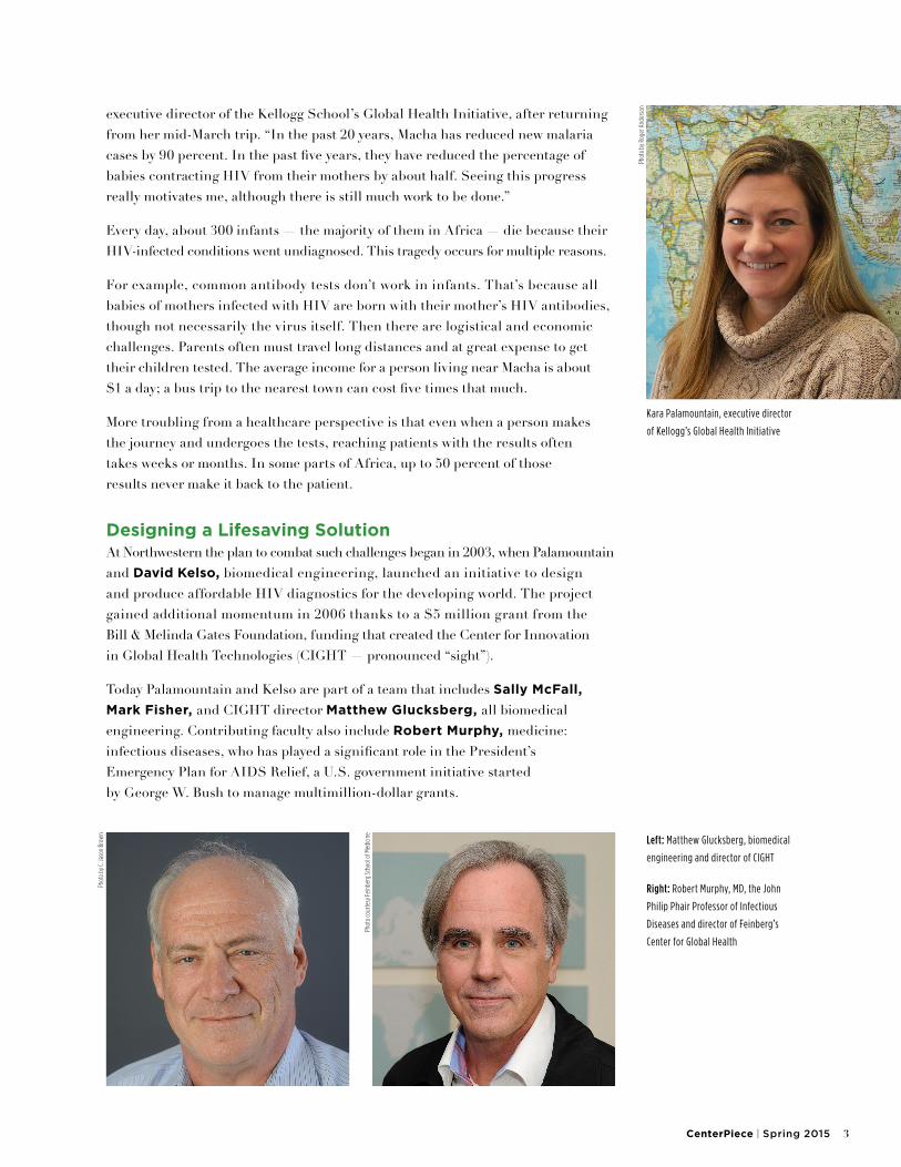
Kara Palamountain awakens to a rooster’s crow. It’s already warm at 6 a.m., and the tropical sun has just begun its daily ascent. She is 8,000 miles from her office at Northwestern’s Kellogg School of Management, working among the scattered homesteads of Macha, Zambia (shown on map, left). More than 125,000 people live here, and one in seven has the virus that causes AIDS. In neighboring Botswana, the statistics are even worse. Palamountain and her Northwestern colleagues are trying to slow the epidemic by harnessing the power of management, medicine, and biomedical design.
“The people of Macha have worked hard to improve health within their community, and they are seeing the fruits of their labor,” says Palamountain,
Designed for LifeA DECADE IN THE MAKING, LOW-COST INFANT–HIV TEST NEARS CLINICAL USE
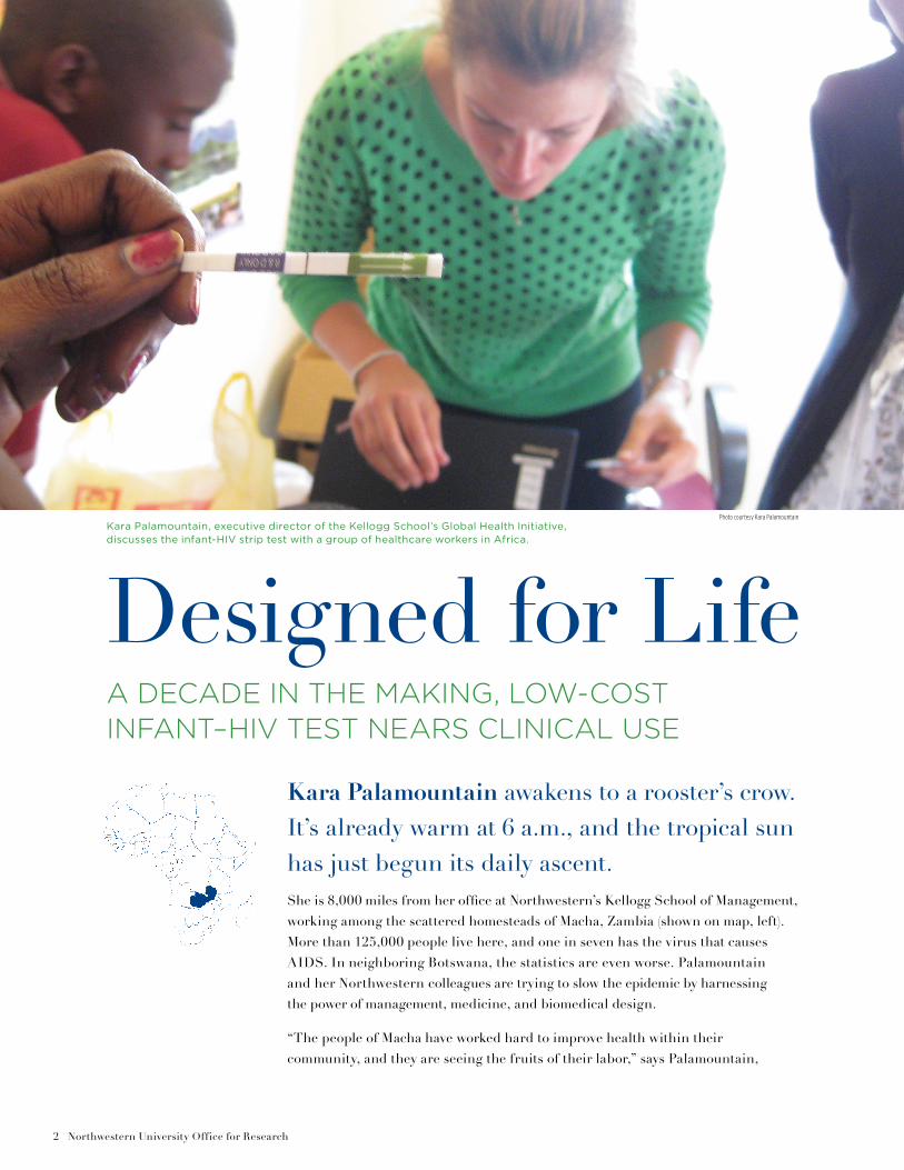
Kara Palamountain, executive director of the Kellogg School’s Global Health Initiative, discusses the infant-HIV strip test with a group of healthcare workers in Africa.
Photo courtesy Kara Palamountain
CenterPiece | Spring 2015 3
executive director of the Kellogg School’s Global Health Initiative, after returning from her mid-March trip. “In the past 20 years, Macha has reduced new malaria cases by 90 percent. In the past five years, they have reduced the percentage of babies contracting HIV from their mothers by about half. Seeing this progress really motivates me, although there is still much work to be done.”
Every day, about 300 infants — the majority of them in Africa — die because their HIV-infected conditions went undiagnosed. This tragedy occurs for multiple reasons.
For example, common antibody tests don’t work in infants. That’s because all babies of mothers infected with HIV are born with their mother’s HIV antibodies, though not necessarily the virus itself. Then there are logistical and economic challenges. Parents often must travel long distances and at great expense to get their children tested. The average income for a person living near Macha is about $1 a day; a bus trip to the nearest town can cost five times that much.
More troubling from a healthcare perspective is that even when a person makes the journey and undergoes the tests, reaching patients with the results often takes weeks or months. In some parts of Africa, up to 50 percent of those results never make it back to the patient.
Designing a Lifesaving SolutionAt Northwestern the plan to combat such challenges began in 2003, when Palamountain and David Kelso, biomedical engineering, launched an initiative to design and produce affordable HIV diagnostics for the developing world. The project gained additional momentum in 2006 thanks to a $5 million grant from the Bill & Melinda Gates Foundation, funding that created the Center for Innovation in Global Health Technologies (CIGHT — pronounced “sight”).
Today Palamountain and Kelso are part of a team that includes Sally McFall, Mark Fisher, and CIGHT director Matthew Glucksberg, all biomedical engineering. Contributing faculty also include Robert Murphy, medicine: infectious diseases, who has played a significant role in the President’s Emergency Plan for AIDS Relief, a U.S. government initiative started by George W. Bush to manage multimillion-dollar grants.
Kara Palamountain, executive director
of Kellogg’s Global Health Initiative
Left: Matthew Glucksberg, biomedical
engineering and director of CIGHT
Right: Robert Murphy, MD, the John
Philip Phair Professor of Infectious
Diseases and director of Feinberg’s
Center for Global Health
Phot
o by
Rog
er A
nder
son
Phot
o by
C. J
ason
Bro
wn
Phot
o co
urte
sy Fe
inbe
rg S
choo
l of M
edici
ne
4 Northwestern University Office for Research
Harnessing the combined thought leadership of faculty from Kellogg, the McCormick School of Engineering and Applied Science, and the Feinberg School of Medicine means CIGHT has accomplished what few academic global health organizations can do: develop and achieve commercialization of innovative medical technologies.
Marketing New IdeasPalamountain plays a critical role in developing each new CIGHT device, and so do students in her Kellogg course Medical Technologies in Developing Countries. Together they conduct the crucial market research to identify community needs and then work with end users to pursue optimal designs, closely assessing any functionality tradeoffs. For instance, a small design tweak may require more manual steps to obtain test
results, or a change could require electricity, a resource not universally or reliably available in some locations. Palamountain collaborates with in-country healthcare providers to determine whether such modifications are acceptable.
In her Evanston office, she sets out an early prototype of an infant HIV test kit. It’s not much bigger than a deck of cards and has no functionality.
“The first time I brought this to Africa, it was difficult to have a real conversation while holding foam core,” she recalls. “But when you return and show them how we’ve improved the design, how we are making progress, and how their suggestions helped advance this project, the dialogue becomes much easier.”
After many iterations, the rudimentary model evolved into an advanced rendering and finally
a working prototype. Today the basic strip test uses a small blood sample to provide an answer to a single question: Does this baby have HIV?
Results take about 45 minutes, and healthcare workers almost anywhere can be trained to conduct the test. Its use will allow parents to leave clinics armed with vital diagnostic insights and a medical plan.
“We are looking at what products should be made and also how they might need to be constructed to benefit a specific group of people,” says Palamountain. “With 54 countries in Africa, our final hurdle will be getting the finished product approved and introduced into each healthcare market.”
Thirteen students joined Palamountain on her recent trip to Zambia. Another 12 students visited Lesotho with course co-instructor Robert Dintruff.
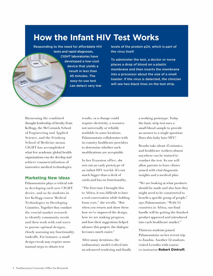
Responding to the need for affordable HIV
tests and rapid diagnoses,
CIGHT laboratories have
developed a low-cost
device that yields a
result in less than
45 minutes. The
easy-to-use test
can detect very low
levels of the protein p24, which is part of
the virus itself.
To administer the test, a doctor or nurse
places a drop of blood on a plastic
membrane and then inserts the membrane
into a processor about the size of a small
toaster. If the virus is detected, the clinician
will see two black lines on the test strip.
How the Infant HIV Test Works
CenterPiece | Spring 2015 5
The teams each researched how and where they might place a second testing platform — a more expensive device that provides information on a patient’s HIV viral load. Such testing is common for individuals living with HIV in the United States; results indicate viral strength based on the amount of HIV in the blood.
CIGHT is working with collaborators on this second testing platform, which can identify not only HIV viral load but also tuberculosis and many other diseases. Although each test is conducted differently, the core equipment adapts to provide accurate disease-specific results. Back in Evanston, engineers at CIGHT’s laboratory space are working to finalize the TB-specific test technique; tuberculosis ranks second only to HIV/AIDS in deaths caused by a single infectious agent.
Advances in EngineeringWhile Palamountain was in Zambia, her colleagues Glucksberg and Murphy were 1,500 miles away in South Africa.
Since its inception, CIGHT has worked closely with the University of Cape Town to provide an academic framework for student-led innovation. This spring 16 students from Northwestern spent nearly three months working on projects that included a device to protect healthcare workers in TB clinics and a low-cost CO2 monitor to aid in placing endotracheal tubes while out in the field.
The team’s ultimate goal is to sustainably design new devices for the developing world while also transforming current medical and manufacturing techniques. Tests that are expensive, take significant time to analyze, or require electricity present significant obstacles throughout Africa.
“CIGHT helps build platforms — the machines that run the tests — using well-understood technologies and developing the assays for specific diseases,”
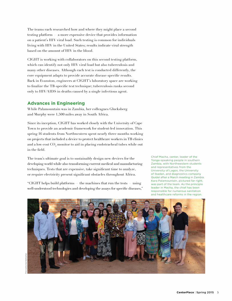
Chief Macha, center, leader of the Tonga-speaking people in southern Zambia, with Northwestern students and representatives from the University of Lagos, the University of Ibadan, and diagnostics company Quidel after a March meeting in Zambia. Kara Palamountain, pictured far right, was part of the team. As the principle leader in Macha, the chief has been responsible for numerous sanitation and healthcare reforms in the region.
Phot
o co
urte
sy K
ara
Pala
mou
ntai
n
6 Northwestern University Office for Research
says Glucksberg. “In Africa we are looking at point-of-care diagnostics because we believe it’s a critical need there. For example, during the process of sending tests to a lab, results frequently go missing.”
Once the team assesses such market needs and solves related chemistry and engineering challenges, prototypes are tested in the field. Then CIGHT’s HIV-testing devices move into the manufacturing stage.
“That is where we are right now with both projects — the HIV strip test as well as the more complicated HIV viral load test. Figuring out how to translate a manmade device into a mass-produced one is something that universities don’t traditionally do,” Glucksberg explains. “We’re finalizing the design and identifying a manufacturer, and will be working with Rob Murphy to ensure our introduction into clinics goes as well as possible.”
Major Impact on HealthIt’s often said that Robert Murphy is the most traveled professor at Northwestern. Director of Feinberg’s Center for Global Health, Murphy recently attended a conference in Seattle before heading to Lima, Peru. He then flew to Cape Town to watch students present their final CIGHT projects.
In the 1990s Murphy was already working in Africa as the AIDS epidemic there began to spread. An expert in viral infections, he has spent the past two decades exploring new antiretroviral drugs and vaccines for HIV and viral hepatitis. He’s also worked to improve therapies for tuberculosis and AIDS.
“The perception by some is that global health involves getting on a plane and taking care of poor people in various stages of disease,” says Murphy. “A big part of the way that our team thinks of global health revolves around doing things to decrease the health disparities that exist between nations. The only way to achieve that vision is by building strong relationships within these communities over time.”
As Northwestern’s infant-HIV test enters the marketplace — a development expected this year — Murphy will play a large role in its implementation.
“Africa is rife with applied engineering teams, groups that work to fix broken Western machines,” he says. “What CIGHT has done is develop products relevant to Africans that clinicians and healthcare workers can use to improve the overall health of those around them. It’s a model that’s soon to make a major impact.”
— Roger Anderson
Other CIGHT ProjectsCIGHT currently has 10 ongoing projects in various stages of
development. Student-led initiatives include an iPad application for
training nurses in integrated management of childhood illness and a
low-energy phototherapy blanket for treating jaundice in newborns.
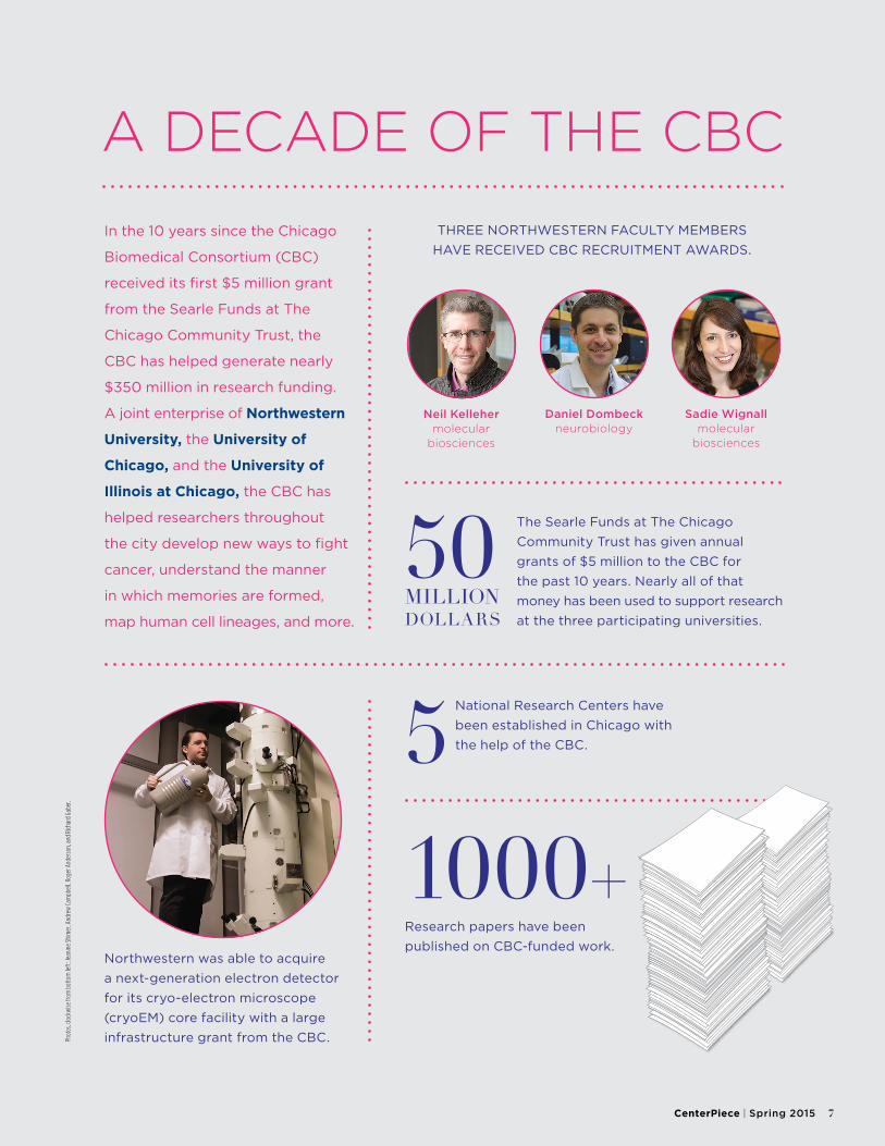
A DECADE OF THE CBC
In the 10 years since the Chicago
Biomedical Consortium (CBC)
received its first $5 million grant
from the Searle Funds at The
Chicago Community Trust, the
CBC has helped generate nearly
$350 million in research funding.
A joint enterprise of Northwestern
University, the University of
Chicago, and the University of
Illinois at Chicago, the CBC has
helped researchers throughout
the city develop new ways to fight
cancer, understand the manner
in which memories are formed,
map human cell lineages, and more.
50 MILLIONDOLLARS
The Searle Funds at The Chicago
Community Trust has given annual
grants of $5 million to the CBC for
the past 10 years. Nearly all of that
money has been used to support research
at the three participating universities.
Neil Kelleher molecular
biosciences
Daniel Dombeck neurobiology
Sadie Wignall molecular
biosciences
THREE NORTHWESTERN FACULTY MEMBERS
HAVE RECEIVED CBC RECRUITMENT AWARDS.
Northwestern was able to acquire
a next-generation electron detector
for its cryo-electron microscope
(cryoEM) core facility with a large
infrastructure grant from the CBC.
1000+Research papers have been
published on CBC-funded work.
National Research Centers have
been established in Chicago with
the help of the CBC.5
Phot
os, c
lock
wise
from
bot
tom
left
: Jea
nine
Shi
mer
, And
rew
Cam
pbel
l, Ro
ger A
nder
son,
and
Rich
ard
Gabe
r.
CenterPiece | Spring 2015 7
8 Northwestern University Office for Research
Imagine sitting in your physician’s office and receiving the results of a genetics test. The results indicate that you have a high risk for Alzheimer’s disease. Right now, there is no cure. What would you do with that information? What could your doctor do?According to Maureen Smith, director of Northwestern’s NUgene Project, this scenario illustrates some of the major ethical issues currently associated with genetics: the problems of returning research results to patients and preparing physicians and other healthcare providers with information and decision-support tools to help patients understand the results, as well as the need for much more research into the basis of and potential therapies for disease.
UntanglingEthical Issues in Genetic Research
DATA ADVANCES PRESENT FRESH CLINICAL
OPPORTUNITIES — AND CHALLENGES
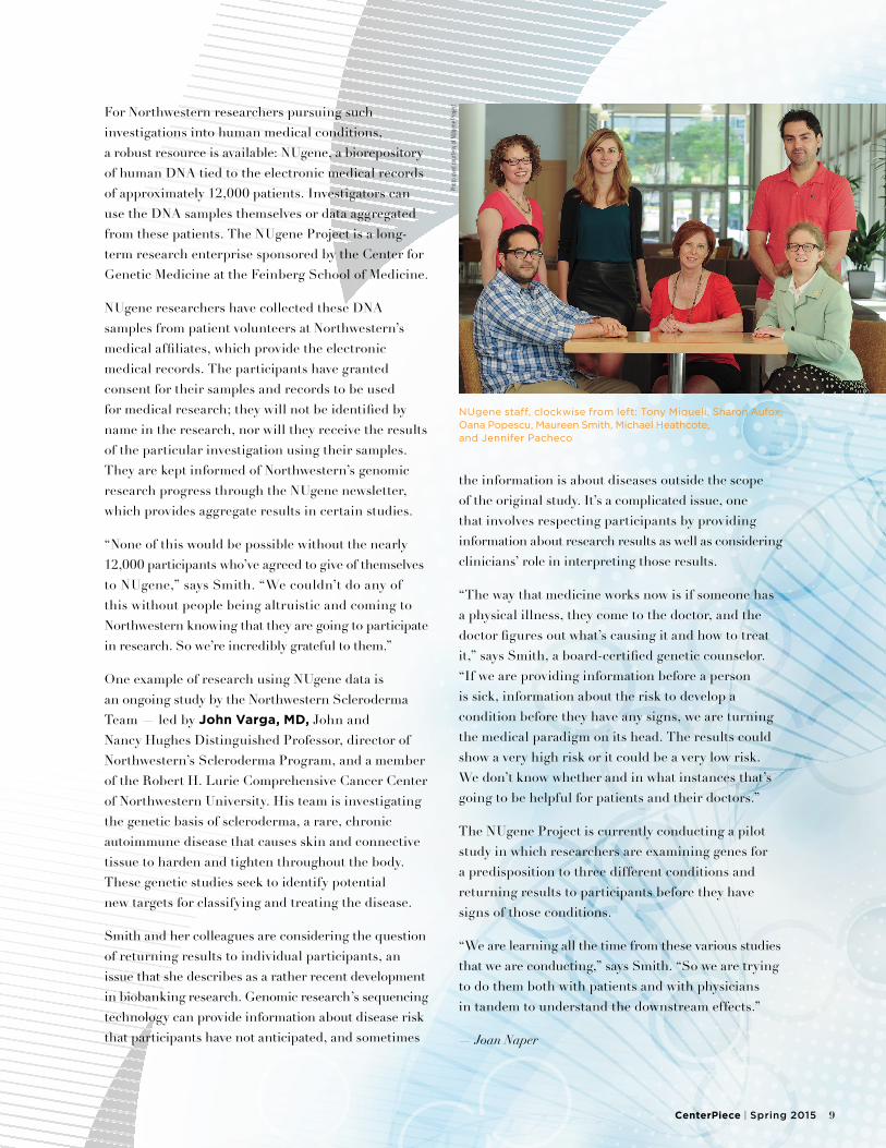
NUgene staff, clockwise from left: Tony Miqueli, Sharon Aufox, Oana Popescu, Maureen Smith, Michael Heathcote, and Jennifer Pacheco
CenterPiece | Spring 2015 9
For Northwestern researchers pursuing such investigations into human medical conditions, a robust resource is available: NUgene, a biorepository of human DNA tied to the electronic medical records of approximately 12,000 patients. Investigators can use the DNA samples themselves or data aggregated from these patients. The NUgene Project is a long-term research enterprise sponsored by the Center for Genetic Medicine at the Feinberg School of Medicine.
NUgene researchers have collected these DNA samples from patient volunteers at Northwestern’s medical affiliates, which provide the electronic medical records. The participants have granted consent for their samples and records to be used for medical research; they will not be identified by name in the research, nor will they receive the results of the particular investigation using their samples. They are kept informed of Northwestern’s genomic research progress through the NUgene newsletter, which provides aggregate results in certain studies.
“None of this would be possible without the nearly 12,000 participants who’ve agreed to give of themselves to NUgene,” says Smith. “We couldn’t do any of this without people being altruistic and coming to Northwestern knowing that they are going to participate in research. So we’re incredibly grateful to them.”
One example of research using NUgene data is an ongoing study by the Northwestern Scleroderma Team — led by John Varga, MD, John and Nancy Hughes Distinguished Professor, director of Northwestern’s Scleroderma Program, and a member of the Robert H. Lurie Comprehensive Cancer Center of Northwestern University. His team is investigating the genetic basis of scleroderma, a rare, chronic autoimmune disease that causes skin and connective tissue to harden and tighten throughout the body. These genetic studies seek to identify potential new targets for classifying and treating the disease.
Smith and her colleagues are considering the question of returning results to individual participants, an issue that she describes as a rather recent development in biobanking research. Genomic research’s sequencing technology can provide information about disease risk that participants have not anticipated, and sometimes
the information is about diseases outside the scope of the original study. It’s a complicated issue, one that involves respecting participants by providing information about research results as well as considering clinicians’ role in interpreting those results.
“The way that medicine works now is if someone has a physical illness, they come to the doctor, and the doctor figures out what’s causing it and how to treat it,” says Smith, a board-certified genetic counselor. “If we are providing information before a person is sick, information about the risk to develop a condition before they have any signs, we are turning the medical paradigm on its head. The results could show a very high risk or it could be a very low risk. We don’t know whether and in what instances that’s going to be helpful for patients and their doctors.”
The NUgene Project is currently conducting a pilot study in which researchers are examining genes for a predisposition to three different conditions and returning results to participants before they have signs of those conditions.
“We are learning all the time from these various studies that we are conducting,” says Smith. “So we are trying to do them both with patients and with physicians in tandem to understand the downstream effects.”
— Joan Naper
Phot
o us
ed co
urte
sy o
f NUg
ene
Proj
ect
10 Northwestern University Office for Research
Photo by Jeffrey Ross
CenterPiece | Spring 2015 11
NORTHWESTERN, RIC LEADING THE WAY IN NEXT-GENERATION PROSTHETICS
ONE SMALL STEP FOR MAN,
ONE GIANT LEAP FOR BIONIC DESIGN
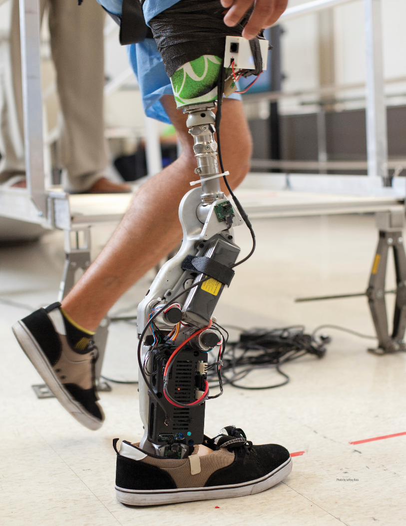
For three days, doctors tried to save his right leg.By the time their attempts had failed, Zac Vawter was already discussing revolutionary prosthetics allowing neural control by amputees.
The problem? No one had ever attempted targeted muscle reinnervation (TMR) — a series of nerve transfers that permit intuitive control of a prosthesis — in a lower- limb case study.
And so Vawter — a software engineer and father of two who crashed his motorcycle on his way home from work in rural Washington — became test pilot number one for the world’s first bionic leg. In 2012 he publicly revealed the work of doctors and engineers in a grand way, climbing the 2,109 steps to the top of Chicago’s Willis Tower. A triumphant photo shows him standing on the Skydeck’s Ledge, 103 floors above the city.
12 Northwestern University Office for Research
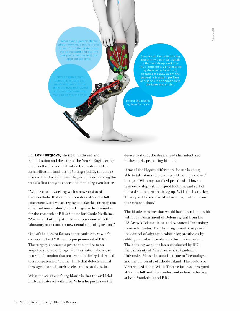
For Levi Hargrove, physical medicine and rehabilitation and director of the Neural Engineering for Prosthetics and Orthotics Laboratory at the Rehabilitation Institute of Chicago (RIC), the image marked the start of an even bigger journey: making the world’s first thought-controlled bionic leg even better.
“We have been working with a new version of the prosthetic that our collaborators at Vanderbilt constructed, and we are trying to make the entire system safer and more robust,” says Hargrove, lead scientist for the research at RIC’s Center for Bionic Medicine. “Zac — and other patients — often come into the laboratory to test out our new neural control algorithms.”
One of the biggest factors contributing to Vawter’s success is the TMR technique pioneered at RIC. The surgery connects a prosthetic device to an amputee’s nerve endings (see illustration above), so neural information that once went to the leg is directed to a computerized “bionic” limb that detects neural messages through surface electrodes on the skin.
What makes Vawter’s leg bionic is that the artificial limb can interact with him. When he pushes on the
device to stand, the device reads his intent and pushes back, propelling him up.
“One of the biggest differences for me is being able to take stairs step over step like everyone else,” he says. “With my standard prosthesis, I have to take every step with my good foot first and sort of lift or drag the prosthetic leg up. With the bionic leg, it’s simple: I take stairs like I used to, and can even take two at a time.”
The bionic leg’s creation would have been impossible without a Department of Defense grant from the US Army’s Telemedicine and Advanced Technology Research Center. That funding aimed to improve the control of advanced robotic leg prostheses by adding neural information to the control system. The ensuing work has been conducted by RIC, the University of New Brunswick, Vanderbilt University, Massachusetts Institute of Technology, and the University of Rhode Island. The prototype Vawter used in his Willis Tower climb was designed at Vanderbilt and then underwent extensive testing at both Vanderbilt and RIC.
telling the bionic leg how to move.
Sensors on the patient's leg detect tiny electrical signals in the hamstring, and then
RIC's intelligently engineered system instantaneously
decodes the movement the patient is trying to perform and sends the commands to
the knee and ankle...
Whenever a person thinks about moving, a neuro signal is sent from the brain down the spinal cord and out the peripheral nerves into the
appropriate limb.
Nerve signals from damaged muscle that previously went to the
ankle are now redirected into healthy hamstring
muscles above the knee.
Phot
o co
urte
sy o
f RIC
CenterPiece | Spring 2015 13
“This bionic leg features incredibly intelligent engineering,” says Hargrove. “It learns and performs activities unprecedented for any leg amputee, including seamless transitions between sitting, walking, ascending and descending stairs and ramps, and repositioning the leg while seated.”
In striving to make the leg even better, Hargrove has collaborated with fellow Northwestern faculty members, including Eric Perreault, biomedical engineering. He is also working with Arun Jayaraman, medical social sciences, to design a comprehensive at-home trial of the bionic system.
Part of Hargrove’s work with Perreault will clarify what happens when a patient wearing a bionic prosthetic slips. Their research is meant to help overcome a major challenge for many lower-limb prosthesis users: adapting to different terrains.
As able-bodied people walk across pavement, grass, sand, or ice, their legs automatically adapt to the changing surface with little cognitive input. “In contrast, the mechanical properties of most prosthetic legs are unable to adapt in an intelligent manner, and certainly not automatically,” says Perreault. “The ultimate goal of our project is to build lower-limb prostheses with this adaptive behavior.”
The starting point for Perreault is learning more about how the central nervous system fine-tunes the properties of an unimpaired limb as it adapts to different surfaces. Interestingly, the research group found a control gradient that changes almost continuously from the hip to the ankle. Initial results showed that the hip stiffens to prevent gross movements of the leg, while the ankle becomes compliant to maximize contact with the ground. These findings have led to innovative ideas that Perreault and Hargrove plan to pursue in the coming years.
— Roger Anderson, The Rehabilitation Institute of Chicago, and Northwestern University's Feinberg School of Medicine contributed to this story.
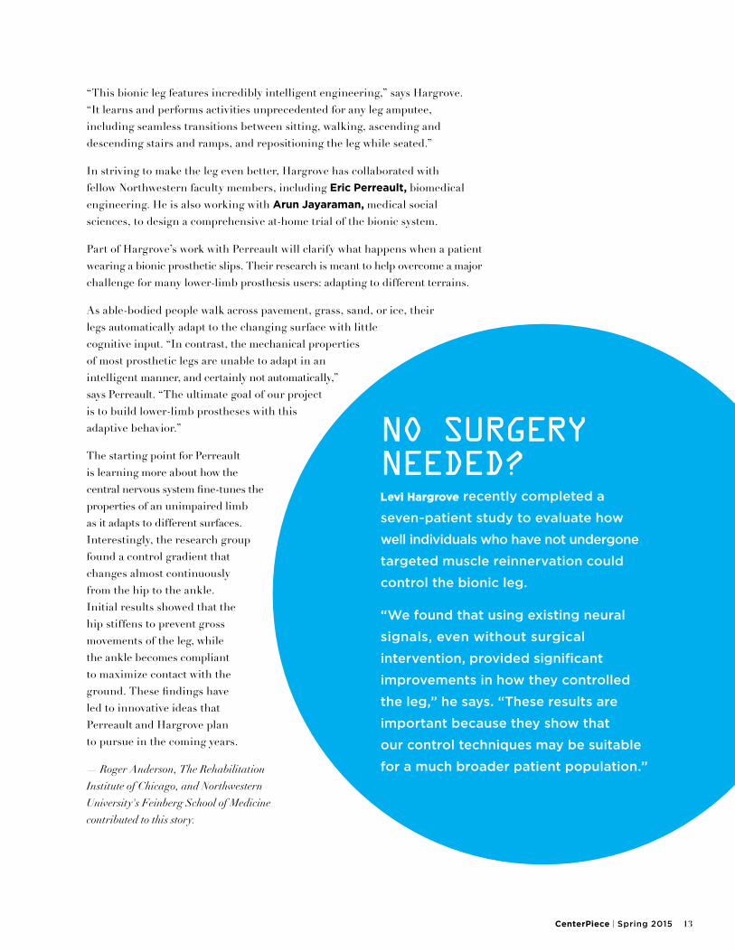
NO SURGERY NEEDED?Levi Hargrove recently completed a
seven-patient study to evaluate how
well individuals who have not undergone
targeted muscle reinnervation could
control the bionic leg.
“We found that using existing neural
signals, even without surgical
intervention, provided significant
improvements in how they controlled
the leg,” he says. “These results are
important because they show that
our control techniques may be suitable
for a much broader patient population.”
14 Northwestern University Office for Research
Call it a pool party of epic proportions. Thanks to the Obama administration’s recent Precision Medicine Initiative, researchers may soon have access to an unprecedented sampling of the human gene pool. In fact, the resulting dataset may include as many as 25 billion genes.
The president’s $215-million initiative, proposed in January, will combine the genetic details of 1 million volunteers with their medical and biographical records. The effort aims to give researchers additional tools to advance customizable healthcare.
At Northwestern University, research into precision medicine has been growing for more than a decade. It started with the founding of the Center for Genetic Medicine (CGM) in 2000, and continued two years later with the formation of the
CLOSING IN ON PRECISION MEDICINENORTHWESTERN SCIENTISTS ARE HELPING MAKE
CUSTOMIZED HEALTHCARE A REALITY
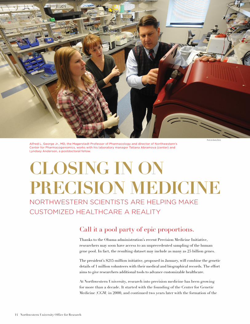
Alfred L. George Jr., MD, the Magerstadt Professor of Pharmacology and director of Northwestern’s Center for Pharmacogenomics, works with his laboratory manager Tatiana Abramova (center) and Lyndsey Anderson, a postdoctoral fellow.
Photo by Randy Belice
CenterPiece | Spring 2015 15
NUgene biobank, which has grown to include approximately 12,000 de-identified patient entries. CGM gained wider exposure in 2007 through its inclusion in the nationwide Electronic Medical Records and Genomics Network (eMERGE), a combination of eight sites — and hundreds of thousands of electronic health records — focused on using that clinical data for complex genomic analysis of disease susceptibility.
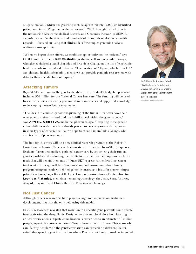
“When we began these efforts, we could see opportunity on the horizon,” says CGM founding director Rex Chisholm, medicine: cell and molecular biology, who also cochaired a panel that advised President Obama on the use of electronic health records in the federal initiative. “The creation of NUgene, which links DNA samples and health information, means we can provide genomic researchers with data for their specific lines of inquiry.”
Attacking TumorsBeyond $130 million for the genetic database, the president’s budgeted proposal includes $70 million for the National Cancer Institute. The funding will be used to scale up efforts to identify genomic drivers in cancer and apply that knowledge to developing more effective treatments.
“The idea is to conduct genome sequencing of the tumor — cancers have their own genetic makeup — and find the Achilles heel within the genetic code,” says Alfred L. George Jr., medicine: pharmacology. “Targeting these genetic vulnerabilities with drugs has already proven to be a very successful approach in some types of cancer, one that we hope to expand upon,” adds George, who also is chair of pharmacology.
The hub for this work will be a new clinical research program at the Robert H. Lurie Comprehensive Cancer of Northwestern University: Onco-SET (Sequence, Evaluate, Treat) personalizes patients’ cancer care by sequencing their tumors’ genetic profiles and evaluating the results to provide treatment options or clinical trials that will benefit them most. “Onco-SET represents the first time cancer treatment in Chicago will be offered in a comprehensive, multidisciplinary program using molecularly defined genomic targets as a basis for determining a patient’s options,” says Robert H. Lurie Comprehensive Cancer Center Director Leonidas Platanias, medicine: hematology/oncology, the Jesse, Sara, Andrew, Abigail, Benjamin and Elizabeth Lurie Professor of Oncology.
Not Just CancerAlthough cancer researchers have played a large role in precision medicine’s development, that isn’t the only field using this model.
In 2010 researchers revealed that variation in a specific gene prevents some people from activating the drug Plavix. Designed to prevent blood clots from forming in critical arteries, this antiplatelet medication is prescribed to an estimated 40 million people, especially those who have suffered a heart attack or stroke. Physicians who can identify people with the genetic variation can prescribe a different, better-suited therapeutic agent in situations where Plavix is not likely to work as intended.
Rex Chisholm, the Adam and Richard
T. Lind Professor of Medical Genetics,
associate vice president for research,
and vice dean for scientific affairs and
graduate educationPhoto courtesy of Feinberg School of Medicine
16 Northwestern University Office for Research
“Precision medicine isn’t just about giving the right drug to the right person at the right time. It’s also about avoiding potentially toxic reactions to drugs,” says George. “We are taking an experimental approach that will result in a new database linking genetic variation with the risk for drug interactions. Such a database would alert physicians about drug combinations that may cause problems in certain genetic backgrounds.” An ongoing eMERGE pharmacogenomics study is examining 86 genes identified as involved in different drug-gene interactions.
Clinical-decision support tools, which provide automatic alerts to physicians at the time of care, can be embedded in electronic health records to enable the implementation of personalized medicine. “We are learning how to better convey genetic information directly to physicians so that it is useful in patient care,” says Maureen Smith, NUgene director and co-principal investigator of the eMERGE study. The study also delivers information to patients about their genetic test results.
Technological RevolutionFifteen years ago, the first human genome was sequenced at a cost of nearly $3 billion. Today, sequencing can be completed for less than $2,000. Cheaper sequencing has brought more answers, though researchers are realizing just how different individuals truly are.
“We can interpret genetic differences and use them to predict risk, diagnose diseases, and better apply our drug therapies. In the future, we will be in the position to fix some of these genetic defects,” says CGM director Elizabeth M. McNally, medicine: cardiology and biochemistry and molecular genetics. “Genetic profiling of heart failure is showing us that heart failure is a lot like cancer, in that it is many diseases with different progression rates and risks of abnormal rhythms. We should modulate treatment based on that profile.”
McNally has seen the clinical applications firsthand. When a patient comes to her with a family history of heart disease, she often orders genetic testing.
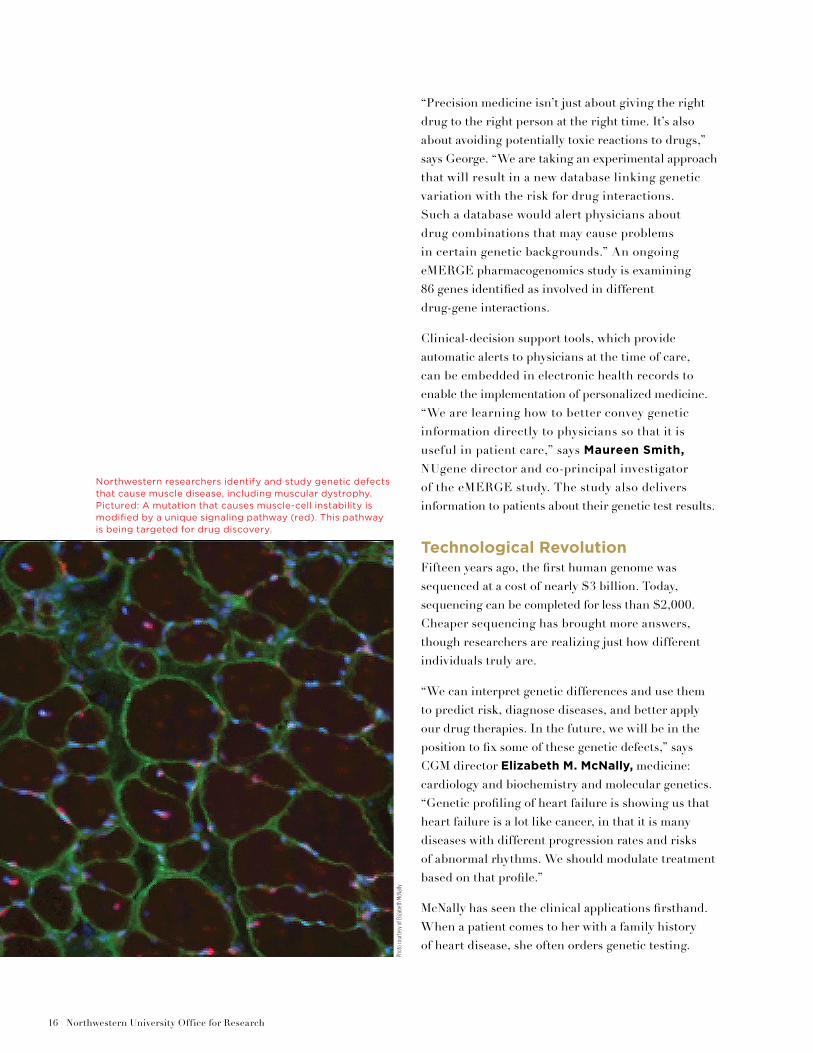
Northwestern researchers identify and study genetic defects that cause muscle disease, including muscular dystrophy. Pictured: A mutation that causes muscle-cell instability is modified by a unique signaling pathway (red). This pathway is being targeted for drug discovery.
Phot
o co
urte
sy o
f Eliz
abet
h Mc
Nally
CenterPiece | Spring 2015 17
If a mutation is discovered, McNally and genetic counselors discuss the nature and consequences of the potential disorder with the patient, explaining that an increased disease risk does not guarantee that everyone predisposed to the illness will develop it.
To illustrate her point, McNally cites a genetic mutation in a 30-year-old patient who has experienced a host of symptoms associated with cardiomyopathy, a disease putting him at increased risk of heart failure. Three of his aunts, however, have the same mutation and have been living normal lives for more than 70 years.
“I always say that it’s really important not to be a genetic determinist,” says McNally. “We still don’t know all of the answers why some people develop disease and some don’t. What we do know is that people who carry the genetic variants are at some increased risk. That means we can change our medical practice to increase their quality of life and better react to the disease.”
Screening at Birth?As sequencing costs continue to fall, some worry that genomic sequencing will proliferate, perhaps
with undesirable consequences. A person may be living with 100 genetic risks, but too few genetic counselors are available to decode and explain what each risk factor means.
Diminishing costs also raise the possibility that one day all babies could have their genome sequenced at birth. The results would give parents knowledge of the specific risks their children face but also create ethical questions about the responsibility to tell the child of those risks. “I don’t think we’re ready to screen every child just yet,” says Smith. “We still have a lot of work to do regarding when and how to tell people their results.”
As researchers and society further define precision medicine’s role, one goal is to solve these ethical and policy challenges along the way.
“Ultimately, we have an ability in many instances to move beyond treating a disease and, instead, to treat a specific person’s disease, taking into account that person’s unique characteristics,” says Chisholm. “Precision medicine is still in its early days, but it’s an exciting time to be a scientist working in this field.”
— Roger Anderson
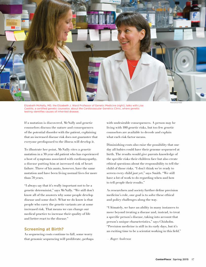
Elizabeth McNally, MD, the Elizabeth J. Ward Professor of Genetic Medicine (right), talks with Lisa Castillo, a certified genetic counselor, about the Cardiovascular Genetics Clinic, where genetic testing identifies causes of inherited disease.
Photo by Bruce Powell
18 Northwestern University Office for Research
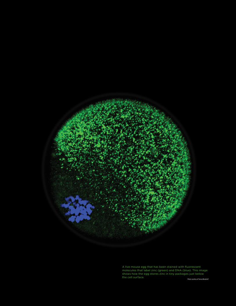
A live mouse egg that has been stained with fluorescent molecules that label zinc (green) and DNA (blue). This image shows how the egg stores zinc in tiny packages just below the cell surface.
Photo courtesy of Teresa Woodruff
CenterPiece | Spring 2015 19
Research collaboration gives birth to “brilliant”
reproductive insights. The advance could
signal a new conception of fertility treatment
Life begins with a tiny, but impressive, light show. After nearly six years,
a 12-person interdisciplinary team led by two Northwestern scientists has
discovered that sparks really do fly when sperm meet an egg.
When reproductive cells connect, vesicles — pouches near the oocyte’s
surface — release billions of zinc atoms in bursts called “zinc sparks.”
These sparks resemble fireworks exploding across the cell. What they
stimulate — the transformation from egg to embryo — is pretty explosive
too. The findings point to zinc’s vital, intricate part in promoting this
healthy development. The team used mouse cells for their research.
“There’s no precedent to study zinc at this point in the reproductive process,
but this fundamental discovery has opened a whole new field of signaling,”
says reproductive biologist Teresa Woodruff, medicine: obstetrics and
gynecology-fertility preservation, one of the project’s lead researchers.
She is the Thomas J. Watkins Memorial Professor in Obstetrics and Gynecology
and director of the Women’s Health Research Institute at the Feinberg School
of Medicine, where she studies hormones and female reproductive health.
IN QUEST FOR A GOOD EGG
SPARKS FLY
20 Northwestern University Office for Research
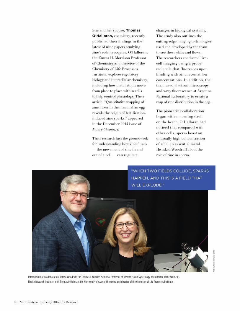
She and her spouse, Thomas
O'Halloran, chemistry, recently published their findings in the latest of nine papers studying zinc's role in oocytes. O’Halloran, the Emma H. Morrison Professor of Chemistry and director of the Chemistry of Life Processes Institute, explores regulatory biology and intercellular chemistry, including how metal atoms move from place to place within cells to help control physiology. Their article, “Quantitative mapping of zinc fluxes in the mammalian egg reveals the origin of fertilization-induced zinc sparks,” appeared in the December 2014 issue of Nature Chemistry.
Their research lays the groundwork for understanding how zinc fluxes — the movement of zinc in and out of a cell — can regulate
changes in biological systems. The study also outlines the cutting-edge imaging technologies used and developed by the team to see these ebbs and flows: The researchers conducted live- cell imaging using a probe molecule that fluoresces upon binding with zinc, even at low concentrations. In addition, the team used electron microscopy and x-ray fluorescence at Argonne National Laboratory to create a map of zinc distribution in the egg.
The pioneering collaboration began with a morning stroll on the beach. O’Halloran had noticed that compared with other cells, sperm boast an unusually high concentration of zinc, an essential metal. He asked Woodruff about the role of zinc in sperm.
“WHEN TWO FIELDS COLLIDE, SPARKS
HAPPEN, AND THIS IS A FIELD THAT
WILL EXPLODE.”
Interdisciplinary collaboration: Teresa Woodruff, the Thomas J. Watkins Memorial Professor of Obstetrics and Gynecology and director of the Women’s
Health Research Institute, with Thomas O’Halloran, the Morrison Professor of Chemistry and director of the Chemistry of Life Processes Institute
Phot
o co
urte
sy o
f Tho
mas
O’H
allo
ran
CenterPiece | Spring 2015 21
Woodruff wasn’t exactly impressed by the observation.
After all, sperm are the smallest cells in the body. Roughly 1,000 sperm are produced each heartbeat. Female reproductive cells, however, are a different matter. A woman is born with all her eggs, about a million, and some of those cells live for 40 or 50 years in the ovary. Over time, they are nurtured by “nurse cells” and become activated, with a limited number going through ovulation. Very little is known about this basic human process.
“Show me what zinc is doing in an egg, and then we can talk,” responded Woodruff.
Six years later, this challenge has led to innovations in biology, chemistry, and beyond. It also has offered new insight into zinc’s importance for fertilization and egg quality.
“Discoveries like this don’t happen every day,” says Woodruff. “When two fields collide, sparks happen, and this is a field that will explode.”
Revolutionary PotentialThis Northwestern collaboration already has led to a fresh understanding of fertility and infertility. The research findings hold promise for many possible applications, including those in the area of in vitro fertilization: The breakthrough could allow doctors to detect the most viable embryo to transfer, instead of transferring two or three. And since zinc sparks occur at fertilization, the ability to observe this phenomenon also would allow physicians to transfer that embryo sooner, instead of waiting days before the fertilized egg is inserted in the body, as is currently done.
O’Halloran says the research could hold practical applications in other areas of cell life and death as well, particularly in understanding cancer cells. Researchers have also noted zinc fluxes in conjunction with the release of insulin and in neurotransmitters associated with memory. Devising a way to follow the cell life cycle in real time could usher in a revolution.
“It’s so cool for us to have a test tube as large as the mammalian egg,” says O’Halloran. “It’s almost a perfect playground to understand metal trafficking and signaling.”
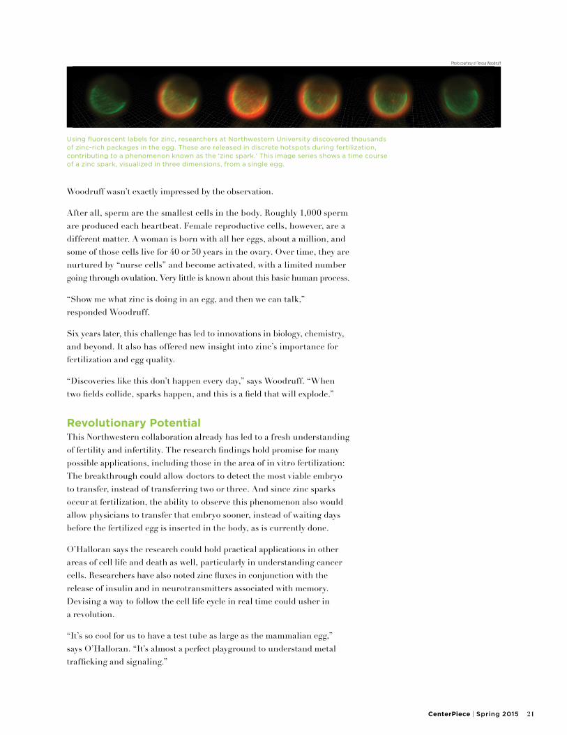
Using fluorescent labels for zinc, researchers at Northwestern University discovered thousands of zinc-rich packages in the egg. These are released in discrete hotspots during fertilization, contributing to a phenomenon known as the ‘zinc spark.’ This image series shows a time course of a zinc spark, visualized in three dimensions, from a single egg.
Photo courtesy of Teresa Woodruff
22 Northwestern University Office for Research
For two early papers, the senior researchers directed then-graduate student Alison Kim, who is today a postdoctoral fellow in obstetrics and gynecology. Kim first documented the movement of billions of zinc atoms during the short time of development. In less than one day, the cell experiences massive zinc flux, an increase of some 20 billion atoms
in the maturing oocyte. That’s more than half of the cell’s total zinc content. If the egg then meets a sperm cell, fertilization triggers the release of roughly 10 billion additional zinc atoms.
While these exciting discoveries opened the field of zinc signaling in oocyte maturation, it wasn’t until the most recent paper that the Northwestern team identified and documented exactly how that release happens.
“These new types of signaling are fundamental for new scientific insights that people will build on to understand the right time for a cell to live or die, particularly in development,” says O’Halloran.
To do so, the pair developed and applied a series of new imaging technologies.
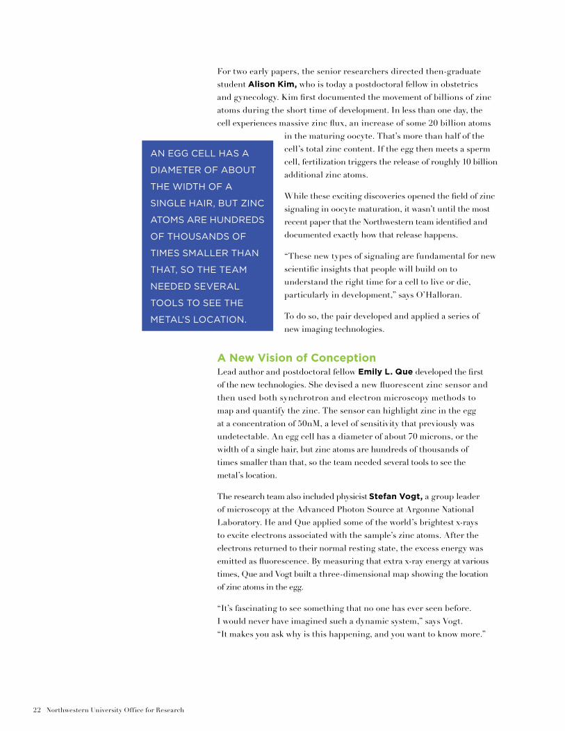
A New Vision of ConceptionLead author and postdoctoral fellow Emily L. Que developed the first of the new technologies. She devised a new fluorescent zinc sensor and then used both synchrotron and electron microscopy methods to map and quantify the zinc. The sensor can highlight zinc in the egg at a concentration of 50nM, a level of sensitivity that previously was undetectable. An egg cell has a diameter of about 70 microns, or the width of a single hair, but zinc atoms are hundreds of thousands of times smaller than that, so the team needed several tools to see the metal’s location.
The research team also included physicist Stefan Vogt, a group leader of microscopy at the Advanced Photon Source at Argonne National Laboratory. He and Que applied some of the world’s brightest x-rays to excite electrons associated with the sample’s zinc atoms. After the electrons returned to their normal resting state, the excess energy was emitted as fluorescence. By measuring that extra x-ray energy at various times, Que and Vogt built a three-dimensional map showing the location of zinc atoms in the egg.
“It’s fascinating to see something that no one has ever seen before. I would never have imagined such a dynamic system,” says Vogt. “It makes you ask why is this happening, and you want to know more.”
AN EGG CELL HAS A
DIAMETER OF ABOUT
THE WIDTH OF A
SINGLE HAIR, BUT ZINC
ATOMS ARE HUNDREDS
OF THOUSANDS OF
TIMES SMALLER THAN
THAT, SO THE TEAM
NEEDED SEVERAL
TOOLS TO SEE THE
METAL’S LOCATION.
CenterPiece | Spring 2015 23
Woodruff and O’Halloran also partnered with longtime friend and scholar Vinayak P. Dravid, materials science. The Abraham Harris Professor of Materials Science and Engineering at the McCormick School of Engineering and Applied Science, Dravid also directs the NUANCE Center, which houses advanced microscopes and develops innovative techniques for materials analysis. For the research team, Dravid designed a new technology that allows for imaging at an unprecedented level.
“It was a challenging problem, because there aren’t many techniques that can detect zinc at that level,” he says. This is because zinc is distributed narrowly at a nanoscale in fertilization, which makes it difficult to image. Relentless pursuit of a breakthrough enabled him to overcome the hurdle.
“Innovation takes a long time and requires collaboration,” says Dravid. “Coming together as we did to solve a problem is what stands out.”
After analyzing the problem, Dravid, a microscopist by trade, came up with a design that could detect zinc at the required sensitivity. Dravid’s design led to a new microscope, which was manufactured by Hitachi with grant funding from the W. M. Keck Foundation. The microscope works by harnessing the power of two detectors and zinc atoms to capture x-rays.
“Think of it like a fingerprint for zinc, because zinc has a very specific energy,” he explains.
The microscope has two detectors aimed at the sample. Once the electron beam penetrates the sample, it stimulates zinc-specific x-ray emission and allows researchers to map images of zinc location.
“It's like Sacagawea exploring a new land with Lewis and Clark," says Dravid, noting that the team redesigned the microscope’s capacity to accommodate multiple x-ray detectors. “The next steps are studying the phenomenon in human cells to understand where the zinc is coming from.”
Woodruff and O’Halloran are continuing their interdisciplinary study of zinc and expect to publish their latest research progress soon. Their ultimate goal is to help improve people’s reproductive lives.
“We’re trying to get at the molecular mechanism,” says Woodruff. “Gaining that control is going to have important implications for human health and for fertility and infertility.”
— Christina Cala
The National Institutes of Health, Eunice Kennedy Shriver National Institute of Child Health and Human Development (grant U54HD076188), and the W.M. Keck Foundation supported the research mentioned in this article.

24 Northwestern University Office for Research
Photo by Chris Campbell via Flickr
CenterPiece | Spring 2015 25
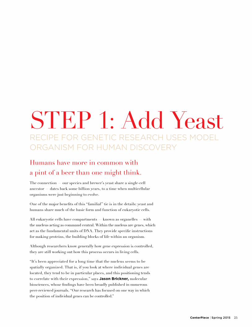
Humans have more in common with a pint of a beer than one might think.The connection — our species and brewer’s yeast share a single-cell ancestor — dates back some billion years, to a time when multicellular organisms were just beginning to evolve.
One of the major benefits of this “familial” tie is in the details: yeast and humans share much of the basic form and function of eukaryotic cells.
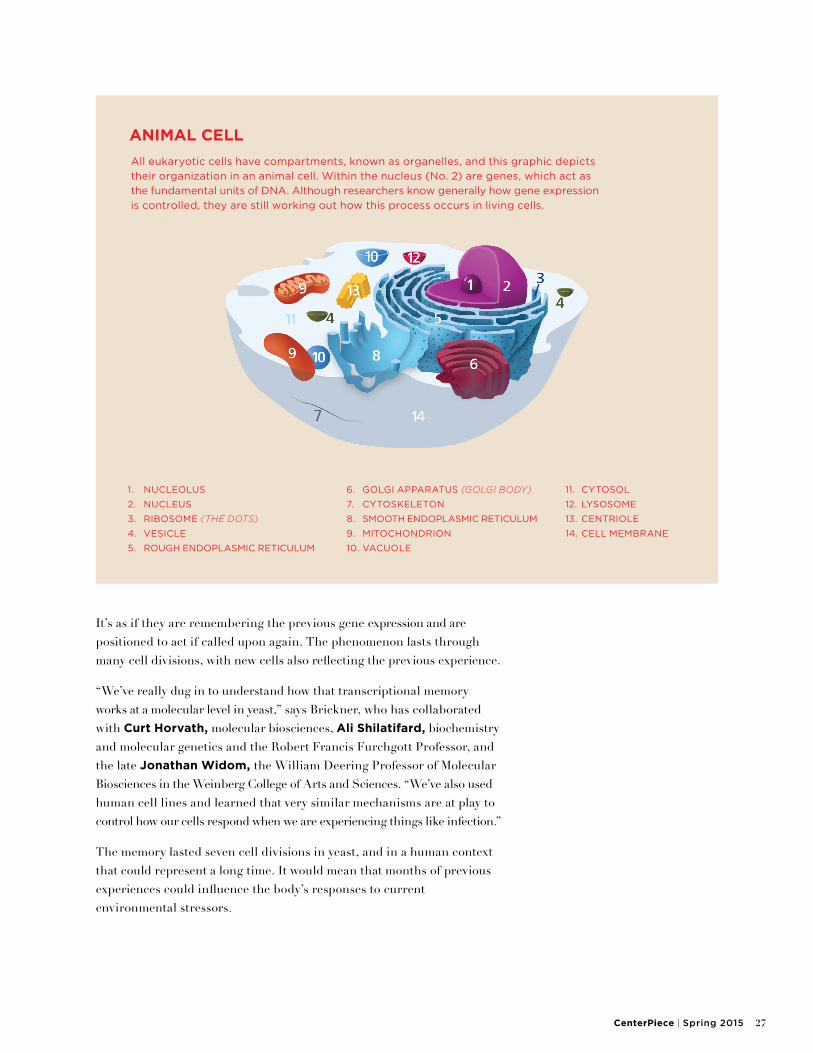
All eukaryotic cells have compartments — known as organelles — with the nucleus acting as command central. Within the nucleus are genes, which act as the fundamental units of DNA. They provide specific instructions for making proteins, the building blocks of life within an organism.
Although researchers know generally how gene expression is controlled, they are still working out how this process occurs in living cells.
“It’s been appreciated for a long time that the nucleus seems to be spatially organized. That is, if you look at where individual genes are located, they tend to be in particular places, and this positioning tends to correlate with their expression,” says Jason Brickner, molecular biosciences, whose findings have been broadly published in numerous peer-reviewed journals. “Our research has focused on one way in which the position of individual genes can be controlled.”
STEP 1: Add YeastRECIPE FOR GENETIC RESEARCH USES MODEL ORGANISM FOR HUMAN DISCOVERY
26 Northwestern University Office for Research
As a PhD student at Stanford, and then as a postdoctoral fellow at UCSF, Brickner began thinking about this spatial organization from the cell’s point of view. How are proteins specifically targeted to particular organelles? How is the size and function of an organelle controlled by the moment-to-moment needs of the cell itself?
To answer these questions, he first sought to understand how certain yeast genes, those that control an organelle’s exterior barrier, are regulated when the organelle’s functioning is insufficient to meet the cell’s needs. “As I was working out the mechanism, I made an unanticipated discovery: One of these genes, when it was turned on, changed its position within the nucleus,” Brickner recalls. “Although this didn’t really get at the question I thought I was asking, it revealed a new and totally fascinating way in which cells change the spatial organization of the genome to regulate the expression of genes.”
Serendipity can be a scientist’s best friend. Brickner began referring to the small DNA elements that define a gene’s positioning within the nucleus as DNA “zip codes.” His findings were first published in Nature Cell Biology in 2010.
A subsequent discovery revealed that certain genes “remember” where they are and how they got there. “In trying to understand how genes move around in the nucleus, we found that some genes, when you turn them on, go to the edge of the nucleus,” says Brickner. “Most genes, when you turn them off, go right back to where they started. But a subset of genes do something very interesting.” Those genes go to the edge of the nucleus when turned on and stay at the periphery even when turned off.
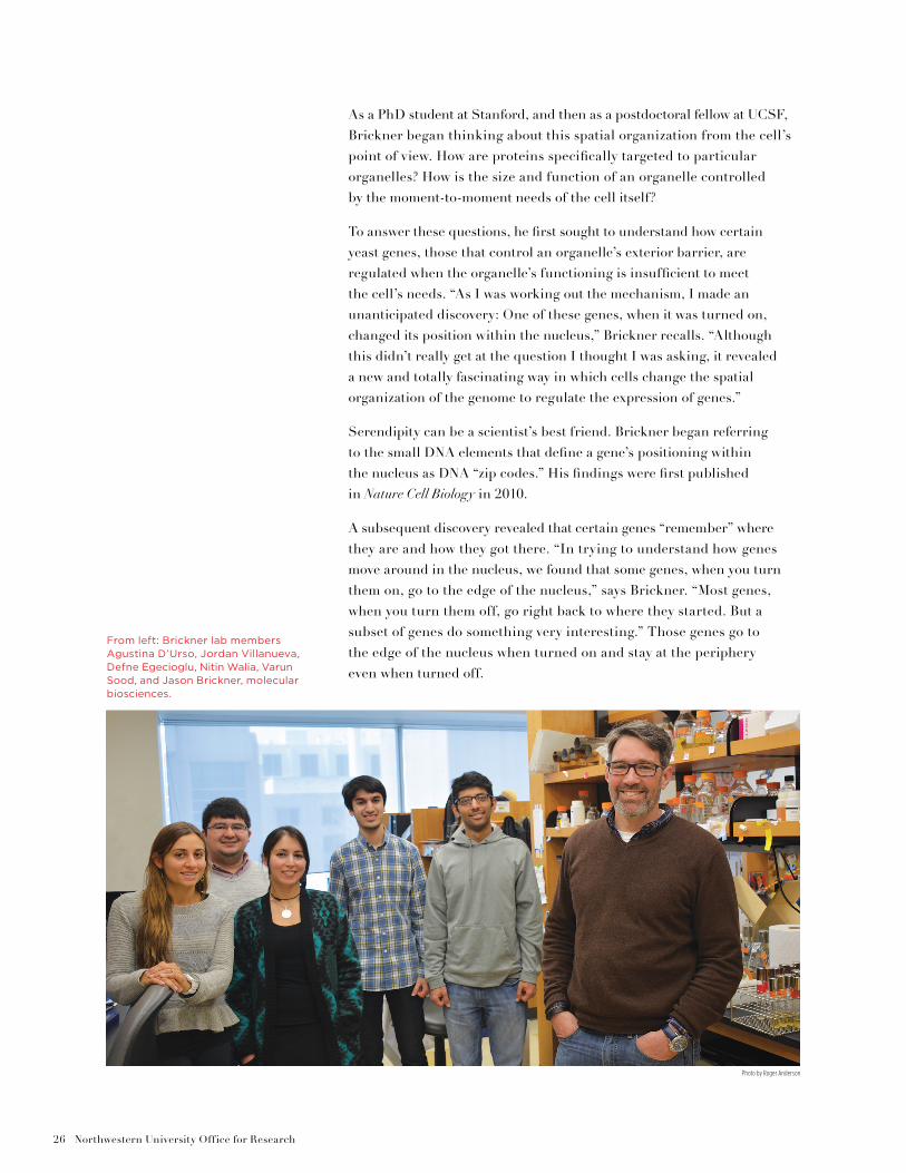
From left: Brickner lab members Agustina D’Urso, Jordan Villanueva, Defne Egecioglu, Nitin Walia, Varun Sood, and Jason Brickner, molecular biosciences.
Photo by Roger Anderson
CenterPiece | Spring 2015 27
It’s as if they are remembering the previous gene expression and are positioned to act if called upon again. The phenomenon lasts through many cell divisions, with new cells also reflecting the previous experience.
“We’ve really dug in to understand how that transcriptional memory works at a molecular level in yeast,” says Brickner, who has collaborated with Curt Horvath, molecular biosciences, Ali Shilatifard, biochemistry and molecular genetics and the Robert Francis Furchgott Professor, and the late Jonathan Widom, the William Deering Professor of Molecular Biosciences in the Weinberg College of Arts and Sciences. “We’ve also used human cell lines and learned that very similar mechanisms are at play to control how our cells respond when we are experiencing things like infection.”
The memory lasted seven cell divisions in yeast, and in a human context that could represent a long time. It would mean that months of previous experiences could influence the body’s responses to current environmental stressors.
1. NUCLEOLUS
2. NUCLEUS
3. RIBOSOME (THE DOTS)
4. VESICLE
5. ROUGH ENDOPLASMIC RETICULUM
6. GOLGI APPARATUS (GOLGI BODY)
7. CYTOSKELETON
8. SMOOTH ENDOPLASMIC RETICULUM
9. MITOCHONDRION
10. VACUOLE
11. CYTOSOL
12. LYSOSOME
13. CENTRIOLE
14. CELL MEMBRANE
ANIMAL CELL
All eukaryotic cells have compartments, known as organelles, and this graphic depicts
their organization in an animal cell. Within the nucleus (No. 2) are genes, which act as
the fundamental units of DNA. Although researchers know generally how gene expression
is controlled, they are still working out how this process occurs in living cells.
28 Northwestern University Office for Research
Brickner’s observations appear to show that transcriptional memory doesn’t just change the speed at which genes get turned on in the face of stressors but also enacts a qualitative change in the cells’ response. In biology, it's thought that when stimulus A is sensed by the cell, the cell comes up with response B. Brickner found that stimulus A may initially produce that reaction, but if one discontinues stimulus A and reintroduces it again later, certain cells actually give response C, a similar but different reaction. The finding implies that the cell has actually incorporated the initial memory into its biology.
“Presumably, this response is adaptive to provide the body with a more optimal response, but when considering the human system, you can imagine it could produce problems,” Brickner points out. “For instance, responses that elicit inflammation are essential, but inflammation is also something that can become pathological. In fact, it is connected to most diseases.”
Researchers know that the spatial arrangement of genes within the nucleus can differ in disease states. Research in 2009 by a team at the National Cancer Institute revealed that cancer cells might have disease-specific gene arrangements, raising the possibility that such patterns
WHAT PERCENT OF THEIR GENES MATCH YOURS?
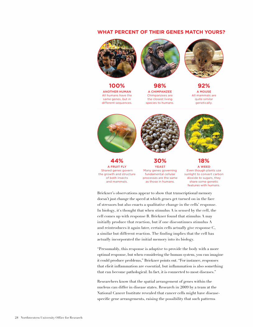
100%ANOTHER HUMANAll humans have the same genes, but in
different sequences.
98%A CHIMPANZEEChimpanzees are the closest living
species to humans.
92%A MOUSE
All mammals are quite similar genetically.
44%A FRUIT FLY
Shared genes govern the growth and structure
of both insects and mammals.
30%YEAST
Many genes governing fundamental cellular
processes are the same as those in humans.
18%A WEED
Even though plants use sunlight to convert carbon
dioxide to sugars, they share some genetic
features with humans.
Why Yeast?Yeast has become a model organism in
the lab for several reasons. Chief among
them are its cell structure, genetic
makeup, capacity for manipulation,
and reproductive rate.
Since its genome was mapped — the first
complete DNA sequencing of a eukaryotic
cell — nearly 20 years ago, yeast has
remained at the forefront of genetics
research. Today Jason Brickner, molecular
biosciences, has access to strains where
every gene in the genome has been tagged
with different fluorescent markers and
collections where every gene has been
knocked out.
It’s estimated that 30 percent of yeast
genes are mammalian homologs, meaning
they have near-equivalent mammalian
counterparts. And yeast cells divide every
90 minutes, rather than every day or week
as with human cells, meaning researchers
can make discoveries at a faster pace.
Brickner’s lab frequently uses cutting-
edge Center for Advanced Microscopy
instrumentation that allows his team
to look at static or even moving images
of the studies as they occur.
“We have the ability to see what is
happening in real time. And because
we can control the growth conditions,
we can manipulate the organism in
various ways and see it respond to
environmental challenges very quickly.
These cells can’t swim or walk away,
so when they see a change in their
environment they have to be able
to immediately adapt,” says Brickner,
snapping his fingers for emphasis.
One disadvantage of using yeast in
experiments is that the microbe leads
an independent existence, whereas a
human cell is invariably part of a system,
such as an organ — so their respective
cells interact and behave in different
ways. Yeast’s simplicity, then, makes
it a less than perfect model.
“But the nice thing is that most of what
we’ve found with yeast turns out to also
be true in humans,” says Brickner. “It’s
why this very simple model organism
has continued to lead the way in the
field of genetics.”
could be used as a diagnostic strategy. Unfortunately, researchers have been unable to verify whether the changes in gene location are causing the problems or merely reflecting them.
“The bottom line is that we can’t do much to intervene or even diagnose the molecular basis of disease without first understanding the molecular basis of life,” says Brickner, whose lab is working to understand both the molecular mechanism of transcriptional memory and the importance of this mechanism in yeast and human physiology. “Our research provides a starting point for much broader investigations.”
— Roger Anderson
30 Northwestern University Office for Research
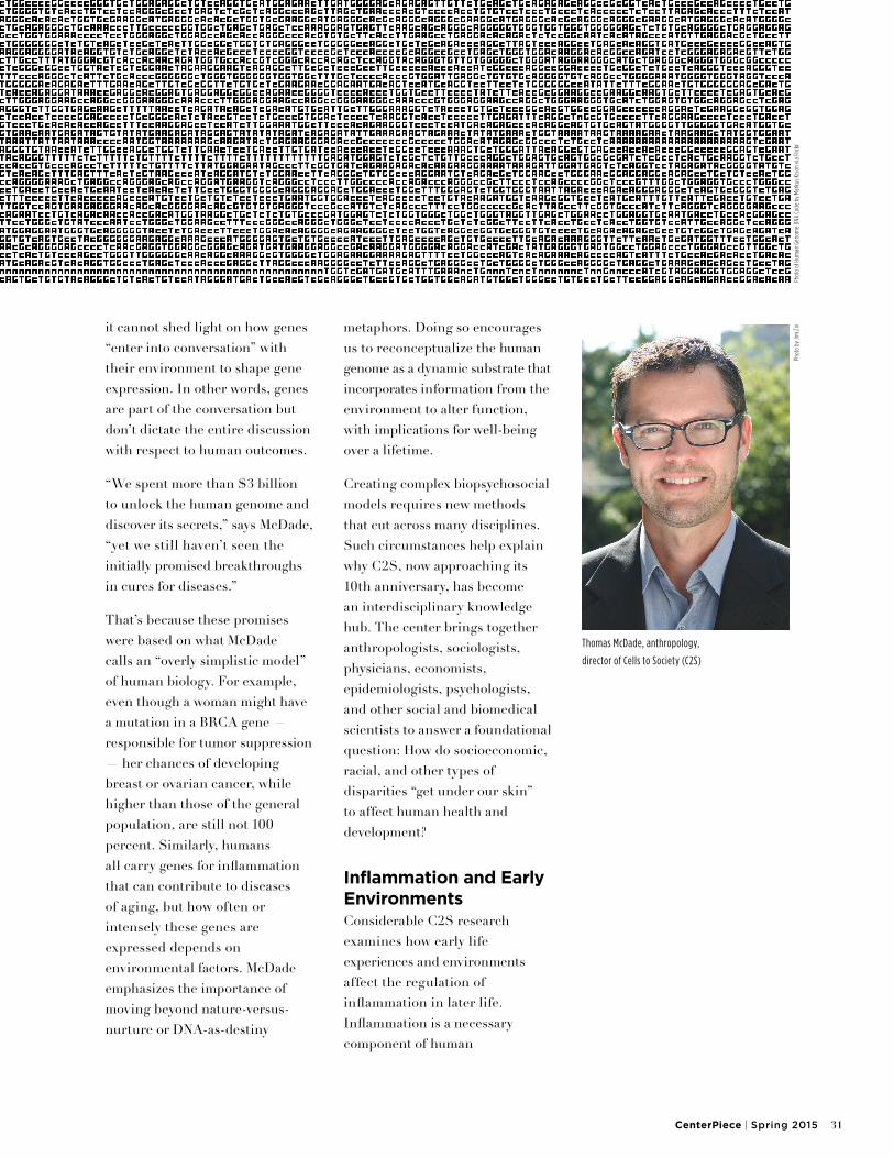
Interdisciplinary scholars at the Institute for Policy Research are decoding complex social impacts on genes, exploring dynamic new models of human developmentWhen the Human Genome Project got underway in 1990, experts believed that people carried an estimated 100,000 or more genes. Since then, the overall count has been revised downward to fewer than 25,000 genes, or about 7,000 fewer than a fleshy tomato. Does this mean that a human being is less complex than a salad ingredient?
No, says Thomas McDade, anthropology, who directs Cells to Society (C2S): The Center for Social Disparities and Health at the Institute for Policy Research (IPR), where he is a fellow. Still, the comparison indicates the subtle complexity of gene-environment interplay.
In McDade’s view, one of the most significant achievements of human genome sequencing, completed in 2003, was pinning down the number of human genes. Gene mapping, however, only points to an organism’s inherited information;
Biologyand Beyond
CenterPiece | Spring 2015 31
it cannot shed light on how genes “enter into conversation” with their environment to shape gene expression. In other words, genes are part of the conversation but don’t dictate the entire discussion with respect to human outcomes.
“We spent more than $3 billion to unlock the human genome and discover its secrets,” says McDade, “yet we still haven’t seen the initially promised breakthroughs in cures for diseases.”
That’s because these promises were based on what McDade calls an “overly simplistic model” of human biology. For example, even though a woman might have a mutation in a BRCA gene — responsible for tumor suppression — her chances of developing breast or ovarian cancer, while higher than those of the general population, are still not 100 percent. Similarly, humans all carry genes for inflammation that can contribute to diseases of aging, but how often or intensely these genes are expressed depends on environmental factors. McDade emphasizes the importance of moving beyond nature-versus-nurture or DNA-as-destiny
metaphors. Doing so encourages us to reconceptualize the human genome as a dynamic substrate that incorporates information from the environment to alter function, with implications for well-being over a lifetime.
Creating complex biopsychosocial models requires new methods that cut across many disciplines. Such circumstances help explain why C2S, now approaching its 10th anniversary, has become an interdisciplinary knowledge hub. The center brings together anthropologists, sociologists, physicians, economists, epidemiologists, psychologists, and other social and biomedical scientists to answer a foundational question: How do socioeconomic, racial, and other types of disparities “get under our skin” to affect human health and development?
Inflammation and Early EnvironmentsConsiderable C2S research examines how early life experiences and environments affect the regulation of inflammation in later life. Inflammation is a necessary component of human
Thomas McDade, anthropology,
director of Cells to Society (C2S)
Phot
o by
Jim
Ziv
Phot
o of
Hum
an G
enom
e DN
A Co
de b
y Ma
rkus
Kiso
n vi
a Fl
ickr
32 Northwestern University Office for Research
development, but a tricky one. Too little inflammation at certain stages can cause harm. So can too much. Inflammation is a culprit implicated in a variety of illnesses, including cardiovascular disease (CVD), type 2 diabetes, and psychosocial conditions, such as depression and anxiety.
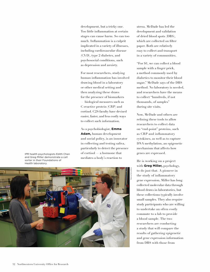
For most researchers, studying human inflammation has involved drawing blood in a laboratory or other medical setting and then analyzing these draws for the presence of biomarkers — biological measures such as C-reactive protein (CRP) and cortisol. C2S faculty have devised easier, faster, and less costly ways to collect such information.
As a psychobiologist, Emma
Adam, human development and social policy, is an innovator in collecting and testing saliva, particularly to detect the presence of cortisol — a hormone that mediates a body’s reaction to
stress. McDade has led the development and validation of dried blood spots (DBS), which are collected on filter paper. Both are relatively easy to collect and transport in a variety of communities.
“For $1, we can collect a blood sample with a finger prick, a method commonly used by diabetics to monitor their blood sugar,” McDade says of the DBS method. No laboratory is needed, and researchers have the means to collect “hundreds, if not thousands, of samples” during site visits.
Now, McDade and others are refining these tools to allow researchers to collect data on “end-point” proteins, such as CRP and inflammatory cytokines, as well as to capture DNA methylation, an epigenetic mechanism that affects how genes are expressed.
He is working on a project with Greg Miller, psychology, to do just that. A pioneer in the study of inflammatory gene expression, Miller has long collected molecular data through blood draws in laboratories, but these collections typically involve small samples. They also require study participants who are willing to undertake an often costly commute to a lab to provide a blood sample. The two researchers are conducting a study that will compare the results of gathering epigenetic and gene expression information from DBS with those from
IPR health psychologists Edith Chen and Greg Miller demonstrate a cell sorter in their Foundations of Health laboratory.
Phot
o by
Pat
ricia
Ree
se
CenterPiece | Spring 2015 33
a traditional venipuncture blood draw. If the DBS and venipuncture results prove consistent, this could lead to many other applications in a range of research settings. The project has received funding from the National Science Foundation and the National Institutes of Health.
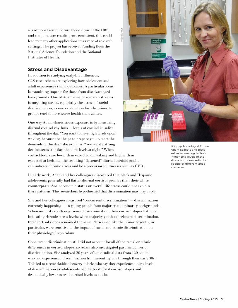
Stress and DisadvantageIn addition to studying early-life influences, C2S researchers are exploring how adolescent and adult experiences shape outcomes. A particular focus is examining impacts for those from disadvantaged backgrounds. One of Adam’s major research streams is targeting stress, especially the stress of racial discrimination, as one explanation for why minority groups tend to have worse health than whites.
One way Adam charts stress exposure is by measuring diurnal cortisol rhythms — levels of cortisol in saliva throughout the day. “You want to have high levels upon waking, because that helps to prepare you to meet the demands of the day,” she explains. “You want a strong decline across the day, then low levels at night.” When cortisol levels are lower than expected on waking and higher than expected at bedtime, the resulting “flattened” diurnal cortisol profile can indicate chronic stress and be a precursor to illnesses such as CVD.
In early work, Adam and her colleagues discovered that black and Hispanic adolescents generally had flatter diurnal cortisol profiles than their white counterparts. Socioeconomic status or overall life stress could not explain these patterns. The researchers hypothesized that discrimination may play a role.
She and her colleagues measured “concurrent discrimination” — discrimination currently happening — in young people from majority and minority backgrounds. When minority youth experienced discrimination, their cortisol slopes flattened, indicating chronic stress levels; when majority youth experienced discrimination, their cortisol slopes remained the same. “It seemed like the minority youth, in particular, were sensitive to the impact of racial and ethnic discrimination on their physiology,” says Adam.
Concurrent discrimination still did not account for all of the racial or ethnic differences in cortisol slopes, so Adam also investigated past incidences of discrimination. She analyzed 20 years of longitudinal data from 120 adults who had experienced discrimination from seventh grade through their early 30s. This led to a remarkable discovery: Blacks who say they experienced high levels of discrimination as adolescents had flatter diurnal cortisol slopes and dramatically lower overall cortisol levels as adults.
IPR psychobiologist Emma Adam collects and tests saliva, examining factors influencing levels of the stress hormone cortisol in people of different ages and races.
Phot
o co
urte
sy o
f SES
P
34 Northwestern University Office for Research
This finding reveals that “subtle discriminatory acts matter for your biology, and also for your health,” says Adam. “It’s a costly societal problem that really needs to be addressed.”
She and her colleagues continue to explore the topic. One of their recent research projects is the first to examine racial and ethnic differences in cortisol rhythms longitudinally. Their five-year Chicago-based study of 229 white, black, and Hispanic adults, 50 and older, reveals that older black adults, like their younger counterparts, were more sensitive to stress than whites.
“We were able to watch the flattening of the slopes unfold in response to chronic stress,” says Adam, “revealing a potential pathway for flattening of these slopes in racial or ethnic minorities.”
Disparities and Adaptations across Generations Another area where C2S faculty have broken ground is in using biomarkers to trace how health disparities can transfer from one generation to the next.
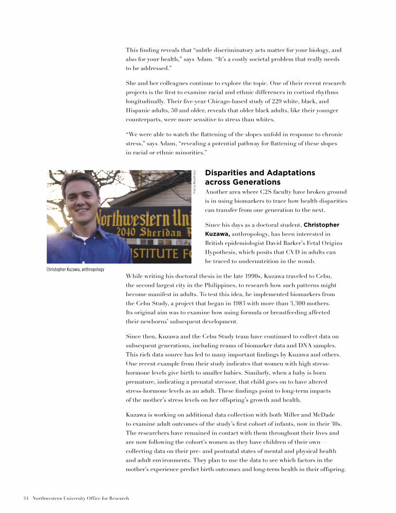
Since his days as a doctoral student, Christopher
Kuzawa, anthropology, has been interested in British epidemiologist David Barker’s Fetal Origins Hypothesis, which posits that CVD in adults can be traced to undernutrition in the womb.
While writing his doctoral thesis in the late 1990s, Kuzawa traveled to Cebu, the second largest city in the Philippines, to research how such patterns might become manifest in adults. To test this idea, he implemented biomarkers from the Cebu Study, a project that began in 1983 with more than 3,300 mothers. Its original aim was to examine how using formula or breastfeeding affected their newborns’ subsequent development.
Since then, Kuzawa and the Cebu Study team have continued to collect data on subsequent generations, including reams of biomarker data and DNA samples. This rich data source has led to many important findings by Kuzawa and others. One recent example from their study indicates that women with high stress-hormone levels give birth to smaller babies. Similarly, when a baby is born premature, indicating a prenatal stressor, that child goes on to have altered stress-hormone levels as an adult. These findings point to long-term impacts of the mother’s stress levels on her offspring’s growth and health.
Kuzawa is working on additional data collection with both Miller and McDade to examine adult outcomes of the study’s first cohort of infants, now in their 30s. The researchers have remained in contact with them throughout their lives and are now following the cohort’s women as they have children of their own — collecting data on their pre- and postnatal states of mental and physical health and adult environments. They plan to use the data to see which factors in the mother’s experience predict birth outcomes and long-term health in their offspring.
Christopher Kuzawa, anthropology
Phot
o by
Mer
edith
Bus
e
A second focus of Kuzawa’s research aims to clarify how humans evolved as a species. For example, his team has explored why fathers’ testosterone levels might drop after the birth of a child as an evolutionary response to male caregiving. More recently, he has documented the energetic costs of human brain development and how this varies across the life cycle.
“Humans managed to pull off an interesting trick,” says Kuzawa. “Although we evolved a large and energetically costly brain, our body’s resting energy expenditure is the same as predicted for a mammal of our size. How was this achieved?”
As part of an NSF-funded study, Kuzawa and his colleagues used PET and MRI brain scan data to estimate how much energy the brain consumes across different developmental stages. Their results explain why, after a certain age, it becomes difficult to guess the age of a toddler or young child by physical size. By childhood, the brain has become “an energy monster,” nearly halting body growth and using twice as much energy as an adult’s brain.
“Our findings suggest that our bodies can’t afford to grow faster during the toddler and childhood years because a huge quantity of resources are required to fuel the developing human brain,” says Kuzawa, indicating why adequate nutrition is critical in childhood. “As humans we have so much to learn, and that learning requires a complex and energy-hungry brain.”
Biology and Policy RelevanceLooking over the last decade of C2S’s growth and development, McDade says it was crucial for the center to be situated within IPR. Yet at the beginning, when faculty discussed launching the center, they raised a lot of questions about how it would fit within IPR’s framework.
“We were at the very early stages of investigation about how social environments and social policy may matter to biology and health,” McDade recalls. There were no easy answers for how C2S was going to realize its aim of policy-relevant research. Yet he says C2S faculty also recognized that even if they were not doing policy research immediately, they would get there a lot faster by being part of IPR and collaborating with colleagues who do policy work.
“And we have gotten there,” he says. “It’s a unique intellectual moment, when the population sciences can make important contributions to our understanding of human biology and its complex, multilevel determinants.”
— Patricia Reese and Sara Kupper, with additional reporting by Erin Spain and Hilary Hurd Anyaso.
“ALTHOUGH WE EVOLVED A LARGE
AND ENERGETICALLY COSTLY BRAIN,
OUR BODY’S RESTING ENERGY
EXPENDITURE IS THE SAME AS
PREDICTED FOR A MAMMAL OF OUR
SIZE. HOW WAS THIS ACHIEVED?”
36 Northwestern University Office for Research
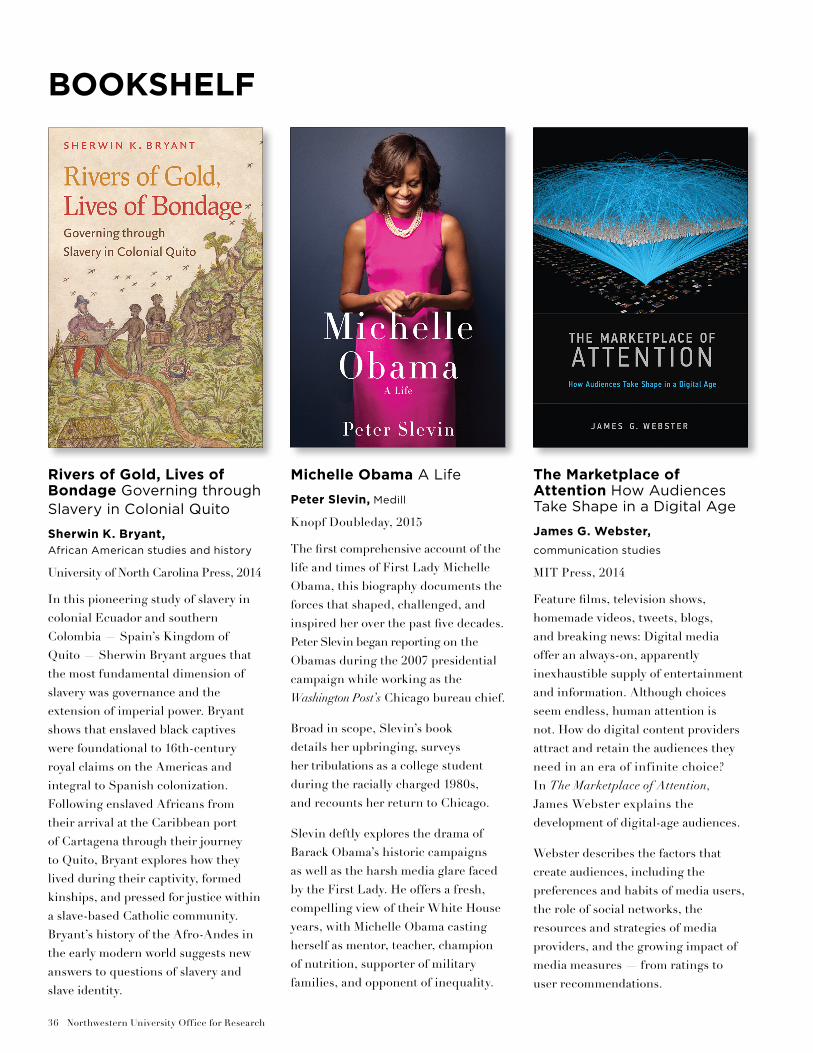
Rivers of Gold, Lives of Bondage Governing through Slavery in Colonial Quito
Sherwin K. Bryant, African American studies and history
University of North Carolina Press, 2014
In this pioneering study of slavery in colonial Ecuador and southern Colombia — Spain’s Kingdom of Quito — Sherwin Bryant argues that the most fundamental dimension of slavery was governance and the extension of imperial power. Bryant shows that enslaved black captives were foundational to 16th-century royal claims on the Americas and integral to Spanish colonization. Following enslaved Africans from their arrival at the Caribbean port of Cartagena through their journey to Quito, Bryant explores how they lived during their captivity, formed kinships, and pressed for justice within a slave-based Catholic community. Bryant’s history of the Afro-Andes in the early modern world suggests new answers to questions of slavery and slave identity.
Michelle Obama A Life
Peter Slevin, Medill
Knopf Doubleday, 2015
The first comprehensive account of the life and times of First Lady Michelle Obama, this biography documents the forces that shaped, challenged, and inspired her over the past five decades. Peter Slevin began reporting on the Obamas during the 2007 presidential campaign while working as the Washington Post’s Chicago bureau chief.
Broad in scope, Slevin’s book details her upbringing, surveys her tribulations as a college student during the racially charged 1980s, and recounts her return to Chicago.
Slevin deftly explores the drama of Barack Obama’s historic campaigns as well as the harsh media glare faced by the First Lady. He offers a fresh, compelling view of their White House years, with Michelle Obama casting herself as mentor, teacher, champion of nutrition, supporter of military families, and opponent of inequality.
The Marketplace of Attention How Audiences Take Shape in a Digital Age
James G. Webster,
communication studies
MIT Press, 2014
Feature films, television shows, homemade videos, tweets, blogs, and breaking news: Digital media offer an always-on, apparently inexhaustible supply of entertainment and information. Although choices seem endless, human attention is not. How do digital content providers attract and retain the audiences they need in an era of infinite choice? In The Marketplace of Attention, James Webster explains the development of digital-age audiences.
Webster describes the factors that create audiences, including the preferences and habits of media users, the role of social networks, the resources and strategies of media providers, and the growing impact of media measures — from ratings to user recommendations.
BOOKSHELF
CenterPiece | Spring 2015 37
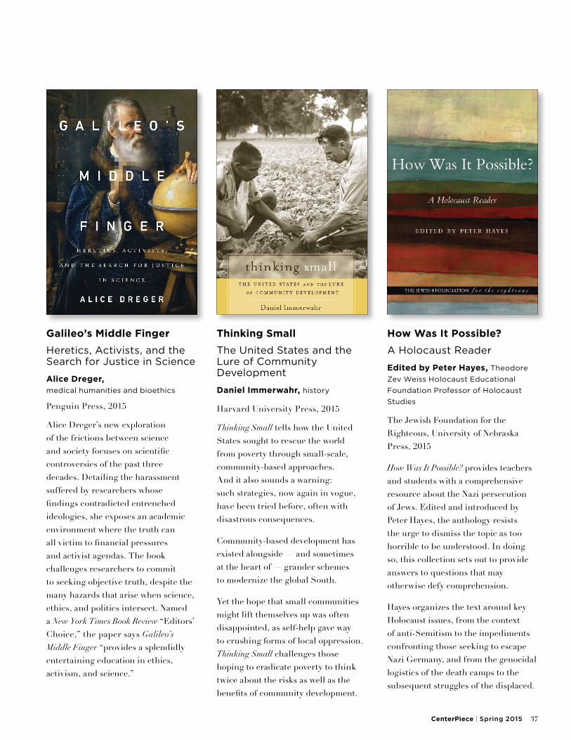
Galileo’s Middle Finger
Heretics, Activists, and the Search for Justice in Science
Alice Dreger, medical humanities and bioethics
Penguin Press, 2015
Alice Dreger’s new exploration of the frictions between science and society focuses on scientific controversies of the past three decades. Detailing the harassment suffered by researchers whose findings contradicted entrenched ideologies, she exposes an academic environment where the truth can all victim to financial pressures and activist agendas. The book challenges researchers to commit to seeking objective truth, despite the many hazards that arise when science, ethics, and politics intersect. Named a New York Times Book Review “Editors’ Choice,” the paper says Galileo’s Middle Finger “provides a splendidly entertaining education in ethics, activism, and science.”
Thinking Small
The United States and the Lure of Community Development
Daniel Immerwahr, history
Harvard University Press, 2015
Thinking Small tells how the United States sought to rescue the world from poverty through small-scale, community-based approaches. And it also sounds a warning: such strategies, now again in vogue, have been tried before, often with disastrous consequences.
Community-based development has existed alongside — and sometimes at the heart of — grander schemes to modernize the global South.
Yet the hope that small communities might lift themselves up was often disappointed, as self-help gave way to crushing forms of local oppression. Thinking Small challenges those hoping to eradicate poverty to think twice about the risks as well as the benefits of community development.
How Was It Possible?
A Holocaust Reader
Edited by Peter Hayes, Theodore
Zev Weiss Holocaust Educational
Foundation Professor of Holocaust
Studies
The Jewish Foundation for the Righteous, University of Nebraska Press, 2015
How Was It Possible? provides teachers and students with a comprehensive resource about the Nazi persecution of Jews. Edited and introduced by Peter Hayes, the anthology resists the urge to dismiss the topic as too horrible to be understood. In doing so, this collection sets out to provide answers to questions that may otherwise defy comprehension.
Hayes organizes the text around key Holocaust issues, from the context of anti-Semitism to the impediments confronting those seeking to escape Nazi Germany, and from the genocidal logistics of the death camps to the subsequent struggles of the displaced.
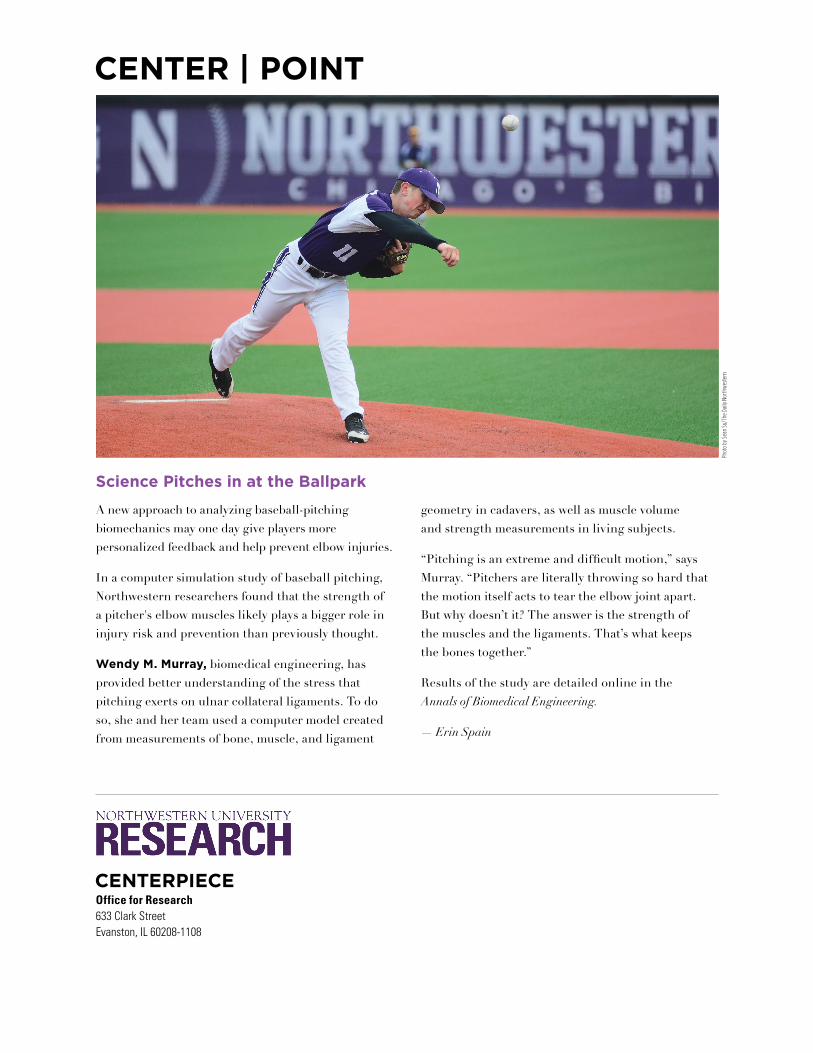
CENTER | POINT
Office for Research633 Clark StreetEvanston, IL 60208-1108
CENTERPIECE
Science Pitches in at the Ballpark
Phot
o by
Sea
n Su
/The
Dai
ly N
orth
wes
tern
A new approach to analyzing baseball-pitching biomechanics may one day give players more personalized feedback and help prevent elbow injuries.
In a computer simulation study of baseball pitching, Northwestern researchers found that the strength of a pitcher's elbow muscles likely plays a bigger role in injury risk and prevention than previously thought.
Wendy M. Murray, biomedical engineering, has provided better understanding of the stress that pitching exerts on ulnar collateral ligaments. To do so, she and her team used a computer model created from measurements of bone, muscle, and ligament
geometry in cadavers, as well as muscle volume and strength measurements in living subjects.
“Pitching is an extreme and difficult motion,” says Murray. “Pitchers are literally throwing so hard that the motion itself acts to tear the elbow joint apart. But why doesn’t it? The answer is the strength of the muscles and the ligaments. That’s what keeps the bones together.”
Results of the study are detailed online in the Annals of Biomedical Engineering.
— Erin Spain