Embed Size (px)
Citation preview
Cellulose Synthase-Like D1 Is Integral to NormalCell Division, Expansion, and Leaf Developmentin Maize1[W][OA]
Charles T. Hunter*, Daniel Hill Kirienko, Anne W. Sylvester, Gary F. Peter,Donald R. McCarty, and Karen E. Koch
Horticultural Sciences (C.T.H., D.R.M., K.E.K.) and School of Forest Resources and Conservation (G.F.P.),University of Florida, Gainesville, Florida 32611; and Department of Molecular Biology, University ofWyoming, Laramie, Wyoming 82071 (D.H.K., A.W.S.)
The Cellulose Synthase-Like D (CslD) genes have important, although still poorly defined, roles in cell wall formation. Here, weshow an unexpected involvement of CslD1 from maize (Zea mays) in cell division. Both division and expansion were altered inthe narrow-organ and warty phenotypes of the csld1mutants. Leaf width was reduced by 35%, due mainly to a 47% drop in thenumber of cell files across the blade. Width of other organs was also proportionally reduced. In leaf epidermis, the deficiency inlateral divisions was only partially compensated by a modest, uniform increase in cell width. Localized clusters of misdividedepidermal cells also led to the formation of warty lesions, with cell clusters bulging from the epidermal layer, and some cellsexpanding to volumes 75-fold greater than normal. The decreased cell divisions and localized epidermal expansions were notassociated with detectable changes in the cell wall composition of csld1 leaf blades or epidermal peels, yet a greater abundanceof thin, dense walls was indicated by high-resolution x-ray tomography of stems. Cell-level defects leading to wart formationwere traced to sites of active cell division and expansion at the bases of leaf blades, where cytokinesis and cross-wall formationwere disrupted. Flow cytometry confirmed a greater frequency of polyploid cells in basal zones of leaf blades, consistent withthe disruption of cytokinesis and/or the cell cycle in csld1 mutants. Collectively, these data indicate a previously unrecognizedrole for CSLD activity in plant cell division, especially during early phases of cross-wall formation.
The ancient, highly conserved family of CelluloseSynthase-Like D (CSLD) proteins are required for cellgrowth and development, yet their biochemical andcellular functions are only now emerging (Richmondand Somerville, 2000, 2001; Favery et al., 2001; Wanget al., 2001; Bernal et al., 2007, 2008; Yin et al., 2009, 2011;Park et al., 2011). CSLDs belong to one of 10 distinctsubfamilies in the Cellulose Synthase superfamily, de-fined by amino acid sequence similarity to CelluloseSynthase (CESA; Richmond and Somerville, 2000;Hazen et al., 2002; Farrokhi et al., 2006; Fincher, 2009;Penning et al., 2009). All members of this superfamilyshare predicted functions based on sequence identity asmembrane-bound, processive glycosyltransferases thatsynthesize b-linked glycan polymers, such as those ofcell wall polysaccharides (Richmond and Somerville,2000, 2001). Known products range from cellulose tohemicellulose backbones and may include additional
b-linked glycan chains (Arioli et al., 1998; Dhugga et al.,2004; Liepman et al., 2005; Burton et al., 2006; Cocuronet al., 2007; Doblin et al., 2009). The CSLDs remainpoorly understood despite their importance in celldevelopment and evidence for their evolution in plantlineages extending back to nonvascular land plants andpossibly before (Roberts and Roberts, 2007).
Of the cellulose synthase-like genes, CslDs are themost closely related to the CesAs themselves, leadingto early suggestions that CSLDs may also function ascellulose synthases (Doblin et al., 2001). The CSLDproteins also share the greatest amino acid sequenceidentity (40%–50%) with CESAs and are similar orslightly larger in size (Richmond and Somerville,2001). Other than CESAs, CSLDs are the onlymembersof the superfamily that have the N-terminal, zinc-finger-like domain thought to function in protein-protein interactions, possibly mediating the formationof complexes or protein turnover (Richmond andSomerville, 2000; Kurek et al., 2002; Gamsjaegeret al., 2007). Also, recent evidence suggests coopera-tive action between CSLD proteins in Arabidopsis(Arabidopsis thaliana; Yin et al., 2011), possibly in asimilar fashion to cellulose synthases (Taylor et al.,2003; Persson et al., 2007b). To date, the strongestevidence that CSLDs function as cellulose synthasescomes from data for (1/ 4)-b-glucan synthase activityfrom a CSLD protein and the successful complementa-tion of a csld3 mutant in Arabidopsis using a chimeric
1 This work was supported by the National Science FoundationPlant Genome Research Program (grant nos. DBI–0217552, DBI–0501862, and DBI–0703273).
* Corresponding author; e-mail [email protected] author responsible for distribution of materials integral to the
findings presented in this article in accordance with the policydescribed in the Instructions for Authors (www.plantphysiol.org) is:Charles Thomas Hunter ([email protected]).
[W] The online version of this article contains Web-only data.[OA] Open Access articles can be viewed online without a sub-
scription.www.plantphysiol.org/cgi/doi/10.1104/pp.111.188466
708 Plant Physiology�, February 2012, Vol. 158, pp. 708–724, www.plantphysiol.org � 2011 American Society of Plant Biologists. All Rights Reserved.
CSLD3 protein with a CESA catalytic domain (Parket al., 2011).The suggestion that the CSLD subfamily may be
ancestral to the entire Cellulose Synthase superfamilyis consistent with the locations and sizes of introns inCslD genes (Richmond and Somerville, 2000, 2001;Yin et al., 2009). The CslD genes are also present in allplant genomes examined thus far, including mosses(Richmond and Somerville, 2000, 2001; Roberts andBushoven, 2007; Yin et al., 2009). In contrast, many ofthe other CSL subfamilies appear only in specific taxa(Farrokhi et al., 2006; Keegstra and Walton, 2006;Vogel, 2008; Fincher, 2009; Penning et al., 2009). Ofthe five CSL subfamilies yet to be assigned a specificpolysaccharide synthase role, only CslD and CslE sub-families are found in both dicot and monocot genomes(unlike CslB, CslG, and CslJ). The broad distributionof the CslD genes across taxa implies a highly con-served function (Richmond and Somerville, 2000, 2001;Roberts and Bushoven, 2007; Yin et al., 2009).Clues to the biological roles of the CSLDs have been
sought by defining their biochemical activity and/orsubcellular localization, but interpretation of this workhas not yet been conclusive. Heterologous expressionstudies demonstrated that CSLA (Liepman et al.,2005), CSLF (Burton et al., 2006), CSLC (Cocuronet al., 2007), and CSLH (Doblin et al., 2009) proteinscatalyze the synthesis of hemicellulose polysaccharidebackbones; consequently, the CslD genes were alsohypothesized to encode hemicellulose synthases(Sandhu et al., 2009). However, similar approacheshave thus far been unsuccessful with CSLDs. Otherimportant lines of evidence have led to alternativeinterpretations. Analysis of cell wall polysaccharides,for example, from key cell types, cell culture treat-ments, or genetic perturbations suggest that CSLDscould function in either the production of cellulose(Manfield et al., 2004; Li et al., 2009) or hemicellulosebackbones (Bernal et al., 2007; Li et al., 2009; Yin et al.,2011). However, interpreting differences in cell wallcomposition is complicated by broad changes in mul-tiple wall constituents that often occur in response togenetic perturbation (Orfila et al., 2005; Bernal et al.,2007; Persson et al., 2007a; Li et al., 2009).Localization studies, which would indicate where
CSLD functions within a cell, have also been incon-clusive to date. Targeting studies show that CSLDproteins appear to localize in the Golgi, where theycould aid hemicellulose biosynthesis (Favery et al.,2001; Bernal et al., 2007, 2008; Zeng and Keegstra, 2008;Li et al., 2009). However, these observations are alsoconsistent with the transit of CSLD proteins throughthe Golgi en route to the plasma membrane, as hasbeen observed for cellulose synthases (Kimura et al.,1999; Crowell et al., 2009; Gutierrez et al., 2009). Recentstudies also demonstrate that CSLD proteins localizeto the plasma membrane in rice (Oryza sativa) suspen-sion culture cells (Natera et al., 2008) and in Arabi-dopsis root hair cells (Park et al., 2011). Polarizedplasma membrane localization of CSLD3 supports a
role in polysaccharide deposition at the growing tip ofArabidopsis root hair cells (Park et al., 2011).
Thus far, the majority of genetic evidence has im-plicated CSLDs in polarized cell expansion associatedwith polar tip growth typical of pollen tubes and roothairs (Favery et al., 2001; Wang et al., 2001; Kim et al.,2007; Bernal et al., 2008; Penning et al., 2009) and theintrusive tip growth typical of xylem and other scle-renchyma fibers (Samuga and Joshi, 2004). In the mossPhyscomitrella patens, where tip growth predominatesin caulonema cells, CslD genes constitute 46% of allESTs from the CESA superfamily, including all CesAsand CSLs (Roberts and Bushoven, 2007). In vascularplants, root hairs and pollen tubes provide classicmodels for cellular tip growth (Hepler et al., 2001; Coleand Fowler, 2006), but xylem and sclerenchyma fibersalso elongate by a form of intrusive tip growth(Mellerowicz et al., 2001; Samuga and Joshi, 2004).The expression of CslD2 in developing xylem ofPopulus is consistent with its proposed influence onxylem fiber length (Samuga and Joshi, 2004). However,the function of CSLDs in other aspects of cell growth,such as cell division, nonpolar cell expansion, or dif-ferentiation, has not been explicitly studied to date.Some mutant phenotypes cannot be readily explainedby polar growth defects (Bernal et al., 2007; Li et al.,2009; Yin et al., 2011), suggesting an expanded role forCSLDs in other cellular processes.
Here, we present evidence for an unexpected, butintegral, role for CSLDs in plant cell division. In csld1mutants of maize (Zea mays), defects in cell divisionwere identified as the underlying basis for the narrow-organ morphology as well as the characteristic epi-dermal warts. Using reverse genetics tools in maize,we identified several mutants whose phenotypes arecaused by transposon insertion in CslD1. The csld1phenotype was uniquely informative for dissectingthe relationship between division and expansion.Through a detailed cellular analysis, we demonstratethat altered cell division in csld1 null mutants reducescell number and is an underlying cause of the narrow-organ morphology. In-depth analysis of wall position-ing, cell shape, nuclear size, cell ploidy,wall architecture,and alterations in cell number indicated that defectivecell division was an early consequence of a CSLD1deficiency. These data provide new insight into thefunction of CSLD proteins in plant growth, extendingour understanding of their roles from tip growth toinclude cell division in most or all organs.
RESULTS
Phylogenetic Analyses Indicate Early Evolution and
Conservation of Divergent Roles for CSLD Proteins
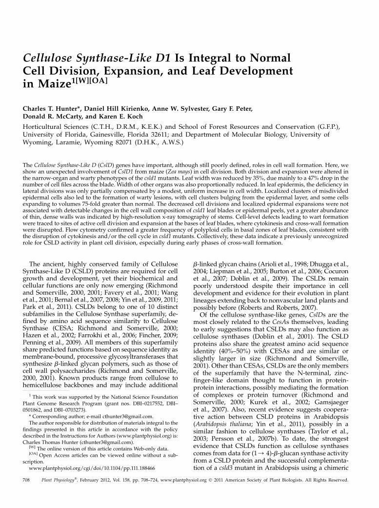
Phylogenetic analyses (Fig. 1A) identified three dis-tinct clades in the CSLD subfamily and found these torepresent three phenotypic classes of csldmutants. Thesethree major phenotypic groups are, for Arabidopsis,
Maize CslD1 Affects Cell Division
Plant Physiol. Vol. 158, 2012 709
pollen tube defects in csld1 and csld4 mutants, (Bernalet al., 2008), root hair defects in csld2 and csld3mutants(Favery et al., 2001; Wang et al., 2001; Bernal et al.,2008), and reduced plant size in csld5 mutants (Bernalet al., 2007). Thus far, mutants in related clades of CslDgenes in rice and maize have yielded phenotypes
similar to those of Arabidopsis. Mutants of riceCslD1 (Kim et al., 2007), for example, and its maizehomolog, CslD5 (Penning et al., 2009), result in roothair-deficient phenotypes. Additionally, alterations inrice CslD4 (the closest homolog of ZmCslD1 andAtCslD5) confer a narrow leaf and dwarf1 phenotype
Figure 1. Comparison of mutant phenotypes with CSLD protein phylogeny, and diagram of the CSLD1 protein and mutant genesin maize. A, Neighbor-joining tree of predicted protein sequences encoded by CslD genes in maize, rice, and Arabidopsis.Reported phenotypes for null alleles are shown in red (ZmCSLD5 [Penning et al., 2009], OsCSLD1 [Kim et al., 2007], AtCSLD2[Bernal et al., 2008], AtCSLD3 [Favery et al., 2001;Wang et al., 2001], AtCSLD5 [Bernal et al., 2007], OsCSLD4 [Li et al., 2009],AtCSLD1 and AtCSLD4 [Bernal et al., 2008]). AtCslD6 and ZmCslD3 are predicted to be pseudogenes. The ZmCSLD1 protein isstarred. Members of distinct clades are grouped by shapes, with dashed lines including proteins for which no genetic evidenceexists. The tree was created using MEGA 4.0 (http://megasoftware.net/mega.html; Tamura et al., 2007) with 2,000 bootstraprepetitions and the pairwise deletion option. Units are amino acid substitutions per site. Supplemental Figure S1 shows theClustalW alignments used in this phylogenetic analysis. B, Diagram of the maize CslD1 gene and protein. Sites of Mu transposoninsertion (triangles) are shown on the CslD1 gene. Lengths of exons and introns are indicated below each region (in bp). Both thecsld1-1 and csld1-2 mutants were obtained from the UniformMu maize population at the University of Florida (http://uniformmu.uf-genome.org [McCarty et al., 2005; Settles et al., 2007]). Smaller triangles represent Pioneer Hi-Bred InternationalTUSC alleles (McCarty and Meeley, 2009). The predicted protein includes 1,218 amino acids, an N-terminal RING-type zinc-finger-like domain, eight transmembrane domains, and the conserved residues of an inverting, processive glycosyltransferase.The protein diagram is modeled after Richmond (2000), and protein domains were identified using SMART (http://smart.embl-heidelberg.de) and TMHMM (http://www.cbs.dtu.dk/services/TMHMM).
Hunter et al.
710 Plant Physiol. Vol. 158, 2012
(Li et al., 2009; Hu et al., 2010; Wu et al., 2010). Similarfunctional roles are indicated by the reduced-growthphenotypes common to all three of these mutants (Fig.1A; Bernal et al., 2007; Li et al., 2009; Hu et al., 2010; Wuet al., 2010). Collectively, these data suggest the con-servation of specific developmental roles for individ-ual CSLD proteins in plants and indicate that thesearose early in plant evolution.
An Allelic Series of csld1 Mutants in Maize Enabled
Functional Analysis
Seven independent loss-of-function mutants for themaize CslD1 gene were identified in reverse geneticscreens, including two from the UniformMu maizepopulation (University of Florida) and five from theTrait Utilities for Screening of Corn (TUSC) lines(Pioneer Hi-Bred International; Meeley and Briggs,1995; McCarty and Meeley, 2009; Fig. 1B). The twoUniformMu alleles, csld1-1 and csld1-2, were examinedin the greatest depth because of their uniform geneticbackground (McCarty et al., 2005). Both of these werenull for detectable expression of CslD1 mRNA byquantitative PCR (data not shown). Phenotypes ofcsld1-1 and csld1-2 homozygous mutants, as well asoffspring from their reciprocal F1 hybrids, were indis-tinguishable, thus demonstrating a causal role for thedysfunctional CslD1 gene. Mutant plants showedoverall reduced growth, narrow leaves, and had arough leaf texture caused by warty protrusions fromthe mature leaf epidermis. Genotypic analysis of over200 individuals from segregating families showed a100% correspondence between this phenotype and ho-mozygosity for the csld1-1 mutation (data not shown).Mendelian segregation ratios were typical of a reces-sive mutation. The five other transposon insertions inCslD1 (csld1-3–csld1-7; courtesy of Pioneer Hi-BredInternational) also resulted in mutant phenotypesthat included narrower organs and wart-like clustersof epidermal cells, regardless of heterologous geneticbackground.
Phenotype of csld1 Mutants: Narrow Organs and
Wart-Like Cell Clusters
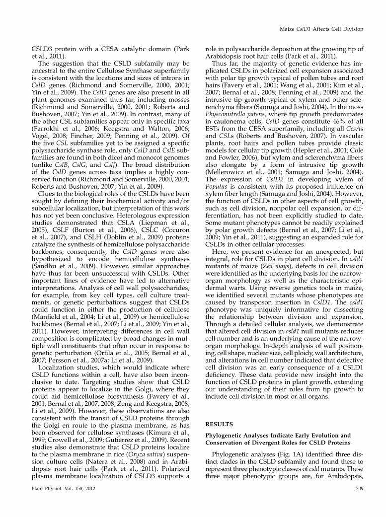
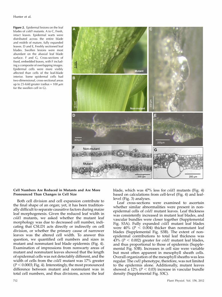
Null mutants of csld1 showed a striking phenotypethat included narrow leaves, reduced stature, andhighly textured leaf blades (Figs. 2 and 3). Lightmicroscopy showed that the visibly rough texture ofcsld1 mutant leaves was due to irregular swelling bygroups of epidermal cells that formed wart-like cellclusters (Fig. 2). Some epidermal cells expanded 75-fold in volume and were generally arranged in linearprofiles along the longitudinal axis of the leaf midriband blade (Fig. 2A) but not on leaf sheaths or stalks(data not shown). The groups of cells that formed so-called warts were interspersed with normal-appearingregions of leaf epidermis along the entire length of theleaf blade. Swollen cells remained filled with fluid
until the onset of leaf senescence (Fig. 2C). Warts werepresent on both surfaces of mutant leaf blades butwere larger and more abundant on the abaxial face(Fig. 2D). These malformed cells consistently lackedchloroplasts (Fig. 2, C and E), indicating an epidermalorigin, as confirmed in serial cross-sections of mutantleaves (Fig. 2, F and G). These ballooned epidermalcells in csld1 mutants often had diameters well over100 mm, at least 5-fold and sometimes 20-fold greaterthan epidermal pavement cells of nonmutant plants(Fig. 2G). Both scanning electron microscopy and op-tical microscopy of fresh, intact leaves showed thatlesions continued to expand throughout leaf matura-tion and that the largest clusters included swollen cellsthat had collapsed. In other instances, cells remainedintact, even in lesions greater than 300 mm across(Fig. 2C).
Potentially analogous, wart-like epidermal swell-ings were described by Burton et al. (2000) in a virus-induced gene silencing experiment in tobacco (Nicotianatabacum). Although a CesA gene was targeted, thehighly similar CslD genes may also have been silenced.The transgenic tobacco had wart-like lesions on theabaxial side of leaves and an overall phenotype strik-ingly similar to the maize csld1 mutants (Burton et al.,2000). This commonalitywould be consistent with somedegree of repression of the ZmCslD1 ortholog in thetobacco experiment. Alternatively, if observed lesionsdid result from the down-regulation of CesA genesalone, then this would support a role for CSLD proteinsin cellulose biosynthesis (Doblin et al., 2001; Park et al.,2011).
Plant Dry Weight and Organ Width Are Reduced incsld1 Mutants
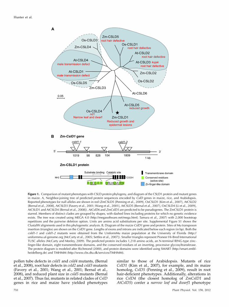
Total growth and organ size were reduced in ho-mozygous csld1 mutants (Fig. 3), even though overallplant architecture, leaf number, and flowering timewere similar for csld1 mutant and nonmutant siblingsunder field and greenhouse conditions (data notshown). At maturity, the mean height of mutant plants(to the auricle of the uppermost leaf) was only 11% less(P , 0.001), whereas dry weight decreased by a morepronounced 44% (P , 0.0025; Fig. 3A). Proportionalreductions in dry weight were also evident for allorgans examined, including ears, tassels, stalks, roots,and leaves (data not shown). Organ width decreased35% (P, 0.0003) for mature leaf blades of csld1mutantplants, but length was only 10% less (P , 0.0003; Fig.3B). This narrow-leaf phenotype was proportional inall leaves examined, indicating a consistent defect inlateral development rather than an ontological effect atspecific leaf positions (Fig. 3B).
Organ width was also reduced in stalks from csld1mutants (Supplemental Fig. S2). The cross-sectionalareas of csld1 stalks were an average of 24% less (P ,0.025). The narrow-organ phenotype extended to cobsand tassels as well (data not shown).
Maize CslD1 Affects Cell Division
Plant Physiol. Vol. 158, 2012 711
Cell Numbers Are Reduced in Mutants and Are MorePronounced Than Changes in Cell Size
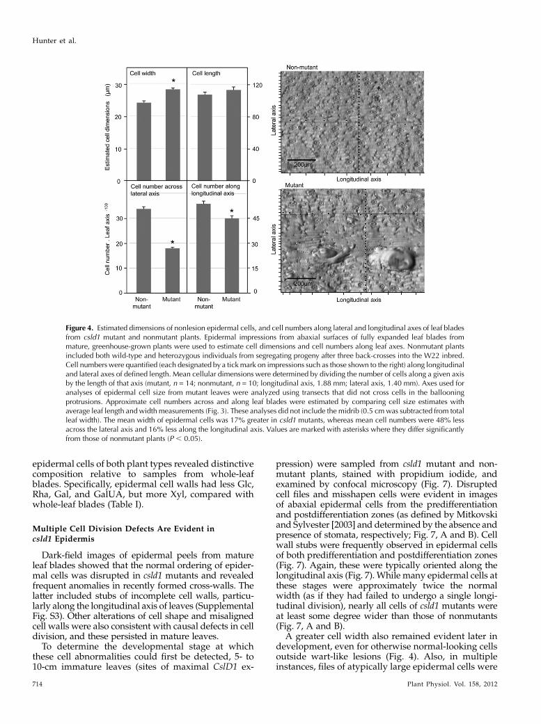
Both cell division and cell expansion contribute tothe final shape of an organ; yet, it has been tradition-ally difficult to separate causative factors duringmaizeleaf morphogenesis. Given the reduced leaf width incsld1 mutants, we asked whether the mutant leafmorphology was due to decreased cell number, indi-cating that CSLD1 acts directly or indirectly on celldivision, or whether the primary cause of narrowerleaves was the altered cell width. To answer thisquestion, we quantified cell numbers and sizes inmutant and nonmutant leaf blade epidermis (Fig. 4).Examination of impressions from nonwarty areas ofmutant and nonmutant leaves showed that the lengthof epidermal cells was not detectably different, and thewidth of cells from the csld1 mutant was 17% greater(P, 0.0003; Fig. 4). Interestingly, the most pronounceddifference between mutant and nonmutant was intotal cell numbers, and thus divisions, across the leaf
blade, which was 47% less for csld1 mutants (Fig. 4)based on calculations from cell-level (Fig. 4) and leaf-level (Fig. 3) analyses.
Leaf cross-sections were examined to ascertainwhether similar abnormalities were present in non-epidermal cells of csld1 mutant leaves. Leaf thicknesswas consistently increased in mutant leaf blades, andvascular bundles were closer together (SupplementalFig. S3A). Fully expanded csld1 mutant leaf bladeswere 40% (P , 0.004) thicker than nonmutant leafblades (Supplemental Fig. S3B). The extent of non-epidermal contributions to total leaf thickness was43% (P , 0.002) greater for csld1 mutant leaf blades,and thus proportional to those of epidermis (Supple-mental Fig. S3B). Increases in cell size were variablebut most often apparent in mesophyll sheath cells.Overall organization of the mesophyll sheaths was lessregular. The csd1 phenotype, therefore, was not limitedto the epidermis alone. Additionally, mutant leavesshowed a 12% (P , 0.03) increase in vascular bundledensity (Supplemental Fig. S3C).
Figure 2. Epidermal lesions on the leafblades of csld1 mutants. A to C, Fresh,intact leaves. Epidermal warts weredistributed across the entire bladeand midrib of mature, fully expandedleaves. D and E, Freshly sectioned leafblades. Swollen lesions were mostabundant on the abaxial leaf bladesurface. F and G, Cross-sections offixed, embedded leaves, with F includ-ing a composite of overlapping images.Epidermal cells were more visiblyaffected than cells of the leaf-bladeinterior. Some epidermal cells hadtwo-dimensional, cross-sectional areasup to 25-fold greater (radius = 100 mmfor the swollen cell in G).
Hunter et al.
712 Plant Physiol. Vol. 158, 2012

Levels of CslD1 mRNA Are Greatest in Regions of Active
Cell Division
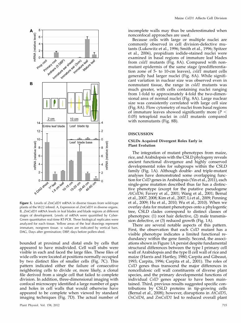
In order to view the phenotypes of csld1 mutants inthe context of where the wild-type gene is expressed,levels of CslD1mRNAwere measured via quantitativereverse transcription (RT)-PCR across diverse tissuesand stages of development (Fig. 5). The CslD1 tran-script levels were greatest in young, preemergentleaves (inside the whorl) and still relatively abundantin young primary root tips and bases of more matureleaves (Fig. 5A). These mRNAs were also detected inbasal zones of leaves by other recent studies of maizeleaf development (Li et al., 2010). To more clearlydefine the pattern of transcript accumulation duringleaf development, staged samples of young to matureleaves were analyzed. The CslD1 mRNA levels werehighest in tissues with actively dividing cells andmaximal in shoots 6 d after germination (Fig. 5B). Laterin development, single-leaf analyses showed thatlevels of CslD1 mRNAwere greatest in basal portionsof blades from expanding leaves, 15 to 25 cm long. Innondividing, fully expanded leaves, CslD1mRNA haddropped below detectable levels and remained so inthe fully differentiated portions of leaves (Fig. 5B).Notably, wart formation and expansion continues inleaves through maturity, even within areas whereCslD1 is no longer expressed.
Cell Wall Composition Was Similar, but Thin, Dense
Walls Were More Abundant in csld1 Mutants
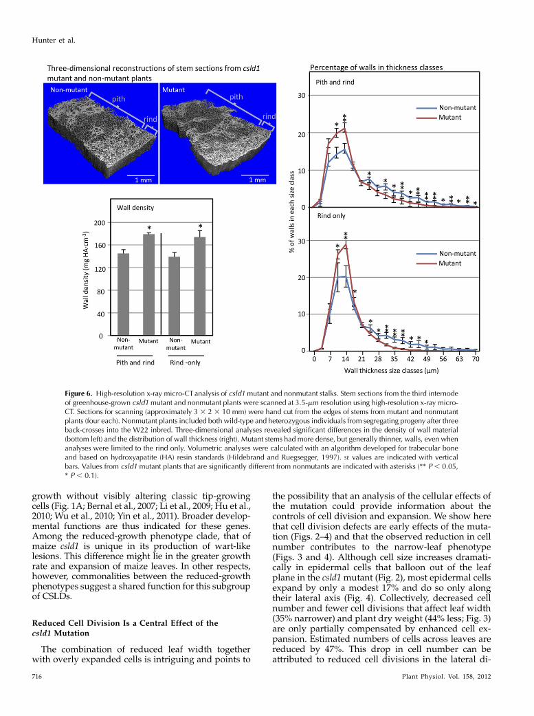
To further characterize differences between mutantand nonmutant tissues, high-resolution x-ray micro-computed tomography (micro-CT) was used to analyzehand-cut sections of stalks, since this tissue was foundamenable to the x-ray approach. Three-dimensionalreconstructions revealed a shift in average wall thick-ness toward thinner walls in the csld1 mutant (Fig. 6).Additionally, this method allowed a comparison ofoverall wall density, which was greater for stalks frommutant plants, regardless of whether the pith andvessel-rich rind were examined together or separately(Fig. 6).
To determine whether these changes were associ-ated with alterations to cell wall polysaccharide com-position, we examined cell walls from mature leafblades as well as isolated leaf epidermis. No signifi-cant differences in alcohol-insoluble cell wall com-position were detected between csld1 mutants andnonmutants for either cellulose or sugar subunits ofnoncellulosic constituents (Table I). Cell walls from
Figure 3. The narrow-leaf phenotype of csld1 mutant plants. A, Plantheight and dry weight (aboveground and belowground) were quantifiedfor field-grown csld1 mutant and nonmutant plants. Plant height was90.9 cm (SE, 2.8; n = 20) for mutants and 102.2 cm (SE, 1.7; n = 35) fornonmutants. For dry weight measurements, whole plants were sampled3 d after ear maturity (40 d post pollination) and did not include matureears or fine roots. Total plant dry weight was 41.9 g (SE, 4.0; n = 4) formutants and 76.5 g (SE, 3.2; n = 4) for nonmutants. Belowground dryweight was 8.7 g (SE, 0.2; n = 4) for mutants and 17.1 g (SE, 1.1; n = 4) fornonmutants. Aboveground and belowground dry weights were reducedby similar amounts (average of 44% and 49%, respectively). SE valuesare indicated with vertical bars and are marked by asterisks wherevervalues for mutant plants differed significantly from those of nonmutants(P , 0.05). B, Leaf blade length and width (at the widest point) werequantified for leaf positions 3 through 5 (as indicated) for field-grownmutant and nonmutant plants. Nonmutant plants included both wild-type and heterozygous individuals from segregating progeny after threeback-crosses into the W22 inbred. Imaged blades were from leaves inposition 3 from the apex. The left-most portion of each graph showscombined data from all leaf positions measured. The leaf blade width-to-length ratio was 27% less for mutant plants. Blade width and length
were reduced 35% and 10%, respectively, relative to those of nonmu-tant plants. Data were similar for csld1-2 (data not shown) and visualappraisals of the five other csld1 mutants. Note that leaf photographswere from greenhouse-grown plants, whereas quantifications werefrom field-grown plants. * Significantly different from the nonmutant(P , 0.05).
Maize CslD1 Affects Cell Division
Plant Physiol. Vol. 158, 2012 713
epidermal cells of both plant types revealed distinctivecomposition relative to samples from whole-leafblades. Specifically, epidermal cell walls had less Glc,Rha, Gal, and GalUA, but more Xyl, compared withwhole-leaf blades (Table I).
Multiple Cell Division Defects Are Evident incsld1 Epidermis
Dark-field images of epidermal peels from matureleaf blades showed that the normal ordering of epider-mal cells was disrupted in csld1 mutants and revealedfrequent anomalies in recently formed cross-walls. Thelatter included stubs of incomplete cell walls, particu-larly along the longitudinal axis of leaves (SupplementalFig. S3). Other alterations of cell shape and misalignedcell walls were also consistent with causal defects in celldivision, and these persisted in mature leaves.
To determine the developmental stage at whichthese cell abnormalities could first be detected, 5- to10-cm immature leaves (sites of maximal CslD1 ex-
pression) were sampled from csld1 mutant and non-mutant plants, stained with propidium iodide, andexamined by confocal microscopy (Fig. 7). Disruptedcell files and misshapen cells were evident in imagesof abaxial epidermal cells from the predifferentiationand postdifferentiation zones (as defined by Mitkovskiand Sylvester [2003] and determined by the absence andpresence of stomata, respectively; Fig. 7, A and B). Cellwall stubs were frequently observed in epidermal cellsof both predifferentiation and postdifferentiation zones(Fig. 7). Again, these were typically oriented along thelongitudinal axis (Fig. 7). While many epidermal cells atthese stages were approximately twice the normalwidth (as if they had failed to undergo a single longi-tudinal division), nearly all cells of csld1 mutants wereat least some degree wider than those of nonmutants(Fig. 7, A and B).
A greater cell width also remained evident later indevelopment, even for otherwise normal-looking cellsoutside wart-like lesions (Fig. 4). Also, in multipleinstances, files of atypically large epidermal cells were
Figure 4. Estimated dimensions of nonlesion epidermal cells, and cell numbers along lateral and longitudinal axes of leaf bladesfrom csld1 mutant and nonmutant plants. Epidermal impressions from abaxial surfaces of fully expanded leaf blades frommature, greenhouse-grown plants were used to estimate cell dimensions and cell numbers along leaf axes. Nonmutant plantsincluded both wild-type and heterozygous individuals from segregating progeny after three back-crosses into the W22 inbred.Cell numbers were quantified (each designated by a tickmark on impressions such as those shown to the right) along longitudinaland lateral axes of defined length. Mean cellular dimensions were determined by dividing the number of cells along a given axisby the length of that axis (mutant, n = 14; nonmutant, n = 10; longitudinal axis, 1.88 mm; lateral axis, 1.40 mm). Axes used foranalyses of epidermal cell size from mutant leaves were analyzed using transects that did not cross cells in the ballooningprotrusions. Approximate cell numbers across and along leaf blades were estimated by comparing cell size estimates withaverage leaf length andwidth measurements (Fig. 3). These analyses did not include the midrib (0.5 cmwas subtracted from totalleaf width). The mean width of epidermal cells was 17% greater in csld1 mutants, whereas mean cell numbers were 48% lessacross the lateral axis and 16% less along the longitudinal axis. Values are marked with asterisks where they differ significantlyfrom those of nonmutant plants (P , 0.05).
Hunter et al.
714 Plant Physiol. Vol. 158, 2012
bounded at proximal and distal ends by cells thatappeared to have misdivided. Cell wall stubs werevisible in each and faced the large files. These files ofwide cells were located at positions normally occupiedby two distinct files of smaller cells (Fig. 7C). Thispattern indicated either the failure of consecutiveneighboring cells to divide or, more likely, a clonalfile derived from a single cell that failed to completedivision. In addition, three-dimensional imaging withconfocal microscopy identified a large number of gapsand holes in cell walls that would otherwise haveappeared to be complete when viewed by standardimaging techniques (Fig. 7D). The actual number of
incomplete walls may thus be underestimated whennonconfocal approaches are used.
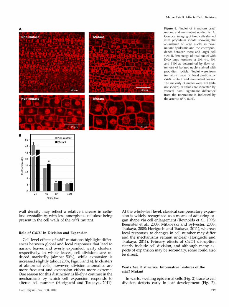
Because cells with large or multiple nuclei arecommonly observed in cell division-defective mu-tants (Lukowitz et al., 1996; Smith et al., 1996; Spitzeret al., 2006), propidium iodide-stained nuclei wereexamined in basal regions of immature leaf bladesfrom csld1 mutants (Fig. 8A). Compared with non-mutant epidermis of the same stage (predifferentia-tion zone of 5- to 10-cm leaves), csld1 mutant cellsgenerally had larger nuclei (Fig. 8A). While signifi-cant variation in nuclear size was observed even innonmutant tissue, the range in csld1 mutants wasmuch greater, with cells containing nuclei rangingfrom 1-fold to approximately 4-fold the two-dimen-sional area of normal nuclei (Fig. 8A). Large nuclearsize was consistently correlated with large cell size(Fig. 8A). Flow cytometry of nuclei from basal regionsof immature leaves showed significantly more (P ,0.05) tetraploid nuclei in csld1 mutants comparedwith nonmutants (Fig. 8B).
DISCUSSION
CSLDs Acquired Divergent Roles Early inPlant Evolution
The integration of mutant phenotypes from maize,rice, and Arabidopsis with the CSLD phylogeny revealsancient functional divergence and highly conserveddevelopmental roles for subgroups within the CSLDfamily (Fig. 1A). Although double- and triple-mutantanalyses have demonstrated some overlapping func-tion forCslD genes in Arabidopsis (Yin et al., 2011), eachsingle-gene mutation described thus far has a distinc-tive phenotype (except for the putative pseudogeneAtCslD6; Favery et al., 2001; Wang et al., 2001; Bernalet al., 2007, 2008; Kim et al., 2007; Li et al., 2009; Penninget al., 2009; Hu et al., 2010; Wu et al., 2010). When weoverlay data for mutant phenotypes onto a phylogentictree, CSLD clades correspond to distinct classes ofphenotypes: (1) root hair defective, (2) male transmis-sion defective, or (3) reduced growth (Fig. 1A).
There are several notable aspects of this pattern.First, the observation that each CslD mutant has avisible phenotype indicates a limited functional re-dundancy within the gene family. Second, the associ-ations shown in Figure 1A persist despite fundamentalstructural differences between the type I primary cellwall of Arabidopsis and the type II cell wall of rice andmaize (Harris and Hartley, 1980; Carpita and Gibeaut,1993; Carpita, 1996; Carpita et al., 2001). The roles ofCslD genes thus transcend the major differences innoncellulosic cell wall constituents of diverse plantspecies, and the primary developmental functions ofindividual CslD genes appear to have been main-tained. Third, previous results suggested specific con-tributions by CSLD proteins in tip-growing cells(Bernal et al., 2008); however, disruptions in AtCslD5,OsCslD4, and ZmCslD1 led to reduced overall plant
Figure 5. Levels of ZmCslD1 mRNA in diverse tissues from wild-typeplants of the W22 inbred. A, Expression of ZmCslD1 in diverse organs.B, ZmCslD1 mRNA levels in leaf blades and blade regions at differentstages of development. Levels of mRNA were quantified by Cyber-Green quantitative real-time RT-PCR. Three biological replicates wereanalyzed for each tissue. Yellow areas of the leaf drawings representimmature, nongreen tissue. SE values are indicated by vertical bars.DAG, Days after germination; DBP, days before pollen-shed.
Maize CslD1 Affects Cell Division
Plant Physiol. Vol. 158, 2012 715
growth without visibly altering classic tip-growingcells (Fig. 1A; Bernal et al., 2007; Li et al., 2009; Hu et al.,2010; Wu et al., 2010; Yin et al., 2011). Broader develop-mental functions are thus indicated for these genes.Among the reduced-growth phenotype clade, that ofmaize csld1 is unique in its production of wart-likelesions. This difference might lie in the greater growthrate and expansion of maize leaves. In other respects,however, commonalities between the reduced-growthphenotypes suggest a shared function for this subgroupof CSLDs.
Reduced Cell Division Is a Central Effect of thecsld1 Mutation
The combination of reduced leaf width togetherwith overly expanded cells is intriguing and points to
the possibility that an analysis of the cellular effects ofthe mutation could provide information about thecontrols of cell division and expansion. We show herethat cell division defects are early effects of the muta-tion (Figs. 2–4) and that the observed reduction in cellnumber contributes to the narrow-leaf phenotype(Figs. 3 and 4). Although cell size increases dramati-cally in epidermal cells that balloon out of the leafplane in the csld1mutant (Fig. 2), most epidermal cellsexpand by only a modest 17% and do so only alongtheir lateral axis (Fig. 4). Collectively, decreased cellnumber and fewer cell divisions that affect leaf width(35% narrower) and plant dry weight (44% less; Fig. 3)are only partially compensated by enhanced cell ex-pansion. Estimated numbers of cells across leaves arereduced by 47%. This drop in cell number can beattributed to reduced cell divisions in the lateral di-
Figure 6. High-resolution x-ray micro-CTanalysis of csld1mutant and nonmutant stalks. Stem sections from the third internodeof greenhouse-grown csld1mutant and nonmutant plants were scanned at 3.5-mm resolution using high-resolution x-ray micro-CT. Sections for scanning (approximately 3 3 2 3 10 mm) were hand cut from the edges of stems from mutant and nonmutantplants (four each). Nonmutant plants included both wild-type and heterozygous individuals from segregating progeny after threeback-crosses into the W22 inbred. Three-dimensional analyses revealed significant differences in the density of wall material(bottom left) and the distribution of wall thickness (right). Mutant stems had more dense, but generally thinner, walls, even whenanalyses were limited to the rind only. Volumetric analyses were calculated with an algorithm developed for trabecular boneand based on hydroxyapatite (HA) resin standards (Hildebrand and Ruegsegger, 1997). SE values are indicated with verticalbars. Values from csld1 mutant plants that are significantly different from nonmutants are indicated with asterisks (** P , 0.05,* P , 0.1).
Hunter et al.
716 Plant Physiol. Vol. 158, 2012
mension, indicating that this specific decrease in divi-sions is a central effect of csld1mutations. Although anexamination of cross-sections suggested that leaf vas-cular bundle number and density were altered inmutant leaves, the size and shape of vascular bundleswere generally unchanged. Instead, the greater thick-ness of csld1 leaf blades typically corresponded mostclosely with irregular increases in size by cells of themesophyll sheath (Supplemental Fig. S3), indicating abroader role for CSLD1 than epidermal developmentalone.A cell division role for CslD1 early in leaf develop-
ment is also supported by its maximal expression inzones of wild-type leaves where cells are most activelydividing (Fig. 5) andwith the occurrence of anomalousdivisions within this zone in the csld1 mutant (Fig. 7).These disrupted divisions result in the formation ofabnormal cell clusters implicated in later wart devel-opment and potentially affect the determination of cellfile numbers across leaf blades. Expansion of theepidermal warts appears to be largely a secondaryeffect of the csld1 mutation, because these cells con-tinue to expand in leaves after CslD1 mRNAs wouldhave dropped below detectable levels (Fig. 5).While our results are compatible with a central
defect in the rate or total number of cell divisions,diverse, indirect effects may also contribute to theobserved phenotype. Narrow leaves of csld1 mutants,for example (Fig. 3), could exacerbate the reductions inplant dry weight by decreasing total plant photosyn-thetic capacity. The smaller csld1 root system (Fig. 3)could further reduce growth. Also, the epidermis mayhave a prominent physical role in organ expansionand meristem geometry (Green, 1980; Moulia, 2000),providing additional potential for secondary or ter-tiary effects of the csld1 mutations.Cell expansion is clearly also altered by direct and/or
indirect effects of the mutation. Much of the cellularoverexpansion is likely secondary, since compensatoryexpansion is a common response to decreases in cellnumber (Reynolds et al., 1998; Beemster et al., 2003;Mitkovski and Sylvester, 2003; Horiguchi and Tsukaya,2011). However, the csld1 mutations could theoreticallyaffect more than cross-wall deposition during divisionand subsequent, compensatory expansion. If the prop-erties of mutant walls are altered, then these too could
contribute to expansion-based aspects of the pheno-types (discussed further below).
Cell Wall Thickness, but Not Composition, Is Altered in
the csld1 Mutant
Although the overall quantity of cell wall material isreduced by 45% in the csld1 mutant plants, cell wallcomposition is unaffected in either whole organs orepidermis alone (Table I). Several possibilities couldaccount for the lack of a detectable difference. First,the CSLD1 polysaccharide product may normally bepresent in only small amounts, particularly if synthesisis restricted to a defined period during cytokinesis ornew cell wall formation. If so, then cell wall contribu-tions from CSLD1 could be masked by more abundantpolymers. As noted above, an early developmentalrole for small amounts of CSLD product would beconsistent with the maximal expression of CslD1 invery young leaves and basal portions of fast-growingblades (Fig. 5), since these are zones of active cell di-vision (Sylvester et al., 1990; Freeling, 1992; Sylvester,2000). One permutation of this suggestion is thatCSLD1 might aid a specialized, directed depositionof early-arriving polymers to the growing cell plateduring division. The CSLD1 product itself could thusbe limited to a narrow point in time.
Another possibility is that the CSLD1 enzyme mightsynthesize a limiting constituent of cell walls, such thata drop in levels of the CSLD1 product would result insimilar decreases for other cell wall polymers. In thisway, csld1 mutants could produce less total cell wallwithout changing the relative proportions of individ-ual cell wall components. This suggestion is consistentwith the reduction in total dry matter. Still another,compatible scenario is that the polysaccharide productof CSLD1 may not differ in a way immediately de-tectable by current analyses but may nonetheless havefunctionally altered properties and/or capacities forinteraction that affect later cell wall behavior. All threeof the above possibilities remain consistent with atemporally limited contribution of a known wall com-ponent.
Cell wall analysis by high-resolution, x-ray, micro-CT(Fig. 6) provides detailed structural information oninternal regions of intact tissue, including the relative
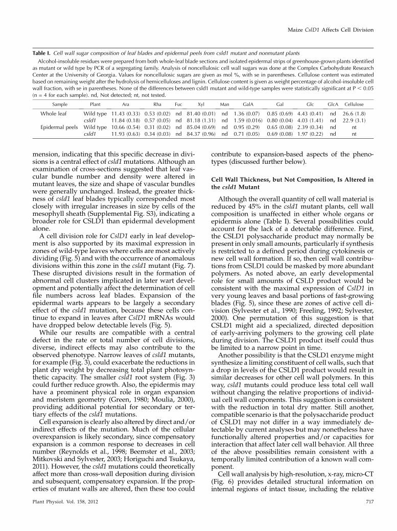
Table I. Cell wall sugar composition of leaf blades and epidermal peels from csld1 mutant and nonmutant plants
Alcohol-insoluble residues were prepared from both whole-leaf blade sections and isolated epidermal strips of greenhouse-grown plants identifiedas mutant or wild type by PCR of a segregating family. Analysis of noncellulosic cell wall sugars was done at the Complex Carbohydrate ResearchCenter at the University of Georgia. Values for noncellulosic sugars are given as mol %, with se in parentheses. Cellulose content was estimatedbased on remaining weight after the hydrolysis of hemicelluloses and lignin. Cellulose content is given as weight percentage of alcohol-insoluble cellwall fraction, with se in parentheses. None of the differences between csld1 mutant and wild-type samples were statistically significant at P , 0.05(n = 4 for each sample). nd, Not detected; nt, not tested.
Sample Plant Ara Rha Fuc Xyl Man GalA Gal Glc GlcA Cellulose
Whole leaf Wild type 11.43 (0.33) 0.53 (0.02) nd 81.40 (0.01) nd 1.36 (0.07) 0.85 (0.69) 4.43 (0.41) nd 26.6 (1.8)csld1 11.84 (0.18) 0.57 (0.05) nd 81.18 (1.31) nd 1.59 (0.016) 0.80 (0.04) 4.03 (1.41) nd 22.9 (3.1)
Epidermal peels Wild type 10.66 (0.54) 0.31 (0.02) nd 85.04 (0.69) nd 0.95 (0.29) 0.65 (0.08) 2.39 (0.34) nd ntcsld1 11.93 (0.63) 0.34 (0.03) nd 84.37 (0.96) nd 0.71 (0.05) 0.69 (0.08) 1.97 (0.22) nd nt
Maize CslD1 Affects Cell Division
Plant Physiol. Vol. 158, 2012 717

density of cell wall material, based on x-ray beamattenuation (Steppe et al., 2004; Dhondt et al., 2010).Stems of csld1mutants (tissues found most amenable tothe x-ray, micro-CT approach) show slight, but consis-tent, increases in wall density and a greater overall
abundance of thin-walled areas, regardless of positionin rind or pith (Fig. 6). These findings indicate analtered cell wall or cellular architecture that is appar-ently independent of changes in wall composition. One,albeit speculative, possibility is that the changes in cell
Figure 7. Confocal images showing defects early in the development of csld1 mutant leaf epidermis. Propidium iodide-stainedcell walls from fresh, immature csld1mutant and nonmutant leaves are shown. A, Predifferentiation zones from the basal 3 mmof nonmutant leaf blades contrasting with the abnormal csld1 cell size, shape, and organization. Leaf blades were approximately10 cm long. B, Postdifferentiation zones (3–10 mm above the base of the blade) showing the persistent effects of altered celldivision, with the presence of large, misshapen, less ordered cells of the csld1-1 leaf epidermis. Leaf blades were approximately10 cm long. C, A typical file of large, irregular cells bounded by individuals with incomplete cell walls protruding into them fromtheir outer edges, consistent with the clonal inheritance of large cell size in the csld1mutants. D, Serial optical sections revealingirregular gaps in cell walls (arrows) that occur frequently in epidermal cells of the csld1 mutant.
Hunter et al.
718 Plant Physiol. Vol. 158, 2012
wall density may reflect a relative increase in cellu-lose crystallinity, with less amorphous cellulose beingpresent in the cell walls of the csld1 mutant.
Role of CslD1 in Division and Expansion
Cell-level effects of csld1 mutations highlight differ-ences between global and local responses that lead tonarrow leaves and overly expanded, warty clusters,respectively. In whole leaves, cell divisions are re-duced markedly (almost 50%), while expansion isincreased slightly (about 20%; Figs. 3 and 4). In clustersof abnormal cells, however, division anomalies aremore frequent and expansion effects more extreme.One reason for this distinction is likely a contrast in themechanisms by which cell expansion responds toaltered cell number (Horiguchi and Tsukaya, 2011).
At the whole-leaf level, classical compensatory expan-sion is widely recognized as a means of adjusting or-gan shape via cell enlargement (Reynolds et al., 1998;Beemster et al., 2003; Mitkovski and Sylvester, 2003;Tsukaya, 2008; Horiguchi and Tsukaya, 2011), whereaslocal responses to changes in cell number may differand the mechanisms remain unclear (Horiguchi andTsukaya, 2011). Primary effects of CslD1 disruptionclearly include cell division, and although many as-pects of expansion may be secondary, some could alsobe direct.
Warts Are Distinctive, Informative Features of thecsld1 Mutant
In warts, swelling epidermal cells (Fig. 2) trace to celldivision defects early in leaf development (Fig. 7).
Figure 8. Nuclei of immature csld1mutant and nonmutant epidermis. A,Confocal imaging of fixed cells stainedwith propidium iodide showing theabundance of large nuclei in clsd1mutant epidermis and the correspon-dence between these and larger cellsize. B, Percentage of total nuclei withDNA copy numbers of 2N, 4N, 8N,and 16N as determined by flow cy-tometry of isolated nuclei stained withpropidium iodide. Nuclei were fromimmature tissue of basal portions ofcsld1 mutant and nonmutant leaves.The majority of nuclei were 2N (datanot shown). SE values are indicated byvertical bars. Significant differencefrom the nonmutant is indicated bythe asterisk (P , 0.05).
Maize CslD1 Affects Cell Division
Plant Physiol. Vol. 158, 2012 719
Assuming that CSLD1 is a wall-synthesizing enzyme,these cellular observations indicate that loss of CSLD1activity disrupts the synthesis of early cell wall com-ponents, and this in turn alters division. The narroworgans result from a decreased number of long-axis celldivisions throughout the plant (discussed above). Wartformation also derives from disrupted divisions, butthe locally extreme cell size may involve additionaleffects of csld1 on wall expansion. Two possible scenar-ios for epidermal wart formation are presented below.
Hypothesis 1 is based on a role for CSLD1 in newcell wall formation during cytokinesis and posits thatepidermal warts would be initiated when rapid elon-gation by leaves outpaces the capacity of dividing cellsto form normal cross-walls. The first set of disruptedcross-walls would lead to misplacement of the next,and the resulting clusters of irregular cells would beprone to anomalous expansion. The extent and irreg-ularity of expansion would be exacerbated by asym-metric support from surrounding cells. Hypothesis 2includes basic tenets of hypothesis 1 but also suggeststhat csld1mutants may have broader alterations to cellwall properties that reduce the control of expansion.This in turn could contribute to both incomplete andmisplaced cross-wall formation. Altered wall proper-ties could reduce the control of cell expansion at eachstep in the development of irregular cell clusters andepidermal warts and could act in a slow, uniformmanner and/or add a threshold component to theballooning of epidermal cells. Under either hypothe-sis, the excessive expansion of cells in wart formationcan be considered a secondary effect of the CslD1deficiency, resulting from a cascade of interactionsbetween altered cell division and expansion.
The sequence of events in hypothesis 1 begins with acentral role for the CSLD1 polysaccharide product(likely cellulose) in the formation of new cross-walls.In csld1 mutants, this cross-wall formation is impli-cated in reducing the number of long-axis cell divi-sions and thus organ width. In leaf blades especially, aCSLD1-based limitation on new wall formation couldhave prominent effects when the demands of rapidgrowth outpace the compromised capacity for cross-wall production. The resulting anomalies in csld1epidermis include instances of nondivided cells anddefective cross-walls. The latter could arise from eitherpartial formation of these walls and/or their expan-sion before full completion. Resulting cells have non-existent or defective cross walls with central holes orcell wall stubs (as observed). A single cell divisionaltered at the base of a growing leaf blade could lead toother disruptions by altering the timing and placementof the subsequent cross-walls. The abnormal divisionswould then lead to the uneven and disrupted cell filesas well as to large and misshapen cells (Fig. 7, A andB). These cells would lack the support otherwiseafforded a normal, highly ordered, brick-like patternof epidermal cells and would be more prone to exces-sive expansion during turgor-driven growth. In addi-tion, hypothesis 1 predicts that the wartyness of csld1
phenotypes will be more extreme under conditionswhere growth is most rapid and cross-walls cannotform correctly. Consistent with this prediction, thephenotypes vary in plants grown under field versusgreenhouse conditions (data not shown).
Another possibility is that csld1 alters cell wallproperties that affect expansion and these in turncause cell division defects that lead to wart formation(hypothesis 2). Such changes could have functionalsignificance even if generated by trace amounts ofmaterials contributed only during a brief period earlyin cell development. Although altered cell walls (miss-ing a CSLD1 product) could be “weaker,” they mightalso be more responsive to effectors of cell expansion,such as endogenous signals or physical aspects ofturgor pressure. Larger or irregularly shaped cellscould result, and this could compromise their capacityto form accurately positioned or complete cross walls.The resulting series of anomalous, failed, or disrupteddivisions would produce warty cell clusters. The sug-gestion that csld1 cells expand more readily would beconsistent with the somewhat larger size of epidermalcells overall. Wall properties might be altered in subtleways throughout development or impart a thresholdcomponent to ballooning cells of warts.
The CslD1 Gene Is Essential for Specific Aspects of
Cell Division
Primary effects of the csld1mutation on cell divisioncan be clearly discerned in the narrow leaves and finestems of the csld1 phenotype. Although the overlyexpanded warty lesions exhibit cascades of anomaloussecondary events, the majority of cells in leaves andstems are well ordered. Reduced cell numbers and celldivisions in these organs are consistent with a globalresponse to the csld1 mutation. In particular, long-axisdivisions are decreased and proportional to reduc-tions in leaf width, other organ diameters, and plantdry weight. Although the narrowness of leaf bladescould theoretically be exacerbated by secondary ef-fects of warts, the same cannot be said of the othernarrow organs of csld1 plants. As a cell wall bio-synthetic gene, CslD1 can have direct and indirecteffects. The latter can include compensatory expansion(Horiguchi and Tsukaya, 2011) as well as wall-basedsignaling (Hematy and Hofte, 2008; Ringli, 2010;Seifert and Blaukopf, 2010; Boisson-Dernier et al.,2011). However, changes in cell number are seldomsecondary consequences of altered expansion at theorgan level (Wang et al., 2003; Cnops et al., 2004; Huet al., 2006; Szecsi et al., 2006; Horiguchi et al., 2011;Horiguchi and Tsukaya, 2011), and a primary role issuggested here for CslD1 in cell division.
The deposition and integrity of new cross-walls areclearly impaired in dividing cells of csld1 mutants.Long-axis divisions are most markedly affected, withnew walls having large central openings or appearingas incomplete stubs. Collective data reveal a newdimension to functions of the CslD gene subfamily,
Hunter et al.
720 Plant Physiol. Vol. 158, 2012
since these were previously considered largely relatedto tip growth (Favery et al., 2001; Wang et al., 2001;Kim et al., 2007; Bernal et al., 2008; Penning et al., 2009;Park et al., 2011). However, both tip growth and cross-wall formation during division share a common, di-rectional aspect to the deposition of new wall material.Bednarek and Falbel (2002) previously suggested thatsuch mechanisms could involve similar components.Our work here is consistent with that possibility aswell as with contributions by CslD genes to targetedwall formation in tip-growing and dividing cells.The csld1 mutation also disrupts cytokinesis and/or
the cell cycle. Larger nuclei were observed by confocalimaging in cells of the predifferentiation zone ofimmature csld1 leaves (Fig. 8A). Similar, large-nucleuscells have been reported in studies of cell division-defective mutants (Lukowitz et al., 1996). Here, flowcytometry also showed a small, but significant, in-crease in endoreduplication (Fig. 8B). Even with 50%of epidermal cells undergoing endoreduplication,these would constitute a relatively small portion ofnuclei from whole-leaf tissue. Whether the increase inendoreduplication and larger sized nuclei reflects aresponse to larger cell size or the arrest of the cell cycleafter DNA replication, but before nuclear division,remains unclear. A more rapid entry of dividing cellsinto an endoreduplicative state is typically observedwhere cell size is increasing as a compensatory re-sponse to limited cell number in a developing organ(Beemster et al., 2005; Ferjani et al., 2007; Horiguchiand Tsukaya, 2011). The delay of cells at the G2 phaseimplies a disruption of the normal cell cycle due to alack of CSLD1 activity.
CONCLUSION
The significance of our collective findings is 3-fold.(1) A previously unrecognized role in cell division isdemonstrated here for a cell wall biosynthetic gene. (2)CslD1 is also found here to be central for the effectiveformation and integrity of new cross-walls during celldivision. (3) Collective roles for the CslD gene sub-family are shown to include not only tip growth bycells but also probable contributions to other aspects ofdirectional wall deposition. Additionally, csld1 maizemutants, besides helping our understanding of thespecific functions of a clearly important cell wall-synthesizing enzyme, will provide valuable tools fordissecting the complex and interconnected processesof cell division and cell expansion. Protein localizationefforts are under way and should further clarify thespecific role of CSLD1 in early cell wall formation.
MATERIALS AND METHODS
Phylogenetic Analyses
Protein sequences predicted from full-length cDNAs for each of the CslD
genes from rice (Oryza sativa), maize (Zea mays), and Arabidopsis (Arabidopsis
thaliana) were used to create a neighbor-joining tree using MEGA 4.0 (Tamura
et al., 2007; http://megasoftware.net), with 2,000 bootstrap repetitions using
the pairwise deletion option. Alignments used to create the tree can be
accessed in Supplemental Figure S1. The nomenclature for proteins encoded
by the CslD gene family in maize was assigned as per van Erp and Walton
(2009). Protein domains were identified using SMART (http://smart.embl-
heidelberg.de) and TMHMM (http://www.cbs.dtu.dk/services/TMHMM).
Identification of csld1 Mutants
The UniformMupopulationwas screened using PCR-based assays to identify
Mutator (Mu) transposon inserts inZmCslD1 as per Penning et al. (2009). Close to
15,000 UniformMu lines were screened using a series of pooled DNA samples,
which were forerunners of the sequence-indexed materials currently available at
MaizeGDB (http://maizegdb.org, http://uniformmu.uf-genome.org). For PCR
screening, CslD1-specific primers (5#-AGTTCGTGCACTACACCGTGCA-
CATCC-3# and 5#-TGCTACCTGTAAGGACTGAGGATGGCCTG-3#) were used
along with the Mu-specific primer TIR6 (5#-AGAGAAGCCAACGCCAWCGCC-
TCYATTTCGTC-3#). The resulting products were separated on 1% agarose gels,
blotted onto nylon membranes, and probed with a CslD1-specific PCR
product. Positive probe-binding samples were identified at X45:Y4 of the
UniformMu Reverse Genetics Grid 6 (of eight total). Seeds from the Uni-
formMu family corresponding to these coordinates (04S-1130-27) were grown
and PCR genotyped to identify individuals homozygous for an insertion in
CslD1. The csld1-1 allele was identified from this family, and a csld1-1 line was
established after three generations of successive back-crosses to the W22
inbred.
A second mutant allele, csld1-2, was identified during a visual screen of
field-grown UniformMu lines. Its phenotype was indistinguishable from
that of csld1-1. PCR primers (5#-ACCAGATCCTCTTCCTCCTCGGTTTGC-3#,5#-ACCTTGTTCCTGAGGAAGTCCCTCTTC-3#, 5#-GTGGTGATCACGCT-
GGCATCATTCAG-3#, and 5#-AGGAGGGCTGATGTAGACCCACAG-3#)were designed to cover nearly the entire length of the CslD1 gene and
identified a Mu insertion in the third exon of CslD1. Homozygous recessive
mutants of this allele were obtained from segregating progeny of this family,
and the csld1-2 line was established after two successive back-crosses into the
W22 inbred.
Additional Mu-insert alleles of csld1 were identified from the TUSC
population of Pioneer Hi-Bred International as per McCarty and Meeley
(2009). Primers used were 5#-ACCAGATCCTCTTCCTCCTCGGTTTGC-3#,5#-ACCTTGTTCCTGAGGAAGTCCCTCTTC-3#, 5#-GTGGTGATCACGCTGG-
CATCATTCAG-3#, and 5#-AGGAGGGCTGATGTAGACCCACAG-3#, which
identified five additional mutant alleles designated csld1-3 through csld1-7.
Overall Phenotypic Analyses and Size Measurements
Plant height was measured from the soil level to the auricles at the base of
the uppermost leaf blades for 55 field-grown, mature plants (25 mutant, 30
nonmutant). These same 55 plants were used to measure width (at the widest
point) and length of leaves at positions 3, 4, and 5 (relative to the apex). For dry
weight measurements, whole-plant samples (including released root mass)
were collected 3 d after ear harvest and did not include mature ears. Samples
were weighed after drying for 4 weeks at 38�C. Belowground and above-
ground dry weights were determined by separating root masses from the
aerial portions of these plants.
Tissue Fixation and Sectioning
One-centimeter squares were excised from leaves of greenhouse-grown
plants and fixed in 10% formaldehyde (Fisher lot no. 992720), 5% acetic acid,
and 50% ethanol. Samples were vacuum infiltrated overnight at 4�C and then
shaken at 4�C during a dehydration series using ethanol in phosphate-
buffered saline (60 min each; progressing from 13 phosphate-buffered saline
with 30% ethanol to 40%, 50%, 60%, 70%, 85%, and finally 95% ethanol).
Samples were stained overnight with eosin in 95% ethanol, followed by four
1-h incubations in 100% ethanol and eosin at 25�C. Wax embedding was
initiated by introducing CitriSolv (Fisher catalog no. 22-143975) into samples
using a series of 1-h incubations (while shaking) in ethanol with increasing
CitriSolv:ethanol content (25:75, 50:50, 75:25, and 100:0). Paraplast wax chips
(Fisher catalog no. 23-021-399; 1 g wax mL21 CitriSolv) were added to the
100% CitriSolv and incubated overnight at 25�C. Additional wax was added,
Maize CslD1 Affects Cell Division
Plant Physiol. Vol. 158, 2012 721
followed by a 2-h incubation at 42�C. Samples were transferred to 60�C, where
wax was poured off and replaced eight times over 4 d before samples were
allowed to harden in molds. Sections (10 mm; cut with a Leitz 1512 microtome)
were dewaxed with three 5-min incubations in xylene (Fisher lot no. 083423)
and then washed twice in 100% ethanol (5 min each) and once in 95% ethanol
(3 min). Slides were dried and examined with an Olympus BH2 light
microscope.
Cell Volume Estimates
The extent of maximal expansion was estimated for ballooning epidermal
cells of the csld1 mutant by comparing their volume with standard epidermal
pavement cells of nonmutant plants. Cells were considered to be roughly
cylindrical (V = pr2 3m), where V is volume and m is length, with nonmutant
pavement cell dimensions approximately 40 mm (diameter)3 200 mm (length;
Figs. 4 and 5). Ballooned epidermal cells of blades from csld1 mutant plants
were often as large as 200 mm (diameter) 3 600 mm (length; Figs. 4 and 5).
Epidermal Impressions and Nonwarty CellSize Determination
Fresh samples from mature leaves of greenhouse-grown plants were cut
into 1-cm2 pieces and firmly pressed into Superglue on glass slides. Glue was
allowed to dry completely before leaf tissue was removed, leaving detailed
epidermal impressions. These were imaged with a light microscope (Olympus
BH2) with an RT SPOT camera (Diagnostic Instruments). Average cell length
and width were determined by quantifying the total number of cells in a given
area (1.88 3 1.40 mm). Longitudinal and lateral transects were used that did
not include warty clusters.
Real-Time Quantitative RT-PCR
For each sample, RNAwas extracted from approximately 200 mg of tissue,
initially frozen in liquid nitrogen, and then homogenized in 1.0 mL of Trizol
(Invitrogen catalog no. 15596-018) using a Q-BIOgene FastPrep 120 with
Lysing Matrix D (MP Biomedicals catalog no. 116913). Samples were incu-
bated for 5 min at 25�C with frequent vortexing. Chloroform (200 mL) was
added, and samples were vortexed 15 s before, and after, a 1-min incubation at
25�C. Phases were separated by centrifuging for 10 min at 15,000g, and 200 mL
of the aqueous layers was transferred to 700 mL of Qiagen RLT buffer (from the
RNeasy Plant Mini kit; Qiagen catalog no. 74904). Ethanol was added (500 mL,
100% ethanol), and samples were vortexed. Half of this volume was used to
clean and elute total RNA as per the RNeasy Plant Mini kit (Qiagen catalog no.
74904). The resulting RNA was treated with DNase-1 (Ambion catalog no.
AM1906) and quantified using a Bio-Rad SmartSpec 3000. The cDNA was
synthesized using the SuperScript One-Step kit and protocol (Invitrogen
catalog no. 10928-042).
Levels of CslD1mRNAwere quantified in diverse maize tissues and in leaf
blades at a range of developmental stages via real-time RT-PCR using a Step
One Plus Real-Time PCR System (ABI). At least three biological replicates
were analyzed for each tissue or time point, and for each of these replicates,
reactions were performed in duplicate. A given reaction included 10 mL of
Fast SYBR Green Master Mix (ABI lot no. 1003024), 5.0 mL of cDNA sample
(diluted 103 from cDNA reaction), and 100 nM of each gene-specific primer
(forward, 5#-GCCGCTCACGTCAATGG-3#; reverse, 5#-CTGGGCATCTT-
CATGGAGTGT-3#) in a final volume of 20 mL. The relative abundance of
transcripts was normalized with 18S rRNA controls (Taqman rRNA Control
Reagents; ABI lot no. 0804133) as per Eveland et al. (2008). Primer pairs for
CslD1 were designed using Primer Express 3.0 (ABI).
High-Resolution X-Ray CT Analysis
Field-grown plants were sampled 3 d after harvesting ears and dried at
38�C for 3 weeks. Sections (approximately 0.5 cm) from the middle of the
second internode of the conditioned stems (approximately 9% moisture
content) were cut using a small band saw and scanned using a Scanco
Medical Ag uCT35 instrument. Initial measurements were taken on whole-
stem sections at 10-mm resolution. Regions including pith and rind (33 4 mm)
were hand cut from the edge of these sections and scanned at 3.5-mm
resolution over a 0.88-mm-high region to quantify cell wall and air space sizes.
The 232 slices from each scan were reconstructed into three-dimensional
images and contoured over whole stems for volumetric analyses. All scans
were conducted with integration times of 600 ms and averaging two times.
Both a fixed, common threshold and an adaptive threshold were used to
segment cell wall from air space, and volumetric analyses were calculated with
an algorithm developed for trabecular bone (Hildebrand and Ruegsegger, 1997).
For rind-only analyses, hand-drawn contours were used to isolate the vascu-
lar bundle-rich region along the edge of the stem prior to three-dimensional
reconstruction.
Cell Wall Composition Analysis
Samples from leaves and epidermal peels were ground in liquid nitrogen
along with 200 mL of extraction buffer (50 mM Tris-Cl with 1% SDS at pH 7.2).
Homogenate was transferred to 14-mL round-bottom polypropylene tubes
(Falcon product no. 352059) along with 9mL of extraction buffer, incubated for
15 min at 80�C, and centrifuged at 3,500 rpm for 5 min (approximately 2,000g)
in a swinging-bucket rotor centrifuge (ThermoForma 1LGP). Supernatant was
removed with an aspirator, and pellets (water-insoluble cell wall fraction)
were washed, resuspended, and repelleted three times in about 10 mL of 80�Cwater. The same process was repeated three times with 50% ethanol at 80�C,followed by three washes with 80�C water. Samples were transferred to 1.5-
mL Eppendorf tubes, alcohol-insoluble cell wall fractions were pelleted and
dried, and noncellulosic sugar composition was analyzed by the Complex
Carbohydrate Research Center at the University of Georgia.
For cellulose content, the alcohol-insoluble cell wall fractions from whole-
leaf samples were isolated in the same way, dried for 30 h at 60�C, transferredto 14-mL round-bottom polypropylene tubes, and weighed. For each sample,
approximately 50 mg of cell wall isolate was used, to which 3 mL of 80%
aqueous acetic acid and 300 mL of 70% nitric acid were added. Tubes were
incubated in an oil bath at 110�C and 120�C for 20 min each to hydrolyze
hemicellulose and lignin (Sun et al., 2004). Samples were cooled, 1.8 mL of
distilled water was added, tubes were centrifuged for 5 min (approximately
2,000g), and supernatant was removed with an aspirator. Cellulose was rinsed
thoroughly with water (three times) and 95% ethanol (three times) and dried
for 30 h at 60�C. Samples were weighed and compared for cellulose content as
a fraction of alcohol-insoluble cell wall isolate.
Propidium Iodide Staining
Immature leaves (10–15 cm) were dissected from whorls of csld1 mutant
and nonmutant plants. The basal portions (2 cm) of these leaves were
immediately submerged in a solution of 0.1 mg mL21 propidium iodide and
allowed to absorb the dye for 5 min at 25�C. Samples were then rinsed
thoroughly in water to remove excess stain and flattened on a glass slide. The
abaxial epidermis was imaged using a Zeiss confocal microscope. For the
visualization of nuclei, the same process was followed, but leaf samples were
first fixed in 10% formaldehyde (Fisher lot no. 992720), 5% acetic acid, and 50%
ethanol before staining with propidium iodide.
Flow Cytometry
The basal 1 cm of immature leaves (2–3 cm) was dissected and finely sliced
(approximately 0.5 mm) with a razor blade in ice-cold chopping buffer (4%
MOPS [0.5 M; pH 7.2], 9% MgCl2 [0.5 M], 6% Na3 citrate [0.5 M], 0.1% Triton
X-100 [Sigma lot no. MKBD6639V], and 1 mg of RNase [Thermo Scientific;
catalog no. AB-0549] in water). Homogenate was filtered through 50-mm
nylon mesh followed by 20-mm nylon mesh and then transferred to a 1.5-mL
microcentrifuge tube. Nuclei were pelleted at 1,000 rpm for 3 min, and the
supernatant was discarded. Pellets were resuspended in staining buffer
(chopping buffer plus 1% propidium iodide [5 mg mL21]) and incubated at
room temperature for 5 min. Nuclei were repelleted at 1,000 rpm for 3 min,
and the supernatant was discarded. Pellets were resuspended in 300 mL of
staining buffer and analyzed on an LSR-II cytometer (BD Biosciences). Nuclei
were excited using a solid-state laser emitting 100 mW at 488 nm. Forward
light scatter and orange fluorescence (575 6 13 nm) were collected on up to
5,000 particles per sample. Small particles of debris were gated out using a
fluorescence versus forward light scatter dot plot. Peaks were identified on a
fluorescence histogram plotted on a logarithmic scale, and the geometric and
median fluorescence values for each peak were calculated. The software used
was Diva 6.1.2 (BD Biosciences).
Accession numbers for each of the gene sequences referred to in thiswork are
as follows:AtCslD1, AT2G33100.1;AtCslD2, AT5G16910.1;AtCslD3, AT3G03050.1;
Hunter et al.
722 Plant Physiol. Vol. 158, 2012
AtCslD4, AT4G38190.1; AtCslD5, AT1G02730; AtCslD6, AT1G32180.1; OsCslD1,
AC027037.6;OsCslD2, Os06g0111800;OsCslD3, AC091687.1;OsCslD4, AK242601.1;
OsCslD5, Os06g0336500; ZmCslD1, GRMZM2G015886; ZmCslD2, GRMZM2-
G052149; ZmCslD3, GRMZM2G061764; ZmCslD4, GRMZM2G044269; and
ZmCslD5, GRMZM2G436299.
Supplemental Data
The following materials are available in the online version of this article.
Supplemental Figure S1. ClustalW alignments of CSLD proteins.
Supplemental Figure S2. Analysis of mutant and nonmutant stalks.
Supplemental Figure S3. Analysis of internal leaf structure.
Supplemental Figure S4. Analysis of epidermal peels from mutant and
nonmutant leaves.
Supplemental Materials and Methods.
Received October 28, 2011; accepted November 26, 2011; published November
28, 2011.
LITERATURE CITED
Arioli T, Peng L, Betzner AS, Burn J, Wittke W, Herth W, Camilleri C,
Hofte H, Plazinski J, Birch R, et al (1998) Molecular analysis of cellulose
biosynthesis in Arabidopsis. Science 279: 717–720
Bednarek SY, Falbel TG (2002) Membrane trafficking during plant cyto-
kinesis. Traffic 3: 621–629
Beemster GT, De Veylder L, Vercruysse S, West G, Rombaut D, Van
Hummelen P, Galichet A, Gruissem W, Inze D, Vuylsteke M (2005)
Genome-wide analysis of gene expression profiles associated with cell cycle
transitions in growing organs of Arabidopsis. Plant Physiol 138: 734–743
Beemster GT, Fiorani F, Inze D (2003) Cell cycle: the key to plant growth
control? Trends Plant Sci 8: 154–158
Bernal AJ, Jensen JK, Harholt J, Sørensen S, Moller I, Blaukopf C,
Johansen B, de Lotto R, Pauly M, Scheller HV, et al (2007) Disruption of
ATCSLD5 results in reduced growth, reduced xylan and homogalactur-
onan synthase activity and altered xylan occurrence in Arabidopsis.
Plant J 52: 791–802
Bernal AJ, Yoo C-M, Mutwil M, Jensen JK, Hou G, Blaukopf C, Sørensen I,
Blancaflor EB, Scheller HV, Willats WGT (2008) Functional analysis of the
cellulose synthase-like genes CSLD1, CSLD2, and CSLD4 in tip-growing
Arabidopsis cells. Plant Physiol 148: 1238–1253
Boisson-Dernier A, Kessler SA, Grossniklaus U (2011) The walls have
ears: the role of plant CrRLK1Ls in sensing and transducing extracel-
lular signals. J Exp Bot 62: 1581–1591
Burton RA, Gibeaut DM, Bacic A, Findlay K, Roberts K, Hamilton A,
Baulcombe DC, Fincher GB (2000) Virus-induced silencing of a plant
cellulose synthase gene. Plant Cell 12: 691–706
Burton RA, Wilson SM, Hrmova M, Harvey AJ, Shirley NJ, Medhurst A,
Stone BA, Newbigin EJ, Bacic A, Fincher GB (2006) Cellulose synthase-
like CslF genes mediate the synthesis of cell wall (1,3;1,4)-beta-D-
glucans. Science 311: 1940–1942
Carpita NC (1996) Structure and biogenesis of the cell walls of grasses.
Annu Rev Plant Physiol Plant Mol Biol 47: 445–476
Carpita NC, Defernez M, Findlay K, Wells B, Shoue DA, Catchpole G,
Wilson RH, McCann MC (2001) Cell wall architecture of the elongating
maize coleoptile. Plant Physiol 127: 551–565
Carpita NC, Gibeaut DM (1993) Structural models of primary cell walls in
flowering plants: consistency of molecular structure with the physical
properties of the walls during growth. Plant J 3: 1–30
Cnops G, Jover-Gil S, Peters JL, Neyt P, De Block S, Robles P, Ponce MR,
Gerats T, Micol JL, Van Lijsebettens M (2004) The rotunda2 mutants
identify a role for the LEUNIG gene in vegetative leaf morphogenesis.
J Exp Bot 55: 1529–1539
Cocuron JC, Lerouxel O, Drakakaki G, Alonso AP, Liepman AH, Keegstra
K, Raikhel N, Wilkerson CG (2007) A gene from the cellulose synthase-
like C family encodes a beta-1,4 glucan synthase. Proc Natl Acad Sci
USA 104: 8550–8555
Cole RA, Fowler JE (2006) Polarized growth: maintaining focus on the tip.
Curr Opin Plant Biol 9: 579–588
Crowell EF, Bischoff V, Desprez T, Rolland A, Stierhof Y-D, Schumacher
K, GonneauM, Hofte H, Vernhettes S (2009) Pausing of Golgi bodies on
microtubules regulates secretion of cellulose synthase complexes in
Arabidopsis. Plant Cell 21: 1141–1154
Dhondt S, Vanhaeren H, Van Loo D, Cnudde V, Inze D (2010) Plant
structure visualization by high-resolution x-ray computed tomography.
Trends Plant Sci 15: 419–422
Dhugga KS, Barreiro R, Whitten B, Stecca K, Hazebroek J, Randhawa GS,
Dolan M, Kinney AJ, Tomes D, Nichols S, et al (2004) Guar seed beta-
mannan synthase is a member of the cellulose synthase super gene
family. Science 303: 363–366
Doblin MS, De Melis L, Newbigin E, Bacic A, Read SM (2001) Pollen
tubes of Nicotiana alata express two genes from different b-glucan
synthase families. Plant Physiol 125: 2040–2052
Doblin MS, Pettolino FA, Wilson SM, Campbell R, Burton RA, Fincher
GB, Newbigin E, Bacic A (2009) A barley cellulose synthase-like CSLH
gene mediates (1,3;1,4)-beta-D-glucan synthesis in transgenic Arabi-
dopsis. Proc Natl Acad Sci USA 106: 5996–6001
Eveland AL, McCarty DR, Koch KE (2008) Transcript profiling by
3#-untranslated region sequencing resolves expression of gene fami-
lies. Plant Physiol 146: 32–44
Farrokhi N, Burton RA, Brownfield L, Hrmova M, Wilson SM, Bacic A,
Fincher GB (2006) Plant cell wall biosynthesis: genetic, biochemical and
functional genomics approaches to the identification of key genes. Plant
Biotechnol J 4: 145–167
Favery B, Ryan E, Foreman J, Linstead P, Boudonck K, Steer M, Shaw P,
Dolan L (2001) KOJAK encodes a cellulose synthase-like protein required
for root hair cell morphogenesis in Arabidopsis. Genes Dev 15: 79–89
Ferjani A, Horiguchi G, Yano S, Tsukaya H (2007) Analysis of leaf
development in fugu mutants of Arabidopsis reveals three compensa-
tion modes that modulate cell expansion in determinate organs. Plant
Physiol 144: 988–999
Fincher GB (2009) Revolutionary times in our understanding of cell wall
biosynthesis and remodeling in the grasses. Plant Physiol 149: 27–37
Freeling M (1992) A conceptual framework for maize leaf development.
Dev Biol 153: 44–58
Gamsjaeger R, Liew CK, Loughlin FE, Crossley M, Mackay JP (2007)
Sticky fingers: zinc-fingers as protein-recognition motifs. Trends Bio-
chem Sci 32: 63–70
Green PB (1980) Organogenesis: a biophysical view. Annu Rev Plant
Physiol 31: 51–82
Gutierrez R, Lindeboom JJ, Paredez AR, Emons AMC, Ehrhardt DW
(2009) Arabidopsis cortical microtubules position cellulose synthase
delivery to the plasma membrane and interact with cellulose synthase
trafficking compartments. Nat Cell Biol 11: 797–806
Harris PJ, Hartley RD (1980) Phenolic constituents of the cell walls of
monocotyledons. Biochem Syst Ecol 8: 153–160
Hazen SP, Scott-Craig JS, Walton JD (2002) Cellulose synthase-like genes
of rice. Plant Physiol 128: 336–340
Hematy K, Hofte H (2008) Novel receptor kinases involved in growth
regulation. Curr Opin Plant Biol 11: 321–328
Hepler PK, Vidali L, Cheung AY (2001) Polarized cell growth in higher
plants. Annu Rev Cell Dev Biol 17: 159–187
Hildebrand T, Ruegsegger P (1997) A new method for the model-independent
assessment of thickness in three-dimensional images. J Microsc 185: 67–75
Horiguchi G, Molla-Morales A, Perez-Perez JM, Kojima K, Robles P,
Ponce MR, Micol JL, Tsukaya H (2011) Differential contributions of
ribosomal protein genes to Arabidopsis thaliana leaf development. Plant J
65: 724–736
Horiguchi G, Tsukaya H (2011) Organ size regulation in plants: insights
from compensation. Front Plant Biol 2: 1–6
Hu J, Zhu L, Zeng D, Gao Z, Guo L, Fang Y, Zhang G, Dong G, Yan M, Liu J,
et al (2010) Identification and characterization of NARROW AND ROLLED
LEAF 1, a novel gene regulating leaf morphology and plant architecture in
rice. Plant Mol Biol 73: 283–292
Hu Y, Poh HM, Chua N-H (2006) The Arabidopsis ARGOS-LIKE gene
regulates cell expansion during organ growth. Plant J 47: 1–9
Keegstra K, Walton J (2006) Plant science. b-Glucans: brewer’s bane,
dietician’s delight. Science 311: 1872–1873
Kim CM, Park SH, Je BI, Park SH, Park SJ, Piao HL, Eun MY, Dolan L,
Han C-D (2007) OsCSLD1, a cellulose synthase-like D1 gene, is required
for root hair morphogenesis in rice. Plant Physiol 143: 1220–1230
Kimura S, Laosinchai W, Itoh T, Cui X, Linder CR, Brown RM Jr (1999)
Maize CslD1 Affects Cell Division
Plant Physiol. Vol. 158, 2012 723
Immunogold labeling of rosette terminal cellulose-synthesizing com-
plexes in the vascular plant Vigna angularis. Plant Cell 11: 2075–2086
Kurek I, Kawagoe Y, Jacob-Wilk D, Doblin M, Delmer D (2002) Dimer-
ization of cotton fiber cellulose synthase catalytic subunits occurs via
oxidation of the zinc-binding domains. Proc Natl Acad Sci USA 99:
11109–11114
Li M, Xiong G, Li R, Cui J, Tang D, Zhang B, Pauly M, Cheng Z, Zhou Y
(2009) Rice cellulose synthase-like D4 is essential for normal cell-wall
biosynthesis and plant growth. Plant J 60: 1055–1069
Li P, Ponnala L, Gandotra N, Wang L, Si Y, Tausta SL, Kebrom TH, Provart
N, Patel R, Myers CR, et al (2010) The developmental dynamics of the
maize leaf transcriptome. Nat Genet 42: 1060–1067
Liepman AH, Wilkerson CG, Keegstra K (2005) Expression of cellulose
synthase-like (Csl) genes in insect cells reveals that CslA family mem-
bers encode mannan synthases. Proc Natl Acad Sci USA 102: 2221–2226
LukowitzW,Mayer U, JurgensG (1996) Cytokinesis in the Arabidopsis embryo
involves the syntaxin-related KNOLLE gene product. Cell 84: 61–71
Manfield IW, Orfila C, McCartney L, Harholt J, Bernal AJ, Scheller HV,
Gilmartin PM, Mikkelsen JD, Paul Knox J, Willats WGT (2004) Novel cell
wall architecture of isoxaben-habituated Arabidopsis suspension-cultured
cells: global transcript profiling and cellular analysis. Plant J 40: 260–275
McCarty DR, Meeley RB (2009) Transposon resources for forward and
reverse genetics in maize. In JL Bennetzen, S Hake, eds, Handbook of
Maize. Springer, New York, pp 561–584
McCarty DR, Settles AM, Suzuki M, Tan BC, Latshaw S, Porch T, Robin K,
Baier J, Avigne W, Lai J, et al (2005) Steady-state transposon mutagenesis in
inbred maize. Plant J 44: 52–61
Meeley RB, Briggs SP (1995) Reverse genetics for maize. Maize Genet
Coop Newsl 69: 67–82
Mellerowicz EJ, Baucher M, Sundberg B, Boerjan W (2001) Unravelling
cell wall formation in the woody dicot stem. Plant Mol Biol 47: 239–274
Mitkovski M, Sylvester AW (2003) Analysis of cell patterns in developing
maize leaves: dark-induced cell expansion restores normal division
orientation in the mutant tangled. Int J Plant Sci 164: 113–124
Moulia B (2000) Leaves as shell structures: double curvature, auto-stresses,
and minimal mechanical energy constraints on leaf rolling in grasses.
J Plant Growth Regul 19: 19–30
Natera SHA, Ford KL, Cassin AM, Patterson JH, Newbigin EJ, Bacic A
(2008) Analysis of the Oryza sativa plasma membrane proteome using
combined protein and peptide fractionation approaches in conjunction
with mass spectrometry. J Proteome Res 7: 1159–1187
Orfila C, Sørensen SO, Harholt J, Geshi N, Crombie H, Truong HN, Reid
JS, Knox JP, Scheller HV (2005) QUASIMODO1 is expressed in vascular
tissue of Arabidopsis thaliana inflorescence stems, and affects homoga-
lacturonan and xylan biosynthesis. Planta 222: 613–622
Park S, Szumlanski AL, Gu F, Guo F, Nielsen E (2011) A role for CSLD3
during cell-wall synthesis in apical plasma membranes of tip-growing
root-hair cells. Nat Cell Biol 13: 973–980
Penning BW, Hunter CT III, Tayengwa R, Eveland AL, Dugard CK, Olek
AT, Vermerris W, Koch KE, McCarty DR, Davis MF, et al (2009) Genetic
resources for maize cell wall biology. Plant Physiol 151: 1703–1728
Persson S, Caffall KH, Freshour G, Hilley MT, Bauer S, Poindexter P, Hahn
MG, Mohnen D, Somerville C (2007a) The Arabidopsis irregular xylem8
mutant is deficient in glucuronoxylan and homogalacturonan, which are
essential for secondary cell wall integrity. Plant Cell 19: 237–255
Persson S, Paredez A, Carroll A, Palsdottir H, Doblin M, Poindexter P,
Khitrov N, Auer M, Somerville CR (2007b) Genetic evidence for three
unique components in primary cell-wall cellulose synthase complexes
in Arabidopsis. Proc Natl Acad Sci USA 104: 15566–15571
Reynolds JO, Eisses JF, Sylvester AW (1998) Balancing division and
expansion during maize leaf morphogenesis: analysis of the mutant,
warty-1. Development 125: 259–268
Richmond T (2000) Higher plant cellulose synthases. Genome Biol 1:
reviews3001.1–reviews3001.6
Richmond TA, Somerville CR (2000) The cellulose synthase superfamily.
Plant Physiol 124: 495–498
Richmond TA, Somerville CR (2001) Integrative approaches to determin-
ing Csl function. Plant Mol Biol 47: 131–143
Ringli C (2010) Monitoring the outside: cell wall-sensing mechanisms.
Plant Physiol 153: 1445–1452
Roberts AW, Bushoven JT (2007) The cellulose synthase (CESA) gene
superfamily of the moss Physcomitrella patens. Plant Mol Biol 63: 207–219
Roberts AW, Roberts E (2007) Evolution of the cellulose synthase (CesA)
gene family: insights from green algae and seedless plants. In RM
Brown, IM Saxena, eds, Cellulose: Molecular and Structural Biology.
Springer, Dordrecht, The Netherlands, pp 17–34
Samuga A, Joshi CP (2004) Cloning and characterization of cellulose
synthase-like gene, PtCSLD2 from developing xylem of aspen trees.
Plant Physiol 120: 631–641
Sandhu AP, Randhawa GS, Dhugga KS (2009) Plant cell wall matrix
polysaccharide biosynthesis. Mol Plant 2: 840–850
Seifert GJ, Blaukopf C (2010) Irritable walls: the plant extracellular matrix
and signaling. Plant Physiol 153: 467–478
Settles AM, Holding DR, Tan BC, Latshaw SP, Liu J, Suzuki M, Li L,
O’Brien BA, Fajardo DS, Wroclawska E, et al (2007) Sequence-indexed
mutations in maize using the UniformMu transposon-tagging popula-
tion. BMC Genomics 8: 116
Smith LG, Hake S, Sylvester AW (1996) The tangled-1 mutation alters cell
division orientations throughout maize leaf development without al-
tering leaf shape. Development 122: 481–489
Spitzer C, Schellmann S, Sabovljevic A, Shahriari M, Keshavaiah C,
Bechtold N, Herzog M, Muller S, Hanisch F-G, Hulskamp M (2006)
The Arabidopsis elch mutant reveals functions of an ESCRT component
in cytokinesis. Development 133: 4679–4689
Steppe K, Cnudde V, Girard C, Lemeur R, Cnudde J-P, Jacobs P (2004) Use
of x-ray computed microtomography for non-invasive determination of
wood anatomical characteristics. J Struct Biol 148: 11–21
Sun JX, Sun XF, Zhao H, Sun RC (2004) Isolation and characterization of
cellulose from sugarcane bagasse. Polym Degrad Stabil 84: 331–339
Sylvester AW (2000) Division decisions and the spatial regulation of
cytokinesis. Curr Opin Plant Biol 3: 58–68
Sylvester AW, Cande WZ, Freeling M (1990) Division and differentiation
during normal and liguleless-1 maize leaf development. Development
110: 985–1000
Szecsi J, Joly C, Bordji K, Varaud E, Cock JM, Dumas C, Bendahmane M
(2006) BIGPETALp, a bHLH transcription factor is involved in the
control of Arabidopsis petal size. EMBO J 25: 3912–3920
Tamura K, Dudley J, Nei M, Kumar S (2007) MEGA4: Molecular Evolu-
tionary Genetics Analysis (MEGA) software version 4.0. Mol Biol Evol
24: 1596–1599
Taylor NG, Howells RM, Huttly AK, Vickers K, Turner SR (2003) Inter-
actions among three distinct CesA proteins essential for cellulose
synthesis. Proc Natl Acad Sci USA 100: 1450–1455
Tsukaya H (2008) Controlling size in multicellular organs: focus on the leaf.
PLoS Biol 6: e174
van Erp H, Walton JD (2009) Regulation of the cellulose synthase-like gene
family by light in the maize mesocotyl. Planta 229: 885–897
Vogel J (2008) Unique aspects of the grass cell wall. Curr Opin Plant Biol 11:
301–307
Wang X, Cnops G, Vanderhaeghen R, De Block S, Van Montagu M, Van
Lijsebettens M (2001) AtCSLD3, a cellulose synthase-like gene impor-
tant for root hair growth in Arabidopsis. Plant Physiol 126: 575–586
Wang Y, Henriksson E, Soderman E, Henriksson KN, Sundberg E,
Engstrom P (2003) The Arabidopsis homeobox gene, ATHB16, regulates
leaf development and the sensitivity to photoperiod in Arabidopsis.
Dev Biol 264: 228–239
Wu C, Fu Y, Hu G, Si H, Cheng S, Liu W (2010) Isolation and character-
ization of a rice mutant with narrow and rolled leaves. Planta 232: 313–324
Yin L, Verhertbruggen Y, Oikawa A, Manisseri C, Knierim B, Prak L,
Jensen JK, Knox JP, Auer M, Willats WGT, et al (2011) The cooperative
activities of CSLD2, CSLD3, and CSLD5 are required for normal
Arabidopsis development. Mol Plant 4: 1024–1037
Yin Y, Huang J, Xu Y (2009) The cellulose synthase superfamily in fully
sequenced plants and algae. BMC Plant Biol 9: 99
Zeng W, Keegstra K (2008) AtCSLD2 is an integral Golgi membrane protein
with its N-terminus facing the cytosol. Planta 228: 823–838
Hunter et al.
724 Plant Physiol. Vol. 158, 2012


![Cell · Web viewTo date two human diseases associated with defective ganglioside biosynthesis have been reported based on GM3 synthase [11, 12] and GM2/GD2 synthase [13, 14]. Both](https://img.pdfslide.us/doc/110x75/60b80db3528ff467166aaba6/cell-web-view-to-date-two-human-diseases-associated-with-defective-ganglioside-biosynthesis.jpg)