Embed Size (px)
Citation preview
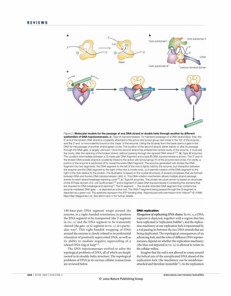
430 | JUNE 2002 | VOLUME 3 www.nature.com/reviews/molcellbio
R E V I E W S
Many a spectator of the linking-ring trick has been fas-cinated by the magician’s seemingly impossible feat oflinking solid rings into a chain, or separating a chaininto individual rings. In reality, the magician creates anillusion of interconversion by a sleight of hand: some ofthe rings are permanently linked and the others are per-manently separate — the one exception being a ‘keyring’ with a hidden opening, through which other ringscan be inserted or removed.
DNA topoisomerases are the true magicians ofthe DNA world. In their presence, DNA strands ordouble helices can pass through each other as if allphysical boundaries had disappeared: the inter-twined parental strands of a replicating DNA ringcan come apart, interlocked double-stranded DNArings (catenanes) can become unlinked and knotscan be introduced or removed from DNA rings. Incontrast to the hocus-pocus of the magician, how-ever, the DNA topoisomerases accomplish their feats bythe simple and elegant chemistry of transesterification.
In the strand-breakage reaction by a DNA topoiso-merase, a tyrosyl oxygen of the enzyme attacks a DNAphosphorus, forming a covalent phosphotyrosine linkand breaking a DNA phosphodiester bond at the sametime (FIG. 1). Rejoining of the DNA strand occurs by asecond transesterification, which is basically thereverse of the first — the oxygen of the DNA hydroxylgroup that is generated in the first reaction attacks the
CELLULAR ROLES OF DNATOPOISOMERASES: A MOLECULARPERSPECTIVEJames C. Wang
DNA topoisomerases are the magicians of the DNA world — by allowing DNA strands or doublehelices to pass through each other, they can solve all of the topological problems of DNA inreplication, transcription and other cellular transactions. Extensive biochemical and structuralstudies over the past three decades have provided molecular models of how the varioussubfamilies of DNA topoisomerase manipulate DNA. In this review, the cellular roles of theseenzymes are examined from a molecular point of view.
Department of Molecularand Cellular Biology,Harvard University,Fairchild Building,7 Divinity Avenue,Cambridge, Massachusetts02138, USA.e-mail:[email protected]:10.1038/nrm831
phosphorus of the phosphotyrosine link, breaking thecovalent bond between the protein and DNA, and re-forming the DNA backbone bond. These reactions cre-ate transient enzyme-mediated gates in the DNA for thepassage of another DNA strand or double helix.
DNA topoisomerases fall into two categories — type Iand type II. For the type I enzymes, the DNA strands aretransiently broken one at a time; for the type IIenzymes, by contrast, a pair of strands in a DNA doublehelix are transiently broken in concert by a dimericenzyme molecule. The two types can be further dividedinto four subfamilies: IA, IB, IIA and IIB (TABLE 1).Members of the same subfamily are structurally andmechanistically similar, whereas those of different sub-families are distinct.
The purpose of this review is to provide a perspectiveof the cellular roles of these remarkable enzymes from avista of their basic reaction characteristics — a morecomprehensive coverage of the literature can be foundin several recent reviews1–3. To provide the necessarybackdrop, some unique aspects of reactions that arecatalysed by the different subfamilies of the DNA topoi-somerases are summarized first. For clarity, reactionsthat are catalysed by these enzymes are often describedfor DNA rings. However, similar reactions occur in lin-ear chromosomes owing to their organization intointracellular structures that contain multiple loops, orbecause their ends are immobile.
© 2002 Nature Publishing Group

NATURE REVIEWS | MOLECULAR CELL BIOLOGY VOLUME 3 | JUNE 2002 | 431
R E V I E W S
NEGATIVELY AND POSITIVELY
SUPERCOILED DNA
A loop of a double-strandedDNA segment will becomecontorted if one end of it isturned around its helical axis atthat end while the other end iskept stationary in space (muchlike the spatial coiling of arubber tubing when similarlyhandled). The number ofsupercoils that are introducedinto this loop is a parameter thatis used to quantify the distortionof the looped DNA segment; onenegative supercoil is said to beintroduced into the DNA loopby each full turn of the rotatingend in the direction that tends tounwind the right-handed doublehelix; one positive supercoil issaid to be introduced by each fullturn of the rotating end in thedirection that tends to overwindthe right-handed double helix.
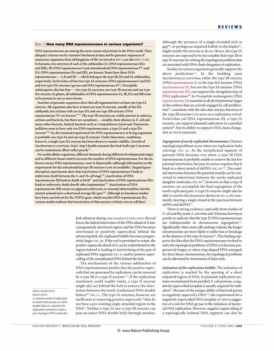
DNA ends relative to each other mediate the openingand closing of the DNA gate7.
Type IB. The type IB enzymes are thought to act by a‘DNA rotation’, rather than by an enzyme-bridging,mechanism3.When a DNA-bound type IB enzyme (FIG.2b)
transiently cleaves one of the DNA strands, only the sideof the DNA double helix that is upstream of the nick —the side containing the protein-linked 3′ end of the bro-ken strand — is tightly bound to the enzyme.Interaction between the downstream side of the dsDNAand the enzyme is mostly ionic in nature, so it presents alow barrier to rotation between the DNA and protein3.The DNA segments that flank a transient nick can there-fore rotate relative to each other by turning around oneof the single bonds that opposes the nick3.
The type IB enzymes are very efficient at relaxingboth positively and negatively supercoiled DNA.Although catenation or decatenation of nicked dsDNArings by a type IB enzyme in vitro has been reported8, itremains unclear how the enzyme carries out intermole-cular strand passage. A linear dsDNA intermediate, withthe enzyme covalently linked to one end of it, could beformed in such a reaction; if so, these reactions are prob-ably not significant in vivo.
There is another mechanistically important differencebetween the two type I enzymes. In the type IA-enzyme-catalysed reactions, breakage and rejoining of the DNAstrand occur in a single-stranded region1,4,5. In the reac-tions that are catalysed by the type IB enzymes, the nickis generated in a dsDNA segment3. Cleavage by a type IBenzyme in the single-stranded region of a dsDNA with asingle-stranded gap could occur, but the 5′ end of thetransiently broken DNA might readily detach from theenzyme, yielding a linear DNA intermediate.
Type II. In contrast to the type IA and type IBenzymes, the type IIA and IIB DNA topoisomerasescatalyse the ATP-dependent transport of one intactDNA double helix through another1–3,9. Before thefirst type IIB enzyme was identified in the archaeonSulfolobus shibatae10, all type II DNA topoisomeraseswere thought to belong to a single subfamily. It is nowclear that both type IIA and type IIB DNA topoiso-merases are widely distributed11 (BOX 1). FIGURE 2c
depicts a molecular model for the transport of oneDNA double helix through another by a type IIAenzyme9,12. The less-extensively studied type IIBenzymes share several common mechanistic featureswith the type IIA enzymes10, but there are distinctstructural differences between the two subfamilies13.
The ATP-dependent transport of one DNA doublehelix through another by a type II DNA topoisomeraseis manifested in several topological transformations,including catenation and decatenation of dsDNA rings,and the relaxation of positively or negatively super-coiled DNA1–3,9,12. The relative efficiencies of a giventype II enzyme in catalysing these reactions depend onthe structural features of the DNA substrates and theenzyme–DNA complexes. Bacterial gyrase (DNAtopoisomerase II), for example, is unique in that a
Reactions catalysed by topoisomerases Type IA. In the relaxation of an underwound or NEGATIVELY
SUPERCOILED DNA by a type IA enzyme, a short stretchof double-stranded (ds)DNA is first unpaired by thebinding of the enzyme, and a transient break isintroduced in this single-stranded region1,4,5. The lessnegatively supercoiled the DNA is, the more difficultit is for the enzyme to unpair the dsDNA; and so, theproficiency of the enzyme progressively decreasesduring the course of the reaction. Overwound orPOSITIVELY SUPERCOILED DNA is refractive to the type IAenzymes unless a pre-existing single-stranded regionis present5.
The type IA DNA topoisomerases can also pass oneDNA double helix through another if at least one of thepair contains a nick or gap6; in this reaction, an enzymeprobably introduces a transient break across from thenick or gap. The type IA enzymes are believed to catal-yse DNA strand passage by an ‘enzyme-bridging’ mech-anism (FIG. 2a), in which the DNA ends that are createdin the DNA breakage reaction are bridged by the topoi-somerase1,7,8, and movements of the enzyme-bound
OH OO DNA
PO
–OO DNAP
O
O–O
DNA
OH
DNA5′
3′
3′
5′
3′
Figure 1 | Catalysis of transient breakage of DNA by DNAtopoisomerases. Transesterification between an enzymetyrosyl and a DNA phosphate group leads to the breakage of a DNA backbone bond and the formation of a covalentenzyme–DNA intermediate. Rejoining of the DNA backbonebond occurs by the reversal of the reaction shown. In thereaction that is catalysed by a type IA or a type II enzyme, a 3′-OH is the leaving group and the active-site tyrosylbecomes covalently linked to a 5′-phosphoryl group, asdepicted. In the reaction that is catalysed by a type IB enzyme(not shown), a 5′-OH is the leaving group and the active-sitetyrosyl becomes covalently linked to a 3′-phosphoryl group.
Table 1 | Subfamilies of DNA topoisomerases
Subfamily Representative members
IA Bacterial DNA topoisomerases I and IIIYeast DNA topoisomerase IIIDrosophila melanogaster DNA topoisomerases IIIα and IIIβMammalian DNA topoisomerases IIIα and IIIβ
IB Eukaryotic DNA topoisomerase IMammalian mitochondrial DNA topoisomerase IPox virus topoisomerase
IIA Bacterial gyrase, DNA topoisomerase IVPhage T4 DNA topoisomeraseYeast DNA topoisomerase IIDrosophila DNA topoisomerase IIMammalian DNA topoisomerases IIα and IIβ
IIB Sulfolobus shibatae DNA topoisomerase VI(subunit A homologous to yeast Spo11)
© 2002 Nature Publishing Group
432 | JUNE 2002 | VOLUME 3 www.nature.com/reviews/molcellbio
R E V I E W S
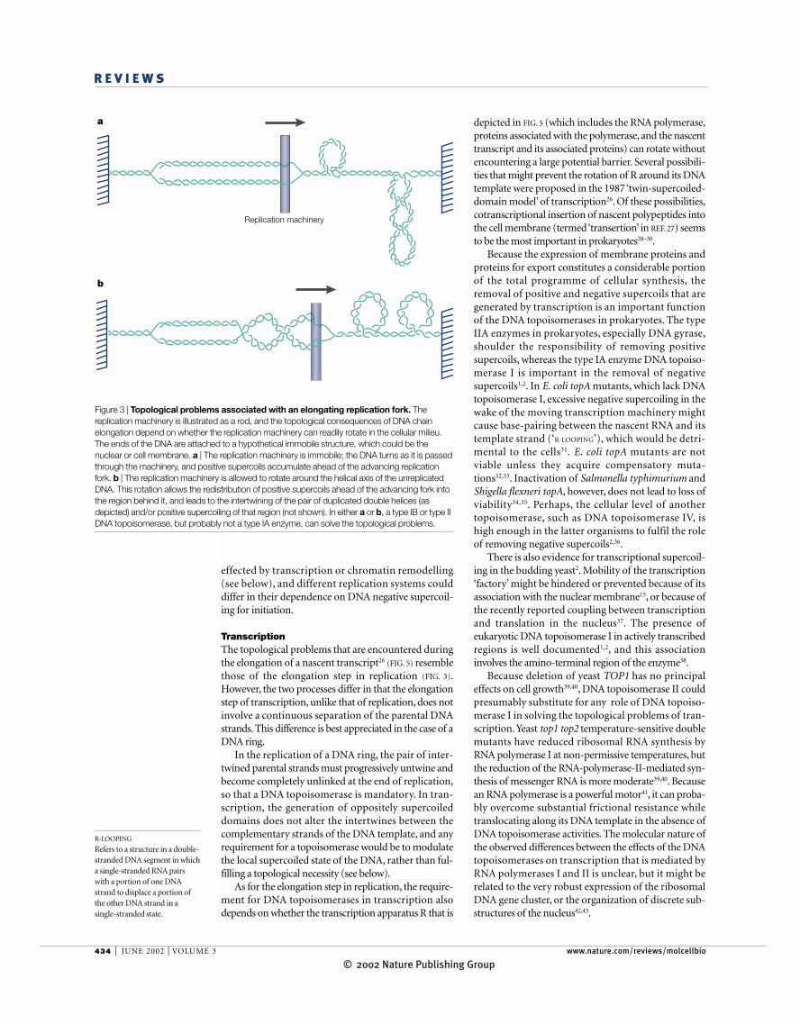
DNA replicationElongation of replicating DNA chains. In FIG. 3a, a DNAsegment is depicted, together with a region that hasbeen replicated (a ‘replication bubble’), and the replica-tion machinery at one replication fork is represented bya rod passing in-between the two DNA strands that arebeing duplicated. The topological consequences of anadvancing fork, and the roles of different DNA topoiso-merases, depend on whether the replication machinery(the blue rod depicted in FIG. 3a) is allowed to rotate inthe cellular milieu.
Imagine that the rod is not allowed to rotate aroundthe helical axis of the unreplicated DNA ahead of thereplication fork (the machinery can be membrane-attached and therefore immobile15). As the replication
140-base-pair DNA segment wraps around theenzyme, in a right-handed orientation, to positionthe DNA segment to be transported (the T-segmentin FIG. 2c) and the DNA segment to be transientlycleaved (the gate- or G-segment in FIG. 2c) in a partic-ular way9. This right-handed wrapping of DNAaround the enzyme is closely related to its preferentialrelaxation of positively supercoiled DNA, as well asits ability to mediate negative supercoiling of arelaxed DNA ring or loop9,14.
The DNA topoisomerases evolved to solve thetopological problems of DNA, all of which are deeplyrooted in its double-helix structure. The topologicalproblems of DNA in its various cellular transactionsare reviewed below.
* ** * * *
T
G
c
ba
3′
5′
3′
3′5′
5′'Lid'
'Base'
Core subdomain I
Core subdomain III
Core subdomain II
Linker
Carboxy-terminal domain
Y
Figure 2 | Molecular models for the passage of one DNA strand or double helix through another by differentsubfamilies of DNA topoisomerases. a | Type IA topoisomerases. On transient breakage of a DNA strand (blue line), the5′ end of the broken DNA strand is covalently attached to the active-site tyrosyl group (red circle) in the ‘lid’ of the enzyme,and the 3′ end is noncovalently bound to the ‘base’ of the enzyme. Lifting the lid away from the base opens a gate in theDNA for the passage of another strand (green circle). The location of the second strand, either before or after its passagethrough the DNA gate, is largely unknown. Once the second strand has entered the central cavity of the enzyme, it must exitthe cavity, after the rejoining of the broken strand, without passing through the rejoined DNA strand7,113. b | Type IB enzymes.The covalent intermediate between a 22-base-pair DNA fragment and a type IB DNA topoisomerase is shown. The 3′ end ofthe broken DNA scissile strand is covalently linked to the active-site tyrosyl group (Y) of the enzyme (red circle). For clarity, aportion of the enzyme is sectioned off to reveal the entire DNA fragment. The enzyme-generated nick divides the DNAfragment into two segments: the DNA segment to the left of the nick is tightly held by the enzyme, but interaction betweenthe enzyme and the DNA segment to the right of the nick is mostly ionic, so it permits rotation of the DNA segment to theright of the nick relative to the protein. The illustration is based on the crystal structures of several complexes that are formedbetween DNA and human DNA topoisomerase I (REF. 3). This DNA-rotation mechanism allows multiple strand-passageevents for each strand breakage–rejoining cycle114. c | Type IIA enzymes. The protein structure shown is based on structuresof the ATPase domain of E. coli GyrB protein115 and a fragment of yeast DNA topoisomerase II containing the domains thatare required for DNA breakage and rejoining12. The G-segment — the double-stranded DNA segment that contains theenzyme-mediated DNA gate — is depicted as a blue rod. The DNA T-segment being passed through the G-segment isdepicted as a green rod. The asterisks represent the ATP-binding sites. Reproduced with permission from Nature12 © (1996)Macmillan Magazines Ltd. See REFS 9 and 12 for further details.
© 2002 Nature Publishing Group
NATURE REVIEWS | MOLECULAR CELL BIOLOGY VOLUME 3 | JUNE 2002 | 433
R E V I E W S
although the presence of a single-stranded nick orgap6,8, or perhaps an unpaired bubble in the duplex17,might enable the enzyme to do so. Hence, the type IAenzymes are expected to be less suitable than type IB ortype II enzymes for solving the topological problems thatare associated with DNA chain elongation in replication.
Studies in various organisms generally support theabove predictions1,2. In the budding yeastSaccharomyces cerevisiae, either the type IB enzyme(DNA topoisomerase I) or the type IIA enzyme (DNAtopoisomerase II), but not the type IA enzyme (DNAtopoisomerase III), can support the elongation step ofDNA replication18. In Drosophila melanogaster, DNAtopoisomerase I is essential in all developmental stagesof the embryo that are actively engaged in cell prolifera-tion19, consistent with the idea that one key function ofthe type IB enzyme is to serve as a replication swivel.Escherichia coli DNA topoisomerase III, a type IAenzyme, can support plasmid replication in a purifiedsystem20, but its ability to support DNA chain elonga-tion in vivo is uncertain.
Segregation of newly replicated chromosomes. Distincttopological problems occur when two replication forksconverge (FIG. 4a). As the unreplicated segment ofparental DNA becomes very short, a type IB DNAtopoisomerase is probably unable to remove the last fewparental intertwines, because its action requires that itbinds to a short stretch of dsDNA (FIG. 2b)3,21. The resid-ual intertwines between the parental strands can be con-verted to intertwines between the newly replicateddaughter molecules (FIG. 4b)22, however, so that a type IIenzyme can accomplish the final segregation of thenewly replicated pair. A type IA enzyme might also beable to resolve the structures shown in FIG. 4a, by tran-siently cleaving a single strand at the junction betweenssDNA and dsDNA23.
There is strong evidence, especially from studies ofE. coli and the yeasts S. cerevisiae and Schizosaccharomycespombe, to indicate that the type II DNA topoisomerasesare indispensable in chromosome segregation1,2.Significantly, when yeast cells undergo mitosis, the longerchromosomes are more likely to suffer loss or breakagein the absence of the type II enzyme24. This finding sup-ports the idea that the DNA topoisomerases evolved tosolve the topological problems of DNA as it became pro-gressively longer, or when ring-shaped DNA emerged;for short linear chromosomes, the topological problemscan be alleviated by movements of their ends.
Initiation of the replication bubble. The initiation ofreplication is marked by the opening of a shortunpaired region of DNA. In plasmid-replication sys-tems reconstituted from purified E. coli proteins, a neg-atively supercoiled template is usually required for initi-ation25. Because of the unique ability of bacterial gyraseto negatively supercoil a DNA1,14, the requirement for anegatively supercoiled DNA template in vitro is sugges-tive of a role for DNA gyrase in the initiation of bacter-ial DNA replication. However, negative supercoiling ofa topologically isolated DNA segment can also be
fork advances during SEMICONSERVATIVE REPLICATION, the rodforces the helical intertwines of the DNA ahead of it intoa progressively shortened region, and the DNA becomesoverwound or positively supercoiled; behind theadvancing fork, the replicated bubble becomes progres-sively larger (FIG. 3b). If the rod is permitted to rotate, thepositive supercoils ahead of it can be redistributed to theregion behind it, leading to intertwining of the pair ofreplicated DNA segments (FIG. 3c) and/or positive super-coiling of the unreplicated DNA behind the fork.
The mechanisms of the various subfamilies ofDNA topoisomerases predict that the positive super-coils that are generated by replication can be removedby a type IB or a type II enzyme1–3. If the replicationmachinery could readily rotate, a type II enzymemight also act behind the fork to remove the inter-twines between the newly synthesized DNA doublehelices16 (FIG. 3c). The type IA enzymes, however, areinefficient at removing positive supercoils1,4 that donot have a pre-existing single-stranded region in theDNA5. Neither a type IA nor a type IB enzyme canpass an intact DNA double helix through another,
Box 1 | How many DNA topoisomerases in various organisms?
DNA topoisomerases are among the most conserved proteins in the DNA world. Theirubiquity is borne out by extensive biochemical data and by genomic sequences ofnumerous organisms from all kingdoms of life (reviewed in REF. 3; see also REFS 11,98).In humans, two enzymes of each of the subfamilies IA (DNA topoisomerases IIIαand IIIβ), IB (DNA topoisomerase I and mitochondrial DNA topoisomerase I99) andIIA (DNA topoisomerases IIα and IIβ), are known.Yeasts have three DNAtopoisomerases — I, II and III — which belong to the type IB, IIA and IA subfamilies,respectively. Escherichia coli has two type IA enzymes (DNA topoisomerases I and III)and two type IIA enzymes (gyrase and DNA topoisomerase IV). Drosophilamelanogaster also has four — two type IA enzymes, one type IB enzyme and one typeIIA enzyme. In plants, all subfamilies of DNA topoisomerases IA, IB, IIA and IIB seemto be present in one or more forms.
Searches of genomic sequences show that all organisms have at least one type IAenzyme. All organisms also have at least one type II enzyme, usually of the IIAsubfamily, but archaea with no type IIA and one type IIB enzyme (DNAtopoisomerase VI) are known10,11. The type IB enzymes are widely present in eukarya,archaea and bacteria, but there are exceptions — notably, their absence in E. coli andmany other bacteria. Indeed, bacteria such as Campylobacter jejuni and Treponemapallidum seem to have only two DNA topoisomerases, a type IA and a type IIAenzyme3,98. So, the minimal requirement for DNA topoisomerases in living organismsis probably one type IA and one type II enzyme. Under laboratory conditions,however, a single type IIA enzyme has been shown to sustain viability. Growth ofSaccharomyces cerevisiae ∆top1 ∆top3 double mutants that lack both type I enzymescan be maintained, albeit rather poorly55.
For multicellular organisms, different demands during different developmental stagesand in different tissues tend to increase the number of DNA topoisomerases. For the sixknown mouse DNA topoisomerases, none is dispensable (although information on therequirement for the mitochondrial type IB enzyme is not yet available). Targeted gene-disruption experiments show that inactivation of DNA topoisomerase I leads toembryonic death between the 4- and 16-cell stage100, inactivation of DNAtopoisomerase IIβ leads to death at birth97 and inactivation of DNA topoisomerase IIIαleads to embryonic death shortly after implantation101. Inactivation of DNAtopoisomerase IIIβ causes no apparent embryonic or neonatal abnormalities, but themutant animals have a shortened average life span102. Although no knockout studieshave been carried out for the TOP2α gene, which encodes DNA topoisomerase IIα,various studies indicate that inactivation of this enzyme is lethal, even in cell lines2.
SEMICONSERVATIVE
REPLICATION
A common mode of replicationin which both strands of a DNAdouble helix are copied by thereplication machinery to give apair of progeny DNA molecules.
© 2002 Nature Publishing Group
434 | JUNE 2002 | VOLUME 3 www.nature.com/reviews/molcellbio
R E V I E W S
R-LOOPING
Refers to a structure in a double-stranded DNA segment in whicha single-stranded RNA pairswith a portion of one DNAstrand to displace a portion ofthe other DNA strand in asingle-stranded state.
depicted in FIG. 5 (which includes the RNA polymerase,proteins associated with the polymerase, and the nascenttranscript and its associated proteins) can rotate withoutencountering a large potential barrier. Several possibili-ties that might prevent the rotation of R around its DNAtemplate were proposed in the 1987 ‘twin-supercoiled-domain model’ of transcription26. Of these possibilities,cotranscriptional insertion of nascent polypeptides intothe cell membrane (termed ‘transertion’ in REF. 27) seemsto be the most important in prokaryotes28–30.
Because the expression of membrane proteins andproteins for export constitutes a considerable portionof the total programme of cellular synthesis, theremoval of positive and negative supercoils that aregenerated by transcription is an important functionof the DNA topoisomerases in prokaryotes. The typeIIA enzymes in prokaryotes, especially DNA gyrase,shoulder the responsibility of removing positivesupercoils, whereas the type IA enzyme DNA topoiso-merase I is important in the removal of negativesupercoils1,2. In E. coli topA mutants, which lack DNAtopoisomerase I, excessive negative supercoiling in thewake of the moving transcription machinery mightcause base-pairing between the nascent RNA and itstemplate strand (‘R-LOOPING’), which would be detri-mental to the cells31. E. coli topA mutants are notviable unless they acquire compensatory muta-tions32,33. Inactivation of Salmonella typhimurium andShigella flexneri topA, however, does not lead to loss ofviability34,35. Perhaps, the cellular level of anothertopoisomerase, such as DNA topoisomerase IV, ishigh enough in the latter organisms to fulfil the roleof removing negative supercoils2,36.
There is also evidence for transcriptional supercoil-ing in the budding yeast2. Mobility of the transcription‘factory’ might be hindered or prevented because of itsassociation with the nuclear membrane15, or because ofthe recently reported coupling between transcriptionand translation in the nucleus37. The presence ofeukaryotic DNA topoisomerase I in actively transcribedregions is well documented1,2, and this associationinvolves the amino-terminal region of the enzyme38.
Because deletion of yeast TOP1 has no principaleffects on cell growth39,40, DNA topoisomerase II couldpresumably substitute for any role of DNA topoiso-merase I in solving the topological problems of tran-scription. Yeast top1 top2 temperature-sensitive doublemutants have reduced ribosomal RNA synthesis byRNA polymerase I at non-permissive temperatures, butthe reduction of the RNA-polymerase-II-mediated syn-thesis of messenger RNA is more moderate39,40. Becausean RNA polymerase is a powerful motor41, it can proba-bly overcome substantial frictional resistance whiletranslocating along its DNA template in the absence ofDNA topoisomerase activities. The molecular nature ofthe observed differences between the effects of the DNAtopoisomerases on transcription that is mediated byRNA polymerases I and II is unclear, but it might berelated to the very robust expression of the ribosomalDNA gene cluster, or the organization of discrete sub-structures of the nucleus42,43.
effected by transcription or chromatin remodelling(see below), and different replication systems coulddiffer in their dependence on DNA negative supercoil-ing for initiation.
Transcription The topological problems that are encountered duringthe elongation of a nascent transcript26 (FIG. 5) resemblethose of the elongation step in replication (FIG. 3).However, the two processes differ in that the elongationstep of transcription, unlike that of replication, does notinvolve a continuous separation of the parental DNAstrands. This difference is best appreciated in the case of aDNA ring.
In the replication of a DNA ring, the pair of inter-twined parental strands must progressively untwine andbecome completely unlinked at the end of replication,so that a DNA topoisomerase is mandatory. In tran-scription, the generation of oppositely supercoileddomains does not alter the intertwines between thecomplementary strands of the DNA template, and anyrequirement for a topoisomerase would be to modulatethe local supercoiled state of the DNA, rather than ful-filling a topological necessity (see below).
As for the elongation step in replication, the require-ment for DNA topoisomerases in transcription alsodepends on whether the transcription apparatus R that is
Replication machinery
a
b
Figure 3 | Topological problems associated with an elongating replication fork. Thereplication machinery is illustrated as a rod, and the topological consequences of DNA chainelongation depend on whether the replication machinery can readily rotate in the cellular milieu.The ends of the DNA are attached to a hypothetical immobile structure, which could be thenuclear or cell membrane. a | The replication machinery is immobile; the DNA turns as it is passedthrough the machinery, and positive supercoils accumulate ahead of the advancing replicationfork. b | The replication machinery is allowed to rotate around the helical axis of the unreplicatedDNA. This rotation allows the redistribution of positive supercoils ahead of the advancing fork intothe region behind it, and leads to the intertwining of the pair of duplicated double helices (asdepicted) and/or positive supercoiling of that region (not shown). In either a or b, a type IB or type IIDNA topoisomerase, but probably not a type IA enzyme, can solve the topological problems.
© 2002 Nature Publishing Group

NATURE REVIEWS | MOLECULAR CELL BIOLOGY VOLUME 3 | JUNE 2002 | 435
R E V I E W S
results are available yet to support these ideas. For avery long transcript, or for closely spaced transcriptsalong a very actively transcribed gene, entanglementbetween a transcript and its template15 (or between thetranscripts themselves) could present a problem. Also,in cases where a stably base-paired region between anascent RNA and its template strand has formed, atopoisomerase-mediated untwining of the transcriptfrom the template strand, rather than the nucleolyticremoval of the DNA-bound RNA by RNase H, couldalso be used to salvage the transcript.
DNA recombinationMany fundamental steps in different cellular processesoften share common features. The structure that isshown in FIG. 4a to illustrate the problem of resolving apair of intertwined parental strands between two con-verging replication forks, for example, can also be usedto illustrate the pairing of two gapped DNA moleculesto form a recombination intermediate. Resolution of apair of newly replicated chromosomes and of an inter-mediate of recombinational repair clearly share com-mon topological features, and might involve the sameDNA topoisomerases49.
The roles of DNA topoisomerases in modulating thefrequency of recombination events have received muchinterest. Inactivation of any one of the three yeast DNAtopoisomerases increases genome instability50, and theeffects of inactivating the type IA enzymes are particu-larly striking (see below). Equally significant is the find-ing that a key player in meiotic recombination is relatedto one subunit of the type IIB DNA topoisomerases10,51.The recombination field itself is undergoing a seachange, and the importance of recombinational repairof DNA damage, and in restoring stalled replicationforks, has received increasing attention52,53.
Type IA enzymes in recombinational repair. As summa-rized in BOX 1, at least one type IA enzyme is present inall living organisms. Why this omnipresence? What arethe cellular roles of the type IA DNA topoisomerases?Studies of the past decade point to an important role forthese enzymes in the resolution of intermediates that arefound in recombinational repair.
E. coli cells that lack both type IA DNA topoisomerasesare non-viable, even in the presence of a topA compen-satory mutation54. These cells have extensive filamenta-tion and possess an abnormal nucleoid structure. Theviability of topA topB double-mutant cells that carry atopA compensatory mutation can be restored, however,by a further deletion of the recA gene54. These findingsindicate that, in bacteria, the type IA enzymes might beinvolved in RecA-mediated recombination and that theycould specifically resolve recombination intermediatesbefore chromosome segregation54.
In the yeasts, inactivating the single type IA enzyme,DNA topoisomerase III, leads to loss of viability inS. pombe but not in S. cerevisiae 55–57. S. cerevisiae top3nulls show a complex phenotype, including slow growthand reduced viability, hyper-recombination betweenrepetitive sequences, hypersensitivity to DNA-damaging
Transcription of a chromatin template dotted withnucleosomes poses additional topological problems.Passage of an advancing RNA polymerase through anucleosome has been postulated to periodicallyenclose a DNA loop containing both the partiallyunravelled nucleosome and the polymerase44. A DNAtopoisomerase might be involved in continued translo-cation of the polymerase in such a loop. Interestingly, arecent study45 indicates that efficient transcription of achromatin — but not a DNA — template requires thepresence of a DNA topoisomerase in the transcriptioncomplex. However, all postulates that invoke a stringenttopoisomerase requirement in transcription must bereconciled with the finding that, in yeast, inactivation ofboth DNA topoisomerases I and II does not drasticallyreduce mRNA synthesis39,40.
DNA topoisomerase I has also been shown to act as aco-activator in purified mammalian transcription sys-tems46–48. The topoisomerase seems to facilitate the for-mation of an active TFIID–TFIIA protein complex onthe promoter48, but the DNA strand breakage andrejoining activity of the topoisomerase is not required inthis co-activator role46,48.
There are further problems of transcription thatmight involve topoisomerases, although no experimental
a
b
c
Figure 4 | A distinct topological problem occurs when two replication forks converge.When the unreplicated DNA segment becomes very short (a), the type IB topoisomerase enzymecannot remove the last few intertwines between the parental strands. These single-strandedintertwines can probably be removed by a type IA enzyme23, or be converted to double-strandedintertwines (b and c) for removal by a type II DNA topoisomerase22. Modified from REF. 22. ©(1981), with permission from Elsevier Science.
© 2002 Nature Publishing Group

436 | JUNE 2002 | VOLUME 3 www.nature.com/reviews/molcellbio
R E V I E W S
HOLLIDAY JUNCTION
A DNA structure named afterRobin Holliday, who firstdescribed it in 1964 as aplausible recombinationintermediate between a pair ofhomologous DNA molecules.
breaks at the hot-spots of meiotic recombination69.Although Spo11 seems to catalyse DNA breakage as wellas rejoining during meiosis, it differs from the archetypaltype IIB DNA topoisomerases in that there is, as yet, noevidence for its involvement in DNA strand passage69.
Interestingly, although the A subunit of a type IIBenzyme seems to contain the counterparts of all struc-tural motifs of the type IIA DNA topoisomerases thatare known to be important in the catalysis of DNAbreakage and rejoining (including the active-site tyro-sine region and a Rossmann fold termed the ‘toprim’), itdoes not cleave DNA in the absence of the B subunit70.This finding raises several questions. Does DNA cleav-age by Spo11 require a second subunit? Does its mecha-nism resemble that of a type IIB DNA topoisomerase, atleast in terms of DNA breakage and rejoining? If theanswer is yes, how is the rejoining reaction preventedwhen a stable double-stranded break is finally formed?
Topoisomerases and chromosome condensationChromatin compaction, chromosome segregation andDNA topology are intricately interrelated71–74. Ineukaryotes, earlier genetic studies of the fission yeast,cytological studies of various cells treated with topoiso-merase inhibitors, and biochemical studies of chromo-some condensation in cell extracts have all implicatedDNA topoisomerase II in chromatin and chromosomecondensation during mitosis1,2,75. Mammalian DNAtopoisomerase II has also been implicated in apoptoticchromatin condensation76.
Eukaryotic DNA topoisomerase I has also been pro-posed to have a role in chromosome condensation77,78.In yeast, DNA topoisomerase I and the product of theTRF4 gene seem to share such a role in mitotic cells77;trf4 top1 double mutants are defective in chromosomecondensation, spindle elongation and nuclear segrega-tion77, and they also fail to establish the condensed stateof the rDNA locus at mitosis78.
The idea that at least one DNA topoisomerase isrequired for chromosome condensation and deconden-sation is consistent with the expected changes in the twistand writhe of a long DNA when it undergoes protein-mediated compaction. Recent studies of the condensins— ubiquitous ATP-dependent protein complexes that arecrucial for chromosome condensation — have alsoimplicated a requirement for a DNA topoisomerase inchromosome condensation74.
In eukaryotes, either a type II or a type IB enzymeshould be able to solve the topological problems of coil-ing a DNA into a compact form (or in decondensation).Historically, eukaryotic DNA topoisomerase II has beenthe focus of attention in chromosome condensation75,but its precise role in the many steps of morphologicallydistinct changes between an interphase chromosome anda highly condensed metaphase chromosome is unclear.Whether DNA topoisomerase II has a role during mei-otic chromosome condensation in S. pombe has alsobeen questioned79.
In bacteria, it seems that DNA supercoiling per se canalso affect chromosome compaction and segregation.The efficiency of E. coli plasmid partition seems to be
agents and inability to sporulate2,55. Genetic screens forsuppressors of S. cerevisiae top3 null mutants led to theidentification of mutations that map to the gene SGS158.This gene was shown to encode a helicase of the RecQfamily58,59, the members of which include E. coli RecQprotein, S. pombe Rqh1 protein, and the human Bloomsyndrome and Werner syndrome proteins, BLM andWRN, respectively60–62. A link between another humanRecQ helicase RECQL4 and the Rothmond–Thomsonsyndrome has also been proposed63. The three humansyndromes show signs of genome instability, and arevariously characterized by growth abnormality, predis-position to different cancers and signs of premature age-ing60–62. There is strong evidence that Sgs1 helicase phys-ically interacts with DNA topoisomerase III58,64,65, andinteraction between other members of the RecQ familyand type IA DNA topoisomerases has also beenreported60–62,66.
Several studies have implicated SGS1 in DNArecombination and repair67,68. Significantly, it is the inac-tivation of SGS1 that suppresses the requirement for atype IA enzyme, which suggests that a type IA enzymemight be needed to resolve a structure that is formedduring recombination or repair58. What might be themolecular nature of such a structure? Why is a type IADNA topoisomerase specifically required? Could thisunique role of the type IA enzymes be related to theiromnipresence in all organisms? In BOX 2, the plausibleinvolvement of a type IA DNA topoisomerase in the res-olution of Holliday structures, especially the doubleHOLLIDAY JUNCTION, is discussed.
A type IIB enzyme in meiotic recombination? Theamino-acid sequence of the type IIB enzyme DNAtopoisomerase VI indicated the presence of its homo-logues in various organisms, of which the SPO11 geneproduct of S. cerevisiae is of particular interest10,69. TheSpo11 protein links covalently to the 5′ ends of dsDNA
Positive supercoils
R
Figure 5 | Generation of oppositely supercoiled domains by transcription. The transcriptionapparatus R — including the RNA polymerase and its associated proteins, the nascent RNA andRNA-bound proteins — is represented by a rod. When R cannot rotate around the helical axis ofthe DNA template, overwinding or positive supercoiling of the DNA template ahead of R isaccompanied by underwinding or negative supercoiling of the DNA template behind R. Thetopology of replicative elongation (FIG. 3) can be viewed as a special case of the generation of twinsupercoiled domains by a machinery tracking along a DNA double helix. The separation of twointertwined strands behind the replication machinery can be considered as a special case ofnegative supercoiling (two unlinked single-stranded DNA rings of complementary sequences, forexample, can be considered as the most negatively supercoiled form of a duplex DNA ring).
© 2002 Nature Publishing Group
NATURE REVIEWS | MOLECULAR CELL BIOLOGY VOLUME 3 | JUNE 2002 | 437
R E V I E W S
ENDONUCLEASE
An enzyme that catalyseshydrolytic cleavage of DNA inthe middle of a DNA strand ordouble helix.
DNA STRAND-TRANSFERASE
An enzyme that transfers adonor group of one DNA strand(for example, a 5′ phosphorylgroup) to a receiving group ofanother DNA strand (forexample, a 3′ hydroxyl group).
BRANCH MIGRATION
The movement of a junctionformed by multiple DNA orRNA strands of complementarynucleotide sequences that resultsfrom nearly simultaneousbreakage and formation of basepairs among them.
DNA topoisomerases and chromosome structureIn general, whenever a long chromatin fibre undergoes astructural change, any accompanying changes in itstwist and writhe could require the catalytic action of oneor more DNA topoisomerases. Several studies have alsoimplicated DNA topoisomerases in chromatin structureand organization. The association of Drosophila DNAtopoisomerase II and human DNA topoisomerase IIβwith ATP-using chromatin remodelling complexes has
increased by the presence of a strong gyrase-binding siteon a plasmid80,81, or by a reduction of the intracellularactivity of DNA topoisomerase I82. A higher degree ofnegative supercoiling was thought to make the plasmidmolecules more compact, and hence more easily segre-gated by random partition82. E. coli topA mutants werealso found to suppress the production of anucleate cellscaused by mutations in the muk genes, which encode pro-teins that resemble the core subunits of the condensins83.
Box 2 | Type IA DNA topoisomerase in Holliday-junction resolution?
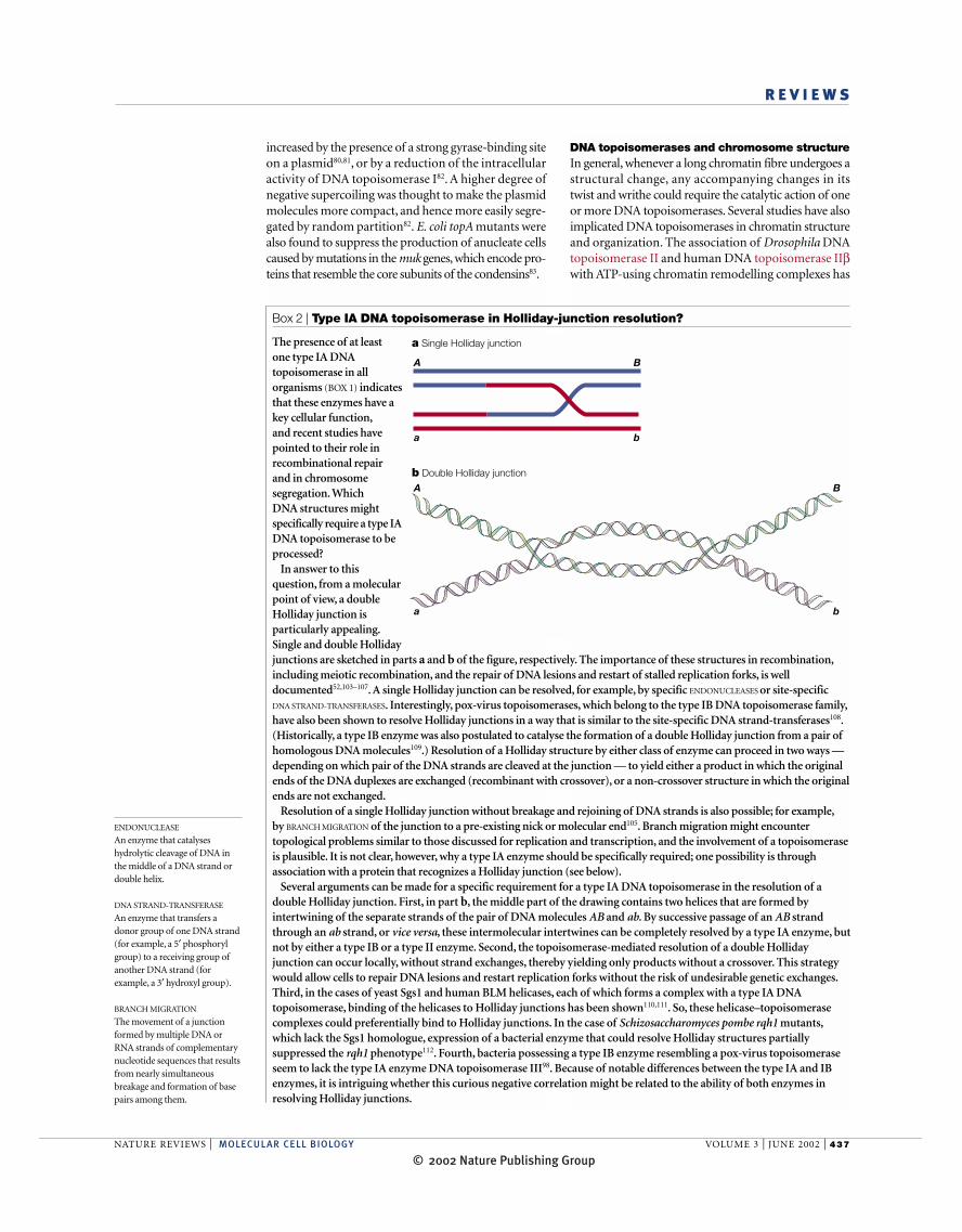
The presence of at least one type IA DNAtopoisomerase in allorganisms (BOX 1) indicatesthat these enzymes have akey cellular function,and recent studies havepointed to their role inrecombinational repairand in chromosomesegregation. Which DNA structures mightspecifically require a type IADNA topoisomerase to beprocessed?
In answer to thisquestion, from a molecularpoint of view, a doubleHolliday junction isparticularly appealing.Single and double Hollidayjunctions are sketched in parts a and b of the figure, respectively. The importance of these structures in recombination,including meiotic recombination, and the repair of DNA lesions and restart of stalled replication forks, is welldocumented52,103–107. A single Holliday junction can be resolved, for example, by specific ENDONUCLEASES or site-specific DNA STRAND-TRANSFERASES. Interestingly, pox-virus topoisomerases, which belong to the type IB DNA topoisomerase family,have also been shown to resolve Holliday junctions in a way that is similar to the site-specific DNA strand-transferases108.(Historically, a type IB enzyme was also postulated to catalyse the formation of a double Holliday junction from a pair ofhomologous DNA molecules109.) Resolution of a Holliday structure by either class of enzyme can proceed in two ways —depending on which pair of the DNA strands are cleaved at the junction — to yield either a product in which the originalends of the DNA duplexes are exchanged (recombinant with crossover), or a non-crossover structure in which the originalends are not exchanged.
Resolution of a single Holliday junction without breakage and rejoining of DNA strands is also possible; for example,by BRANCH MIGRATION of the junction to a pre-existing nick or molecular end105. Branch migration might encountertopological problems similar to those discussed for replication and transcription, and the involvement of a topoisomeraseis plausible. It is not clear, however, why a type IA enzyme should be specifically required; one possibility is throughassociation with a protein that recognizes a Holliday junction (see below).
Several arguments can be made for a specific requirement for a type IA DNA topoisomerase in the resolution of adouble Holliday junction. First, in part b, the middle part of the drawing contains two helices that are formed byintertwining of the separate strands of the pair of DNA molecules AB and ab. By successive passage of an AB strandthrough an ab strand, or vice versa, these intermolecular intertwines can be completely resolved by a type IA enzyme, butnot by either a type IB or a type II enzyme. Second, the topoisomerase-mediated resolution of a double Hollidayjunction can occur locally, without strand exchanges, thereby yielding only products without a crossover. This strategywould allow cells to repair DNA lesions and restart replication forks without the risk of undesirable genetic exchanges.Third, in the cases of yeast Sgs1 and human BLM helicases, each of which forms a complex with a type IA DNAtopoisomerase, binding of the helicases to Holliday junctions has been shown110,111. So, these helicase–topoisomerasecomplexes could preferentially bind to Holliday junctions. In the case of Schizosaccharomyces pombe rqh1 mutants,which lack the Sgs1 homologue, expression of a bacterial enzyme that could resolve Holliday structures partiallysuppressed the rqh1 phenotype112. Fourth, bacteria possessing a type IB enzyme resembling a pox-virus topoisomeraseseem to lack the type IA enzyme DNA topoisomerase III98. Because of notable differences between the type IA and IBenzymes, it is intriguing whether this curious negative correlation might be related to the ability of both enzymes inresolving Holliday junctions.
a Single Holliday junction
b Double Holliday junction
A B
a b
A B
a b
© 2002 Nature Publishing Group

438 | JUNE 2002 | VOLUME 3 www.nature.com/reviews/molcellbio
R E V I E W S
convert DNA topoisomerases to DNA-damagingagents also underscores nature’s dilemma — it mustsolve the topological problems of DNA that comewith its double-helical structure, yet the solution uti-lizing the DNA topoisomerases comes with the risk ofcreating weak spots in the DNA. Although, on the onehand, DNA topoisomerases are key targets for thedevelopment of better therapeutics, on the other handthese enzymes might also suffer assaults by naturaland synthesized products, sometimes with devastat-ing consequences. A plausible role for DNA topoiso-merase II in carcinogenesis, for example, has beenproposed96. Further studies of a potential role for theDNA topoisomerases in preventive medicine areurgently needed.
The many tasks of the DNA topoisomerases oftenmake it a real challenge to establish the links betweentheir molecular roles and the physiological conse-quences of their inactivation. For example, deletionof the mouse TOP2β gene, which encodes DNAtopoisomerase IIβ, leads to defects in the formationof neuromuscular junctions97. How does an enzymethat acts on DNA in the nucleus affect the formationof neuromuscular junctions? We still do not know.
Interactions between various DNA topoiso-merases and other proteins — for example, betweena type IA DNA topoisomerase and a RecQ-familyhelicase58,60–62,64–66 — and the mechanistic and func-tional consequences of these interactions, have notyet been sufficiently explored to shed light on themany questions still remaining. Clearly, the study ofthese fascinating enzymes has not yet passed thepoint of diminishing returns.
been reported84,85, although the functionality of thisassociation is yet to be established.
A structural role for eukaryotic DNA topoisomeraseII in the higher-order organization of chromosomes hasalso been postulated86. Mammalian DNA topoisomeraseII (presumably the α-isoform) is a main non-histoneprotein in the axial core or scaffold of metaphase chro-mosomes87. The co-localization of Drosophila DNAtopoisomerase II with a protein called Barren, which isinvolved in chromosome condensation88, is also consis-tent with a direct role for the topoisomerase in chromo-some organization. However, recent studies of fluores-cently tagged human DNA topoisomerases IIα and IIβindicate that neither enzyme is an immobile structuralcomponent of the chromosomal scaffold89.
Topoisomerases as targets of therapeutic agentsDNA topoisomerases have been shown to be the molec-ular targets of many antimicrobial and anticanceragents. Among topoisomerase-targeting drugs in clini-cal use at present, most (if not all) act by trapping thecovalent DNA–enzyme intermediates to convert a nor-mal cellular enzyme to a DNA-damaging agent. Severalrecent reviews of DNA topoisomerases in pharmacol-ogy and clinical medicine have been published90–92.
ConclusionsIn the three decades since the discovery of the firstDNA topoisomerase4, extensive studies of theseenzymes have led to a much better understanding oftheir reaction mechanisms and cellular roles. Theidentification of many chemically distinct toxins93–95,natural products and synthetic compounds90–92 that
1. Wang, J. C. DNA topoisomerases. Annu. Rev. Biochem. 65,635–692 (1996).
2. Nitiss, J. L. Investigating the biological functions of DNAtopoisomerases in eukaryotic cells. Biochim. Biophys. Acta1400, 63–81 (1998).A comprehensive review of the cellular functions ofeukaryotic DNA topoisomerases.
3. Champoux, J. J. DNA topoisomerases: structure, function,and mechanism. Annu. Rev. Biochem. 70, 369–413 (2001).
4. Wang, J. C. Interaction between DNA and an Escherichia coli protein ω. J. Mol. Biol. 55, 523–533 (1971).This reports the discovery of the first DNAtopoisomerase, and proposes the transesterificationmechanism of DNA strand breakage and rejoining.
5. Kirkegaard, K. & Wang, J. C. Bacterial DNA topoisomerase Ican relax positively supercoiled DNA containing a single-stranded loop. J. Mol. Biol. 185, 625–637 (1985).
6. Tse, Y. & Wang, J. C. E. coli and M. luteus DNAtopoisomerase I can catalyze catenation or decatenation ofdouble-stranded DNA rings. Cell 22, 269–276 (1980).
7. Lima, C. D., Wang, J. C. & Mondragon, A. Three-dimensional structure of the 67K N-terminal fragment of E. coli DNA topoisomerase I. Nature 367, 138–146 (1994).
8. Brown, P. O. & Cozzarelli, N. R. Catenation and knotting ofduplex DNA by type 1 topoisomerases: a mechanisticparallel with type 2 topoisomerases. Proc. Natl Acad. Sci. USA 78, 843–847 (1981).
9. Wang, J. C. Moving one DNA double helix through anotherby a type II DNA topoisomerase: the story of a simplemolecular machine. Q. Rev. Biophys. 31, 107–144 (1998).
10. Bergerat, A. et al. An atypical topoisomerase II from Archaeawith implications for meiotic recombination. Nature 386,414–417 (1997).A report on the discovery of the type IIBtopoisomerase.
11. Forterre, P. Genomics and early cellular evolution. The origin ofthe DNA world. C. R. Acad. Sci. III 324, 1067–1076 (2001).
12. Berger, J. M., Gamblin, S. J., Harrison, S. C. & Wang, J. C.Structure and mechanism of DNA topoisomerase II. Nature379, 225–232 (1996).
13. Nichols, M. D., DeAngelis, K., Keck, J. L. & Berger, J. M.Structure and function of an archaeal topoisomerase VIsubunit with homology to the meiotic recombination factorSpo11. EMBO J. 18, 6177–6188 (1999).
14. Kampranis, S. C., Bates, A. D. & Maxwell, A. A model for themechanism of strand passage by DNA gyrase. Proc. NatlAcad. Sci. USA 96, 8414–8419 (1999).
15. Cook, P. R. The organization of replication and transcription.Science 284, 1790–1795 (1999).
16. Postow, L., Crisona, N. J., Peter, B. J., Hardy, C. D. &Cozzarelli, N. R. Topological challenges to DNA replication:conformations at the fork. Proc. Natl Acad. Sci. USA 98,8219–8226 (2001).A detailed review of the topological problems of DNAchain elongation during replication, and theirplausible solutions by the DNA topoisomerases,especially the type II enzymes. Much of thediscussions, however, deal with cases in which rotation of the replication machinery isunconstrained.
17. Harmon, F. G., DiGate, R. J. & Kowalczykowski, S. C. RecQhelicase and topoisomerase III comprise a novel DNA strandpassage function: a conserved mechanism for control ofDNA recombination. Mol. Cell 3, 611–620 (1999).
18. Kim, R. A. & Wang, J. C. Function of DNA topoisomerasesas replication swivels in Saccharomyces cerevisiae. J. Mol.Biol. 208, 257–267 (1989).
19. Zhang, C. X., Chen, A. D., Gettel, N. J. & Hsieh, T. S.Essential functions of DNA topoisomerase I in Drosophilamelanogaster. Dev. Biol. 222, 27–40 (2000).An elegant study of the cellular roles of DNAtopoisomerase I through its controlled expressionfrom a heat-shock-gene promoter.
20. Hiasa, H. & Marians, K. J. Topoisomerase III, but not
topoisomerase I, can support nascent chain elongationduring θ-type DNA replication. J. Biol. Chem. 269,32655–32659 (1994).
21. Redinbo, M. R., Stewart, L., Kuhn, P., Champoux, J. J. &Hol, W. G. Crystal structures of human topoisomerase I incovalent and noncovalent complexes with DNA. Science279, 1504–1513 (1998).
22. Sundin, O. & Varshavsky, A. Arrest of segregation leads toaccumulation of highly intertwined catenated dimers:dissection of the final stages of SV40 DNA replication. Cell25, 659–669 (1981).The first demonstration of the formation of multipleintertwined pairs of newly replicated DNA rings in theabsence of activities that can resolve them.
23. Hiasa, H., DiGate, R. J. & Marians, K. J. Decatenatingactivity of Escherichia coli DNA gyrase and topoisomerases Iand III during oriC and pBR322 DNA replication in vitro. J. Biol. Chem. 269, 2093–2099 (1994).
24. Spell, R. M. & Holm, C. Nature and distribution ofchromosomal intertwinings in Saccharomyces cerevisiae.Mol. Cell. Biol. 14, 1465–1476 (1994).This report shows clearly that chromosome lossand/or breakage in the absence of a type II DNAtopoisomerase is particularly severe for largerchromosomes.
25. Crooke, E., Hwang, D. S., Skarstad, K., Thony, B. &Kornberg, A. E. coli minichromosome replication: regulationof initiation at oriC. Res. Microbiol. 142, 127–130 (1991).
26. Liu, L. F. & Wang, J. C. Supercoiling of the DNA templateduring transcription. Proc. Natl Acad. Sci. USA 84,7024–7027 (1987).This proposes a ‘twin-supercoiled-domain’ modelwhen rotation of a transcription assembly in thecellular milieu is hindered, and discusses how variousDNA topoisomerases might act to remove the positiveand negative supercoils that are generated in the DNAtemplate.
© 2002 Nature Publishing Group

NATURE REVIEWS | MOLECULAR CELL BIOLOGY VOLUME 3 | JUNE 2002 | 439
R E V I E W S
27. Norris, V. Hypothesis: chromosome separation inEscherichia coli involves autocatalytic gene expression,transertion and membrane-domain formation. Mol.Microbiol. 16, 1051–1057 (1995).
28. Lodge, J. K., Kazic, T. & Berg, D. E. Formation ofsupercoiling domains in plasmid pBR322. J. Bacteriol.171, 2181–2187 (1989).
29. Cook, D. N., Ma, D., Pon, N. G. & Hearst, J. E. Dynamics ofDNA supercoiling by transcription in Escherichia coli. Proc.Natl Acad. Sci. USA 89, 10603–10607 (1992).
30. Lynch, A. S. & Wang, J. C. Anchoring of DNA to thebacterial cytoplasmic membrane through cotranscriptionalsynthesis of polypeptides encoding membrane proteins orproteins for export: a mechanism of plasmid hypernegativesupercoiling in mutants deficient in DNA topoisomerase I. J. Bacteriol. 175, 1645–1655 (1993).
31. Drolet, M. et al. Overexpression of RNase H partiallycomplements the growth defect of an Escherichia coli ∆topAmutant: R-loop formation is a major problem in the absenceof DNA topoisomerase I. Proc. Natl Acad. Sci. USA 92,3526–3530 (1995).
32. Pruss, G. J., Manes, S. H. & Drlica, K. Escherichia coliDNA topoisomerase I mutants: increased supercoiling iscorrected by mutations near gyrase genes. Cell 31, 35–42(1982).
33. DiNardo, S., Voelkel, K. A., Sternglanz, R., Reynolds, A. E. & Wright, A. Escherichia coli DNA topoisomerase I mutantshave compensatory mutations in DNA gyrase genes. Cell31, 43–51 (1982).
34. Richardson, S. M., Higgins, C. F. & Lilley, D. M. The geneticcontrol of DNA supercoiling in Salmonella typhimurium.EMBO J. 3, 1745–1752 (1984).
35. Bhriain, N. N. & Dorman, C. J. Isolation and characterizationof a topA mutant of Shigella flexneri. Mol. Microbiol.7, 351–358 (1993).
36. Kato, J. et al. New topoisomerase essential forchromosome segregation in E. coli. Cell 63, 393–404(1990).
37. Iborra, F. J., Jackson, D. A. & Cook, P. R. Coupledtranscription and translation within nuclei of mammaliancells. Science 293, 1139–1142 (2001).
38. Shaiu, W. L. & Hsieh, T. S. Targeting to transcriptionallyactive loci by the hydrophilic N-terminal domain ofDrosophila DNA topoisomerase I. Mol. Cell. Biol.18, 4358–4367 (1998).
39. Uemura, T. & Yanagida, M. Isolation of type I and II DNAtopoisomerase mutants from fission yeast: single anddouble mutants show different phenotypes in cell growthand chromatin organization. EMBO J. 3, 1737–1744 (1984).
40. Thrash, C., Bankier, A. T., Barrell, B. G. & Sternglanz, R.Cloning, characterization, and sequence of the yeast DNAtopoisomerase I gene. Proc. Natl Acad. Sci. USA82, 4374–4378 (1985).
41. Wang, M. D. et al. Force and velocity measured for singlemolecules of RNA polymerase. Science 282, 902–907(1998).
42. Matera, A. G. Nuclear bodies: multifaceted subdomains ofthe interchromatin space. Trends Cell Biol. 9, 302–309(1999).
43. Misteli, T. Protein dynamics: implications for nucleararchitecture and gene expression. Science 291, 843–847(2001).
44. Felsenfeld, G., Clark, D. & Studitsky, V. Transcription throughnucleosomes. Biophys. Chem. 86, 231–237 (2000).
45. Mondal, N. & Parvin, J. D. DNA topoisomerase IIα isrequired for RNA polymerase II transcription on chromatintemplates. Nature 413, 435–438 (2001).
46. Merino, A., Madden, K. R., Lane, W. S., Champoux, J. J. &Reinberg, D. DNA topoisomerase I is involved in bothrepression and activation of transcription. Nature 365,227–232 (1993).
47. Kretzschmar, M., Meisterernst, M. & Roeder, R. G.Identification of human DNA topoisomerase I as a cofactorfor activator-dependent transcription by RNA polymerase II.Proc. Natl Acad. Sci. USA 90, 11508–11512 (1993).
48. Shykind, B. M., Kim, J., Stewart, L., Champoux, J. J. &Sharp, P. A. Topoisomerase I enhances TFIID–TFIIAcomplex assembly during activation of transcription. GenesDev. 11, 397–407 (1997).
49. Wang, J. C. DNA topoisomerases: why so many? J. Biol.Chem. 266, 6659–6662 (1991).
50. Wang, J. C., Caron, P. R. & Kim, R. A. The role of DNAtopoisomerases in recombination and genome stability: a double-edged sword? Cell 62, 403–406 (1990).
51. Keeney, S., Giroux, C. N. & Kleckner, N. Meiosis-specificDNA double-strand breaks are catalyzed by Spo11, amember of a widely conserved protein family. Cell 88,375–384 (1997).
52. Cox, M. M. et al. The importance of repairing stalledreplication forks. Nature 404, 37–41 (2000).
53. Kuzminov, A. DNA replication meets genetic exchange:chromosomal damage and its repair by homologousrecombination. Proc. Natl Acad. Sci. USA 98, 8461–8468(2001).A summary of a recent colloquium on the roles ofrecombination in DNA replication. This issue of thejournal contains many other contributions to thecolloquium, including reference 16.
54. Zhu, Q., Pongpech, P. & DiGate, R. J. Type I topoisomeraseactivity is required for proper chromosomal segregation inEscherichia coli. Proc. Natl Acad. Sci. USA 98, 9766–9771(2001).
55. Wallis, J. W., Chrebet, G., Brodsky, G., Rolfe, M. &Rothstein, R. A hyper-recombination mutation in S. cerevisiae identifies a novel eukaryotic topoisomerase.Cell 58, 409–419 (1989).The first report of a type IA DNA topoisomerase ineukaryotes.
56. Maftahi, M. et al. The top3+ gene is essential inSchizosaccharomyces pombe and the lethality associatedwith its loss is caused by Rad12 helicase activity. NucleicAcids Res. 27, 4715–4724 (1999).
57. Goodwin, A., Wang, S. W., Toda, T., Norbury, C. & Hickson, I. D.Topoisomerase III is essential for accurate nuclear division inSchizosaccharomyces pombe. Nucleic Acids Res. 27,4050–4058 (1999).
58. Gangloff, S., McDonald, J. P., Bendixen, C., Arthur, L. &Rothstein, R. The yeast type I topoisomerase Top3 interactswith Sgs1, a DNA helicase homolog: a potential eukaryoticreverse gyrase. Mol. Cell. Biol. 14, 8391–8398. (1994).This study reported the identification of the firstsuppressor of top3 and established physical andfunctional interactions between a type IA DNAtopoisomerase and a RecQ family helicase.
59. Bennett, R. J., Sharp, J. A. & Wang, J. C. Purification andcharacterization of the Sgs1 DNA helicase activity ofSaccharomyces cerevisiae. J. Biol. Chem. 273, 9644–9650(1998).
60. van Brabant, A. J., Stan, R. & Ellis, N. A. DNA helicases,genomic instability, and human genetic disease. Annu. Rev.Genomics Hum. Genet. 1, 409–459 (2000).
61. Shen, J. C. & Loeb, L. A. The Werner syndrome gene: themolecular basis of RecQ helicase-deficiency diseases.Trends Genet. 16, 213–220 (2000).
62. Wu, L. & Hickson, I. D. RecQ helicases andtopoisomerases: components of a conserved complex forthe regulation of genetic recombination. Cell. Mol. Life Sci.58, 894–901 (2001).
63. Kitao, S., Lindor, N. M., Shiratori, M., Furuichi, Y. &Shimamoto, A. Rothmund-Thomson syndrome responsiblegene, RECQL4: genomic structure and products. Genomics61, 268–276 (1999).
64. Bennett, R. J., Noirot-Gros, M. F. & Wang, J. C. Interactionbetween yeast Sgs1 helicase and DNA topoisomerase III. J.Biol. Chem. 275, 26898–26905 (2000).
65. Fricke, W. M., Kaliraman, V. & Brill, S. J. Mapping the DNAtopoisomerase III binding domain of the Sgs1 DNA helicase.J. Biol. Chem. 276, 8848–8855 (2001).
66. Shimamoto, A., Nishikawa, K., Kitao, S. & Furuichi, Y.Human RecQ5β, a large isomer of RecQ5 DNA helicase,localizes in the nucleoplasm and interacts withtopoisomerases 3α and 3β. Nucleic Acids Res. 28,1647–1655 (2000).
67. Gangloff, S., de Massy, B., Arthur, L., Rothstein, R. & Fabre, F.The essential role of yeast topoisomerase III in meiosisdepends on recombination. EMBO J. 18, 1701–1711(1999).
68. Kaliraman, V., Mullen, J. R., Fricke, W. M., Bastin-Shanower, S. A. & Brill, S. J. Functional overlapbetween Sgs1-Top3 and the Mms4-Mus81 endonuclease.Genes Dev. 15, 2730–2740 (2001).
69. Keeney, S. Mechanism and control of meiotic recombinationinitiation. Curr. Top. Dev. Biol. 52, 1–53 (2001).
70. Buhler, C., Gadelle, D., Forterre, P., Wang, J. C. & Bergerat, A.Reconstitution of DNA topoisomerase VI of the thermophilicarchaeon Sulfolobus shibatae from subunits separatelyoverexpressed in Escherichia coli. Nucleic Acids Res. 26,5157–5162 (1998).
71. Koshland, D. & Strunnikov, A. Mitotic chromosomecondensation. Annu. Rev. Cell Dev. Biol. 12, 305–333(1996).
72. Gordon, G. S. & Wright, A. DNA segregation in bacteria.Annu. Rev. Microbiol. 54, 681–708 (2000).
73. Nordstrom, K. & Dasgupta, S. Partitioning of the Escherichia coli chromosome: superhelicity andcondensation. Biochimie 83, 41–48 (2001).
74. Losada, A. & Hirano, T. Shaping the metaphasechromosome: coordination of cohesion and condensation.Bioessays 23, 924–935 (2001).
75. Uemura, T. et al. DNA topoisomerase II is required for
condensation and separation of mitotic chromosomes in S. pombe. Cell 50, 917–925 (1987).
76. Durrieu, F. et al. DNA topoisomerase IIα interacts with CADnuclease and is involved in chromatin condensation duringapoptotic execution. Curr. Biol. 10, 923–926 (2000).
77. Castano, I. B., Heath-Pagliuso, S., Sadoff, B. U., Fitzhugh,D. J. & Christman, M. F. A novel family of TRF (DNAtopoisomerase I-related function) genes required for propernuclear segregation. Nucleic Acids Res. 24, 2404–2410(1996).
78. Castano, I. B., Brzoska, P. M., Sadoff, B. U., Chen, H. &Christman, M. F. Mitotic chromosome condensation in the rDNA requires TRF4 and DNA topoisomerase I inSaccharomyces cerevisiae. Genes Dev. 10, 2564–2576(1996).
79. Hartsuiker, E., Bahler, J. & Kohli, J. The role oftopoisomerase II in meiotic chromosome condensation andsegregation in Schizosaccharomyces pombe. Mol. Biol. Cell9, 2739–2750 (1998).
80. Miller, C. A., Beaucage, S. L. & Cohen, S. N. Role of DNAsuperhelicity in partitioning of the pSC101 plasmid. Cell62, 127–133 (1990).
81. Conley, D. L. & Cohen, S. N. Effects of the pSC101 partition(par) locus on in vivo DNA supercoiling near the plasmidreplication origin. Nucleic Acids Res. 23, 701–707 (1995).
82. Austin, S. J. & Eichorn, B. G. Random diffusion can accountfor topA-dependent suppression of partition defects in low-copy-number plasmids. J. Bacteriol. 174, 5190–5195(1992).
83. Sawitzke, J. A. & Austin, S. Suppression of chromosomesegregation defects of Escherichia coli muk mutants bymutations in topoisomerase I. Proc. Natl Acad. Sci. USA97, 1671–1676 (2000).
84. Varga-Weisz, P. D. et al. Chromatin-remodelling factorCHRAC contains the ATPases ISWI and topoisomerase II.Nature 388, 598–602 (1997).
85. LeRoy, G., Loyola, A., Lane, W. S. & Reinberg, D.Purification and characterization of a human factor thatassembles and remodels chromatin. J. Biol. Chem.275, 14787–14790 (2000).
86. Warburton, P. E. & Earnshaw, W. C. Untangling the role ofDNA topoisomerase II in mitotic chromosome structure andfunction. Bioessays 19, 97–99 (1997).
87. Gasser, S. M., Laroche, T., Falquet, J., Boy de la Tour, E. &Laemmli, U. K. Metaphase chromosome structure.Involvement of topoisomerase II. J. Mol. Biol. 188, 613–629(1986).
88. Bhat, M. A., Philp, A. V., Glover, D. M. & Bellen, H. J.Chromatid segregation at anaphase requires the barrenproduct, a novel chromosome-associated protein thatinteracts with topoisomerase II. Cell 87, 1103–1114 (1996).
89. Christensen, M. O. et al. Dynamics of human DNAtopoisomerases IIα and IIβ in living cells. J. Cell Biol. 157, 31–44 (2002).
90. Lewis, R. J., Tsai, F. T. & Wigley, D. B. Molecularmechanisms of drug inhibition of DNA gyrase. Bioessays18, 661–671 (1996).
91. Maxwell, A. DNA gyrase as a drug target. Biochem. Soc.Trans. 27, 48–53 (1999).
92. Li, T. K. & Liu, L. F. Tumor cell death induced bytopoisomerase-targeting drugs. Annu. Rev. Pharmacol.Toxicol. 41, 53–77 (2001).
93. Vizan, J. L., Hernandez-Chico, C., del Castillo, I. & Moreno,F. The peptide antibiotic microcin B17 induces double-strand cleavage of DNA mediated by E. coli DNA gyrase.EMBO J. 10, 467–476 (1991).
94. Miki, T., Park, J. A., Nagao, K., Murayama, N. & Horiuchi, T.Control of segregation of chromosomal DNA by sex factor Fin Escherichia coli. Mutants of DNA gyrase subunit Asuppress letD (ccdB) product growth inhibition. J. Mol. Biol.225, 39–52 (1992).
95. Bernard, P. & Couturier, M. Cell killing by the F plasmid CcdBprotein involves poisoning of DNA-topoisomerase IIcomplexes. J. Mol. Biol. 226, 735–745 (1992).
96. Wang, H. et al. Stimulation of topoisomerase II-mediatedDNA damage via a mechanism involving protein thiolation.Biochemistry 40, 3316–3323 (2001).
97. Yang, X., Li, W., Prescott, E. D., Burden, S. J. & Wang, J. C.DNA topoisomerase IIβ and neural development. Science287, 131–134 (2000).
98. Krogh, B. O. & Shuman, S. A poxvirus-like type IBtopoisomerase family in bacteria. Proc. Natl Acad. Sci. USA99, 1853–1858 (2002).The first report of the presence of a type IB DNAtopoisomerase in prokaryotes.
99. Zhang, H. et al. Human mitochondrial topoisomerase I.Proc. Natl Acad. Sci. USA 98, 10608–10613 (2001).A report of the discovery of the newest DNAtopoisomerase gene in humans.
100. Morham, S. G., Kluckman, K. D., Voulomanos, N. &
© 2002 Nature Publishing Group
440 | JUNE 2002 | VOLUME 3 www.nature.com/reviews/molcellbio
R E V I E W S
Smithies, O. Targeted disruption of the mousetopoisomerase I gene by camptothecin selection. Mol. Cell.Biol. 16, 6804–6809 (1996).
101. Li, W. & Wang, J. C. Mammalian DNA topoisomerase IIIα isessential in early embryogenesis. Proc. Natl Acad. Sci. USA95, 1010–1013 (1998).
102. Kwan, K. Y. & Wang, J. C. Mice lacking DNA topoisomeraseIIIβ develop to maturity but show a reduced mean lifespan.Proc. Natl Acad. Sci. USA 98, 5717–5721 (2001).
103. Szostak, J. W., Orr-Weaver, T. L., Rothstein, R. J. & Stahl, F. W.The double-strand-break repair model for recombination.Cell 33, 25–35 (1983).
104. Schwacha, A. & Kleckner, N. Identification of double Hollidayjunctions as intermediates in meiotic recombination. Cell 83,783–791 (1995).An elegant study that establishes the formation of thedouble Holliday junction in meiotic recombination.The potential requirement for a type I DNAtopoisomerase in resolving a subset of suchstructures was also raised. The idea that a type II DNAtopoisomerase might be able to resolve some of thedouble Holliday junctions, however, differed from theview that is presented in this review.
105. Gilbertson, L. A. & Stahl, F. W. A test of the double-strandbreak repair model for meiotic recombination inSaccharomyces cerevisiae. Genetics 144, 27–41 (1996).
106. Zou, H. & Rothstein, R. Holliday junctions accumulate in
replication mutants via a RecA homolog-independentmechanism. Cell 90, 87–96 (1997).
107. Michel, B. Replication fork arrest and DNA recombination.Trends Biochem. Sci. 25, 173–178 (2000).
108. Sekiguchi, J., Seeman, N. C. & Shuman, S. Resolution ofHolliday junctions by eukaryotic DNA topoisomerase I. Proc. Natl Acad. Sci. USA 93, 785–789 (1996).
109. Champoux, J. J. Renaturation of complementary single-stranded DNA circles: complete rewinding facilitated by theDNA untwisting enzyme. Proc. Natl Acad. Sci. USA 74,5328–5332 (1977).
110. Bennett, R. J., Keck, J. L. & Wang, J. C. Binding specificitydetermines polarity of DNA unwinding by the Sgs1 protein ofS. cerevisiae. J. Mol. Biol. 289, 235–248 (1999).
111. Karow, J. K., Constantinou, A., Li, J. L., West, S. C. &Hickson, I. D. The Bloom’s syndrome gene productpromotes branch migration of Holliday junctions. Proc. NatlAcad. Sci. USA 97, 6504–6508 (2000).
112. Doe, C. L., Dixon, J., Osman, F. & Whitby, M. C. Partialsuppression of the fission yeast rqh1– phenotype byexpression of a bacterial Holliday junction resolvase. EMBO J.19, 2751–2762 (2000).
113. Chen, S. J. & Wang, J. C. Identification of active siteresidues in Escherichia coli DNA topoisomerase I. J. Biol.Chem. 273, 6050–6056 (1998).
114. Stivers, J. T., Shuman, S. & Mildvan, A. S. Vaccinia DNAtopoisomerase I: single-turnover and steady-state kinetic
analysis of the DNA strand cleavage and ligation reactions.Biochemistry 33, 327–339 (1994).
115. Wigley, D. B., Davies, G. J., Dodson, E. J., Maxwell, A. &Dodson, G. Crystal structure of an N-terminal fragment ofthe DNA gyrase B protein. Nature 351, 624–629 (1991).
AcknowledgementsI thank A. Bergerat, K. Kwan and E. Marcotte for their help in con-firming the presence of at least one type IA DNA topoisomerase-coding region in genomes of known nucleotide sequences. Work inmy laboratory on DNA topoisomerases has been supported mainlyby grants from the National Institutes of Health.
Online links
DATABASESThe following terms in this article are linked online to:Flybase: http://flybase.bio.indiana.edu/Barren | topoisomerase I | topoisomerase II LocusLink: http://www.ncbi.nlm.nih.gov/LocusLinkBLM | RECQL4 | topoisomerase IIα | topoisomerase IIβ | WRNSaccharomyces Genome Database: http://genome-www.stanford.edu/Saccharomyces/SGS1 | SPO11 | topoisomerase I | topoisomerase II |topoisomerase III | TRF4 |Access to this interactive links box is free online.
© 2002 Nature Publishing Group
O N L I N E
Corrections (shown in red)
Box 2 | Type IA DNA topoisomerase in Holliday-junction resolution?
The presence of at least one type IA DNAtopoisomerase in allorganisms (BOX 1) indicatesthat these enzymes have akey cellular function,and recent studies havepointed to their role inrecombinational repairand in chromosomesegregation. Which DNA structures mightspecifically require a type IADNA topoisomerase to beprocessed?
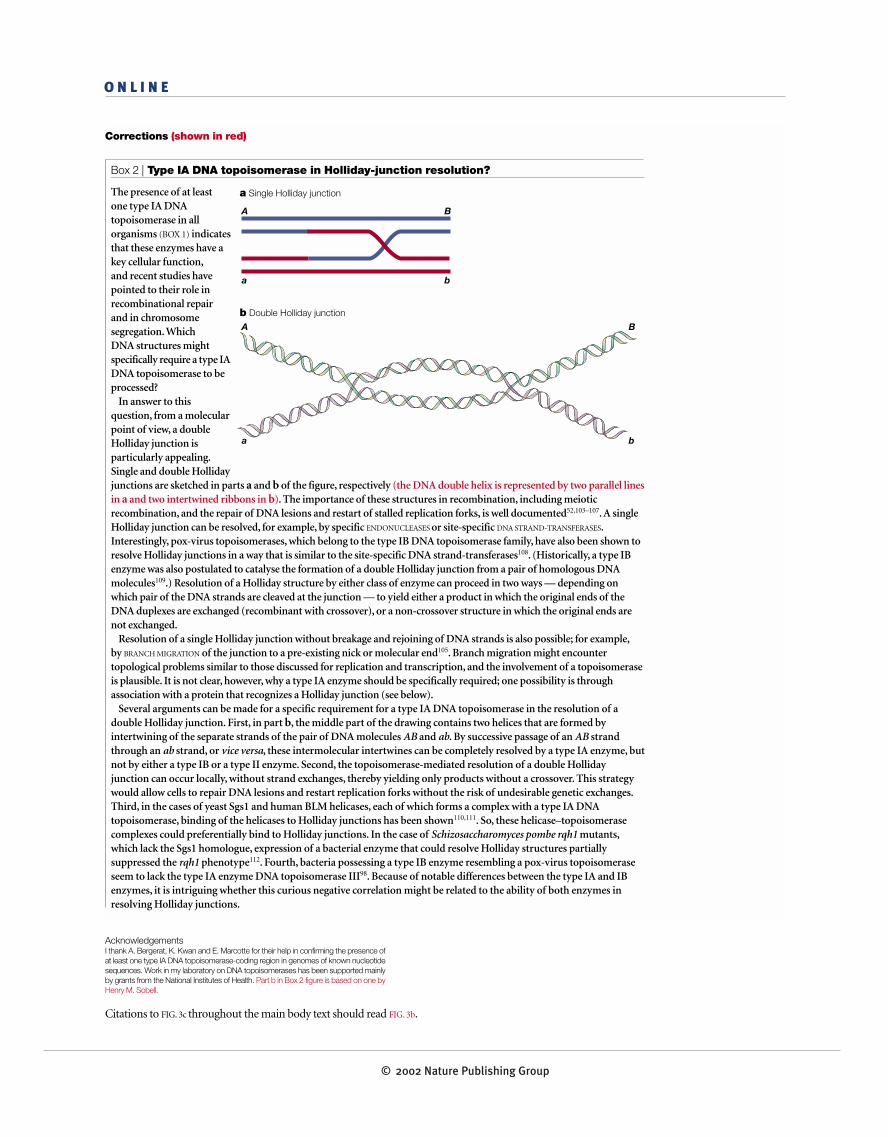
In answer to thisquestion, from a molecularpoint of view, a doubleHolliday junction isparticularly appealing.Single and double Hollidayjunctions are sketched in parts a and b of the figure, respectively (the DNA double helix is represented by two parallel linesin a and two intertwined ribbons in b). The importance of these structures in recombination, including meioticrecombination, and the repair of DNA lesions and restart of stalled replication forks, is well documented52,103–107. A singleHolliday junction can be resolved, for example, by specific ENDONUCLEASES or site-specific DNA STRAND-TRANSFERASES.Interestingly, pox-virus topoisomerases, which belong to the type IB DNA topoisomerase family, have also been shown toresolve Holliday junctions in a way that is similar to the site-specific DNA strand-transferases108. (Historically, a type IBenzyme was also postulated to catalyse the formation of a double Holliday junction from a pair of homologous DNAmolecules109.) Resolution of a Holliday structure by either class of enzyme can proceed in two ways — depending onwhich pair of the DNA strands are cleaved at the junction — to yield either a product in which the original ends of theDNA duplexes are exchanged (recombinant with crossover), or a non-crossover structure in which the original ends arenot exchanged.
Resolution of a single Holliday junction without breakage and rejoining of DNA strands is also possible; for example,by BRANCH MIGRATION of the junction to a pre-existing nick or molecular end105. Branch migration might encountertopological problems similar to those discussed for replication and transcription, and the involvement of a topoisomeraseis plausible. It is not clear, however, why a type IA enzyme should be specifically required; one possibility is throughassociation with a protein that recognizes a Holliday junction (see below).
Several arguments can be made for a specific requirement for a type IA DNA topoisomerase in the resolution of adouble Holliday junction. First, in part b, the middle part of the drawing contains two helices that are formed byintertwining of the separate strands of the pair of DNA molecules AB and ab. By successive passage of an AB strandthrough an ab strand, or vice versa, these intermolecular intertwines can be completely resolved by a type IA enzyme, butnot by either a type IB or a type II enzyme. Second, the topoisomerase-mediated resolution of a double Hollidayjunction can occur locally, without strand exchanges, thereby yielding only products without a crossover. This strategywould allow cells to repair DNA lesions and restart replication forks without the risk of undesirable genetic exchanges.Third, in the cases of yeast Sgs1 and human BLM helicases, each of which forms a complex with a type IA DNAtopoisomerase, binding of the helicases to Holliday junctions has been shown110,111. So, these helicase–topoisomerasecomplexes could preferentially bind to Holliday junctions. In the case of Schizosaccharomyces pombe rqh1 mutants,which lack the Sgs1 homologue, expression of a bacterial enzyme that could resolve Holliday structures partiallysuppressed the rqh1 phenotype112. Fourth, bacteria possessing a type IB enzyme resembling a pox-virus topoisomeraseseem to lack the type IA enzyme DNA topoisomerase III98. Because of notable differences between the type IA and IBenzymes, it is intriguing whether this curious negative correlation might be related to the ability of both enzymes inresolving Holliday junctions.
a Single Holliday junction
b Double Holliday junction
A B
a b
A B
a b
AcknowledgementsI thank A. Bergerat, K. Kwan and E. Marcotte for their help in confirming the presence ofat least one type IA DNA topoisomerase-coding region in genomes of known nucleotidesequences. Work in my laboratory on DNA topoisomerases has been supported mainlyby grants from the National Institutes of Health. Part b in Box 2 figure is based on one byHenry M. Sobell.
Citations to FIG. 3c throughout the main body text should read FIG. 3b.
© 2002 Nature Publishing Group