Embed Size (px)
Citation preview
Cellular & Molecular BiologyGraduate Studies Program 2011 – 2012

CONTENTS
The Cellular and Molecular Biology Program . . . . . . . . . . . . . . . . . . . …. . . . . . . . . . . . . . . . . . . . . .2 About Buffalo, New York . . . . . . . . . . . . . . . . . . . . . . . . . . . . . . . . . . . . . . . . . . . . . . . . . . . . . . . . . .3 Map – Directions to Roswell Park Cancer Institute . . . . . . . . . . . . . . … . . . . . . . . . . . . . . . . . . . . . .3
Faculty Research Marina P. Antoch.......................................................................4 Circadian Proteins as Modulators of Stress Response
Andrei V. Bakin .........................................................................5 Transforming Growth Factor β in Tumor Invasion and Metastasis Heinz Baumann..........................................................................5 Molecular Mechanisms of Gene Regulation by Cytokines and Hormones Michael J. Buck ..........................................................................6 Dissecting the Epigenetics Roles for Transcription Factor Targeting William C. Burhans ...................................................................7 Oxidative Stress and DNA Replication Stress in a Yeast Model of Aging Kailash Chadha ..........................................................................8 The Biology of the Interferon System in Health and Disease Peter Demant ............................................................................10 Cancer Susceptibility Genes and Their Role in Cancer Development Rosemary W. Elliott .................................................................11 The Genetic Map of the Mouse
Irwin H. Gelman .......................................................................12 Suppression of Prostate Cancer Oncogenesis and Metastasis by Regulators of Cytoskeletal and Signaling Pathways Vita M. Golubovskaya...............................................................12 Focal Adhesion Kinase Expression and Signaling in Cancer
Richard Gronostajski................................................................13 Transcription Factor Networks Regulate Metazoan Development Kenneth W. Gross ....................................................................15 The Renin-expressing Cell and Development of the Renal Vasculature Katerina V. Gurova .................................................................16 Anti-cancer Drug Delivery Through Modulation of Transcriptional Factors Marc S. Halfon .........................................................................17 Genetic Regulatory Networks
Michael J. Higgins ............................................................... 18 Epigenetics and Cancer
Yurij Ionov........................................................................... 20 Genomewide Analysis of Markers of Cancerogenesis Joseph T.Y. Lau .................................................................. 20 Molecular Glycobiology and Cellular Regulation
Thomas Melendy.................................................................. 21 Mechanisms and DNA Damage Regulation of HPV DNA Replication Norma J. Nowak .................................................................. 22 A Genomic Approach to Identifying Aberrations in Cancer
Roberto Pili........................................................................... 23 Tumor Microenvironment in Animal Models
Steven C. Pruitt ................................................................... .23 Stem Cells, Cancer and Aging
Nicoletta Sacchi.................................................................... .24 Epigenetic Mechanisms of Cancer Development
Dominic J. Smiraglia ........................................................... .25 DNA Methylation in Cancer and Normal Cells
John L. Yates..................................................................... 26 EpsteinBarr Virus; DNA Replication; Viral Oncology Y. Eugene Yu........................................................................ 27 Modeling Human Chromosomal Disorders in Mice
Jianmin Zhang..................................................................... 28 Dysregulation of the Hippo Pathway and Epithelialto-Mesenchymal Transition (EMT) in Tumorigenesis and Metastasis Shahriar Koochekpour ………………………….……... 29 Identification and Biological Characterization of Biomarkers Of Prostate Cancer Aggressiveness and Progression Toru Ouchi …………………………………………..….. 31 Molecular and Systems Biology of Carcinogenesis
2
The Cellular & Molecular Biology Program at Roswell ParkCancer Institute (RPCI) offers intensive training in basic andapplied research to doctoral candidates and postdoctoralassociates. The faculty is drawn primarily from two researchdepartments at RPCI, Molecular & Cellular Biology and CancerGenetics. The doctoral program is designed for dedicatedstudents with a substantial background in biology and chemistry.Faculty research interests cover a broad spectrum of cellular,molecular and cancer biology, with exceptional strengths in theareas of isolation and characterization of cancer genes, somaticcell genetics, high-throughput genomics, mouse genetics andmouse models of cancer, oncogenic viruses, DNA replication andgenetic and structural approaches to regulation of geneexpression.
Roswell Park Cancer Institute, one of the oldest cancer researchinstitutes in the world, is located on several blocks near otherhospitals within a mile of downtown Buffalo. RPCI provides anexcellent hospital for the care and treatment of cancer patientsas well as laboratories for basic and applied research relevant tocancer. Excellent support facilities are available, including DNAand protein sequencing, peptide synthesis, mass spectrometry,Affymetrix and cDNA Microarray analysis, quantitative PCR andDNA-HPLC, tissue histology, bioinformatics, statistics, facilitiesfor mouse genetic studies and transgenic/knockout mice, a cellsorter and electron microscopes. Related programs at RPCIinclude Molecular Immunology, Molecular & Cellular Biophysics,Pharmacology & Therapeutics, and Cancer Prevention &Population Science.
The graduate programs at RPCI are affiliated with the StateUniversity of New York at Buffalo (UB), and graduates receivetheir degrees from the State University of New York. Studentsenrolled at RPCI may take courses offered at RPCI or at either ofUB’s campuses. Courses offered within the Cellular & MolecularBiology Program include Molecular Genetics, RegulatoryMechanisms of Eukaryotic Cells, Viral Oncology, Structure andMolecular Interactions of DNA and Interferons. In addition,special student seminar courses provide advanced training in
analysis and oral presentation of research results from scientificliterature. A student seminar series provides an interactivetraining experience with staff members to develop skills for thepresentation of scientific data.
The Cellular & Molecular Biology program offers a low faculty-to-student ratio and an accessible faculty. In their first year,students are required to take Oncology for Scientists, laboratoryrotations and the student seminar. Beyond that, each student isencouraged (in consultation with an advisor) to take whatevercourses necessary to obtain the broad background of a capableand independent research scientist. In January of their secondyear, students are expected to pass a written qualifyingexamination which may include questions in molecular and cellbiology, genetics, virology, development and cancer biology.
Laboratory research is the major emphasis of the graduateprogram. In consultation with faculty, students arrange to work inlaboratories of their choice. A second qualifying examination atthe end of the second year requires the preparation of aproposal for the thesis project and its oral presentation anddefense. The thesis project involves independent research at thefrontiers of current research. The results of the research and theirsignificance for biological science are reported in an oral defenseof the doctoral dissertation. Completion of the thesis requiresthat the student possess or develop skills in thinking, organizing,writing and speaking, as well as laboratory skills. Most studentscomplete their course work and thesis research in about fiveyears and then traditionally go on to postdoctoral training inresearch institutions throughout the world.
All doctoral students in the Cellular & Molecular Biology Programreceive financial support. The current stipend for incoming newstudents is $24,000 per year. The stipend usually rises each yearor so, and students who have completed all prelims (usually bythe end of the second year) receive $1,000 more per year.
Information about application procedures, as well as applicationforms, is on-line at www.roswellpark.org/education.htm.
The Cellular & Molecular Biology Program
3
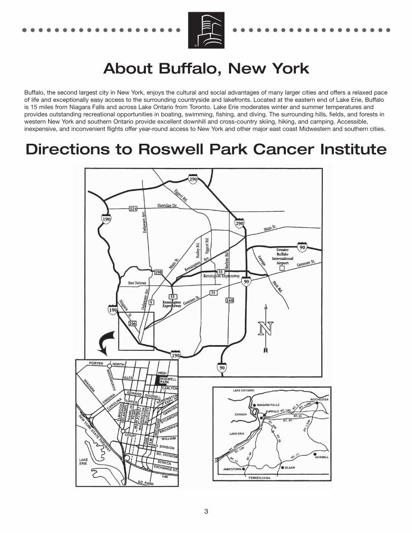
About Buffalo, New YorkBuffalo, the second largest city in New York, enjoys the cultural and social advantages of many larger cities and offers a relaxed paceof life and exceptionally easy access to the surrounding countryside and lakefronts. Located at the eastern end of Lake Erie, Buffalois 15 miles from Niagara Falls and across Lake Ontario from Toronto. Lake Erie moderates winter and summer temperatures andprovides outstanding recreational opportunities in boating, swimming, fishing, and diving. The surrounding hills, fields, and forests inwestern New York and southern Ontario provide excellent downhill and cross-country skiing, hiking, and camping. Accessible,inexpensive, and inconvenient flights offer year-round access to New York and other major east coast Midwestern and southern cities.
Directions to Roswell Park Cancer Institute
4
Circadian Proteins asModulators of StressResponse
Marina P. Antoch, PhD, Associate Professor of Oncology,Department of Molecular and Cellular Biology
Virtually all aspects of an animals’ biochemical, physiological andbehavioral functions are linked to circadian regulation. Circadianrhythms (i.e. 24-hr oscillation in various processes) are generatedendogenously and function under genetic control. In mammals,the basic molecular oscillator consists of two transcriptionalactivators - CLOCK and BMAL1 - and their transcriptionaltargets, CRYPTOCHROMES and PERIODS, which function asnegative regulators of the CLOCK/BMAL1 activity, thus formingthe major circadian autoregulatory feedback loop. The intrinsiccircadian clock regulates a variety of fundamental processesincluding cell cycle control, cellular response to genotoxic stressas well as regulation of components of the immune system. Themajor goal of our research program is to identify pathways,which cross-talk with the circadian clock and which can bemodulated by the activity of core circadian proteins.
The key role of major circadian proteins in genotoxic stressresponse was first demonstrated in our laboratory by testing thesensitivity of wild type, Clock mutant, Bmal1-/- knockout andCry1-/-Cry2-/- double-knockout animals to toxicity induced bychemotherapeutic drug cyclophosphamide. We showed that wildtype mice display a robust daily rhythm in sensitivity to the drug.Importantly, the morbidity and mortality associated withtreatment are at their highest levels when cyclophosphamide isadministered at the times of day corresponding to minimalfunctional activity of the CLOCK/BMAL1 complex and the lowestat the peak times of its activity. Consistently, animals with themutations or the targeted disruption of Clock or Bmal1 genesthat are characterized with the constant low levels ofCLOCK/BMAL1 transcriptional activity show high levels of drugsensitivity at all times tested. Moreover, animals with constanthigh levels of CLOCK/BMAL1 functional activity due to the lackof circadian repressors (Cryptochrome double-knockout animals)are extremely resistant to the treatment. These data suggest thatdrug sensitivity is affected by the functional status of majorcircadian transactivation complex, which translates into differentgene expression pattern of its direct and indirect targets.Currently, we are trying to understand the underlying mechanismfor circadian modulation of stress response pathways.
Importantly, this work identified circadian CLOCK/BMAL1complex as promising pharmacological target that maypotentially be used in combination with conventional anti-cancer
treatments to ameliorate side effects. This prompted us todevelop a cell-based readout system that we used to screenseveral libraries of small molecules for modulators ofCLOCK/BMAL1 functional activity. We were able to identifyseveral compounds that could modulate response to genotoxicstress through activating clock proteins. These compounds arecurrently under investigation.
Among other systems, circadian variations in the symptomintensity of infectious diseases have been described and linkedto variations in immune response. Thus, many immuneparameters exhibit daily variations, including the number ofspecific immune cells in circulation and plasma levels ofcytokines. However, the molecular details as well as majorplayers of the cross-talk between these two fundamentalsystems are still poorly understood. Our recent studies identifiedtwo potential players of this cross talk – circadian regulatorsCLOCK and BMAL1 and major regulator of immune responsetranscription factor NFkB. This interaction is bi-directional andinvolves transcriptional and post-translational regulatorymechanisms. Deciphering the molecular details of this interactioncan ultimately result in development of new immunomodulators(both stimulators and inhibitors) that are based on tuning NFkBresponse via circadian mechanism and, vice-versa, adjustingprocesses that are under circadian control by NFkB modulators.Importantly, the involvement of both clock- and NFkB-basedmechanisms in determining sensitivity to genotoxic stresses (i.e.,gamma radiation of chemotherapeutic drugs) suggests that byanalyzing the cross-talk between them we may get new insightsin the mechanisms controlling radio- and chemo-resistance.
REPRESENTATIVE PUBLICATIONS:Kondratov R, Chernov MV, Kondratova A, Gorbacheva V, Gudkov AV, and AntochMP. BMAL1-Dependent Circadian Oscillation of Nuclear CLOCK. Genes andDevelopment 17: 1921-1932, 2003.
Gorbacheva VY, Kondratov RV, Zhang R, Cherukuri S, Gudkov AV, Takahashi JS,and Antoch MP. Circadian sensitivity to the chemotherapeutic agentcyclophosphamide depends on the functional status of the CLOCK/BMAL1transactivation complex. Proc Natl Acad Sci USA 102(9): 3407-3412, 2005.
Kondratov RV, Shamanna RK, Kondratova AA, Gorbacheva VY, and Antoch MP.Dual role of the CLOCK/BMAL1 circadian complex in transcriptional regulation.FASEB J. 20(3): 530-532, 2006.
Kondratov RV, Kondratova AA, Lee C, Gorbacheva VY, Chernov MV, and AntochMP. Post-translational regulation of circadian transcriptionalCLOCK(NPAS2)/BMAL1 complex by CRYPTOCHROMES. Cell Cycle 5(8): 890-895,2006.
Kondratov RV, and Antoch MP. Circadian proteins in the regulation of cell cycle andgenotoxic stress responses. Trends Cell Biol. 17(7): 311-317, 2007.
Kondratov RV, and Antoch MP. The clock proteins, aging, and tumorigenesis. ColdSpring Harb Symp Quant Biol. 72: 477, 2007.
Antoch MP, and Chernov MV. Pharmacological modulators of the circadian clock aspotential therapeutic drugs. Mutat Res 679(1-2): 17-23, 2009.
Spengler ML, Kuropatwinski KK, Schumer M, and Antoch MP. A serine clustermediates BMAL1-dependent CLOCK phosphorylation and degradation. Cell Cycle8(24): 4138-4146, 2009.
Antoch MP, Kondratov RV. Circadian proteins and genotoxic stress response. CircRes. 106(1): 68-78, 2010.
Faculty Research
5
Transforming GrowthFactor β in TumorInvasion and Metastasis
Andrei V. Bakin, PhD, Assistant Professor of Oncology,Department of Cancer GeneticsThe research program explores tumor physiology and tumormicroenvironment, two major aspects of tumor biology that areultimately linked to the cancer progression and metastases. Ourresearch is primarily focused on breast cancer, the third mostcommon cause of cancer death in the United States. Theprogression of cancer is critically dependent on themicroenvironment in which a tumor originates. Themicroenvironment controls tumor growth, invasion andmetastasis. It also impacts the efficiency of cancer treatment andthe development of drug resistance. Tumor and host cellssecrete and activate various factors that ultimately affect allcomponents of the microenvironment, including vasculature,lymphatic system, extracellular matrix (ECM), immune system,and inflammatory response.
We investigate the role of transforming growth factor beta (TGF-β), a major cytokine in the tumor microenvironment. TGF-β playsa prominent role in cancer as well as in normal development andhomeostasis of nearly all tissues, including breast. TGF-β is apotent inhibitor of cell growth and can induce apoptosis inresponsive cells. In early-stage cancers, TGF-β functions as atumor suppressor, and alterations in the TGF-β pathway areimplicated in cancer development. Paradoxically, advancedtumors secrete abnormally high levels of TGF-β, and this isassociated with tumor invasion and metastases. Understandingthe molecular mechanism behind the oncogenic TGF-β functionis a major focus of our research. The goal is to identify keyfactors responsible for the oncogenic function of TGF-β in orderto design better diagnostic tools and cancer treatment.
The research program includes three directions: (i) TGF-β-induced epithelial-mesenchymal transition (EMT) as a means ofthe acquisition of motility and invasiveness; (ii) TGF-β-mediatedchanges in tumor microenvironment and angiogenesis; (iii) TGF-βin control of metabolic pathways involving glutathione.
REPRESENTATIVE PUBLICATIONS:Safina A, Ren MQ, Vandette E and Bakin AV. TAK1 is required for TGFb1-mediatedregulation of matrix metalloproteinase-9 and metastasis. Oncogene 27(9): 1198-1207, 2008.
Safina A, Vandette E and Bakin AV. ALK5 promotes tumor angiogenesis byupregulating matrix metalloproteinase-9 in tumor cells. Oncogene 26(17): 2407-2422, 2007.
Varga AE, Quan L, Stourman NV, Safina A, Li X, Sossey-Alaoui K and Bakin AV.Silencing of the tropomyosin 1 gene by DNA methylation alters tumor suppressorfunction of TGF beta. Oncogene, 24 (32): 5043-5052, 2005.
Bakin AV, Sekhar KR, Stourman NV, Rinehart C, Yan X, Meredith MJ, Arteaga CLand Freeman ML. Smad signaling suppresses Phase II gene expression. FreeRadical Biol. Med. 38(3): 375-87, 2005.
Bakin AV, Rinehart C, Safina A, Daroqui C, Darbary H and Helfman D. A critical roleof tropomyosins in TGF-β regulation of the actin cytoskeleton and cell motility inepithelial cells. Mol. Biol. Cell 15(10): 4682-4694, 2004.
Bakin AV, Rinehart C, Tomlinson AK and Arteaga CL. p38 mitogen-activated proteinkinase is required for TGFβ-mediated fibroblastic transdifferentiation and cellmigration. J. Cell. Sci. 115(15): 3193-3206, 2002.
Shin I, Yakes FM, Rojo F, Shin N-Y, Bakin AV, Baselga J and Arteaga CL. PKB/Aktmediates cell-cycle progression by phosphorylation of p27Kip1 at threonine 157and modulation of its cellular localization. Nature Med. 8(10): 1145-1152, 2002.
Shin I, Bakin AV, Rodeck U, Brunet A, Arteaga CL. TGFβ enhances epithelial cellsurvival via Akt-dependent regulation of FKHRL1. Molecular Biology of Cell, 12 (11):3328-3339, 2001.
Bhowmick NA, Ghiassi M, Bakin A, Aakre M, Lundquist CA, Engel ME, Arteaga CL,Moses HL. TGFβ mediates epithelial to mesenchymal transdifferentiation through aRhoA-dependent mechanism. Molecular Biology of Cell, 12: 27-36, 2001.
Bakin AV, Tomlinson AK, Bhowmick NA, Moses HL, Arteaga CL. Phosphatidylinositol3-kinase function is required for TGFbeta -mediated epithelial to mesenchymaltransition and cell migration. J. Biol. Chem., 275 (47): 36803-36810, 2000.
Bakin A, Curran T. Cell transformation by the fos oncogene is mediated by DNA 5-methylcytosine transferase. Science, 283 (5400): 387-390, 1999.
Molecular Mechanismsof Gene Regulation byCytokines & Hormones
Heinz Baumann, PhD, Professor of Oncology, Department of Molecular and Cellular Biology
A network of cytokines, growth factors, hormones, and variousbiologically active metabolites control systemic homeostasis inthe postnatal organism. Cytokines are major regulators of thoseevolutionarily conserved processes that determine, amongothers, the tissue response to injury, mediate inflammation,activate systemic acute phase reaction and fever, and direct theinnate and adaptive immune responses. This laboratory pursuestwo long-term research programs. The first program has theoverall goal of characterizing the cellular and molecular action ofcytokines that mediated the local and systemic response totissue damage and modulated tumor growth. The secondprogram focuses on the genetics and biology of the hepaticacute phase response and the physiological role of the plasmaproteins induced by inflammation.
Current work on the first program concerns the identification ofthe mechanisms by which members of the hematopoietic cytokinefamily control proliferation and expression of differentiation genesin various normal and transformed cell types. The studies addresstwo questions: (1) What are the mechanisms by which theheteromeric receptors for the interleukin 6 class cytokines –namely IL-6, IL-31, leukemia inhibitory factor (LIF) and oncostatinM (OSM) – control cell proliferation and the expression ofdifferentiated genes? And (2) how is the responsiveness to theseIL-6 class cytokines controlled during malignant transformation of
6
epithelial cells? The biochemical analysis of signaling employsvarious tissue culture cell lines in which the specific functions ofthe receptor subunits for the cytokines are reconstituted. Thesignaling action of the OSM receptor has been defined by thesuppression of proliferation through mediating G1 arrest in part bythe induction of cyclin kinase inhibitors and to trigger induction aswell as suppression of genes through transcription factors thatinclude STAT3 and STAT5, AP-1/ATFs, C/EBP and CREB. Thesecellular responses are used to characterize the structural motifswithin the signal transducing receptor subunits (gp130, LIFR-alpha, OSMR-beta, IL-31R-alpha [or GPL]) that are required forengaging the intracellular signaling pathways, in particular leadingto the activation of the JAKs, PI3K, and the MAPK pathwaysengaging ERK and JNK. Genetic targets through which thereceptor signals induce transcription have been established in theexamples of acute phase plasma protein genes in liver and othercell types. The altered profile of cytokine responses in cancer celltypes compared to the normal counterparts has allowed theidentification of changes in the expression of signaling proteins (asin the case of myeloid leukemias) and cytokine receptors (as inthe case of lung epithelial cells) as function of malignanttransformation. The tumor-specific expression of cytokine receptorsubunits is in part correlated with epigenetic alterations in DNAmethylation and histone acetylation. The information gain oninflammatory mediators in tissue culture model system arecurrently applied to the identification of regulatory processesoccurring in vivo following photodynamic therapy in the lung,head/neck and skin.
The work on the second program investigates how the acutephase response in various organs is controlled and what thephysiological role of the acute phase proteins is. In the last fewyears, mouse models have been developed in which theexpression of the major acute phase proteins, haptoglobin(hemoglobin binding protein), hemopexin (heme-binding protein)and alpha-1-acid glycoprotein (ligand-binding protein), are alteredeither by silencing through gene knockout or by overproductionthrough introducing constitutively expressed transgenes. Theseplasma proteins are effective regulators of three major processesassociated with inflammation: inhibition of extracellular proteases,reduction of oxidative damages to tissue, and attenuation of theactivity of inflammatory and immune cells. The recent studies onthe biology of haptoglobin have focused on the anti-inflammatoryfunction of this acute phase protein and its role in directingprogression of tissue injury response, stimulated proliferation oftumor cells at site of inflammation, and directing the immuneresponse through direct regulation of development of lymphoidorgans and differentiation of lymphocytes. The hypothesis thatinflammatory mediators are promoting proliferation of epithelialtumor cells is addressed by the effects of haptoglobin deficiencyor overexpression in the intestinal tumor model provided by theApc+/MIN mouse.
REPRESENTATIVE PUBLICATIONS:Huntoon KM, Wang Y, Eppolito CA, Barbour KW, Berger FG, Shrikant PA andBaumann H. The acute phase protein regulates host immunity. J. Leukocyte Biol.84: 170-181, 2008.
Chattopadhyay S, Tracy E, Liang P, Robledo O, Rose-John S and Baumann, H.Interleukin-31 and oncostatin-M mediate distinct signaling reactions and responsepatterns in lung epithelial cells. J. Biol. Chem. 282: 3014-3026, 2007.
Henderson BW, Daroqui C, Tracy E, Vaughan LA, Loewen GM, Cooper MT andBaumann H. Cross-linking of signal transducer and activator of transcription 3 – a
molecular marker for the photodynamic reaction in cells and tumors. Clin. CancerRes. 13: 3156-3163, 2007.
Loewen GM, Tracy E, Blanchard F, Tan D, Yu J, Raza S, Matsui S and Baumann H.Transformation of human bronchial epithelial cells alters responsiveness toinflammatory cytokines. BMC Cancer 5: 145, 2005.
Dissecting theEpigenetics Roles forTranscription FactorTargeting
Michael J. Buck, PhD, Assistant Professor, Department ofBiochemistry, State University of New York at Buffalo
The Buck lab integrates experimental and computationalapproaches to determine the rules dictating transcription factor(TF) targeting in a Eukaryotic genome and apply our findings tohuman clinical samples. To address this question we us modelsystems (mice and yeast) and human cell culture. Specifically, weinvestigate TF binding selection in response to environmentalstress, characterize the chromatin mediated mechanisms directingTF target selection, determine how developmental signals reshapethe epigenetic landscape during cellular development, anddevelop bioinformatics tools to analyze and interpret next-generation chromatin datasets. The lab uses state of the artgenomic techniques including ChIP-seq, FAIRE-seq, and MNase-seq and is composed of both a molecular and computational lab.Currently the lab is focusing on the following projects:
Determining how the Tup1 co-repressor is targeted and howit regulates chromatin structure in budding yeastUp-regulation of Transducin-like Enhancer of Split (TLE) proteinsis associated with astrocytoma, meningioma, pituitary adenoma,synovial sarcoma, and lung adenocarcinoma. TLE1, the humanhomolog of fly Groucho and yeast Tup1, represses transcriptionby recruiting chromatin remodeling proteins which establishesrepressive chromatin architecture, and is involved in severalsignal-transduction cascades, such as Notch, Wingless/Wnt, andDPP/BMP. The budding yeast homolog Tup1 has been a modelfor studying similar repressor complexes in multicellulareukaryotes. Tup1-Ssn6 does not bind DNA directly, but isdirected to individual promoters by one or more DNA-bindingproteins, referred to as Tup1 recruiters. The goal of this project isto determine how Tup1 identifies its sites across the genome andhow it regulates chromatin structure at it targets.
Nucleosome inhibition of transcription factor bindingChromatin structure and nucleosome positioning has beenpostulated as the most important factor directing TFs to theirappropriate binding sites. The goal of this project is to determinethe principals and characteristics of nucleosome inhibition of TFbinding. TF binding sites, located within DNA that is tightlywrapped within a nucleosome are typically inaccessible.Nucleosome inhibition of TF binding depends on at least sevenfactors: the type of TF and its concentration, nucleosome
7
occupancy, histone tail modifications, binding site affinity,binding site location and site rotational setting within anucleosome. Because these variables are not independent ofeach other, they need to be explored simultaneously orcontrolled stringently when studying regulation of TF targeting.However, this is highly impractical and requires thousands ofexperiments. To overcome this limitation, a unique approachcombining yeast genetics with next-generation sequencing isapplied, which allows us to study the relationships between theabove factors and TF binding.
Identification of Epigenetic BiomarkersEpigenetic alterations have been associated with cancer-specificexpression differences in development of human tumors. Theability to recognize and detect the progression of epigeneticevents occurring during tumorigenesis is critical to developingstrategies for therapeutic intervention. Key epigenetic alterations,leading to silencing or activation, are associated with changes innucleosome occupancy. We use a straightforward, reproducible,genomic approach for measuring chromatin accessibilityFormaldehyde-Assisted Isolation of Regulatory Elements (FAIRE)combined with next generation sequencing (FAIRE-seq). FAIREisolates nucleosome-depleted genomic regions, which are thefunctionally active regions, and these regions represent ideallocations to identify chromosomal aberrations or SNP’sassociated with tumor formation.
Identification of shared chromatin architecturesCurrently the only way to characterize chromatin architecture isto have an accurately mapped functional element in the genome.Functional elements include genes for protein and non-codingRNAs, and regulatory sequences that direct essential functionssuch as gene expression, DNA replication, and chromosomeinheritance. With an accurately mapped functional element,chromatin structural data is aligned by the genomic coordinatesand an average profile is created. To determine the chromatinarchitecture at unknown or at inaccurately mapped functionalelements we are developing chromatin alignment algorithms andapplying them to genome wide chromatin datasets.
REPRESENTATIVE PUBLICATIONS:Hanlon, S.E., Rizzo, J.M., Tatomer, D.C., Lieb, J.D., Buck, M.J. (2011). The stressresponse factors Yap6, Cin5, Phd1, and Skn7 direct targeting of the conserved co-repressor Tup1-Ssn6. PLOS One, 6(4):e19060.
Buck, M.J., and Lieb, J.D. (2006). A chromatin-mediated mechanism forspecification of conditional transcription factor targets. Nature Genetics Dec;38(12): 1446-51.
Lai, W., and Buck, M.J. (2010) ArchAlign: Coordinate-free alignment of chromatindatasets reveals novel architectures. Genome Biology Dec 23; 11(12):R126.
Oxidative stress andDNA replication stress ina yeast model of aging
William C. Burhans, PhD, Associate Professor ofOncology, Department of Molecular and Cellular Biology
Oxidative stress and DNA replication stress (i.e., inhibition ofDNA replication) cause oncogene-induced senescence at earlystages of neoplastic disease. Senescence also promotes aging.The Burhans laboratory employs a yeast model of senescenceand aging to investigate how these stresses arise downstream ofsustained growth signaling implicated in cancer and other age-related diseases. We are also investigating how caloric restrictionand caloric restriction mimetics mitigate these stresses.
During the past year we and collaborators in Portugal discovereda novel mechanism by which caloric restriction inhibitssenescence in this yeast model. This hormesis-relatedmechanism involves induction by caloric restriction of thereactive oxygen species hydrogen peroxide (H2O2). Onceinduced, H2O2 activates oxidative stress defenses that reduceintracellular levels of superoxide anions, which are a proximalcause of senescence. The results of our study (Mesquita et al.(2010)) also establish that genetic or pharmacologicalinactivation of catalases – which also elevates intracellular levelsof H2O2 - mimics the senescence-inhibiting effects of caloricrestriction.
Since our study was published evidence has emerged that thismechanism operates in metazoans as well. It was recentlyreported that a dramatically increased lifespan induced byenvironmental cues in the social insect Harpegnathos saltator isaccompanied by a reduction in catalase activity and enhancedoxidative stress defenses. A similar mechanism may underlierecent reports that catalase inactivation in mice protects againstacute inflammatory responses in the lung that depend onsuperoxide anions. A broader implication of our findings is that afundamental tenet of the longstanding oxidative stress theory ofaging - which posits H2O2 and other reactive oxygen species asstrictly pro-aging factors in all organisms - requires modification.
In a related study, we discovered that superoxide anionspromote senescence in part by inhibiting DNA replication, thuscausing replication stress (Weinberger et al. (2010)). Our studiesalso demonstrate that replication stress caused by hypomorphicmutations in DNA replication proteins elevates intracellular levelsof superoxide anions. Thus, replication stress can trigger a“vicious cycle” that amplifies both oxidative stress andreplication stress leading to senescence.
Our recent findings also suggest that replication stress initiallydevelops as a consequence of the normal reduction in dNTPpools that occurs in quiescent cells coupled to sustained growthsignaling in these cells (by oncogenes, for example) through
8
some, but not all growth signaling pathways. Sustainedactivation of a subset of these pathways inappropriately drivescells that are approaching quiescence into S phase, but in theabsence of sufficient dNTPs to efficiently replicate DNA. Thisleads to replication stress that triggers the self-amplifying cycledescribed above. Replication stress is also triggered bysustained growth signaling in response to excess glucose.Similar induction of replication stress by hyperglycemia inhumans may be a factor that links diet with cancer and otherage-related diseases.
We also recently completed a high-throughput screen of the NIHcompound library for small molecules that mimic caloricrestriction. A primary goal of this screen was to identifymolecules that reduce intracellular levels of superoxide anions byinducing H2O2 and/or inhibiting growth signaling. Approximately800 primary hits were identified in the screen. Confirmed hitsinclude inhibitors of TOR signaling pathways, tyrosine kinaseinhibitors and suirtuin activators. Structure-activity relationshipstudies of these compounds are ongoing.
REPRESENTATIVE PUBLICATIONS:Burhans WC, and Weinberger M. DNA replication stress, genome instability andaging. Nuc Acids Res 35: 7545-7556, 2007.
Burhans WC, and Heintz NH. The cell cycle is a redox cycle; linking phase-specifictargets to cell fate. Free Radicals in Biology and Medicine 47(9): 1282-1293, 2009.
Mesquita A, Weinberger M, Silva A, Sampaio-Marques B, Almeida B, Leao C, CostaV, Rodrugues F, Burhans WC, and Ludovico P. Caloric restriction or catalaseinactivation extends yeast chronological lifespan by inducing H2O2 and superoxidedismutase activity. Proc Natl Acad Sci USA 107: 15123-15128, 2010.
Weinberger, M. et al. Growth signaling promotes chronological aging in buddingyeast by inducing superoxide anions that inhibit quiescence. Aging 2: 1-8, 2010.
Burhans, W.C. and Weinberger, M. Histone genes, DNA replication, apoptosis andaging – what are the connections? Cell Cycle 9: 4047-4048, 2010.
The Biology of theInterferon System inHealth and Disease
Kailash Chadha, PhD, Associate Professor of Oncology,Department of Molecular and Cellular Biology
The major emphasis in our laboratory is in understanding of theunderlying cause(s) of immune suppression often associatedwith viral infections, tumor growth and in cases of substanceabuse. Since healthy immune surveillance is the key to goodhealth, we have been involved in the areas that have stronginfluence on the overall immune system.
Our laboratory has interest in the following three areas: 1)Interferons; 2) Prostate Cancer; and 3) Multiple Sclerosis. Thefollowing is the brief description of various research activities inthe laboratories:
I. We have observed that late stage cancer patients, andpatients with full blown AIDS, have interferons and interferoninhibitory activity in their blood circulation. Also, the response oftheir WBC to interferon induction is poor, and they have lowlevels of NK cell activity as compared to normal healthyindividuals. The inhibitory activity is neither due to antibody, tointerferon, nor due to any defective interferon in their bloodcirculation. Work carried out in our laboratory has shown thatinterferon inhibitory activity is due to: a) a new 70 kd interferoninhibitory protein that is not present in normal healthy individuals;b) due to high levels of PGE2 in patient blood that is inhibitory tointerferon action; c) due to free floating interferon receptors inthese patients’ blood; or d) any combination of these.
Such interferon inhibitory activity has also been seen inindividuals who have late stage cancer, are heavy cigarettesmokers or are chronic alcoholics. However, interferon inhibitorylevels will significantly decline when one quits smoking, goesthrough proper rehabilitation for drinking, or when patientsundergo successful debulking of tumors as a result of surgery orradiation treatment.
II. We were first to report that 20% of natural human interferon αis glycosylated, and a fraction of natural human interferon α isacid-labile. These observations were made before any clonedinterferons were available. Our earlier observations have nowbeen confirmed by others. We have also reported that themajority of interferons produced by polymorphonuclear cells areacid-labile alpha type.
III. For the past several years, we have been working with amurine model of AIDS. BM5MuLV produces symptoms inC57BL/6 mice that are very similar to early HIV infection inhumans. The virus can infect 100% of the animals in a shortperiod (12-16 weeks). Furthermore, animals used are an inbredstrain, and this avoids any genetic variation. Both chronicalcoholism and MAIDS virus infection decreases T cellpopulations including CD4+ cells; their ability to produce IFN inresponse to poly rIrC is significantly reduced. Also, theirsplenocytes produce low levels of IFNα and γ when challengedin vitro. Significant improvements in the levels of immunesuppression were seen when virus infected animals were treatedwith drugs like Meclomen and Pentoxifylline. Their CD4+/CD8+ratios were restored to essentially hormonal levels if animalswere treated with these drugs within 3-5 days after MAIDS virusinfection.
IV. We have shown in our studies that mild hyperthermiasignificantly modulates a variety of biological activities ofinterferons in both human and mouse cell culture systems. Mildhyperthermia (39°C) significantly enhances antiviral activity of allthree human interferons (α, β, γ). Also, antitumor activity ofhuman interferons on a variety of tumor cells is significantlyincreased. However, interferon production and interferonmediated enhancement of natural killer cell activity is suppressed.
The enhancement of antiviral activity is due to prolonged life ofmRNA and also due to early release of interferon. No significantincrease in level of interferon mRNA was seen in cells treatedwith hyperthermia and induced for interferon β synthesis.
In a clinical setting, a high dose interferon therapy is oftenassociated with many undesirable side effects. These
9
undesirable side effects can be minimized by combining lowdose interferon treatment with use of mild hyperthermia.
V. It is now well established that parental drug abuse is asignificant risk factor for contracting HIV 1 infection. In ourstudies, we have shown that drugs of abuse, like morphine andcocaine, can act as cofactors in susceptibility and progression ofHIV 1 infection. Our results demonstrate that HIV 1 protein-induced lymphocyte proliferation responses are significantlyinhibited by morphine in a dose dependent manner. Morphinealso significantly inhibits IFNα and IFNβ production and inducesapoptosis of normal lymphocytes. Inhibition of IFNα productionby morphine could be reversed by the opiatic receptorantagonist, naloxone. This suggests that immunomodulatoryeffects of morphine are mediated through opioid receptors.
Although cocaine has been linked to the immunopathogenesis ofHIV 1 infection, the corresponding cellular and molecularmechanism(s) have not been well defined. We hypothesize thatcocaine mediates its immunosuppressive effects throughdownregulation of HIV 1 suppressing chemokines and/orupregulating the HIV 1 entry co-receptors in HIV 1 infectedsubjects, resulting in disease progression. Our results show thatcocaine selectively downregulates endogenous MIP 1β secretionby PBMC. Cocaine also selectively suppresses LPS-induced MIP1β production by PBMC. Further, cocaine significantlydownregulates endogenous MIP 1β gene expression while itupregulates HIV 1 entry co-receptors CCR5 by PBMC. Thesestudies suggest a role for cocaine as a cofactor in HIV 1pathogenesis.
VI. Prostate cancer has the highest incidence of any non-cutaneous malignancy in the western world and is the secondleading cause of cancer related deaths in men. Prostate-SpecificAntigen (PSA) is a well recognized biomarker for the earlydiagnosis and management of prostate cancer. However, PSAtest is neither disease specific nor tissue specific and results in>70% false positive. There are two major areas of research in mylaboratory: Project I: It involves identification of new serumbiomarkers that will be more selective and specific, either aloneor in conjunction with PSA, in improving the early diagnosis andin the management of prostate cancer. At present, we areinvestigating the relevance of PSMA, IL-8, TGF-β, TNF-alpha andsTNFR1 etc as potentially new biomarkers. Our preliminaryresults strongly suggest that serum levels of IL-8, TNF-alpha andsTNFR1 will provide powerful tools in differentiating between thebenign and malignant tumors; which is the major disadvantageof currently available PSA test. Project II: It involvesdetermination of the “physiological role PSA” in overall prostatetumor growth and metastasis. Initially, we have documented thatPSA has a significant effect in modulating expression of variouspro- and anti-angiogenic growth factors in prostate tumor celllines. In our preliminary studies we have shown that humanprostate cells that are highly malignant have higher levels ofexpression of pro-angiogenic growth factors like VEGF, IL-8,TGF-β, bFGF etc and low levels of anti-angiogenic factors likeinterferons and angiostatin. The treatment of these cells withPSA results in down regulation of pro-angiogenic factors and up-regulation of anti-angiogenic factors. In a gene array analysis, wehave shown that PC3M cells treated with PSA results in up-regulation of 136 genes and down-regulation of 137 genes.Many of these genes are known to be involved in prostate tumor
growth and metastasis. Gene expression analysis has beenconfirmed by RT-QPCR analysis. In in vivo studies, we have alsodocumented that PSA administration to nude mice, bearinghuman prostate tumor xenografts, significantly reduces growth ofprostate tumors.
VII. Interferon inhibitory activities [IIA] in multiple sclerosispatients. The objective here is to determine the role of seruminterferon inhibitory activity and soluble interferon α/β receptorsin multiple sclerosis patients who are partially responsive tointerferon-β (IFN-β) therapy. Approximately 30% of MS patientsrespond well to treatment with IFN-β whereas the remainingexhibit varying extents of partial responsiveness. Neutralizingantibodies, which occur in 5-25% of IFN-1βa treated MSpatients, provide a biologically intuitive mechanistic explanationfor partial responsiveness for many protein drugs, including IFN-β therapy of MS. Generally, patients who develop anti-IFN-βNAB have less favorable treatment outcomes than those who areNAB negative. However, the majority of MS patients who arepartially responsive to IFN-β tend to be NAB negative. Themolecular mechanisms underlying NAB-negative IFN-β non-responsiveness are not well understood. The overall aim of thisstudy is to characterize multiple sclerosis (MS) patients forpossible such molecular mechanisms capable of causingheterogeneity of response to interferon-β (IFNβ) therapy.
Our hypothesis is that the heterogeneity of interferon responsesin patients with malignancies was caused by a circulating“interferon-inhibitory activity” (IIA) in non-responsive patients. Wehave identified 4 molecular mechanisms that contributed to theobserved IIA:
1. An interferon inhibitory protein (IIP)
2. Free-soluble IFN-α/β receptors (sIFNAR).
3. High prostaglandin E2 levels.
4. High levels of cAMP phosphodiesterases.
Normal, healthy individuals do not have significant IIA incirculation. We will be studying two groups of MS patients(Responders and partial responders) and identify which of thesefour molecular mechanisms are operative in MS patients that arepartial responders.
REPRESENTATIVE PUBLICATIONS:Satheesh Babu AK, Vijayalakshmi MA, Smith GJ, and Chadha KC. Thiophilic-interaction Chromatography of Enzymatically active Tissue Prostate-SpecificAntigen (T-PSA) and its Modulation by Zinc Ions. J. Chromatog B. 861: 227-235,2008.
Sternberg Z, Weinstock-Guttman B, Hojnaki D, Zamboni P, Zivadinov R, Chadha K,Lieberman A, Kazim L, Drake A, Rocco P, Grazioli E, and Munschauer F. Solublereceptor for advanced glycation end products in multiple sclerosis: a potentialmarker of disease severity. Mult Sclerosis 14(6): 759-763, 2008 (PMID 18505774).
Bindukumar B, Schwartz S, Aalinkeel R, Mahajan S, Lieberman A and Chadha K.Proteomic profiling of the effect of prostate-specific antigen on prostate cancercells. Prostate 68(14): 1531-1545, 2008.
Aalinkeel R, Bindukumar B, Reynold JL, Sykes DE, Mahajan SD, Chadha K, andSchwartz SA. The dietary bioflavonoid, quercetin, selectively induces apoptosis ofprostate cancer cells by down-regulating the expression of heat shock protein 90.Prostate 68(16): 1773-1389, 2008.
Bindukumar B, Schwartz SA, Nair MP, Aalinkeel R, Kawinski E, and Chadha KC.Prostate-specific antigen modulates the expression of genes involved in prostatetumor growth. Neoplasia 7: 241-252, 2005.
10
Chadha KC, Weinstock-Guttman B, Zivadinov R, Bhasi K, Muhitch J, Feichter, JTamano-Blanco M, Abdelrahman N, Ambrus Sr J, Munschaeur F and RamanathanM. Interferon Inhibitory Activity in Multiple Sclerosis Patients. Arch. Neurology 63:1579-1584, 2006.
Ambrus JL Sr, Chadha KC, Islam A, Akhter S and Ambrus JL Jr. Treatment of Viraland Neoplastic Diseases with Double-Stranded RNA Derivatives and Other NewAgents. Mini Review: Soc Expt Biology & Medicine 231: 1283-1286, 2006.
Chadha, KC. (ed.) Interferons: Currect Status Research Signpost (ISBN # 81-7736-256-9), 2007
Sternberg Z, Weinstock-Guttman B, Hojnaki D, Zamboni P, Zivadinov R, Chadha K,Lieberman A, Kazim L, Drake A, Rocco P, Grazioli E, and Munschauer F. Solublereceptor for advanced glycation end products in multiple sclerosis: a potentialbiomarker of disease severity. Mult Scler. 14: 759-763, 2008.
Aalinkeel R, Bindukumar B, Reynolds JL, Sykes DE, Mahajan SD, Chadha KC, andSchwartz SA. The dietary bioflavonoid, quercetin, selectively induces apoptosis ofprostate cancer cells by down-regulating the expression of heat shock protein 90.Prostate 68(16):1773-89, 2008.
Bindukumar B, Schwartz S, Aalinkeel R, Mahajan S, Lieberman A, and Chadha K.Proteomic profiling of the effect of prostate-specific antigen on prostate cancercells. Prostate 68(14):1531-45, 2008.
Satheesh Babu AK, Vijayalakshmi MA, Smith GJ, and Chadha KC. Thiophilic-interaction chromatography of enzymatically active tissue prostate-specific antigen(T-PSA) and its modulation by zinc ions. J Chromatogr B Analyt Technol BiomedLife Sci. 861(2):227-35, 2008.
Sternberg Z, Chadha K, Lieberman A, Hojnacki D, Drake A, Zamboni P, Rocco P,Grazioli E, Weinstock-Guttman B, and Munschauer F. Quercetin and interferon-betamodulate immune response(s) in peripheral blood mononuclear cells isolated frommultiple sclerosis patients. J Neuroimmunol. 205 (1-2): 142-7, 2008.
Sternberg Z, Hennies C, Sternberg D, Bistulfi G, Kazim L, Benedict R, Chadha,K,Leung C, Weinstock-Guttman B, and Munschauer F. Plasma Pentosidine: Apotential Biomarker in the management of Multiple Sclerosis. Multiple Sclerosis17(2): 157-163, 2011. PMID: 20965962. -7, 2008.
Nicotera TM, Schuster DP, Bourhim M, Chadha K, Klaich G, and Corral DA.Regulation of PSA secretion and survival signaling by calcium-independentphospholipase A2 β in prostate cancer cells. The Prostate 69: 1270-1280, 2009.
Sternberg Z, Chadha K, Lieberman A, Drake A, Hojnacki D, Weinstock-Guttman B,and Munschauer F. Immunomodulatory responses of peripheral blood mononuclearcells from multiple sclerosis patients upon in vitro incubation with the flavonoidluteolin: additive effects of IFN-beta. J Neuroinflammation 6:28, 2009. PMID:19825164.
Nicotera TM, Schuster DP, Bourhim M, Chadha K, Klaich G, Corral DA. Regulationof PSA secretion and survival signaling by calcium-independent phospholipase A2β in prostate cancer cells. The Prostate, 69: 1270-1280, 2009.
Aalinkeel R, Bindukumar B, Schwartz SA, Smith GJ and Chadha, KC. Role ofProstate Specifdic Antigen (PSA) in Patholohgical Angiogenesis and Prostate TumorGrowth. In Horizons in Cancer Research. Volume 42 (edi) Hiroto S Watanabe. NovaScience Publishers, Inc ISBN: 978-1-61761-111-7. 2010.
Sternberg Z, Hennies C, Sternberg D, Bistulfi G, Kazim L, Benedict R, Chadha,K,Leung C, Weinstock-Guttman B. Plasma Pentosidine: A potential Biomarker in themanagement of Multiple Sclerosis. Multiple Sclerosis. 17(2): 157-163, 2011.
Aalinkeel R, Bindukumar B, Reyonld JL, Sykes DE, Mahajan SD, Chadha, K. andSchwartz SA. Over expression of MMP-9 contributes to invasiveness in ProstateCancer cells. Immunological Investigations, In Press, 2011.
Chadha K, Nair B, Chakravarthi S, Zhou R, Mohler JL, Schwartz SA, Aalinkeel R,Smith GJ. Enzymatic activity of free-prostate specific antigen is not required for itsphysiological activities. The PROSTATE. In press,2011
Cancer SusceptibilityGenes and Their Role inCancer Development
Peter Demant, MD, PhD, Distinguished Professor ofOncology, Department of Molecular and Cellular Biology
A very large number of apparently "sporadic" or “common”cancers, even though they do not occur in an obviously inheritedmanner in families, develop in persons with hereditarypredisposition to cancer. For example, one half of all breastcancers in the population will occur in the most predisposed oneeighth of women. Such strong concentration of cancer risk in arelatively small part of the population could be used to effectivelyfocus the preventive measure on the most susceptible women.To find the genes, which determine such susceptibility, andultimately to identify the persons at high risk is the main themeof our research. To achieve this, we apply the strategy of definingthese genes first in experimental animals, mainly mice, and thenidentifying the homologous genes in humans.
a. Individual Susceptibility to Common cancers.This strategy has been applied with success, partly due toapplication of novel powerful genetic tools we developed for thispurpose. Our studies led to successful mapping of more than 40novel lung cancer susceptibility genes, 15 novel colon cancersusceptibility genes, as well as new genetic information onleukemia and breast cancer. In fact, the lung and colon cancergenes discovered in this laboratory represent the majority of allpresently known susceptibility genes for these cancers. We usethis strategy also for collaborative studies of genetics of otherdiseases (infectious disease, atherosclerosis, bone diseases, etc.).
During this research, we discovered several unexpected featuresof cancer susceptibility genes, for example their mutualcollaborative and antagonistic interactions. One of the genes wediscovered, Scc1, has been cloned in the laboratory andidentified as a receptor protein tyrosine phosphatase, Ptprj.Subsequently, we have shown that its human counterpart isinvolved in colorectal cancers, breast cancers and lung cancers.Since then several independent epidemiological studies inhumans confirmed that this gene co-determines susceptibility tocommon breast cancer and common colon cancer. We haveshown that its alteration in human colorectal cancers predictsthe further molecular pathway, along with such cancers mostlikely to progress towards malignancy. To establish the molecularbasis of these processes, we are using genome-wide expression
Presently, we are engaged in defining additional genes whichcontrol progression of cancers from a benign early stage towardsthe fully expressed malignant phenotype, in the expectation thatunderstanding of these genes and their function will help tobetter predict the prognosis of the patients, to devise, and applyindividually the most suitable therapies.
11
b. Genetics of Immune Defense Against Cancer.It has been shown in numerous clinical studies with differenttypes of cancer that presence of CD8+ lymphocytes insidetumors is an important favorable prognostic indicator. Althoughmany studies have been devoted to molecular mechanisms oflymphocyte infiltration, nobody addressed the question whycancers in some patients are massively infiltrated and in othersnot at all. We have found using the lung tumors in mice as amodel, that this is primarily determined by an inherited capacityof the tumor-bearing individual rather than by the properties ofthe tumor. Indeed we identified four chromosomal regions thatcontain genes that control the capacity to infiltrate tumors(Kakarlapudi et al., 2008). We are now proceeding to identifythese genes. As most types of immunotherapy of cancer requiremigration of immune lymphocytes into the cancer tissue, and aslack of migration of the administered immunized lymphocytesinto the tumor is the most frequent cause of failure ofimmunotherapy, the benefits of predicting, on the basis ofgenetic predisposition for tumor infiltration, in which patients thelymphocytes will readily infiltrate their cancers and in whichpatients not, would be a great improvement, as proper form ofimmunotherapy could be selected according to each patient’spredisposition to tumor infiltration. Moreover, identification ofthese genes and understanding of the mechanisms of theiroperation would provide novel possibilities of manipulation ofimmune response so that it can more effectively counteractcancer growth and progression.
c. A New Strategy for Optimal Individualized Cancer Chemotherapy.One of the major obstacles of the therapy of cancer and manyother diseases are adverse drug reactions (ADRs). ADRsaccompanying cancer chemotherapy limit the dose that can beadministered, leading to sub-optimal results of therapy. Thesignificant ADR-obstacles to cancer chemotherapy areexemplified by ADRs to Irinotecan (Camptosar, CPT-11), atopoisomerase I inhibitor. Irinotecan is one of the most powerfuldrugs in the treatment of advanced colon cancer and some othercancers. However, ADRs (most frequently myelotoxicity ordiarrhea) caused by this drug are very frequent and affect 35 –40 % of all patients. Until now the attempts to understand theseADRs concentrated on variation in enzymes involved inactivation and inactivation of this drug, however, this failed topredict most cases of toxicity.The title of an editorial byM.J.Ratain expresses the problem succintly: Irinotecan Dosing:Does the CPT in CPT-11 Stand for “Can’t Predict Toxicity?” Wediscovered large individual differences in susceptibility toIrinotecan among mouse strains that are caused by unknowngenes, not related to drug metabolism. Identification of thesegenes will provide qualitatively new strong markers foridentification of individual patients that are ADR-susceptible, whocan receive from the start a different therapy. Our approach toIrinotecan toxicity can be extended to a general strategy ofidentification of genetic markers for personalized chemotherapy.
REPRESENTATIVE PUBLICATIONS:Kakarlapudi N, Vernooy.JHJ, Quan L, Fijneman RJA and Demant P. Control oflymphocyte infiltration of lung tumors in mice by host's genes - mapping of fourLynf (Lymphocyte infiltration) loci, Cancer Immunology and Immunotherapy, 57:217-25, 2008.
Lipoldova M and Demant P. Genetic susceptibility to infectious disease: lessonsfrom mouse models of leishmaniasis. Nature Reviews Genetics 7: 294-305, 2006.
Demant, P. Cancer Susceptibility in the mouse: genetics, biology and implications
for human research. Nature Reviews Genetics 4: 721-734, 2003.
Bodnar JS, Chatterjee A, Castellani LW, Ross DA, Ohmen J, Cavalcoli J, Wu C,Dains KM, Catanese J, Chu M, Sheth SS, Charugundla K, Demant P, West DB, deJong P and Lusis AJ. Positional cloning of the combined hyperlipidemia geneHyplip1. Nature Genetics 30: 110-116, 2002.
Ruivenkamp CA, van Wezel T, Zanon C, Stassen AP, Vicek C, Csikos T, Klous A,Tripodis N, Perrakis A, Boerrigter L, Groot PC, Lindeman J, Mooi WJ, Meijer GA,Scholten G, Dauwerse H, Paces V, van Zandwijk N, van Ommen GJ, and Demant P.Ptprj is a candidate for the mouse colon-cancer susceptibility locus, Scc1 and isfrequently deleted in human cancers. Nature Genetics 31: 295-300, 2002.
Castellani LW, Weinreb A, Bodnar J, Goto AM, Doolittle M, Mehrabian M, Demant P,and Lusis AJ. Mapping a gene for combined hyperlipidaemia in a mutant mousestrain. Nature Genetics 18: 374-377, 1998.
van Wezel T, Stassen AP, Moen CJ, Hart AA van der Valk MA, and Demant P. Geneinteraction and single gene effects in colon tumor susceptibility in mice. NatureGenetics 14: 468-470, 1996.
Fijneman RJ, de Vries SS, Jansen RC, and Demant P. Complex interactions of newquantitative trait loci, Sluc1, Sluc2, Sluc3, and Sluc4, that influence thesusceptibility to lung cancer in the mouse. Nature Genetics 14: 465-467, 1996.
The Genetic Map of the Mouse
Rosemary W. Elliott, PhD, Professor Emeritus, Ex-Directorof Graduate Studies, Department of Molecular and Cellular Biology
Dr. Elliott previously worked on the genetics of colon tumorsusceptibility in mouse, hybrid infertility in F1 male mice andfeatures of the mouse genetic map.
REPRESENTATIVE PUBLICATIONS:Singh U., Sun T, Shi W, Schultz R, Nuber U, Varanou K, Hemberger MC, Elliott RW,Wakayama T, Fundele R. Expression and functional analysis of genes deregulatedin mouse placental over growth models (1): Car2 and Ncam1. Dev Dyn 234:1034-1045, 2005.
Elliott RW, Poslinski D, Tabaczynski D, Hohman C, Pazik J. Loci affecting malesterility in hybrids between Mus macedonicus and C57BL/6. Mamm Genome15:704-710, 2004.
Lipkin SM, Wang V, Jacoby R, Banerjee-Basu S, Baxevanis AD, Lynch HT, ElliottRW, Collins FS. MLH3: A DNA mismatch repair gene associated with mammalianmicrosatellite instability. Nat. Genet. 24: 27-35, 2000.
12
Suppression of ProstateCancer Oncogenesis andMetastasis by Regulatorsof Cytoskeletal andSignaling Pathways
Irwin H. Gelman, PhD, Professor of Oncology, Chair,Department of Cancer Genetics and Department ofCellular and Molecular Biology Graduate ProgramMy research interests revolve around understanding the geneticsof cancer metastasis. We have focused on studying the role oftwo tyrosine kinase families, Src and FAK/Pyk2, in regulatingsignaling and cytoskeletal pathways that govern metastaticbehavior such as invasiveness, survival, and neovascularization.Currently, I have several active research programs in mylaboratory: i) the role of the SSeCKS/Gravin/AKAP12 kinasescaffolding protein in metastasis suppression and mitogeniccontrol in prostate cancer, ii) control of cytoskeletal architecture,mitogenic signaling and cell survival by Src-family kinases andthe focal adhesion kinase, FAK, in normal and cancer cells, iii)the identification of novel FAK substrates involved in cancerprogression, iv) the role of Src-family kinase (SFK) tyrosinephosphorylation of the androgen receptor in the progression tocastration-resistant disease, v) the characterization of smallmolecule inhibitors of SFK as therapeutics against recurrent andmetastatic cancer, and vi) the identification and characterizationof novel metastasis-regulating genes.
REPRESENTATIVE PUBLICATIONS:Bu Y, Gao L, and Gelman IH, Role For Transcription Factor TFII-I in the Suppression ofSSeCKS/Gravin/Akap12 Transcription By Src. Int. J. Cancer 128(8): 1836-1842, 2011.
Su B, Bu Y, Engelberg D, and Gelman IH. SSeCKS/Gravin/AKAP12 inhibits cancercell invasiveness and chemotaxis by suppressing a PKC-Raf/MEK/ERK pathway. JBiol Chem 285(7):4578-86, 2010.
Sachdev S, Bu Y, and Gelman IH. Paxillin-Y118 phosphorylation contributes to thecontrol of Src-induced anchorage-independent growth by FAK and adhesion. BMCCancer 9(1): 12-26, 2009.
Lau GM, Yu G, Gelman IH, Gutowski A, Hangauer D, and Fang JWS. Expression ofSrc and FAK in hepatocellular carcinoma and the effect of Src inhibitors onhepatocellular carcinoma in vitro. Dig Dis Sci 54: 1465-1474, 2009.
Akakura S, Huang C, Nelson PJ, Foster B and Gelman IH. Loss of thessecks/gravin/akap12 gene results in prostatic hyperplasia. Cancer Research 68:5096-5103, 2008.
Gelman IH. Metastasis suppression by SSeCKS/Gravin/AKAP12 through thespatiotemporal control of oncogenic signaling mediators. In: Adaptor Proteins andCancer, Maria-Magdalena Georgescu, Ed., Transworld Research Network, Kerala,India, 2008, pp. 83-101.
Bu Y and Gelman IH. v-Src-mediated downregulation of the SSeCKS metastasissuppressor gene promoter by the recruitment of HDAC1 into a USF1/Sp1/Sp3complex. J. Biol. Chem. 282: 26725-26739, 2007.
Gelman IH and Gao L. The SSeCKS/Gravin/AKAP12 Metastasis Suppressor InhibitsPodosome Formation Via RhoA- and Cdc42-Dependent Pathways. Mol Cancer Res4(3): 151-158, 2006.
Su B, Zheng Q, Vaughan MM, Bu Y and Gelman IH. SSeCKS metastasis-suppressing activity correlate with VEGF inhibition. Cancer Research 66: 5599-
5607, 2006.
Liu Y and Gelman IH. SSeCKS/Gravin/AKAP12 ReprogramsProliferative/Angiogenic Gene Expression During Suppression of v-Src-InducedOncogenesis. BMC Cancer 6: 105, 2006.
Moissoglu K and Gelman IH. Enhanced v-Src-Induced Oncogenic Transformation inthe Absence of Focal Adhesion Kinase is Mediated by Phosphatidylinositol 3-Kinase. Biochem. Biophys. Res. Commun. 330: 673-684, 2005.
Lee SW, Kim WJ, Choi YK, Song HS, Son MJ, Gelman IH, Kim YJ and Kim KW.SSeCKS regulates angiogenesis and tight junction formation in blood-brain barrier.Nature Medicine 9: 900-906, 2003.
Moissoglu K and Gelman IH. v-Src Rescues Actin-Based Cytoskeletal Architectureand Cell Motility, and Induces Enhanced Anchorage-Independence DuringOncogenic Transformation of FAK-Null Fibroblasts. J. Biol. Chem. 278: 47946-47959, 2003.
Lin X and Gelman IH. Calmodulin and cyclin D anchoring sites on the Src-suppressed C kinase substrates. SSeCKS. Biochem. Biophys. Res. Commun. 290:1368-1375, 2002.
Xia W and Gelman IH. Mitogen-induced, FAK-dependent tyrosine phosphorylationof the SSeCKS scaffolding protein. Exp. Cell Res. 277(2): 139-151, 2002.
Gelman IH. The role of SSeCKS/Gravin/AKAP12 scaffolding proteins in thespaciotemporal control of signaling pathways in oncogenesis and development.Front. Biosci. 7: d1782-1797, 2002.
Lin X, Nelson P and Gelman IH. SSeCKS, a major protein kinase C substrate withtumor suppressor activity regulates G1→S progression by controlling theexpression and cellular compartmentalization of cyclin D. Molec. Cell Biol. 20(19):7259-7272, 2000.
Xia W, Unger P, Miller L, Nelson J and Gelman IH. The Src-suppressed C kinasesubstrate, SSeCKS, is a potential metastasis inhibitor in prostate cancer. CancerRes. 61: 5644-5651, 2001.
Focal Adhesion KinaseExpression andSignaling in cancer
Vita M. Golubovskaya, PhD, Associate Professor ofOncology, Department of Surgical Oncology
The research focus is to understand the role and function ofFocal Adhesion Kinase in survival pathways duringtumorigenesis. Focal adhesion Kinase is overexpressed in manytypes of tumors and is involved in many intracellular processes:adhesion, motility, invasion, proliferation, angiogenesis andmetastasis. To understand regulation of Focal Adhesion Kinaseexpression we have cloned promoter of Focal Adhesion Kinaseand found p53 and NF-kappaB transcription factors in theregulatory sequence of promoter. One of the projects is tounderstand the mechanism of up-regulation of Focal AdhesionKinase in different types of tumors. We found that p53 inhibitedFAK expression through repression of FAK promoter, andanalysis of 600 breast tumors with mutant p53 demonstratedhigh correlation between p53 mutations and FAK overexpression.In addition, we demonstrated direct interaction of FAK and p53proteins and that FAK inhibit p53-transcriptional activity. We are
13
studying interaction of FAK and p53 pathways. One of theprojects is to target this interaction with small molecule inhibitorsto decrease survival of cancer cells.
Another direction is to target Focal Adhesion Kinaseautophosphorylation activity with novel small molecule inhibitorstargeting autophosphorylation Y397 site. Recently, we developednovel inhibitor of FAK autophosphorylation by computermodeling, virtual screening of small molecule compounds andfunctional studies. This strategy has been applied to breast,pancreatic, neuroblastoma and colon cancer and we were ableto decrease tumorigenesis in mice xenograft models with theseFAK inhibitors. Inhibition of FAK autophosphorylation and itsdown-regulation with FAKsiRNA are used to reveal the functionof Focal Adhesion Kinase in survival signaling, interaction withother signaling pathways, involving Src and PI3-Kinase, invasionand metastasis.
REPRESENTATIVE PUBLICATIONS:Fonar Y, Gutkovich YE, Root H, Malyarova A, Aamar E, Golubovskaya VM, Elias S,Elkouby YM, Frank D. Focal adhesion kinase protein regulates Wnt3a geneexpression to control cell fate specification in the developing neural plate. Mol BiolCell. 2011 May 5; [Epub ahead of print]. PMID: 21551070.
Dunn KB, Heffler M, Golubovskaya VM. Evolving therapies and FAK inhibitors for thetreatment of cancer. Anticancer Agents Med Chem. (review) 10(10): 722-34, 2010.
Golubovskaya VM. Focal adhesion kinase as a cancer therapy target. AnticancerAgents Med Chem. (review) 10(10):735-41, 2010.
Beierle EA, Ma X, Trujillo A, Stewart J, Nyberg C, Trujillo A, Cance WG, andGolubovskaya VM. Inhibition of focal adhesion kinase decreases tumor growth inhuman neuroblastoma. Cell Cycle 9(5): 1005-1015, 2010.
Beierle EA, Ma X, Trujillo A, Kurenova EV, Cance WG, and Golubovskaya VM.Inhibition of focal adhesion kinase and src increases detachment and apoptosis inhuman pancreatic cancer. Mol Carcinog 49: 224-234, 2010.
Hochwald SN, Nyberg C, Zheng M, Zheng D, Wood C, Massoll NA, Magis A,Ostrov D, Cance WG, and Golubovskaya V. A novel small molecule inhibitor of FAKdecreases growth of human pancreatic cancer. Cell Cycle 8(15): 2435-2443, 2009.
Golubovskaya VM, Nyberg C, Zheng M, Kweh F, Magis A, Ostrov D, and CanceWG. A small molecule inhibitor, 1,2,4,5-benzenetetraamine tetrahydrochloride,targeting the y397 site of focal adhesion kinase decreases tumor growth. J MedChem. 51(23): 7405-7416, 2008.
Cance WG, and Golubovskaya VM. FAK vs p53, Survival or Apoptosis? ScienceSign, 1/20/22,2008
Bieierle EA, Trujillo A, Nagaram A, Cance WG, Kurenova E, and Golubovskaya V. N-Myc regulates Focal Adhesion Kinase (FAK) expression. J Biol Chem, 282, 12503-12516, 2007.
Golubovskaya VM, and Cance WG. Focal adhesion kinase and p53 signaling incancer cells. Internl Review of Cytology 263: 103-153, 2007.
Garces CA, Kurenova EV, Golubovskaya VM, and Cance WG. Vascular EndothelialGrowth Factor Receptor-3 (VEGFR-3) and Focal Adhesion Kinase (FAK) Bind andSuppress Apoptosis in Breast Cancer Cells. Cancer Res 66: 1446-1454, 2006.
Golubovskaya VM, Finch R, and Cance WG. Direct Interaction of the N-terminaldomain of focal adhesion kinase with the N-terminal transactivation domain of p53.J Biol Chem 280: 25008-25021, 2005.
Golubovskaya V, Kaur A., and Cance W. Cloning and characterization of thepromoter region of human Focal Adhesion Kinase gene: nuclear factor kappa B andp53 binding sites. BBA 1678(2-3): 111-125, 2004.
Golubovskaya V, Gross S, Kaur AS, Wilson R, Xu LH, and Cance WG. Simultaneousinhibition of focal adhesion kinase (FAK) and Src enhances detachment andapoptosis in colon cancer cell lines. Mol. Cancer Research 1: 755-764, 2003.
Golubovskaya V, Beviglia L, Xu LH, Earp HS, Craven R, and Cance W. Dualinhibition of focal adhesion kinase (FAK) and epidermal growth factor receptor(EGFR) pathways cooperatively induces death receptor-mediated apoptosis in
human breast cancer cells. J Biol Chem. 277(41): 38978-38987, 2002.
Watson J, Hurding T, Golubovskaya VM, Hunter D, Li X, Earp HS, and Haskill JS.CADTK is critical for monocyte spreading and motility. J Biol Chem 276(5): 3536-3542, 2001.
Transcription FactorNetworks RegulateMetazoan Development
Richard Gronostajski, PhD, Professor, State University ofNew York at Buffalo, Department of Biochemistry
The goal of our laboratory is to gain a better understanding ofhow. Our focus is on the structure and function of the NuclearFactor I (NFI) family of site-specific DNA binding proteins. Invertebrates, NFI family members function in both the replicationof viral DNA and the transcription of viral and cellular genes. Weare currently analyzing the role of the NFI gene family in bothvertebrate and C. elegans development. Studies on mouse NFIgenes can be divided into 2 major themes: (1) biochemicalanalysis of NFI protein structure and function and (2) moleculargenetic studies on NFI's role in cell growth, differentiation anddevelopment. We are also assessing the function of the single C.elegans NFI gene (nfi-1, (3)) and have constructed and areannotating the NFIRegulome database, which contains all genesfor which there is published evidence for regulation by NFItranscription factors (4).
(1) The DNA-binding domain of NFI differs from those found inother well characterized DNA-binding proteins. Four majorquestions being addressed in the laboratory are: What is thestructure of the NFI DNA-binding domain? How does NFIrecognize and interact with DNA? Does NFI change the structureof DNA when it binds? What proteins interact with NFI tostimulate RNA transcription and/or DNA replication? We areasking these questions both in our laboratory and incollaboration with a number of talented investigators.
We have shown that the NFI-C protein represses theglucocorticoid-dependent expression of the MMTV promoter.This repression can be overcome by overexpression of the co-activator proteins CBP, p300 or SRC-1, suggesting a role ofthese co-activators in MMTV expression. Surprisingly, NFI-Cdoesn't repress progesterone stimulation of MMTV. We arecurrently working out the biochemical mechanism for thisrepression by NFI-C and the roles of co-activators, histoneacetylase activity and chromatin remodeling activity in theprocess.
(2) We've been generating targeted mutations in mouse NFIgenes to determine the roles of the different NFI family membersin development.
The NFI-A deficient mouse we generated (Nfia-) has major
14
neurological defects including agenesis of the corpus callosum,hydrocephalus and defects, in the generation of specific midlineglial cell populations. We're now studying the biochemicalpathways leading to these developmental defects with the goalof determining how loss of a single transcription factor results inmajor neuroanatomical changes. We're focusing on whether lossof NFI-A causes changes in: 1) cell proliferation or death, 2) cellmigration or differentiation, 3) axonal outgrowth, 4) axonalpathfinding, 5) glial cell differentiation and 6) patterns of neuronalor glial cell gene expression.
The NFI-C deficient mouse we created (Nfic-) has novel defectsin tooth development. Although NFI-C was one of the firsttranscription factors cloned and is expressed in many embryonicand adult tissues, the only defect seen in mice lacking Nfic isthat the molar roots fail to develop and the incisors aredysmorphic and poorly developed. This defect is severe enoughthat most mutant mice die within a few months if fed a standardlab chow, but have a normal lifespan and are fertile if fed a softdough diet. Since this is the first mutation that affects primarilytooth root formation, it should allow us to determine themolecular pathways needed for this important postnataldevelopmental process. Our recent work has shown that Nficfunctions through affects on the TGFb signal transductionpathway and we are currently examining how Nfic functions.
The NFI-B deficient mouse we made (Nfib-) has both majorneuroanatomical defects and defects in lung maturation. Thebrain defects are more extensive then seen in the Nfia- mouseabove and include agenesis of the corpus callosum, loss of thebasilar pons, and hippocampal defects. The lung defects are ofinterest since lung immaturity is a major problem in prematurenewborns. We are determining the biochemical and geneticpathways by which Nfib regulates lung maturation. We're alsodetermining the specific cell type in the lung in which Nfib isrequired for normal lung maturation.
Most recently, the NFI-X knockout mouse we've made (Nfix-) hasan ENLARGED brain and abnormal cells that contain markers ofneural stem cells within the normally empty ventricles. We'recharacterizing these cells and how they relate to the increasedbrain size. These animals also have defects in intestinemorphogenesis and physiology that we are examining.
(3) While all vertebrates examined contain 4 highly conservedNFI genes (NFI-A, -B, -C and -X), the nematode Caenorhabditiselegans has only a single NFI gene (nfi-1). Unlike the case invertebrates, where all 4 NFI genes are expressed in many tissuesduring both embryogenesis and throughout adult life, the C.elegans nfi-1 gene is expressed primarily during embryogenesis.We've shown that worms lacking nfi-1 are viable, but haveseveral interesting phenotypes including a shortened lifespan.We've demonstrated the first cell-autonomous function of NFI byshowing that expression of the protein specifically in pharyngealmuscle cells rescues the pharyngeal pumping defect andshortened lifespan of nfi-1 deficient animals. We're also recentlypublished the mapping the in vivo binding sites of NFI-1 in wholeworms, the first whole genome analysis of in vivo NFI bindingsites in any organism.
(4) We have recently created the NFIRegulome database, whichcontains all genes for which there is published evidence that NFItranscription factors regulate their expression. This database is a
work in progress with several dozens of genes being annotatedwith a few hundred to go. We will soon be able to query thisdatabase for tissue- and cell-type specific genes regulated byknown transcription factors that cooperate with NFI proteins.
REPRESENTATIVE PUBLICATIONS:Messina G, Biressi S, Monteverde S, Magli A, Cassano M, Perani L, Roncaglia E,Tagliafico E, Starnes L, Campbell CE, Grossi M, Goldhamer DJ, Gronostajski RM,and Cossu G. Nfix regulates fetal-specific transcription in developing skeletalmuscle. Cell 140: 554-66, 2010.
Schneegans T, Borgmeyer U, Hentschke M, Gronostajski RM, Schachner M, andTilling T. Nuclear factor I-A represses expression of the cell adhesion molecule L1.BMC Mol Biol 10: 107, 2009.
Piper M, Moldrich RX, Lindwall C, Little E, Barry G, Mason S, Sunn N, Kurniawan ND,Gronostajski RM, and Richards LJ. Multiple non-cell-autonomous defects underlieneocortical callosal dysgenesis in Nfib-deficient mice. Neural Dev 4: 43, 2009.
Whittle CM, Lazakovitch E, Gronostajski RM, and Lieb JD. DNA-binding specificityand in vivo targets of Caenorhabditis elegans nuclear factor I. Proc Natl Acad SciUSA 106: 12049-12054, 2009.
Wang W, Crandall JE, Litwack ED, Gronostajski RM, and Kilpatrick DL. Targets ofthe nuclear factor I regulon involved in early and late development of postmitoticcerebellar granule neurons. J Neurosci Res 88(2): 258-265, 2010.
Lee DS, Park JT, Kim HM, Ko JS, Son HH, Gronostajski RM, Cho MI, Choung PH,and Park JC. Nuclear factor I-C is essential for odontogenic cell proliferation andodontoblast differentiation during tooth root development. J Biol Chem 284: 17293-303, 2009.
Lee TY, Lee DS, Kim HM, Ko JS, Gronostajski RM, Cho MI, Son HH, and Park JC.Disruption of Nfic causes dissociation of odontoblasts by interfering with theformation of intercellular junctions and aberrant odontoblast differentiation. JHistochem Cytochem 57(5): 469-476, 2009.
Kumbasar A, Plachez C, Gronostajski RM, Richards LJ, and Litwack ED. Absenceof the transcription factor Nfib delays the formation of the basilar pontine and othermossy fiber nuclei. J Comp Neurol 513: 98-112, 2009.
Plachez C, Lindwall C, Sunn N, Piper M, Moldrich RX, Campbell CE, Osinski JM,Gronostajski RM, and Richards LJ. Nuclear factor I gene expression in thedeveloping forebrain. J Comp Neurol 508: 385-401, 2008.
Mason S, Piper M, Gronostajski RM, and Richards LJ. Nuclear factor onetranscription factors in CNS development. Mol Neurobiol 39: 10-23, 2008.
Lazakovitch E, Kalb JM, and Gronostajsk RMi. Lifespan extension and increasedpumping rate accompany pharyngeal muscle-specific expression of nfi-1 in C.elegans. Dev Dyn 237: 2100-2107, 2008.
Campbell CE, Piper M, Plachez C, Yeh YT, Baizer JS, Osinski JM, Litwack ED,Richards LJ, and Gronostajski RM. The transcription factor Nfix is essential fornormal brain development. BMC Dev Biol 8: 52, 2008.
Barry G, Piper M, Lindwall C, Moldrich R, Mason S, Little E, Sarkar A, Tole S,Gronostajski RM, and Richards LJ. Specific glial populations regulate hippocampalmorphogenesis. J Neurosci 28: 12328-12340, 2008.
15
The Renin-expressingCell and Development ofthe Renal Vasculature
Kenneth W. Gross, PhD, Professor of Oncology,Department of Molecular and Cellular Biology
Our research program encompasses several projects focused onelucidation of the function of the renin-angiotensin system (RAS)and its regulation. Classically, the RAS is known for its regulationof blood pressure and electrolyte homeostasis through reninrelease from juxtaglomerular(JG) cells. More recently it hasbecome apparent that the RAS is required for normal renaldevelopment and that renin expression is evident throughout thedeveloping renal vasculature, being restricted to the JG cell onlyupon maturation.
A long term project centers on understanding transcriptionalregulation of the renin gene. Our approach in these studiesinvolved development of a renin-expressing cell line bytransgene-targeted tumorigenesis in mice. Transient transfectionassays in this cell line were used as a first pass assay todelineate functionally important regions of the promoter.Following identification of specific DNA recognition sequencesand interacting transcription factors by a variety of in vitroapproaches, validation is currently being undertaken in vivoemploying transgenesis assays. For these studies renin BACconstructs are modified by homologous recombination inbacteria. These studies have unexpectedly revealed that reningene transcription is regulated as an immediate downstreamtarget of class I Hox genes and the Notch pathway. Theseclassical developmental pathways appear to play key roles indirecting tissue specific expression and facilitating interaction ofan evolutionarily conserved downstream enhancer with the basaltranscriptional machinery. Their involvement in regulation of a“blood pressure gene” raises interesting issues in regards to theorigins and role of the RAS.
A second project focuses on rigorously identifying componentsof the RAS in more primitive vertebrates, particularly thoseutilizing evolutionarily primitive kidneys, the pronephros andmesonephros, which are not high pressure kidneys. While manycomponents of the RAS had been verified as present in lowervertebrates, unequivocal evidence for the presence of renin wasunavailable outside mammals. Molecular hybridizationapproaches lacked the ability to discriminate renin from othermore abundant aspartyl protease members. In collaboration withPing Liang we identified multiple exons for the putative zebrafishand pufferfish orthologs in silico using the rapidly expandingsequence databases for these species. The in silico approachallowed introduction of an enhanced degree of specificity intothe homology search, or ‘virtual’ hybridization reaction andeffectively ‘normalized’ for abundance relative to the estdatabases. The zebrafish gene was recovered within a BAC and
direct sequencing was undertaken to determine the sequence ofthe remainder of the exons and the homologous cDNA probewas recovered from opisthonephric(mesonephric) tissue of adultfish, confirming the utilization of the encoded exons. Thiszebrafish renin probe was used in additional whole mounthybridization studies to identify the earliest time and pattern ofexpression. This led to the interesting finding that the reninexpression first observed in association with the larvalpronephric kidney is in fact localized to intermingledadrenocortical tissue. Our goal is to use a similar approach toidentify other components of the RAS which will ultimatelyenable analysis of the functional role of this signaling system inthese evolutionarily primitive vertebrates which are so amenableto developmental genetic analysis.
A third project comprises the main thrust of our laboratory’sefforts to understand the functional role of the renin-expressingcell during mammalian kidney organogenesis. In these studieswe are using BAC transgenics in which the renin promoter isbeing used to drive expression of an enhanced green fluorescentprotein reporter to isolate renin-expressing cells from differentstages of mouse kidney development by flow cytometry. Incollaboration with Ping Liang the isolated cells are beingexpression-profiled using several platforms, including Affymetrixmicroarrays and Massively Parallel Signature Sequencing. Themicroarray studies have been designed in such a way that onecan discern what expressions are co- or de-enriching with renin.The results of these studies strongly support the suggestion thatthe renin-expressing cell that is transiently found in associationwith the developing renal arterial system is in fact an ‘activatedpericyte’, i.e. has the phenotype of a mural cell that is activelyengaged in communicating with endothelial and other cells tobuild blood vessels. Once the vessel is established thisexpression signature is extinguished and a quiescent statuscharacteristic of the mature vessel pertains. These findings nicelyaccount for a number of features of renin expression and theimpact of perturbations of RAS signaling on vessel elaboration.Defects in pericyte function have been postulated to underlievascular pathology associated with hypertension, atherosclerosisand diabetes. We are currently analyzing the cellular lineage andexpression profile of the renin-expressing pericyte of kidneyfollowing pathophysiological perturbations with the aim ofgaining insight into aberrant cellular and vascular function in theindicated disease states.
Finally, during the course of these studies on angiogenesisassociated with normal kidney development it was noted that anumber of gene expressions previously found to be associatedwith the expression profile for Clear Cell Renal Cell Carcinomawere evident within the renin-expressing cell expressionsignature. We hypothesize that this reflects the re-activation ofthe developmental program for renal vascular development inadult kidney by the ‘wounding process’ associated withtumorigenesis. There is considerable interest in developing anti-angiogenic approaches for treatment of this highly vascularizedtumor. Given the recent realization that such approaches mayneed to target the pericyte compartment as well as theendothelial compartment, we are engaged in discerning in depththe human pericyte-specific expression profile with the aim ofidentifying markers with diagnostic or prognostic utility, or aspotential targets for therapeutic intervention.
16
REPRESENTATIVE PUBLICATIONS:Gross KW, Gomez RA, and Sigmund CD. Twists and turns in the search for theelusive renin processing enzyme. Editorial Focus on Cathepsin B is not theprocessing enzyme for mouse prorenin. Am J Physiol Regul Integr Comp Physiol.298(5): 1212-1216, 2010. PMID: 20237305
Glenn ST, Jones CA, Pan L, and Gross KW. In vivo analysis of key elements withinthe renin regulatory region. Physiol Genomics 35(3): 243-253, 2008.
Glenn ST, Jones JA, Liang P, Kaushik D, Gross KW, and Kim HL. Expressionprofiling of archival renal tumors by quantitative PCR to validate prognosticmarkers. BioTechniques 43: 639-647, 2007.
Pan L, and Gross KW. Transcriptional regulation of renin: an update. Hypertension45: 3-8, 2005.
Pan L, Glenn ST, Jones CA, and Gross KW. Activation of the rat renin promoter byHOXD10·PBX1b·PREP1, Ets-1, and the intracellular domain of Notch. J Biol Chem280: 20860-20866, 2005.
Liang P, Jones CA, Bisgrove BW, Song L, Glenn ST, Yost HJ, and Gross KW.Genomic characterization and expression analysis of the first non-mammalian reningenes from zebrafish and pufferfish. Physiol Genomics 16: 314-322, 2004.
Pan L, Xie Y, Black TA, Jones CA, Pruitt, SC, and Gross KW. An Abd-B ClassHOX/PBX recognition sequence is required for expression from the mouse Ren-1cgene. J Biol Chem 276: 32489-32494, 2001.
Jones CA, Hurley MI, Black TA, Kane CM, Pan L, Pruitt SC, and Gross KW.Expression of a renin/GFP transgene in mouse embryonic, extra-embryonic andadult tissues. Physiol Genomics 4: 75-81, 2000.
Piccini N, Knopf JL and Gross KW. A DNA polymorphism, consistent with geneduplication, correlates with high renin levels in the mouse submaxillary gland. Cell30: 205-213, 1982.
Anti-cancer drugdiscovery throughmodulation oftranscriptional factorsactivity in tumor cells
Katerina V. Gurova, MD, Ph.D., Assistant Professor ofOncology, Department of Cell Stress Biology
Major goal of our lab is the discovery of new anti-cancer agentsthrough different approaches, their testing and early stagedevelopment as well as understanding of the mechanisms oftheir activity. The preferred way of discovery of new compoundsis through identification and genetic validation of pathways,critical for survival of different types of tumor cells, generation ofcell based readout monitoring the state of a critical pathway intumor cells and screening of small molecule libraries forcompounds capable of modulation of a critical signaling pathwayin a desired direction. Right now we have two parallel studies inthe lab which are based on this approach. Both projects alloweddiscovery of active in vivo anti-tumor compounds with lowtoxicity profile and good perspectives of drug development.Besides this they also helped us to uncover new mechanisms ofregulation of two very important pathways in tumor cells.
One project was initially focused on p53 activation by non-genotoxic stress in tumor cells with wild type but inactive p53.
We have isolated several compounds which activate p53, butsimultaneously inhibit NF-kB in tumor cells. NF-kB was alsofound to be responsible for p53 inhibition in many different tumortypes. These compounds bind DNA and RNA and causeprofound effect on some types of transcription and translation,not being general inhibitors of transcription or translation. Theyalso do not cause any mutagenic effect or structuralmodifications of DNA. Most probably they interfere with theactivity of transcription elongation complexes responsible fornucleosome assembly in cells. As we believe this leads in thefirst turn to the inhibition of stress-related transcription andtherefore hit predominantly tumor cells, which are dependent onstress signaling permanently in contrast to normal cells. Weobserve that NF-kB and unfolded protein response relatedtranscription in inhibited in tumor cells treated with thesecompounds. We believe that this class of compounds maybecame a new effective and safe type of tumor therapy. Severallines of research in the lab is devoted to the testing of thesecompounds in the most difficult to cure cancer types, likepancreatic cancer, understanding of the compounds effect onnucleosome chaperones (FACT, HMG domain proteins) andtranscription of different genes, mechanism of p53 activation andNF-kB and heat shock factor 1 repression as well as effect onother signaling pathways.
Another project was aimed on the inhibition of androgen receptor(AR) activity in androgen insensitive advanced prostate cancer(PC). We have shown that AR controls death and proliferation ofPC cells on different stages, including androgen-refractorydisease. Therefore targeting of AR may be approach for thetreatment of even androgen –insensitive disease. AR on thisstage does not respond anymore to androgen stimulation, but isstill active. It is usually mutated in ligand binding domain toaccommodate stimulation by other ligands in the absence ofandrogens, which were depleted in the course of initial anti-androgen therapy. We generated a readout system which allowsselection of compounds acting downstream of AR – ligandinteractions. Several groups of compounds were isolated andtested. Some of them are very specific and potent AR inhibitorsin androgen sensitive and insensitive PC in vitro and in vivo.Compounds we isolated in this screening have differentmechanism of activity, one acting only against AR-dependenttransactivation and others disturbing stability of AR mRNA.Experiment with these two groups of compounds allowedproposing some transcription independent role of AR in thecontrol of PC cells survival, as well as new mechanisms of ARregulation. More detailed investigation of these questions, as wellas development of anti-cancer agents based on thesecandidates, are the major focuses of the research in thisdirection.
REPRESENTATIVE PUBLICATIONS:Gurova KV and Gudkov AV. Paradoxical role of apoptosis in tumor progression.(Review) J Cell Biochem 88: 128-137, 2003.
Gurova KV, Hill JE, Guo C, Prokvolit A, Burdelya LG, Samoylova E, Khodyakova AV,Ganapathi R, Ganapathi M, Tararova ND, Bosykh D, Lvovskiy D, Webb TR, StarkGR, and Gudkov AV. Small molecules that reactivate p53 in renal cell carcinomareveal a NF-kappaB-dependent mechanism of p53 suppression in tumors. ProcNatl Acad Sci USA 102: 17448-17453, 2005.
Guo C, Gasparian AV, Zhuang G, Bosykh DA, Komar AA, Gudkov AV, and GurovaKV. 9-Aminoacridine-based anticancer drugs target the PI3K/AKT/mTOR, NF-kappaB and p53 pathways. Oncogene 28(8): 1151-1161, 2009.
17
Tararova ND, Narizhneva N, Krivokrisenko V, Gudkov AV, and Gurova KV. Prostatecancer cells tolerate a narrow range of androgen receptor expression and activity.Prostate 67(16): 1801-1815, 2007.
Jung KJ, Dasgupta A, Huang K, Jeong SJ, Pise-Masison C, Gurova KV, Brady JN.Small molecule inhibitor which reactivates p53 in HTLV-1 transformed cells. J Virol82(17): 8537-8547, 2008.
Narizhneva N, Tararova ND, Ryabokon P, Shyshynova I, Prokvolit A, Komarov PG,Purmal AP, Gudkov AV, and Gurova KV. Small molecule screening reveals atranscription-independent pro-survival function of androgen receptor in castration-resistant prostate cancer. Cell Cycle 8: 24, 2009.
Gurova KV. New hopes from old drugs: revisiting DNA-binding small molecules asanti-cancer agents. Future Oncology. 5(10): 1685-1704, 2009.
Genetic RegulatoryNetworks
Marc S. Halfon, PhD, Associate Professor, StateUniversity of New York at Buffalo, Departments ofBiochemistry and Biological Sciences
The driving biological question in my laboratory is understandingthe genetic regulatory circuitry that determines how cell fates aredetermined during development. We focus on two key aspects,intercellular signaling and transcriptional regulation. Our primarymodel organism is Drosophila melanogaster due to its extremelywell-annotated genome and amenability to experimentalmanipulation. However, all conclusions are expected to relatedirectly to mammalian gene regulation. A defining feature of mylaboratory is that we take both wet-lab andcomputational/bioinformatics approaches to studying the sameset of problems about development and transcriptional regulation;hypotheses and ideas generated using one set of methods aretested and explored using the other. Current research in thelaboratory falls into two main areas: (a) discovery andcharacterization of transcriptional cis-regulatory modules (CRMs),and (b) mechanisms of specificity for receptor tyrosine kinase(RTK) signaling. The combined results of our studies will provideinsight into gene regulation, genome structure, intercellularsignaling, and the regulatory networks that govern embryonicdevelopment. Our work has broad applicability to understandinghuman development and disease, including cancer.
Current research:
1) RTK signaling specificityReceptor tyrosine kinase (RTK) signaling pathways are involvedin many cancers. The RTKs are often believed to signal viaidentical downstream pathways, but we have shown that thereare significant points of divergence and are investigating themechanisms behind this. In Drosophila, the main effector of theRTK pathways is the transcription factor Pnt, but we haveidentified genes that are Pnt-independent in their regulation and
are working to identify the relevant transcriptional effector. Wealso plan to use RNAi-based forward genetics to find upstreammediators of differential RTK signaling. These studies will allowus to construct a comprehensive picture of how RTK signalingspecificity is generated.
2) Enhancer discoveryGenes are regulated in part by DNA sequences called cis-regulatory modules (CRMs, or “enhancers”). We are learning howto predict CRMs computationally based on DNA sequence. Wealso have a strong experimental focus on understanding CRMfunction. We are developing a flexible suite of methods for theprediction, identification and characterization of regulatorysequences when beginning with different degrees of knowledgeof the underlying regulatory network—gene coexpression, knownCRMs, transcription factor binding sites, etc. Empirical testing ofour predictions in vivo in both fly and mouse has yielded anunprecedented >90% success rate, proving the strength of ourmethods and their applicability to mammalian genomes.
3) Enhancer-Promoter InteractionsCRMs work in concert with a gene's promoter. Although thereare known cases in which certain CRMs only influence geneswith a certain promoter sequence composition, this phenomenonis not understood mechanistically. Data we have collected in theREDfly database, a major database of gene regulatory elementsmaintained by our laboratory, along with promotercharacterizations from a recent genome wide study weperformed, are allowing us to explore this questionsystematically. Using a system we have developed to rapidly pairCRMs with different promoters in a transgenic reporter geneassay, we have started a series of experimental investigations,and we look forward to important new insights in the future.
UBwebsite: www.ccr.buffalo.edu/halfon
REPRESENTATIVE PUBLICATIONS:Zhu Q, Miecznikowski JC and Halfon MS. Preferred analysis methods for AffymetrixGeneChips. II. An expanded, balanced, wholly-defined spike-in dataset. BMCBioinformatics 11:285, 2010. PMID: 20507584
Kantorovitz MR, Kazemian M, Kinston S, Miranda-Saavedra D, Zhu Q, RobinsonGE, Göttgens B, Halfon MS* and Sinha S* Motif-blind, genome-wide discovery ofcis-regulatory modules in Drosophila and mouse. Developmental Cell 17: 568-579,2009. PMID: 19853570. *co-corresponding authors
Leatherbarrow JR and Halfon MS. Identification of receptor tyrosine kinasesignaling target genes reveals receptor-specific activities and pathway branchpointsduring Drosophila development. Genetics 181:1335-1345, 2009. PMID: 19189950
Zhu Q, and Halfon MS. Complex organizational structure of the genome revealedby genome-wide analysis of single and alternative promoters in Drosophilamelanogaster. BMC Genomics 10:9, 2009 PMID: 19128496
Yakoby N, Bristow CA, Gong D, Schafer XL, Lembong J, Zartman JJ, Halfon MS,Schüpbach T, and Shavartsman SY. A combinatorial code for pattern formation inDrosophila oogenesis. Developmental Cell 15: 725-737, 2008. PMID: 19000837
Ivan, A., Halfon, M. S. and Sinha, S. Computational discovery of cis-regulatorymodules in Drosophila without prior knowledge of motifs. Genome Biology 9: R22,2008. PMID: 18226245
Griffith OL, Montgomery SB, Bernier B, Chu B, Aerts S, Sleumer MC, Bilenky M,Haeussler M, Griffith M, Gallo SM, Giardine B, Mahony S, Hooghe B, Van Loo P,Blanco E, Ticoll A, Lithwick S, Portales-Casamar E, Donaldson IJ, Robertson G,Wadelius C, De Bleser P, Vlieghe D, Halfon MS, Wasserman W, Hardison R,Bergman CM, Jones SJM, and The Open Regulatory Annotation Consortium.ORegAnno: an open-access community-driven resource for regulatory annotation.Nucleic Acids Res 36(suppl_1):D107-113, 2008. PMID: 18006570
Halfon MS, Gallo SM. and Bergman CM. REDfly 2.0: an integrated database of cis-
18
regulatory modules and transcription factor binding sites in Drosophila. NucleicAcids Res 36(suppl_1):D594-598, 2008. PMID: 18039705.
Halfon MS, and Arnosti DN. New tools, resources for gene regulatory analysis inDrosophila. Fly 1:123-124, 2007. PMID: 18690057
Li L, Zhu Q, He X, Sinha S, and Halfon MS. Large-scale analysis of transcriptionalcis-regulatory modules reveals both common features and distinct subclasses.Genome Biology 8: R101, 2007. PMID: 17550599
Zhu Q, and Halfon MS. Vector-dependent gene expression driven by insulated P-element reporter vectors. Fly 1(1) 55-56, 2007.
Estrada B, Choe SE, Gisselbrecht S, Michaud S, Raj L, Busser BW, Halfon MS,Church GM, and Michelson AM. An integrated strategy for analyzing thedevelopmental programs of different myoblast subtypes. PLoS Genetics 2:e16,2006. PMID: 16482229
Halfon MS. (Re)Modeling the transcriptional enhancer. Nature Genetics 38: 1102-1103, 2006. PMID: 17006462.
Gallo SM, Li L, Hu Z, and Halfon MS. REDfly: a regulatory element database forDrosophila. Bioinformatics 22: 381-383, 2006. PMID: 16303794
Choe SE, Boutros M, Michelson AM, Church GM, and Halfon MS. Preferredanalysis methods for Affymetrix GeneChips revealed by a wholly-defined controldataset. Genome Biology 6: R16, 2005. PMID: 15693945
Grad Y, Roth FP, Halfon MS, and Church GM. Prediction of similarly-acting cis -regulatory modules by subsequence profiling and comparative genomics in D.melanogaster. Bioinformatics 20: 2738-2750, 2004. PMID: 15145800
Halfon MS, and Michelson AM. Exploring genetic regulatory networks in metazoandevelopment: methods and models. Physiological Genomics 10: 131-143, 2002.PMID: 12209016
Halfon MS, Gisselbrecht S, Lu J, Estrada B, Keshishian H, and Michelson AM. Newfluorescent protein reporters for use with the Drosophila Gal4 expression systemand for vital detection of balancer chromosomes. Genesis 34:135-138, 2002. PMID:12324968
Halfon MS, Grad Y, Church G, and Michelson AM. Computation-based discovery ofrelated transcriptional regulatory modules and motifs using a combinatorial model.Genome Res. 12: 1019-1028, 2002. PMID 12097338
Carmena A, Buff E, Halfon MS, Gisselbrecht S, Jiménez F, Baylies MK. andMichelson AM. Reciprocal regulatory interactions between the Notch and Rassignaling pathways in Drosophila. Dev. Biol. 244: 226-242, 2002. PMID: 11944933
Halfon MS, Carmena A, Gisselbrecht S, Sackerson CM, Jiménez F, Baylies MK, andMichelson A. Ras pathway specificity is determined by the integration of multiplesignal-activated and tissue-restricted transcription factors. Cell 103: 63-74, 2000.PMID 11051548
Epigenetics and Cancer
Michael J. Higgins, PhD, Associate Professor ofOncology, Department of Molecular and Cellular Biology
The primary area of research in my lab is epigenetics, the studyof heritable gene regulatory mechanisms involving DNAmethylation, chromatin structure, and DNA replication, and itsrelationship with tumor progression. By definition, epigeneticmechanisms affect gene expression without altering the primary
DNA sequence. The relatively recent interest in epigenetics andcancer stems from several observations: (1) several pediatric andadult tumors, as well as patients with the cancer predispositioncondition Beckwith-Wiedemann syndrome (BWS), exhibit loss ofimprinting (LOI) at one or more genes; (2) many tumorsuppressor and mismatch repair genes are transcriptionallysilenced due to pathologic methylation at their promoters duringcarcinogenesis; (3) the activity of chromatin modifying orremodeling enzymes such as histone acetylases anddeacetylases is altered in certain cancers potentially affecting theexpression of multiple genes; (4) in some tumors, mutations havebeen found in the gene encoding CTCF (CCTC-binding factor), aubiquitously expressed, multifunctional gene repressor/activatorthat functions by altering chromatin structure. An understandingof how genes are regulated by genomic imprinting and otherepigenetic mechanisms could have therapeutic relevance since,unlike genetic alterations; epigenetic changes do not involve theprimary DNA sequence. Indeed, aberrant epigeneticmodifications have been shown to be reversible in some casesby treatment with DNA demethylating agenets and/or histonedeacetlyase inhibitors. Furthermore, the detection of abnormalgenomic imprinting and other epigenetic changes may providemolecular tools for early detection and diagnosis of certaincancers, as well as markers that may help in both the choice oftherapy and in monitoring its effectiveness.
Genomic ImprintingMajor effort in the lab is focused on epigenetic mechanismsinvolved in genomic imprinting and how defects in this regulatoryprocess may contribute to overgrowth and cancer. Studiesinclude the genetic dissection of the KvDMR1 imprinting controlregion (ICR) through the generation and analysis of mousemutants. These mutants are being tested for imprinting defectsmanifested as abnormal gene expression and aberrant chromatinstructure and nuclear organization.
Functional analysis of the BORIS epigenetic regulatorRecently, genome-wide analyses have identified thousands ofputative CTCF binding sites throughout the human genomesuggesting that CTCF plays a major role in genome organizationand function. CTCF is expressed in all somatic cells but isdownregulated during a specific developmental stage ofspermatogenesis; in spermatocytes, a paralogue of CTCF,namely BORIS (Brother of the Regulator of Imprinted Sites) isexpressed instead. BORIS and CTCF have almost completelyidentical DNA binding domains consisting of 11 Zn-fingersindicating that the two proteins could bind to a very similar set ofbinding sites. However, the N and C-terminal domains of BORISand CTCF are totally unrelated suggesting that, once bound, thetwo proteins would mediate drastically different effects.Interestingly, the sequential upregulation of BORIS inspermatocytes and of CTCF later in spermatids takes place atthe same time that DNA methylation and other epigenetic marksare erased and re-established in the germline. This finding hasled to the hypothesis that BORIS is a critical mediator ofepigenetic reprogramming in the germline.
Several lines of evidence link abnormal expression of BORIS totumorigenesis. Our hypothesis is that ectopic expression ofBORIS early in carcinogenesis leads to competition betweenCTCF and BORIS for common binding sites throughout thegenome. Displacement of CTCF by BORIS at tens or hundreds
of binding sites is postulated to result in multiple changes inDNA methylation and chromatin structure leading to genomeinstability and consequent alterations in gene expressioneventually leading to transformation. We are studying the role ofBORIS in carcinogenesis in two ways. First, we are attempting toprovide direct evidence, in an animal model, that ectopicexpression of BORIS can result in either spontaneous tumorformation or accelerated tumor formation in cancer-prone mice.Transgenic mouse lines that conditionally express BORIS havebeen generated in Roswell Park’s Gene Targeting andTransgenics Core. These transgenic lines are being crossed withmice expressing Cre-recombinase in mammary epithelium.These mice, or those generated by crossing the transgene intocancer-prone mouse strains, will then be monitored formammary tumor formation. In parallel, we plan to demonstratethat ectopic expression of BORIS in normal human cells canresult in multiple changes in methylation, chromatin structure,and gene expression. This is being carried out by conditionallyexpressing BORIS in human primary cells and then analyzingmRNA, miRNA, chromatin, and DNA from these cells by abattery of chromatin immunoprecipitation (ChIP) and microarrayand/or “next generation sequencing” (e.g. Illumina Solexa, ABISolid) approaches. Results from these studies have significanttranslational ramifications. If a role as a transforming onogene issubstantiated for BORIS, it may be an exceptionally good targetfor a vaccine or other therapy especially considering that it isnormally expressed only in the germline. Furthermore, theidentification of multiple targets that are disrupted by ectopicexpression of BORIS in normal cells, should provide many newpossibilities for biomarkers for early diagnosis, prevention, andtherapeutic intervention.
Characterization of the potential regulator of miRNAregulator LIN28B in cancerWe have identified the LIN28B gene as the probable target ofrecurrent translocations in Wilms’ tumors. The LIN28B proteinhas been shown to be involved in microRNA (miRNA)biosynthesis, and several studies suggest that this gene isimportant in establishing and/or maintaining the “stemness” inpluripotent cell populations. Our working hypothesis is thatinappropriate expression of LIN28B during early metanephrickidney development results in a blockage of normaldifferentiation and consequent hyperproliferation of renalprecursor cells. To test this model, we have transgenic mouselines to ectopically express LIN28B in the condensingmetanephric mesenchyme. Kidney development andtumorigenesis in these mutant mice are being monitored atdifferent developmental time points. These studies will potentiallyprovide definitive evidence of a role for LIN28B in thedevelopment of Wilms’ tumor thus facilitating our understandingof this disease and identifying a novel therapeutic target.
Racial differences in breast cancer DNA methylationAlthough the overall prevalence of breast cancer is lower inAfrican-American (AA) women compared to European-American(EA) women, when diagnosed, AA women often present withtumors of higher grade and stage. Using the Illumina Infinium450K bead-array, we are carrying out genome-wide methylationanalysis of breast tumors from AA and EA women with the aim ofdetermining epigenetic differences that may account for thedifferences in tumor aggressiveness in these two populations.
REPRESENTATIVE PUBLICATIONS:Oh-McGinnis R, Bogutz AB, Lee KY, Higgins MJ, and Lefebvre L. Rescue ofplacental phenotype in a mechanistic model of Beckwith-Wiedemann syndrome.BMC Dev Biol 10: 50-63, 2010
Wood MD, Hiura H, Tunster SJ, Arima T, Shin JY, Higgins MJ, and John RM.Autonomous silencing of the imprinted Cdkn1c gene in stem cells. Epigenetics 5:214-221, 2010.
Oh R, Ho R, Gertesenstein M, Paderova J, Hsein J, Squire J, Higgins MJ, Naga A,Lefebvre L: Epigenetic and phenotypic consequences of a site-specific truncationdisrupting the imprinted domain on distal mouse chromosome 7. Mol Cell Biol28(3): 1092-1103, 2008.
Shin JY, Fitzpatrick GV, and Higgins MJ. Two distinct mechanisms of silencing bythe KvDMR1 imprinting control region. EMBO J. 27(1): 168-178, 2008.
Fitzpatrick GV, Pugacheva EM, Shin JY, Abdullayev Z, Yang Y, Khatod K,Lobanenkov VV and Higgins MJ. Allele-specific binding of CTCF binds to themultipartite imprinting control region KvDMR1. Mol Cell Biol 27: 2636-2647, 2007.
Diaz-Meyer N, Yang Y, Sait S. Maher ER and Higgins MJ. Alternative mechanismsassociated with silencing of CDKN1C in Beckwith-Wiedmann syndrome. J. Med.Genet. 42: 648-655, 2005.
Diaz-Meyer N, Day CD, Khatod K, Maher ER, Cooper W, Reik W, Junien C, GrahamG, Algar E, Der Kaloustian VM and Higgins MJ. Silencing of CDKN1C (p57KIP2) isassociated with hypomethylation at KvDMR1 in Beckwith-Wiedmann. J Med Genet40: 797-801, 2003.
Kanduri C, Fitzpatrick G, Mukhopadhyay R, Kanduri M, Lobanenkov V, Higgins M,and Ohlsson R. A differentially methylated imprinting control region within theKcnq1 locus harbors a methylation-sensitive chromatin insulator. J Biol Chem 277:18106-18110, 2002.
Fitzpatrick GV, Soloway PD and Higgins MJ. Regional loss of imprinting and growthdeficiency in mice with a targeted deletion of KvDMR1. Nat Genet 32: 426-431, 2002.
Engel JR, Smallwood A, Harper A, Higgins MJ, Oshimura M, Reik W, Schofield PN,and Maher ER. Epigenotype-phenotype correlations in Beckwith-Wiedemannsyndrome. J Med Genet 37: 921-926, 2000.
Day CD, Smilinich NJ, deJong PJ, Shows TB and Higgins MJ. A large-insertbacterial clone contig of the imprinted domain in distal mouse chromosome 7.Mammalian Genome 10: 182-185, 1999.
Smilinich NJ, Day CD, Fitzpatrick GV, Caldwell G, Lossie A, Cooper PR, SmallwoodAC, Joyce JA, Schofield PN, Reik W, Nicholls RD, Driscoll DJ, Maher ER, ShowsTB, and Higgins MJ. A maternally methylated CpG-island in KvLQT1 is associatedwith an antisense paternal transcript and loss of imprinting in Beckwith-Wiedemannsyndrome. Proc Natl Acad Sci USA 96: 8064-8069, 1999.
Cooper PR, Smilinich NJ, Day CD, Nowak NJ, Reid LH, Pearsall RS, Reece M,Prawitt D, Landers J, Housman DE, Winterpacht A, Zabel BU, Pelletier J, WeissmanBE, Shows TB, and Higgins MJ. Divergently transcribed overlapping genesexpressed in liver and kidney and located in the 11p15.5 imprinted domain.Genomics 49: 38-51, 1998.
Gabriel JM, Higgins MJ, Gebhur T, Shows TB, Willard HF, Saitoh S and Nicholls RD.A model system to study genomic imprinting of human genes. Proc Natl Acad SciUSA 95: 14857-14862, 1998.
19
Genome-wide Analysisof Markers ofCancerogenesis
Yurij Ionov, PhD, Assistant Professor of Oncology,Department of Cancer Genetics
The Ionov laboratory focuses on identifying novel genes relatedto carcinogenesis. Tumor suppressor genes (TSG) play a criticalrole in cancer development but are difficult to identify due totheir negative effect on cell survival and proliferation. Inactivatingnonsense or frameshift mutation in one allele of a gene and theloss of the other allele or mutations in both alleles of a gene arethe markers of potential TSG. Nonsense and frameshiftmutations frequently cause a rapid degradation of mutant mRNAtranscripts through the nonsense-mediated mRNA decay (NMD)pathway. We developed a highly efficient genome-wide approachto identify mutant genes in cancer cell lines using inhibition ofthe NMD pathway followed by gene-expression microarrayanalysis. Since the method allows efficient identification of genesonly when both alleles are inactivated by mutations it providesan experimental model to study the functional significance of themutations. Analyzing alterations in tumorigenic potential ofcancer cells following introduction of functional copy of themutant gene provides evidence for tumor suppressor role ofmutated genes. This approach led to identifying novel tumorsuppressor genes in the prostate and colon cancers (Kunnev,Ivanov et al. 2009; Leontieva and Ionov 2009).
Gene-expression profiling of tumor RNA using microarrays is usedto predict clinical outcome of cancer. These predictions are notalways correct since mRNA and corresponding protein levels donot always correlate. It is known that the immune system of thecancer patient produces autoantibodies against proteins that areover expressed in tumors. Thus, integrative analysis of the geneexpression and serum antibody repertoire profiling can be used fordeveloping of better models for predicting clinical outcomes andresponse to therapies. Current high throughput methods ofanalysis of serum antibody repertoires, such as antigenmicroarrays, can reliably detect only high affinity/high titer antibodyreactivities. However, the immune system can react to overexpressed proteins of tumor cells by producing low affinity andlow titer antibodies. Recently we demonstrated that overexpressed by tumor cells proteins, which are recognized by lowaffinity/low titer serum antibodies, can be identified usingbioinformatic analysis of peptide sequences from random peptidephage display libraries (RPPDL) selected for binding to serumantibodies of cancer patients (Ionov 2010). Combining RPPDLenrichment on serum antibodies with NextGen sequencing willallow analysis of serum antibody repertoires with extremely highresolution. The method can be used for early detection of cancerand development of prognostic models for clinical outcomes.
REPRESENTATIVE PUBLICATIONS:Ionov Y. A high throughput method for identifying personalized tumor-associatedantigens. Oncotarget 1(2): 148-155, 2010.
Ionov Y, Le X, Tunquist BJ, Sweetenham J, Sachs T, Ryder J, Johnson T, Lilly MB, andKraft AS. Pim-1 protein kinase is nuclear in Burkitt's lymphoma: nuclear localization isnecessary for its biologic effects. Anticancer Res 23(1A): 167-178, 2003.
Ionov Y, Matsui S, and Cowell JK. A role for p300/CREB binding protein genes inpromoting cancer progression in colon cancer cell lines with microsatelliteinstability. Proc Natl Acad Sci USA 101(5): 1273-1278, 2004.
Ionov Y, Nowak N, Perucho M, Markowitz S, and Cowell JK. Manipulation ofnonsense mediated decay identifies gene mutations in colon cancer Cells withmicrosatellite instability. Oncogene 23(3): 639-45, 2004.
Ionov Y, Yamamoto H, Krajewski S, Reed JC, and Perucho M. Mutationalinactivation of the proapoptotic gene BAX confers selective advantage during tumorclonal evolution. Proc Natl Acad Sci USA 97(20): 10872-10877, 2000.
Ivanov I, Lo KC, Hawthorn L, Cowell JK, and Ionov Y. Identifying candidate coloncancer tumor suppressor genes using inhibition of nonsense-mediated mRNAdecay in colon cancer cells. Oncogene 26(20): 2873-2884, 2007.
Kunnev D, Ivanov I, and Ionov Y. Par-3 partitioning defective 3 homolog (C. elegans)and androgen-induced prostate proliferative shutoff associated protein genes aremutationally inactivated in prostate cancer cells. BMC Cancer 9: 318, 2009.
Leontieva OV, and Ionov Y. RNA-binding motif protein 35A is a novel tumorsuppressor for colorectal cancer. Cell Cycle 8(3): 490-497, 2009.
Rossi MR., Hawthorn L, Platt J, Burkhardt T, Cowell JK and Ionov Y. Identificationof inactivating mutations in the JAK1, SYNJ2, and CLPTM1 genes in prostatecancer cells using inhibition of nonsense-mediated decay and microarray analysis.Cancer Genet Cytogenet 161(2): 97-103, 2005.
Rossi MR, Ionov Y, Bakin AV, and Cowell JK. Truncating mutations in the ACVR2gene attenuates activin signaling in prostate cancer cells. Cancer Genet Cytogenet163(2): 123-9, 2005.
Zientek-Targosz H, Kunnev D, Hawthorn L, Venkow M, Matsui, S, Cheney RT, andIonov Y. Transformation of MCF-10A cells by random mutagenesis with frameshiftmutagen ICR191: a model for identifying candidate breast-tumor suppressors. MolCancer 7: 51, 2008.
Molecular Glycobiologyand Cellular Regulation
Joseph T.Y. Lau, PhD, Associate Professor of Oncology,Department of Molecular and Cellular Biology
The current research focus is the elucidation of the roles of sialicacid-containing glycans and the sialyltransferases that mediatetheir construction in immunity and in the maintenance ofhematopoietic cells, including stem cells. In 2009, we focused onST6Gal-1, the sialyltransferase mediating the synthesis of thesialic acid α2,6 to Gal(β1,4)GlcNAc- termini on glycoproteins. Wehave discovered a novel role of this sialyltransferase in thecontrol of inflammatory cell production with implications ininflammatory pathologies such as allergic asthma. We furthershowed that ST6Gal-1, specifically the ST6Gal-1 present in thesystemic circulation, attenuates production of leukocytes in the
20
21
bone marrow. The bulk of the circulatory ST6Gal-1 originatesfrom the liver, and current efforts are taken to understanding themechanistic link between hepatic produced ST6Gal-1 and themyelopoietic events occurring in the bone marrow. In doing so,we have established compelling evidence for a non-canonicalglycosylation pathway. In the canonical understanding ofglycosylation, glycosyltransferases tethered within the lumen ofcellular secretory apparatus glycosylate newly synthesizedglycoproteins and glycolipids destined for secretion or display onthe cell surface. In the non-canonical mode of glycosylation,glycosyltransferases that are released into circulation have thecapability to glycosylate distal target substrates that are notproducts of the same cells that express the glycosyltransferases.The implications of this non-canonical, or extrinsic pathway ofglycosylation, as a systemic factor in the regulation of bonemarrow hematopoietic events are currently under investigation.
REPRESENTATIVE PUBLICATIONS:Jones MB, Nasirikenari M, Feng L, Migliore MT, Choi KS, Kazim L, Lau JTY. Role forhepatic and circulatory ST6Gal-1 sialyltransferase in regulating myelopoiesis. J BiolChem 285: 25009-25017, 2010.
Nasirikenari M, Chandrasekaran EV, Matta KL, Segal BH, Bogner PN, Lugade AA,Thanavala Y, Lee JJ, Lau JTY. Altered eosinophil profile in mice with ST6Gal-1deficiency: an additional role for ST6Gal-1 generated by the P1 promoter in regulatingallergic inflammation. J Leuk Biol, 87: 457-66, 2009.
Nasirikenari M, Segal BH, Ostberg JR, Urbasic A, Lau JT. Altered granulopoieticprofile and exaggerated acute neutrophilic inflammation in mice with targeteddeficiency in the sialyltransferase, ST6Gal I. Blood 108: 3397-3405, 2006.
Mechanisms and DNADamage Regulation ofHPV DNA Replication
Thomas Melendy, PhD, Assistant Professor, StateUniversity of New York at Buffalo, Department ofMicrobiology and Immunology
Our laboratory studies the basic mechanisms of DNA replication ofhuman cells and of the small DNA viruses that infect human cells. Inaddition, we are also interested in how the DNA replication processis regulated; specifically, how cellular DNA damage responses act toinhibit DNA synthesis. We are studying two basic areas:
Papillomavirus (HPV) DNA replicationWe utilize both in vitro and in vivo HPV DNA replication and otherbiochemical assays to study how human papillomaviruses (HPVs)take over the cellular replication machinery to replicate their ownviral genomes. Study of these viral systems is not only importantto understand these important cancer-causing human pathogens,but they are also important model systems for understandinghuman DNA replication. In addition, identification of vitalinteractions between HPV and cellular DNA replication proteinsidentify novel targets for potential anti-HPV therapeutics.
DNA damage responsesDNA damage triggers wide ranging cellular responses that eitherstop the cell cycle, to allow time for DNA repair, or, if the damageis too great, to send cells into apoptosis. Members of thelaboratory have been using several anti-cancer drugs (most suchdrugs are known DNA damaging agents) as well as other DNAdamaging agents to investigate how different types of DNAdamage or DNA replication fork blockage arrest DNA synthesis (Sphase checkpoint response). By using different types of drugs,we have found that there exist multiple DNA replication arrestpathways. Recent work in the laboratory has shown that one ofthe viruses we work on, SV40, stops its synthesis in response toDNA damage, and that this pathway is dependent on a knowncellular DNA damage signal transduction protein, ATR. We arecontinuing to investigate the interaction between the DNAdamage response proteins and pathways and the DNA replicationfork proteins, and Mismatch Repair proteins and replication forkproteins. Along the way we have also established improvedassays for evaluating the relative function of the DNA damagekinases, ATM, ATR, and DNA-PK in vitro in crude cell lysates.
REPRESENTATIVE PUBLICATIONS:King LE, Fisk JC, Dornan ES, Donaldson MM, Melendy T*, Morgan IM.* Humanpapillomavirus E1 and E2-mediated DNA replication is not arrested by DNAdamage signaling. Virology. 406: 95-102, 2010. PMID: 20673941 * - co-corresponding authors
Tezal M, Sullivan MA, Stoler DL, Melendy T, Hyland A, Smaldino PJ, Rigual, NR,and Loree TR. Chronic periodontitis-human papillomavirus synergy in base oftongue cancers. Arch Otol Head Neck Surg. 135: 391-396, 2009. PMID: 19380363
Masih PM, Kunnev D, and Melendy T. Mismatch repair proteins are recruited toreplicating DNA through interaction with PCNA. Nucleic Acids Research 36: 67-75,2008.
Clower RC, Fisk JC, and Melendy T. Papillomavirus E1 protein binds to andstimulates human topoisomerase I. J. Virology 80: 1584-1587, 2006.
Liu JS, Kuo SR, and Melendy T. Phosphorylation of replication protein A by SPhase checkpoint kinases. DNA Repair 5: 369-80, 2006.
Hu Y, Clower RC, and Melendy T. Cellular topoisomerase I modulates origin bindingby bovine papillomavirus type 1 E1. J Virology 80: 4363-4371, 2006.
Clower RC, Hu Y and Melendy T. The PV E2 protein interacts with and stimulatesDNA topoisomerase I. Virology 348: 13-18, 2006.
Narahari J, Fisk JC, Melendy T and Roman A. Interactions of the cellular CCAATdisplacement protein and human papillomavirus E2 proteins with the viral origin ofreplication can regulate DNA replication. Virology 350: 302-311, 2006.
Liu JS, Kuo SR and Melendy T. DNA damage-induced RPA focalization isindependent of γ-H2AX and RPA hyper-phosphorylation. J Cell Biochem 99: 1452-1462, 2006.
Loo YM and Melendy T. Recruitment of replication protein A by the papillomavirusE1 protein and modulation by single-stranded DNA. J. Virology 78: 1605-1615, 2004.
Tu LC, Melendy T, and Beerman TA. DNA damage responses triggered by a highlycytotoxic monofunctional DNA alkylator, hedamycin. Mol Cancer Therapeutics 3:577-585, 2004.
Liu JS, Kuo SR, Beerman TA, and Melendy T. Induction of DNA damage responsesby adozelesin is S Phase-specific and dependent on active replication forks. MolCancer Therapeutics 2: 41-47, 2003.
*Liu JS, Kuo SR, and Melendy T. Comparison of checkpoint responses triggered byDNA polymerase inhibition versus DNA damaging agents. Mutation Research 532:215-226, 2003. * - cover article for special edition on S phase checkpoints
Liu JS, Kuo SR, Yin X, Beerman TA, and Melendy T. DNA damage by the enediyneC-1027 results in the inhibition of DNA replication by loss of replication protein Afunction and activation of DNA-dependent protein kinase. Biochemistry 40: 14661-14667, 2001.
*Dimitrova DS, Todorov IT, Melendy T and Gilbert DM. Mcm-2, but not RPA, is acomponent of the mammalian early G1-Phase pre-replication complex. J CellBiology 146: 709-722, 1999. * - cover article
*Melendy T, Sedman J and Stenlund A. Cellular factors required for papillomavirusDNA replication. J Virology 69: 7857-7867, 1995. * - corresponding and senior author
Melendy T and Stillman B. An interaction between replication protein A and Tantigen appears essential for primosome assembly during SV40 DNA replication. JBiol Chem 268: 3389-3395, 1993.
Tsurimoto T, Melendy T, and Stillman B. Sequential initiation of lagging and leadingstrand synthesis by two different polymerase complexes at the SV40 DNAreplication origin. Nature 346: 534-539, 1990.
A Genomic Approach toIdentifying Aberrationsin Cancer
Norma J. Nowak, PhD, Associate Professor, Departmentof Biochemistry, State University of New York at Buffalo
Current Program
The working draft of sequence for the human genome has beencompleted, yet we are still facing the biggest challenge indetermining the function of the vast majority of genes that will beuncovered. Described below are projects for global comparativegenomic and differential gene expression analyses.
Description of ResearchTo assist in the effort to discover the genetic alterations thatresult in cancer, we are generating a genome-wide resource ofmapped BAC clones from the RPCI-11 human BAC library, fortheir application as tools for FISH (Fluorescence In SituHybridization) analysis of chromosomal rearrangements inhuman cancer. Each clone in the resource will have beenassigned a sequence tag mapped relative to cytogenetic bands.The short-term goal is to provide cytogeneticists with an evenlyspaced set of clones with which to analyze tumorrearrangements. This set can be used as probes for FISHanalyses of tumor chromosomes and as immobilized DNAtargets for high-resolution CGH (Comparative GenomicHybridization) analysis of tumor DNA versus normal DNA. Theinformation from these analyses will define the genetic signaturefor each tumor type and stage analyzed.
Approximately 500,000 new cases of head and neck squamouscell carcinoma (HNSCC), including the oral cavity, pharynx andlarynx, are reported each year. Compelling evidence exists for adysplasia-carcinoma sequence for HNSCC whereby a series ofgenetic ‘hits’ are required to allow cells to progress throughhyperplasia, dysplasia, carcinoma in situ, invasive tumor, andmetastatic tumor. Genetic instability is the sine qua non of mostepithelial cancers with HNSCC following form. Similar to otherforms of epithelial neoplasia such as colon cancer, geneticinstability measured by loss of heterozygosity (allelic imbalance) is
an early carcinogenic event in the epithelium lining theaerodigestive tract, and may be a key driving force in theemergence of clonal populations. The ability to recognize anddetect the progression of genetic events occurring in theepithelium lining of the aerodigestive tract during tumorigenesis iscritical to developing strategies for therapeutic intervention. Weare applying a discovery-driven approach to perform acomprehensive, genome-wide analysis of dysplasia of the oralcavity and HNSCC in our effort to identify and validate thesegenetic events. We will examine cases of precursor lesions ordysplasia (phenotypically observed as oral leukoplakia), earlystage disease (primary tumor, no cervical lymph nodemetastases), recurrent disease and late stage (primary tumor andcervical lymph node metastases with/without distant metastases)for genomic copy number changes using BAC array basedComparative Genomic Hybridization (aCGH). We have developedthis approach and demonstrated its effectiveness in identifyingcopy number changes. Our intent is to identify a set ofbiomarkers, observed as genomic copy number aberrations, thatcan be used for early disease detection and monitoring forrecurrent disease. Clearly, the analysis will also lead to a betterunderstanding of the genetic events leading to invasive HNSCC.
Advances in technology, fostered through the genomicsrevolution, in conjunction with a greater understanding of thegenetic mechanisms of tumorigenesis, have set the stage forcapitalizing on the mouse as a powerful model to dissect thecomplexity of cancer. The human/mouse comparative map ishighly sophisticated due to the comprehensive genetic analysesthat have been conducted for these two mammalian species.Greater than 1,800 orthologous gene pairs have been mapped inboth species with indications for several hundred conservedgenomic segments. The mouse is already a rich source ofpotential models for human disease, as hundreds of existingmutant loci have already been well characterized. More recently,the ability to create transgenic mice using large-insert DNAclones such as bacterial artificial chromosomes (BACs) and P1artificial chromosomes (PACs) and targeted gene knockoutsallows the specific manipulation of any genomic segment (Bedellet al., 1997, Yang et al, 1999). Hundreds of homologousmouse/human genes have been identified in which mutationscause a disease state in both species, often producing similarphenotypes (Bedell et al., 1997).
Techniques for high-resolution genome-wide analysis such as BACarray CGH will detect chromosomal imbalances in human andmouse tumors. Since non-random, recurrent abnormalities arebest identified through massive screening of specific tumor types,these resources and their application in array based technologywill greatly advance our genotyping and phenotyping abilitytowards comprehensive profiling of tumors. This will inevitablyallow us to identify disease subtypes within traditional pathologicalclassifications that likely will be diagnostically and prognosticallyuseful. While tumor rearrangements in mice tend to be lesscomplex than those observed in humans, they do mirror theaffected conserved chromosomal regions observed in humans.Application of large insert BAC clone resources for generation oftransgenics and as tools for studying at high-resolution mousemodels of human cancer will provide insight into the mechanismsdirecting and controlling development, differentiation andproliferation within the human and mouse genomes.
22
REPRESENTATIVE PUBLICATIONS:Xiang B, Li A, Valentin D, Nowak NJ, Zhao H and Li P. Analytical and clinical validityof whole-genome oligonucleotide array comparative genomic hybridization forpediatric patients with mental retardation and developmental delay. Am. J. Med.Genet. 146A(15): 1942-1954, 2008.
Ronan A, Fagan K, Christie L, Conroy JM, Nowak NJ and Turner G. Familialduplication of Down syndrome critical Region (21q22) detected by interphase FISHand array comparative genomic hybridization. J. Med. Genet. 44(7): 448-451, 2007.
Stoler DL, Nowak NJ, Matsui SI, Wiseman SM, Chen N, Dutt S, Bartos JD, LoreeTR, Rigual NR, Hicks WL, Sait SN and Anderson GR. Comparative genomicinstabilities of thyroid and colon cancers. Arch. Otolaryngol. Head Neck Surg.133(5): 457-463, 2007.
Bartos JD, Gaile DP, McQuaid D, Conroy JM, Darbary H, Nowak NJ, Block A,Petrelli NJ, Mittelman A, Stoler DL and Anderson GR. aCGH local numberaberrations associated with overall copy number genomic instability in colorectalcancer: coordinate involvement of the regions inclcuding BCR and ABL. Mutat.Res. 615(1-2): 1-11, 2007.
Auer H, Newsom DL, Nowak NJ, McHugh KM, Singh S, Yu CY, Yang Y, Wenger GD, Gastier-Foster JM and Kornacker K. Gene-resolution analysis of DNA copynumber variation using oligonucleotide expression microarrays. BMC Genomics 8:111, 2007.
Nowak NJ, Miecznikowski J, Moore S, Gaile D, Bobadilla D, Smith DD, Kernstine K,Forman SJ, Mhawech-Fauceglia P, Reid M, Stoler D, Loree T, Rigual N, Sullivan M,Weiss LM, Hicks D and Slovak ML. Challenges in array CGH for the analysis ofcancer samples. Genet. Med. 9(9): 585-595, 2007.
Goidts V, Armengol L, Schempp W, Conroy J, Nowak N, Muller S, Cooper DN,Estivill X, Enard W, Szamalek JM, Hameister H, Kehrer-Sawatzki H. Identification oflarge-scale human-specific copy number differences by inter-species arraycomparative genomic hybridization. Hum Genet. 5: 1-14, 2006.
Goidts V, Armengol L, Schempp W, Conroy J, Nowak N, Muller S, Cooper DN,Estivill X, Enard W, Szamalek JM, Hameister H and Kehrer-Sawatzki H. Identificationof large-scale human-specific copy number differences by inter-species arraycomparative genomic hybridization. Hum Genet 119(1-2): 185-198, 2006.
Swede H, Bartos J, Chen N, Shaukat A, Dutt S, McQuaid D, Natarajan N, Rodriguez-Bigas M, Nowak N, Wiseman S, Alrawi S, Brenner B, Petrelli N, Cummings KM, StolerDL and Anderson GR. Genomic profiles of colorectal cancers differ based on patientsmoking status. Cancer Genet. Cytogenet. 168(2): 98-104, 2006.
TumorMicroenvironment inAnimal Models
Roberto Pili, MD, Professor of Oncology, Department ofMedicine
Our laboratory has been involved in defining the mechanism ofaction of histone deacetylase inhibitors such as antiangiogenicand immunomodulatory properties by targeting the tumormicroenvironment in animal models of prostate, kidney andbladder cancer. We have an active program for the preclinical andclinical development of these agents for the treatment ofgenitourinary malignancies. Our expertise in translational researchand interprogramatic collaborative efforts will contribute to thesuccessful preclinical and clinical development of MSC as a noveladjuvant agent in rational antitumor combination strategies.
REPRESENTATIVE PUBLICATIONS:Pili R, Rosenthal M, Mainwaring P , Van Hazel G, Srinivas S, Dreicer R, Goel S,Leach J, Wong S, and Clingan P. Phase II study of the addition of ASA404(Vadimezan, 5,6- dimethylxanthenone-4-acetic acid/DMXAA) to docetaxel incastration-refractory metastatic prostate cancer. Clinical Cancer Res 16(10): 2906-2914, 2010.
Hammers H, Verheul H, Salumbides B, Sharma R, Rudek M, Jaspers J, Shah P,Ellis L, Shen L, Paesante S, Dykema K, Furge K, The B , Netto G, and Pili R.Reversible epithelial to mesenchymal transition and acquired resistance to sunitinibin patients with renal cell carcinoma: evidence from a xenograft study. Mol CancerTher 9(6): 1525-1535, 2010.
Ellis L, Hammers H, and Pili R. Targeting tumor angiogenesis with histonedeacetylase inhibitors. Cancer Lett 280(2): 145-153, 2008
Pili R. Recent investigations of histone deacetylase inhibitors in renal cellcarcinoma. Clin Adv Hematol Oncol 7(4): 252-254, 2009.
Stem Cells, Cancer and Aging
Steven C. Pruitt, PhD, Associate Professor of Oncology,Department of Molecular and Cellular BiologyThe overarching theme of our work is to understand themechanisms by which somatic stem cells maintain tissuehomeostasis and the consequences of dysfunction in thesemechanisms for age related disease including cancer. Inparticular, we are addressing fundamental issues concerning therelationship between cell proliferation, the effect of replication ongenetic damage accumulation and the mechanisms by whichcells and tissues manage this damage. Since somatic stem cellsare critical for ongoing tissue maintenance and regeneration afterinjury in most tissues of vertebrates, this area of researchintersects with a large number of basic biological processes anddisease states.
A central concept underlying much current thinking on therelationship between caner and other age related dysfunction isthat there is a trade-off, which can be considered a form ofantagonistic pleiotropy, between the benefits of cell proliferationin tissue maintenance and the negative consequence ofreplication on genetic damage accumulation. The majority of ourwork focuses on two mouse model systems that directly addressthis issue. In one model, the rate of replication related geneticdamage accumulation is accelerated due to a reduction in theexpression of a key component of the DNA replication licensingcomplex, mini-chromosome maintenance protein 2 (Mcm2). In thesecond, the rate of cell proliferation can be conditionallysuppressed across multiple tissue types through tetracyclinedependent expression of the cyclin dependent kinase inhibitorCdkn1b (p27kip1).
Mcm2 is a component of a hexamer of Mcm proteins, the Mcmcomplex, which is assembled onto chromatin during the G1-
23
phase of the cell cycle and functions as the replicative helicasecomponent of the replication machinery during the subsequent S-phase. In most cells, the Mcm complex is loaded at a 3-10 foldexcess over the number of locations that will be used to initiateDNA replication. Nonetheless, Mcm2 deficient mice (Mcm2IRES-CreERT2) in which Mcm2 expression is reduced to approximately1/3 of wild type levels, have a remarkably shortened life-spanwith death occurring by approximately 12 weeks of age. Thereduced longevity is accompanied by deficiency of stem cells inmultiple tissue types and cancers. Although the excess ofchromatin bound Mcm complex is not utilized under mostconditions, these sites can be utilized in the event of replicationfork stalling and recruitment of these otherwise dormant origins isconsidered to be a first line of defense against replication relatedgenetic damage.
Development of a mouse model, tet-inducible p27kip1 mice, inwhich cell proliferation can be conditionally suppressed, allows usto extend these studies in several important ways. Specifically, itis anticipated that many of the physiological changes associatedwith aging might be recapitulated simply by a reduced rate of celldivision and, when treated with doxycyline, these mice show mayof the characteristics associated with aged mice withinapproximately one month. This observation is consistent with theidea that many age related change do in fact result as aconsequence of insufficient cellular proliferation to allow tissuemaintenance.
REPRESENTATIVE PUBLICATIONS:Pruitt SC, Freeland A, and Kudla A. Cell-cycle Heterogeneity in the Small IntestinalCrypt and Maintenance of Genome Integrity. Stem Cells 28(7):1250-9, 2010.
Kunnev D, Rusiniak ME, Kudla A, Freeland A, Cady GK, and Pruitt SC. DNAdamage response and tumorigenesis in Mcm2-deficient mice. Oncogene 29: 3630-8, 2010.
Hastie AR, and Pruitt SC. Yeast two-hybrid interaction partner screening through invivo cremediated binary interaction Tag generation. Nucleic Acids Res 35: e141,2007.
Pruitt SC, Bailey KJ, and Freeland A. Reduced Mcm2 expression results in severestem/progenitor cell deficiency and cancer. Stem Cells 25: 3121-3132, 2007.
Maslov AY, Bailey KJ, Mielnicki LM, Freeland AL, Sun X, Burhans WC, and PruittSC. Stem/progenitor cell specific EGFP expression driven by the endogenousMcm2 promoter. Stem Cells 25(1): 132-8, 2007.
Zhu BK, and Pruitt SC. Determination of transcription factors and their possibleroles in the regulation of Pax3 gene expression in the mouse B16 F1 melanoma cellline. Melanoma Research 15(5): 363-73, 2005.
Maslov AY, Barone TA, Plunkett RJ, and Pruitt SC. Neural stem cell detection,characterization and age related changes in the sub-ventricular zone of mice. J.Neurosci 24:1726-1733, 2004.
Bailey KJ, Maslov AY and Pruitt SC. Accumulation of mutations and somaticselection in aging neural stem/progenitor cells. Aging Cell 3: 391-397, 2004.
Epigenetic Mechanismsof Cancer Development
Nicoletta Sacchi, PhD, Distinguished Professor ofOncology, Department of Cancer Genetics
The human genome is influenced by both internal and externalfactors in the prenatal period and throughout the lifetime.External factors such as diet, smoking, alcohol use, exposures tocertain viruses, exposure to pollutants in the water air and food,ionizing and UV light, may collectively conspire with internalinherited genetic variation and acquired somatic geneticmutations to destabilize normal checks and balances of cells,thus predisposing/leading to cancer.
Cancer prevention and treatment could be greatly improved byunderstanding the relative contribution of both genetic andenvironmental factors to epigenetic modifications of the genome.These modifications, differently from genetic modifications, donot affect the sequence of DNA, but they can affect geneexpression. Epigenetic modifications recognized for playing afundamental role in human development, are also relevant in theinitiation and progression of many complex degenerativediseases and cancer. Remarkably, according to recent studiesepigenetic changes can have also transgenerational effects.
Our laboratory applies different molecular/ cell biology strategiesto address the general hypothesis that “intrinsic and extrinsicfactors, by inducing specific epigenetic changes, can trigger andpromote the process of tumorigenesis”.
Ongoing research is aimed at understanding:
How genetic and environmental factors affect the epigenomeof human somatic cells
How genes sense internal and environmental cues at themolecular level
The fine molecular mechanisms involved in the generation ofstable epigenetic modifications at single/ multiple genes
The effect of epigenetic changes in relationship to differentmicroenvironments and cell contexts
The effect of loss of epigenomic control factors in cancerinitiation and progression
The outcome of our research is currently exploited for devisingcancer prevention approaches, early detection tests, and newtreatments for cancer.
24
25
REPRESENTATIVE PUBLICATIONS:Rossetti S, Hoogeveen AT, Esposito J, and Sacchi N. Loss of MTG16a (CBFA2T3),a novel rDNA repressor, leads to increased ribogenesis and disruption of breastacinar morphogenesis. J Cell Mol Med. 14(6A):1358-70, 2010.
Brioschi M, Fischer J, Cairoli, Rossetti S, Pezzetti L, Nichelatti M, Turrini M,Corlazzoli F, Scarpati B, Morra E, Sacchi N, and Beghini A. Down-regulation ofmicroRNAs 222/-221 in acute myelogenous leukemia with deranged core bindingfactor subunits. Neoplasia 12(11): 866-76, 2010.
Corlazzoli F, Rossetti S, Bistulfi G, Ren M, Sacchi N. Derangement of a FactorUpstream of RAR alpha triggers the Repression of a Pleiotropic EpigeneticNetwork. PLoS ONE 4(2): e430118, 2009.
Rossetti S, van Unen L, Sacchi N, Hoogeveen AT. Novel RNA-binding properties ofthe MTG chromatin regulatory proteins. BMC Mol Biol. 9(1): 93, 2008.
Somenzi G, Sala G, Rossetti S, Ren M, Ghidoni R, Sacchi N. Disruption of retinoicacid receptor alpha reveals the growth promoter face of retinoic acid. PLoS ONE.2(9):e836, 2007.
Rossetti S, Hoogeveen AT, Liang P, Stanciu C, van der Spek P, Sacchi N. A distinctepigenetic signature at targets of a leukemia protein. BMC Genomics 8: 38, 2007.
Bistulfi G, Pozzi S, Ren M, Rossetti S, Sacchi N. A repressive epigenetic dominoeffect confers susceptibility to breast epithelial cell transformation: implications forpredicting breast cancer risk. Cancer Res 66:10308-10314, 2006.
Chomczynski P, Sacchi N. The single-step method of RNA isolation by acidguanidinium thiocyanate- phenol-chloroform extraction. Twenty something yearson. Nature Protocols 581-585, 2006.
Pozzi S, Rossetti S, Bistulfi G, Sacchi N. RAR-mediated epigenetic control of thecytochrome P450 Cyp26a1 in embryocarcinoma cells. Oncogene 2: 1400-1407,2006.
Ren M, Pozzi S, Bistulfi G, Somenzi G, Rossetti S, Sacchi N. Impaired retinoic acid(RA) signal leads to RARbeta2 epigenetic silencing and RA resistance. Mol. CellBiol. 25: 10591-10603, 2005.
Rossetti S, Van Unen L, Touw IP, Hoogeveen At, Sacchi N. Myeloid maturationblock by AML1-MTG16 is associated with Csf1r epigenetic downregulation.Oncogene 11: 5325-5332, 2005.
Qian DZ, Ren M, Wei Y, Wang X, van de Geijn F, Rasmussen C, Nakanishi O,Sacchi N, Pili R. In vivo imaging of retinoic acid receptor beta2 transcriptionalactivation by the histone deacetylase inhibitor MS-275 in retinoid-resistanceprostate cancer cells. Prostate 15: 20-28, 2005.
Rossetti S, Hoogeveen AT, Sacchi N. The MTG proteins: Chromatin repressionplayers with a passion for networking. Genomics 84: 1-9, 2004.
Mehrotra J, Vali M, McVeigh M, Kominsky SL, Fackler MJ, Lahti-Domenici J,Plolyak K, Sacchi N, Garrett-Mayer E, Argani P, Sukumar S. Very high frequency ofhypermethylated genes in breast cancer metastasis to the bone, brain and lung.Clin. Cancer Res. 1: 3104-3109, 2004.
Hoogeveen AT, Rossetti S, Stoyanova V, Schonkeren J, Fenaroli A, Schiaffonati L,van Unen L, Sacchi N. The transcriptional corepressor MTG 16a contains a novelnucleolar targeting sequence deranged in t(16;21)-positive myeloid malignancies.Oncogene 21: 6703-6712, 2002.
Sirchia SM, Ren M, Pili R, Sironi E, Somenzi G, Ghidoni R, Toma S, Nicolo’ G,Sacchi N. Endogenous reactivation of the RARbeta2 tumor suppressor geneepigenetically silenced in breast cancer. Cancer Res. 62: 2455-2461, 2002.
Evron E, Dooley WC, Umbricht CB, Rosenthal D, Sacchi N, Gabrielson E, Soito AB,Hung DT, Ljung B, Davidson NE, Sukumar S. Detection of breast cancer cells inductal lavage fluid by methylation-specific PCR. The Lancet 357: 1335-1336, 2001.
Virmani AK, Rathi A, Zochbauer-Muller S, Sacchi N, Fukuyama Y, Bryant D, MaitraA, Heda S, Fong KM, Thunnissen F, Minna JD, Gazdar AF. Promoter methylationand silencing of the retinoic acid receptor beta gene in carcinomas. J. Natl. CancerInst. 92: 1303-1307, 2000.
Sirchia SM, Ferguson AT, Sironi E, Subramanyan S, Orlandi R, Sukumar S, SacchiN. Evidence of epigenetic changes affecting the chromatin state of the retinoicacid receptor beta2 promoter in breast cancer cells. Oncogene 19: 1556-1563,2000.
DNA Methylation inCancer and NormalCells
Dominic J Smiraglia, PhD, Associate Professor ofOncology, Department of Cancer Genetics, and Directorof Graduate Studies
The attachment of a methyl (CH3) group to the 5' carbon of thecytosine residue in a CpG dinucleotide is a universal, required,and tightly regulated DNA modification found in mammals. Thisis referred to as an epigenetic modification, as it does notchange the coding sequence of the DNA. Methylation of CpGdinucleotides is prevalent in bulk chromatin, particularly inrepetitive elements found throughout mammalian genomes. Thisaccounts for approximately 90% of the CpG dinucleotides in thegenome. The other 10% are found in dense clusters called CpGislands. DNA methylation in combination with various histonemodifications accounts for the majority of epigenetic regulationof the genome.
CpG islands are found primarily in the promoters of genes andare almost exclusively unmethylated, with the notable exceptionsof most genes on the inactive X chromosome and someimprinted genes. In cancer, the tight regulation of DNAmethylation breaks down, and the distribution of methyl-cytosinechanges. The heavy methylation found in the bulk chromatin isreduced, while the normally unmethylated CpG islands becomehypermethylated. The effect of such promoter hypermethylationcan be analogous to genetic loss of function mutations such aspoint mutations and deletions. DNA hypermethylation of apromoter is thought to suppress transcription of the associatedgene through the recruitment of chromatin remodelingcomplexes. Thus, promoter hypermethylation of a tumorsuppressor gene is one way in which a cell may acquire one ormore of the "hits" that must accumulate in order to progresstowards malignancy and/or metastasis.
We have two major themes over-arching our research program.The first is the idea that epigenetic modifications provide anexceptional route for cancer cell ‘evolution’ as cancers progressto advanced phenotypes. The second is the idea that epigeneticregulation of the genome is the means by which the genome canbe responsive to the environment. These themes overlap in thesense that the environmental challenges to a cancer cell changeduring tumor progression as the cells are required to adapt tometabolic pressures, such as hypoxia, stress on nucleotide poolsand reduced efficiency of mitochondrial function (the Warburgeffect), as well as changes in cell-cell interactions as cells leavetheir normal stromal interactions and metastasize. In the case of ahormone responsive tumor like prostate cancer, the environmentalchanges also include changes in hormonal stimulation. Suchenvironmental stresses, or challenges, are key to providing theselective pressures that are required to drive ‘evolution’ of
26
cancers cells, making them adept at progressing to moreadvanced phenotypes. Epigenetic regulation gives the genomethe plasticity required to adapt to changing environmental stressin a way that genetic changes cannot match. A key reason forthis is the simple fact that genetic changes are permanent (for themost part), while epigenetic changes are highly plastic. Indeed,the more we learn about epigenetics, the more plasticity werecognize in this system. Only a few years ago, it was thoughtthat histone methylation was too energetically stable for there tobe enzymes that could de-methylate them; suggesting thatepigenetic plasticity did not extend to histone methylation.However, in the last five years there has been an explosion in thediscovery of histone de-methylases.
We currently have two major research projects that deal withdifferent aspects of these two themes. One project hypothesizesthat intake of folate is particularly relevant to prostate epithelialcells due to their extraordinarily high level of polyaminebiosynthesis. Polyamine production draws on S-adenosylmethionine (SAM) pools, putting extra stress on the 1-carbon metabolic pathways in prostate cells. We are studying ifthe extra stress on these metabolic pathways makes prostateepithelial cells more susceptible to DNA damage due to reducedability to generate dTTP, and epigenetic damage due to reducedmethyl donor pools (SAM). Lastly, we hypothesize that uniqueaberrant DNA methylation could contribute to prostate cancer’s(CaP) ability to recur after androgen deprivation therapy (ADT).We have found that not only are there unique hypermethylationevents in androgen deprivation therapy recurrent prostate cancer(ADT-RCaP), but there is more than double the total amount ofCpG island hypermethylation compared to the initial presentingandrogen stimulated prostate cancer (AS-CaP). The difference isso striking that it must fundamentally change the epigeneticlandscape of the genome with which androgen receptor (AR) caninteract. We aim to study how the dramatically increased CpGisland methylation restricts access to the genome of activatedandrogen receptor in an environment with castrate levels ofandrogens, thereby allowing the cells to adapt to and thrivewithin this new environment.
REPRESENTATIVE PUBLICATIONS:Bistulfi G, Diegelman P, Foster BA, Kramer DL, Porter CW, and Smiraglia DJ.Polyamine Biosynthesis Impacts Cellular Folate Requirements necessary tomaintain s-adenosylmethionine and nucleotide pools. FASEB J 23(9): 2888-97,2009. PMID: 19417083
Bistulfi G, VanDette E, Matsui S, and Smiraglia DJ. Mild folate deficiency inducesgenetic and epigenetic instability and phenotype changes in prostate cancer cells.BMC Biology 8(1): 6, 2010. PMID: 20092614
Smiraglia DJ, Kulawiec M, Bistulfi GL, Ghoshal S, and Singh KK. A novel role formitochrondria in regulating epigenetic modification in the nucleus. Cancer Biol Ther7(8):1182-1190, 2008.
Bennett KL, Karpenko M, Lin M-T, Smiraglia DJ, and Plass C. Frequentlymethylated tumor suppressor genes in head and neck squamous cell carcinoma(HNSCC). Cancer Res 68(12): 4494-4499, 2008.
Camoriano M, Morey Kinney SR, Moser MT, Foster BA, Mohler JL, Trump DL, KarpfAR, and Smiraglia DJ. Phenotypic-specific CpG island methylation events in amurine model of prostate cancer. Cancer Res 68(11): 4173-4182, 2008.
Smiraglia DJ, Kazhiyur-Mannar R, Oakes CC, Wu Y, Liang P, Ansari T, Su J, RushLJ, Smith LT, Yu L, Liu C, Dai Z, Chen S, Wang S, Costello JF, Ioshikhes I, DawsonDW, Hong JS, Teitell MA, Szafranek A, Camoriano M, Song F, Elliott R, Held W,Trasler JM, Plass C, and Wenger R. Restriction landmark genomic scanning (RLGS)spot identification by second generation virtual RLGS in multiple genomes withmultiple enzyme combinations. BMC Genomics 8:446, 2007.
Smith LT, Lin M, Brena RM, Lang JC, Schuller DE, Otterson GA, Morrison CD,Smiraglia DJ, and Plass C. Epgienetic regulation of the tumor suppressor geneTCF21 on 6q23-q24 in lung and head and neck cancer. Proc Natl Acad Sci USA103:4: 982-987, 2006.
Smiraglia DJ, and Plass C. The development of CpG island methylation biomarkersusing restriction landmark genomic scanning. Ann NY Acad Sci 983: 110-119,2003.
Smiraglia DJ, Smith LT, Lang JC, Rush LJ, Dai Z, Schuller DE, and Plass C.Differential targets of CpG island hypermethylation in primary and metastatic headand neck squamous cell carcinoma (HNSCC). J Med Genet 40: 25-33, 2003.
Smiraglia DJ, and Plass C. The study of aberrant methylation in cancer viarestriction landmark genomic scanning. Oncogene 21: 5414-5426, 2002.
Smiraglia DJ, Szymanska J, Kraggerud SM, Lothe RA, Peltomäki P, and Plass C.Distinct epigenetic phenotypes in seminomatous and nonseminomatous testiculargerm cell tumors. Oncogene 21: 3909-3916, 2002.
Smiraglia DJ, Rush LJ, Frűhwald MC, Dai Z, Held WA, Costello JF, Lang JC, Eng C,Li B, Wright FA, Caligiuri MA, Plass C. Excessive CpG island hypermethylation incancer cell lines versus primary human malignancies. Hum Mol Genet 10: 1413-1419, 2001.
Costello JF, Frűhwald MC, Smiraglia DJ, Rush LJ, Robertson GP, Gao X, W rightFA, Feramisco JD, Peltomaki P, Lang JC, Schuller DE, Yu L, Bloomfield CD,Caligiuri MA, Yates A, Nishikawa R, Su Huang H, Petrelli NJ, Zhang X, O'DorisioMS, Held WA, Cavenee WK, and Plass C. Aberrant CpG-island methylation hasnon-random and tumour-type-specific patterns. Nat Genet 24: 132-138, 2000.
Epstein-Barr Virus; DNA Replication; Viral Oncology
John L. Yates, PhD, Professor, Department of Cancer GeneticsEpstein-Barr virus (EBV) is a human herpesvirus, with a largegenome (~170 kilobase pairs) containing approximately 80 genesand a correspondingly rich biology. EBV is associated with avariety of human cancers, mostly of lymphoid or epithelial origin.EBV has a strong tendency to establish latent infections of cells,meaning without producing virus particles or destroying the cells,and it is such latently infected cells that cause disease. Duringlatent infection, the circular EBV chromosome has a nucleosomalstructure and is replicated once during S phase by the cellularreplication machinery in much the same manner by which humanchromosomes are duplicated prior to each cell division. Latentlyinfected B cells are transformed by EBV into proliferating blastcells and cause lymphoproliferative disease if not controlled bythe immune system. EBV encodes proteins that regulatetranscription of viral and cellular genes, that engage the hostchromosomal replication machinery, that support maintenance ofthe viral episome, that initiate or block signal transduction, andthat initiate or block programmed cell death. Thus research intoEBV has the potential to revel much about basic cell biology,particularly in the areas of B-cell proliferation and the replicationof cellular episomes and chromosomes.
Our research concerns three broad questions. Our primary effortis to learn how DNA replication is initiated on the EBV
27
chromosome during latent infection, to learn how multiplecellular replication initiation factors act at oriP on the EBVchromosome. Second, we hope to learn whether similarmechanisms operate at replication origins of a distantly relatedherpesvirus, KSHV (Kaposi's sarcoma herpesvirus, or HHV8),and on human chromosomes. Third, we want to learn throughgenetic approaches how six EBV proteins, six nuclear and one atthe cytoplasmic membrane, cooperate to transform human Bcells into proliferating lymphoblasts.
In recent years, our lab has made considerable progress towardunderstanding how a complex of cellular replication initiationproteins assembles at a replication origin on the EBVchromosome. OriP is an 1800-bp locus on the EBV chromosomethat supports stable replication and maintenance of plasmids inhuman cells when a single EBV-encoded protein, EBNA1, ispresent. EBNA1 supports two distinct functions of oriP. At oneend of oriP, EBNA1 binds to 20 sites within a family of 30-bprepeats (FR) to anchor the circular viral chromosome to the cell'schromosomes during mitosis, preventing loss of the viralepisome to the cytoplasm. At the other end of oriP, EBNA1 bindsto 4 sites at an element called DS, which establishes areplication origin. A precise structure of two contacting EBNA1dimers, bound to adjacent sites on DS DNA, allows a six-proteincomplex called ORC (origin recognition complex) to associate atDS. ORC, together with at least two more proteins, Cdt1 andCdc6, consumes ATP and loads another 6-protein complex,MCM, during G1 of the cell cycle. The MCM complex allowsreplication to initiate and is believed to act as a DNA helicasethat moves with replication forks. Once S phase begins, MCMcannot be loaded onto chromosomes and is removed by DNAreplication; this prevents replication from initiating on any regionof a chromosome that has already been replicated, until the nextcell cycle.
It is not known how replication origins are determined on humanchromosomes, but in some manner they must be determined byORC. In yeast and in Drosophila, ORC has been shown to binddirectly to specific sites at replication origins, and such resultshave been widely anticipated for human chromosomes. Thus, ithas come as a surprise that at DS of EBV, ORC appears toassociate through another protein, EBNA1, while making littleapparent contact with DNA, as indicated by our in-vivo-footprinting data revealing nucleosomes close to the boundEBNA1 molecules at DS. Through future studies, we hope tolearn how EBNA1 recruits ORC, through what molecular contacts.We will also investigate when during the cell cycle replicationinitiation factors such as CDC6, Cdt1, and Mcm 10 associate anddisassociate with DS, as well as whether DNA sequencesneighboring the EBNA1 binding sites contribute to origin activitythrough any of these factors. MCM complexes are loaded ontocellular chromosomes at 10-fold or greater molar excess overORC; we would like to know if this occurs at DS and, if so, wherethese molecules go. MCM complexes are believed to allowinitiation of replication, so a related question that we plan toanswer is precisely where at DS do leading nascent DNA strandsinitiate synthesis at their 5' ends. These studies should contributeto our understanding of mammalian DNA replication as well as toour knowledge of EBV and similar viruses.
REPRESENTATIVE PUBLICATIONS:Chaudhuri B, Xu H, Todorov I, Dutta A, Yates JL. Human DNA replication initiationfactors, ORC and MCM, associate with oriP of Epstein-Barr Virus. Proc. Natl. Acad. Sci.USA 98: 10085-10089, 2001.
Bashaw JM, Yates JL. Replication from oriP of Epstein-Barr virus requires exact spacingof two bound dimmers of EBNA1, which bend DNA. J. Virol. 75: 10603-10611, 2001.
Dhar SK, Yoshida K, Machida Y, Kaira P, Chaudhuri B, Wohlschlegel JA, Leffak M, YatesJ, Dutta A. Replication from oriP or Epstein-Barr virus requires human ORC and isinhibited by geminin. Cell 106: 287-296, 2001.
Lee M-A, Diamond ME, Yates JL. Genetic evidence that EBNA1 is needed for efficient,stable latent infection by Epstein-Barr virus. J. Virol. 73: 2974-2982, 1999.
Modeling HumanChromosomal Disordersin Mice
Y. Eugene Yu, PhD, Associate Professor of Oncology,Department of Cancer Genetics
One of the current focuses of Dr. Yu’s laboratory is the moleculargenetic analysis of human trisomy 21 (Down syndrome). Trisomy21 is the most frequent live-born human aneuploidy. In theUnited States, trisomy 21 occurs in approximately 1 in 733births, and more than 400,000 people have Down syndrome.Trisomy 21 is the most common genetic cause of congenitalheart disease and cognitive deficits. It is a leading cause ofmegakaryoblastic leukemia and causes early-onset Alzheimer-type neurodegeneration in nearly every individual with trisomy21. The mouse is the premier model organism for Downsyndrome because of the existence of highly conserved syntenicregions between human chromosome 21 and three segments ofthe mouse genome on mouse chromosomes 10, 16 and 17.Using the powerful tool of Cre/loxP-mediated chromosomeengineering, Dr. Yu’s laboratory is in the process of generatingand analyzing new mouse mutants that carry the desiredduplications and deletions of the human chromosome 21syntenic regions in order to narrow down the genomic regionsassociated with the aforementioned trisomy 21 phenotypes.Their goal is to identify the causative genes.
Dr. Yu’s laboratory is also interested in the mouse-based geneticdissection of tumor-associated chromosomal rearrangements,such as deletions and translocations. Such efforts shouldfacilitate the establishment of critical genetic alterations in theformation of various types of human tumors.
REPRESENTATIVE PUBLICATIONS:Liu C, Szurek PF, and Yu YE. MICER targeting vectors for manipulating the mousegenome. Methods Mol Biol 693: 245-256, 2011.
Yu T, Clapcote SJ, Li Z, Liu C, Pao A, Bechard AR, Carattini-Rivera S, Matsui SI,Roder JC, Baldini A, Mobley WC, Bradley A, and Yu E. Deficiencies in the regionsyntenic to human 21q22.3 cause cognitive deficits in mice. Mamm Genome 21(5-6): 258-267, 2010.
28
Yu T, Li Z, Jia Z, Clapcote SJ, Liu C, Li S, Asrar S, Pao A, Chen R, Fan N, Carattini-Rivera S, Bechard AR, Spring S, Henkelman RM, Stoica G, Matsui SI, Nowak NJ,Roder JC, Chen C, Bradley A, and Yu E. A mouse model of Down Syndrometrisomic for all human chromosome 21 syntenic regions. Hum Mol Genet 19(14):2780-2791, 2010
Yu YE, Wen L, Silva J, Li Z, Head K, Sossey-Alaoui K, Pao A, Mei L, and Cowell JK.Lgi1 null mutant mice exhibit myoclonic seizures and CA1 neuronalhyperexcitability. Hum Mol Genet 2010; 19:1702-1711.
Yu YE*, Ijuin T*, Mizutani K, Pao A, Tateya S, Tamori Y, Bradley A, and Takenawa T.Increased insulin action in SKIP heterozygous knockout mice. Mol Cell Biol28:5184-5195, 2008. *Contributed equally.
Li Z, Yu T, Morishima M, Pao A, Laduca J, Conroy J, Nowak N, Matsui SI, ShiraishiI, and Yu YE. Duplication of the entire 22.9 Mb human chromosome 21 syntenicregion on mouse chromosome 16 causes cardiovascular and gastrointestinalabnormalities. Hum Mol Genet 16:1359-1366, 2007.
Yu YE, Morishima M, Pao A, Wang DY, Wen XY, Baldini A, and Bradley A. Adeficiency in the region homologous to human 17q21.33-q23.2 causes heartdefects in mice. Genetics 173:297-307, 2006.
Hentges KE, Nakamura H, Furuta Y, Yu Y, Thompson DM, O'Brien W, Bradley A,and Justice MJ. Novel lethal mouse mutants produced in balancer chromosomescreens. Gene Exp Patterns 6:653-665, 2006.
Li Z, Szurek PF, Jiang C, Pao A, Bundy BN, Le W-D, Bradley A, and Yu YE.Neuronal differentiation of NTE-deficient embryonic stem cells. Biochem BiophysRes Comm 330:1103-1109, 2005.
Adams DJ, Biggs PJ, Cox T, Davies R, van der Weyden L, Jonkers J, Smith J,Plumb B, Taylor R, Nishijima I, Yu Y, Rogers J, and Bradley A. Mutagenic insertionand chromosome engineering resource (MICER). Nat Genet 36:867-871, 2004.
Chung Y-J, Jonkers J, Kitson H, Fiegler H, Humphray S, Scott C, Hunt S, Yu Y,Nishijima I, Velds A, Holstege H, Carter N, and Bradley A. A whole-genome mouseBAC microarray with 1 Mb resolution for analysis of DNA copy number changes byarray comparative genomic hybridization. Genome Res 14:188-196, 2004.
Yu Y and Bradley A. Engineering chromosomal rearrangements in mice. Nat RevGenet 2:780-790, 2001.
Zhao M, Shirley CR, Yu YE, Mohapatra B, Zhang Y, Unni E, Deng GM, Arango NA,Weil M, Russell LD, Behringer RR, and Meistrich ML. Targeted disruption of thetransition protein 2 gene affects sperm chromatin structure and reduces fertility inmice. Molec Cell Biol 21:7243-7255, 2001.
Yu YE, Unni E, Zhang Y, Christian C, Deng GM, Russell LD, Weil M, Behringer RR,and Meistrich ML. Abnormal spermatogenesis and reduced fertility in transitionnuclear protein 1-deficient mice. Proc Natl Acad Sci 97:4683-4688, 2000.
Dysregulation of theHippo pathway andepithelial-to-mesenchymal transition(EMT) in tumorigenesisand metastasis
Jianmin Zhang, PhD, Assistant Professor of Oncology,Department of Cancer Genetics
Tumorigenesis in humans is a multi-step process, which reflectsvarious genetic and epigenetic
alterations. Tumor invasion and metastasis account for ~90% ofall cancer deaths, and the process involves transitions betweenthe epithelial and mesenchymal states (EMT and MET, which
also occur during normal organ development). In particular, theEMT (epithelial-to-mesenchymal transition) process has beenimplicated in promoting carcinoma invasion and metastasis.Initiation and progression of the EMT programming involvesextensive crosstalk between various extracellular andintracellular signaling pathways, as well as regulatorycomponents such as transcription factors; and the underlyingmechanisms are yet to be fully elucidated.
The Hippo signaling pathway is regarded as being critical in theregulation of organ size and tumorigenesis in both mammals andDrosophila. Dysregulation of Hippo pathway components, suchas MST1/2, LATS1/2 and YAP, has been observed in humancancers, including hepatocellular carcinoma (HCC), oralsequamous cell carcinoma, sarcomas, astrocytomas and breastcancer.
My lab focuses on: 1) study of the mechanisms underlying EMTin tumorigenesis and metastasis; 2) elucidation of thephysiological regulation of the Hippo pathway and itsdysregulation in cancer; 3) epigenetic regulation of EMT; and 4)the biology of cancer stem cells (CSCs). Current projects includeidentification of the novel EMT and Hippo pathway componentsusing molecular, cellular and biochemical approaches as well as3-D cell culture system and mouse models; to investigate theinvolved signal transduction mechanisms and development ofpromising diagnostic and therapeutic applications.
The ultimate goal of my lab is to unravel the mechanisms oftumor development and metastasis, in the hope of revealingimportant new diagnostic and prognostic biomarkers, and moreimportantly, therapeutic strategies for the treatment of humancancer.
REPRESENTATIVE PUBLICATIONS:Smolen GA*, Zhang J*, Zubrowski MJ, Edelman EJ, Luo B, Yu M, Ng LW, ScherberCM, Schott BJ, Ramaswamy S, Irimia D, Root DE, and Haber DA. A genome-wideRNAi screen identifies multiple RSK-dependent regulators of cell migration. GenesDev. 24(23):2654-65, 2010. (*equal contribution)
Zhang J, Ji JY, Yu M, Overholtzer M, Smolen GA, Wang R, Brugge JS, Dyson NJ,and Haber DA. YAP-dependent induction of amphiregulin identifies a non-cell-autonomous component of the Hippo pathway. Nat Cell Biol. 11(12):1444-1450,2009.
Zhang J, Song YH, Brannigan BW, Wahrer DC, Schiripo TA, Harris PL, Haserlat SM,Ulkus LE, Shannon KM, Garber JE, Freedman ML, Henderson BE, Zou L, Sgroi DC,Haber DA, Bell DW. Prevalence and functional analysis of sequence variants in theATR checkpoint mediator claspin. Mol Cancer Res 7(9): 1510-1516, 2009.
Lande-Diner L*, Zhang J,* and Cedar H. Shifts in replication timing actively affecthistone acetylation during nucleosome reassembly. Mol Cell 34(6): 767-774, 2009.(*equal contribution)
Zhang J, Smolen GA, and Haber DA. Negative regulation of YAP by LATS1underscores evolutionary conservation of the Drosophila Hippo pathway. CancerRes 68(8):2789-94, 2008.
Overholtzer M*, Zhang J*, Smolen GA*, Muir B, Li W, Sgroi DC, Deng CX, BruggeJS, and Haber DA. Transforming properties of YAP, a candidate oncogene on thechromosome 11q22 amplicon. Proc Natl Acad Sci USA 103(33): 12405-12410,2006. (*equal contribution)
Zhang J*, Xu F*, Hashimshony T, Keshet I, and Cedar H. Establishment oftranscriptional competence in early and late S phase. Nature 420(6912):198-202,2002. (*equal contribution)
29
Shahriar Koochekpour, Associate Professor of Genetics, Urology, and Oncology, Department of Cancer Genetics, MD, PhD
The laboratory of Dr. Koochekpour is dedicated to basic-translational prostate cancer (PCa) research, focusing on identification of soluble, genetic, or epigenetic biomarkers which reflect PCa biological aggressiveness at its early organ-confined stage or androgen-independent and/or metastatic castrate resistant state.
Biomarker potential of prosaposin (PSAP): PSAP is a dual function protein. It is the lysosomal precursor of sphingolipid activator proteins (i.e., saposins) involved in metabolism of sphingolipids and ceramides. PSAP also exists as an extracellular soluble molecule found in culture supernatant, serum, and prostatic secretions. During our search for a PCa biomarker, we cloned PSAP as a secreted protein overexpressed in the highly invasive and metastatic PCa cell line, PC-3. Previously, we discovered that 1) PSAP is exclusively overexpressed in androgen-independent prostate cancer cells; 2) PSAP is genomically amplified in the metastatic androgen-independent prostate cancer cell lines (PC-3, DU-145, MDA-PCa 2b, M-12, and NCI-H660), LuCaP-58 and -96 xenografts and in punch biopsy samples of lymph node metastases; 3) PSAP or its neurotrophic domain (saposin C) stimulates prostate cancer cells migration and invasion, activates several interacting signal transduction pathways (e.g., PI3K/Akt, p42/44 MAPK, p38 MAPK); and 4) PSAP not only regulates AR/PSA expression and activity, but also is an androgen-regulated gene.
We also demonstrated that, in metastatic PCa cells, stable down-modulation of PSAP by RNA-interference via a lysosomal proteolysis-dependent pathway decreased beta1A-integrin expression, its cell-surface clustering, and adhesion to basement membrane proteins; led to disassembly of focal adhesion complex; and decreased phosphorylative activity of focal adhesion kinase and its downstream adaptor molecule, paxillin. Cathepsin D (CathD) expression and proteolytic activity, migration, and invasion were also significantly decreased in PSAP knock-down cells. Transient-transfection studies with beta1A integrin- or CathD-siRNA oligos confirmed the cause and effect relationship between PSAP and CathD or PSAP and Cer-beta1A integrin, regulating PCa cell migration
and invasion. Our findings suggested that by a coordinated regulation of Cer levels, CathD and beta1A-integrin expression, and attenuation of "inside-out" integrin-signaling pathway, PSAP is involved in PCa invasion and therefore might be used as a molecular target for PCa therapy.
Recently, we examined the potential of tissue expression and serum PSAP level in 500 cases of normal individuals and patients with primary or metastatic/hormone-refractory prostate cancers. For the first time, we discovered that in the normal male population, the lowest serum PSAP level was detected before puberty, peaked at the most reproductive age group (20- to 39-year-olds), and then decreased to a range between the two groups for men above 40 years old. Univariate and multivariate analyses revealed a significant and inverse association between PSAP expression and clinical stage II and III tumors, dominant Gleason patterns 3 and 4, and seminal vesicle invasion. Regardless of age and when compared with normal individuals, serum PSAP levels significantly decreased in primary organ-confined PCa, but increased in those with metastatic castrate-resistant PCa. From a translational point of view, PSAP has the potential to be used as a prognostic tissue and serum biomarker for discriminating low-volume, low-grade clinically insignificant PCa from more aggressive and higher-stage tumors. Other potential applications include using serum levels to monitor the response to hormonal ablation and possibly as a therapeutic target for PCa.
Biology and clinical significance of the “hypermutator” phenotype in African-American men with PCa: Our preliminary investigation has identified a higher incidence of somatic or inherited genetic variations/alterations (e.g., mutation, SNPs, polymorphisms, etc.) in androgen receptors (AR) in African-Americans compared with Caucasians. Following this line of investigation, we investigate the incidence and clinicohistopathological significance of somatic and germline mutations in tissue and other biological specimens derived from African-Americans with PCa. Using in vitro site-directed mutagenesis, transfections, and reporter gene assays, the functional characteristics of mutant ARs will be determined by analyzing their binding affinity to specific hormones and androgen-response elements and transcriptional activity and transactivation potential in response to androgens, steroid hormones and non-steroidal anti-androgens in AR-negative PCa and other cell types (e.g., PC-3, COS-7). In addition, we are conducting additional studies to evaluate our hypothesis that hormonal-deprivation or hormonal-induction increases gene-specific and/or genome-wide genetic instability in normal or malignant prostate cells. We are currently characterizing the underlying molecular mechanisms behind such genetic instabilities and in identifying specific target(s) affected by this phenomenon.
Other ongoing projects: a) Clinical significance of TMPRSS2/ERG gene fusion in African Americans with PCa; b) Molecular signature of intra-focal progression of clinically aggressive PCa; c) identification of tissue-specific microsatellite as a sensor of genomic instability in PCa; and d) determination of epigenetic signature to investigate the biology of interracial disparity of PCa.
Identification and Biological Characterization of Biomarkers of Prostate Cancer Aggressiveness and Progression

30
REPRESENTATIVE PUBLICATIONS:
Koochekpour S, Zhuang YJ, Beroukhim R, Hsieh C-L, Hofer MD, Zhau HE, Hiraiwa M, Pattan D, Ware JL, Luftig R, Sandhoff K, Sawyers CL, Pienta KJ, Rubin MA, Vessella RL, Sellers WR, Sartor O. Amplification and overexpression of prosaposin in prostate cancer and other malignant cells. Gene Chromosomes Cancer 2005; 44: 351-64
Koochekpour S. PSAP (Prosaposin (variant Gaucher disease and variant metachromatic leukodystrophy)). Atlas Genet Cytogenet Oncol Haematol 2006; 10: 370-84 URL:http://www.infobiogen.fr/services/chromcancer/Genes/PSAPID42980ch10q22.html
Koochekpour S, Lee T-J, Sun Y, Hu S, Grabowski GA, Liu Z, Garay J. Prosaposin is an AR-target gene and its neurotrophic domain upregulates AR expression and activity in prostate stromal cells. J Cell Biochem 2008; 101: 631-41
Hu SY, Liu T, Liu ZZ, Ledet E, Velasco-Gonzalez C, Mandal DM, Koochekpour S. Identification of a novel germline missense mutation of the androgen receptor in African American men with familial prostate cancer. Asian J Androl. 2010; 12(3):336-43.
Hu S, Delorme N, Liu Z, Liu T, Velasco-Gonzalez C, Garai J, Pullikuth A, Koochekpour S. Prosaposin down-modulation decreases metastatic prostate cancer cell adhesion, migration, and invasion. Mol Cancer 2010; 9: 30
D’Antonio JM, Vander Griend DJ, Antony L, Ndikuyeze G, Dalrymple SL, Koochekpour S, Isaacs JT. Loss of androgen receptor-dependent growth suppression by prosate cancer cells can occur independently from acquiring oncogenic addition to androgen receptor signaling. PLoS One 2010; 5(7): e11475 Koochekpour S, Hu S, Vellasco-Gonzalez C, Bernardo R, Azabdaftari G, Zhu G, Zhau HE, Chung LW, Vessella RL. Serum prosaposin levels are increased in patients with advanced prostate cancer. Prostate 2012; 72(3):253-69.
31
Toru Ouchi, PhD, Professor of Oncology, Department of Cancer Genetics and Department of Cellular and Molecular Biology Graduate Program
Our lab has been working on the mechanism of human carcinogenesis and genome instability. The goal of our research is to understand the molecular mechanisms of human tumorigenesis using cell culture model, mice model and systems biology/bioinformatics. Our recent research program also includes translational studies of mammary tumors with chemical compounds that are being used in Phase I, II and III trial under collaborative efforts with both Merck Inc and National Health Research Institute in Taiwan. PROJECT 1: BRCA1 Pathway
I first demonstrated that BRCA1, breast cancer tumor suppressor protein, functions as a co-activator of p53 tumor suppressor protein numbers of years ago. Soon after that, our group discovered that BRCA1 also regulates STAT1 transcription factor, establishing that BRCA1 is involved in the regulation of gene expression. Recently, our lab isolated a gene encoding a protein named BRAT1, BRCA1 Associated ATM Activator-1, which is required for the activation of
ATM/DNA-PK kinase under conditions of cell stress, providing a model that BRCA1 is also involved in DNA damage/ATM/DNA-PK pathway. [1] Novel function/activity of STAT1 in tumor/immune surveillance system
We discovered that STAT1-mediated p21WAF1 induction requires wild type BRCA1. Thus, p21WAF1 is not induced in BRCA1-mutant breast cancer cells after IFNγ treatment. Interestingly, IRF1 induction does not need wild type BRCA1, proposing a promoter selectivity of STAT1 that is determined by BRCA1 status. We also found that STAT1 physically binds to BRCA1 after IFNγ treatment. Given the tumor suppressive activities of both proteins, we are now generating mutant mice carrying either conditional alleles of BRCA1 and STAT1-null or STAT1-conditional allele. Conditional knockout is targeted in mammary gland using MMTV-Cre. Our goal is to test the hypothesis that STAT1 plays a crucial role not only in virus infection response but also tumor/immune surveillance system with BRCA1, particularly in breast cancer. In addition, we are starting to investigate radiationimmunotherapy network using these mutant mice. [2] BRAT1
One of our on-going projects is to generate the mutant mice lacking BRAT1 gene by taking advantage of BRCA1- conditional knockout mice to study the roles of BRCA1-BRAT1 pathway under conditions of cell stress. The long time goals of
this project are to understand, in molecular terms, how signaling networks regulate the cellular response to DNA damage. We use a combination of extensive biochemistry and molecular cell biology to explore the signal transduction mechanisms involved in BRAT1/ATM activation after DNA damage induced by ionizing radiation (IR) and chemicals, and examine how BRAT1/ATM pathway functions together with other checkpoint proteins such as NBS1, to control cell cycle progression and DNA repair after genotoxic stress in cell culture. PROJECT 2: Roles of ATM pathway in bone marrow differentiation
We discovered recently that ATM pathway is inactivated in wild type bone marrow cells. Significantly, when dendritic cell (DC) differentiation is induced by GM-CSF and LPS, levels of these proteins are induced in time-dependent manner. Thus, matured DCs and macrophage express high levels of these proteins. Similar induction of the proteins was observed in human HL60, U937, and THP1 cells during their differentiation. We are studying how levels of ATM and its related proteins are important for bone marrow’s differentiation to DCs and macrophage by taking advantage of bone marrow cells obtained from AT mice in which ATM gene is disrupted. Differentiation and maturation of T cells are being studied using bone marrow cells from AT mice, too. PROJECT 3: Project Summary of Translational Research (1) A subset of p53 targets determines chemosensitivities to Aurora-A small compounds.
We generated MMTV-Aurora-A transgenic mice, and found that their mammary tumorigenesis is accelerated on p53(+/-) background. We also found that mammary tumors developed in these mice lost the remaining allele of p53 locus. These results suggest that p53 plays a crucial checkpoint in Aurora-A-induced carcinogenesis. On the basis of these observations, we are testing a role of p53 pathway in Aurora-A’s tumorigenesis with mice xenograft assay. We initiated using HCT116 human colorectal cancer cell line, and its isogenic cell lines that are deficient for p53, Bax, PUMA, Chk2, p21, ATR and DNA-PKcs in collaboration with Dr. Bert Vogelstein’s group at Johns Hopkins. We are working on the combinatory therapeutic strategies to treat Aurora-A positive tumors with Aurora-A inhibitors. (2) Impact of loss of BRCA1 in multicentric breast cancer
The clinical and biological significance of multiple tumor development in the same breast of the patient remains an unsolved puzzle. Important features include bilateral/ipsilateral multiplicity and multifocal/multicentric
breast cancer development. Among them, multifocal breast cancer represents tumors that have arisen from one original tumor in the breast, but the multicentric breast cancer contains multi tumors from separate origins, which are likely to be in different quadrants of the breast. Statistically, the upper outer quadrant of the breast develops more malignancies than other quadrants, suggesting tissue microenvironment provide advantage for the tumor cells. BRCA1 has been involved in
regulation of genome instability after cell stress, and BRCA1- associated breast cancer is multicentric. Recent studies have demonstrated that each of the quadrants shows differential genome instability. Taken together, it is strongly suggested that
Molecular and Systems biology of Carcinogenesis
32
loss of BRCA1 tumor suppressor impacts each breast quadrants differentially. Investigation of gene expression signature of each of multi BRCA1 tumors developed in the different quadrants would provide useful information of how loss of BRCA1 causes tumor malignancy. These results can be studied together with gene expression signature of the sentinel lymph node from the same patient to identify metastasis-associated genes. These studies cannot be achieved with conventional BRCA1-mutant cell lines such as HCC1937, or a single sample from a BRCA1 patient, and would have tremendous impact on the identification of genes that are essential for BRCA1 tumor development and their metastasis. PROJECT 4: Analyze global DNA methylation in BRCA1-associated tumors
Global DNA hypomethylation at CpG islands coupled with local hypermethylation is a hallmark for breast cancer, yet the mechanism underlying this change remains elusive. There are approximately 30,000 CpG islands in the genome and 50-60% of these are found within the promoter region of genes. Previous studies have shown that subsets of cellular proteins, such as MeCP2, selectively recognizes methylated CpGs, leading to an altered chromatin structure, which subsequently prevents the binding of transcription machinery, and thus precludes gene expression. The abnormal methylation causes transcriptional repression of numerous genes, leading to tumor growth and development.
We have recently discovered that DNMT1, which encodes a methylation maintenance enzyme, is a transcriptional target of BRCA1. In mammary tumors developed in conditional BRCA1 knockout mice, levels of DNMT1 are significantly decreased, and subsequently global DNA methylation is also reduced. Several protooncogenes, such as c-myc, c-fos and Ha-ras, are induced in those tumors, suggesting that altered gene expression due to hypomethylation of genomic DNA contributes to carcinogenesis caused by BRCA1 deficiency. From these observations, we are working on the global methylation patterns of genomic DNA of BRCA1 tumors, and will identify the BRCA1-effector genes whose altered expression is essential for tumorigenesis. cDNA microarray analysis will be performed as an alternative backup. We will identify the genes whose increased or decreased expression is correlated with promoter methylation identified above.
Finally, functions of the identified gene products are studied in mice xenograftand/or mice knockout, by taking advantage of our existing BRCA1 conditional knockout mice. PROJECT 5: Investigate signaling network essential for Aurora-A tumorigenesis
Elevated expression of Aurora-A correlates with aneuploidy and chromosomal abnormalities in a wide variety of naturally occurring human cancers including those of breast, bladder, colon, esophagus, ovary, pancreas and prostate. Nevertheless, remarkably little mechanistic information is available regarding the mechanism of carcinogenesis caused by Aurora-A-overexpression. We have recently discovered that levels of PTEN tumor suppressor is decreased, and Akt/mTOR pathway is activated in Aurora-A-induced mammary tumors, which were established in our MMTV-Aurora-A mice model. Our hypothesis is that PTEN/Akt/mTOR pathway plays a
crucial role in Aurora-A tumorigenesis, and our goal of this project is to identify cellular network that regulates these pathways. The expected results will also be beneficial for guiding molecular epidemiological studies of Aurora Associated tumors. REPRESENTATIVE PUBLICATIONS: Lee, S.W., Fang, L., Igarashi, M., Ouchi, T., Lu, K.P. and Aaronson, S.A. (2000) Cross-talk between the tumor suppressor p53 and the Ras/Raf/MAPK cascade, Proc. Natl Acad. Sci. USA. 97, 8302-8305. Ouchi, M., Fujiuchi, N., Sen, S., Deng, C., Lee, S.W. and Ouchi, T., (2004) Aurora-A phosphorylation of BRCA1 regulates entry into mitosis, J. Bio. Chem. 279, 19643-19648. Wang, X., Zhou, Y.-X., Qiao, W., Tominaga, Y., Ouchi, M., Ouchi, T. and Deng, C. (2006) Over expression of Aurora kinase A in mouse mammary epithelium induces genetic instability precedes mammary tumor formation, Oncogene 25, 7148-7158. Martin, A. S. and Ouchi, T. (2008) Cellular commitment for reentry to the cell cycle after stalled DNA is determined by site-specific phosphorylation of chk1 and PTEN, Molecular Cancer Therapeutics, in press. Tawara, H., Fujiuchi, N., Sironi, J., Martin, Sarah, Aglipay, J., Ouchi, M., Taga, M. and Ouchi, T. (2008) Loss of p53-regulatory protein IFI16 induces NBS1 leading to activation of p53-mediated checkpoint by phosphorylation of p53 Ser37, Frontiers in Bioscience 13, 240-248. Ouchi, M. and Ouchi, T. (2008) Roles of IFI16 in DNA damage and checkpoint, Frontiers in Bioscience 13, 236-239. Saeki, T., M. Ouchi and T. Ouchi. (2009) Physiological and Oncogenic Activity of Aurora-A, International Journal of Biological Sciences 5, 758-762. Taga, M. E. Hirooka and Ouchi, T. (2009) Essential Roles of mTOR/Akt Pathway in Aurora-A Cell Transformation, International Journal of Biological Sciences 19, 444-450. Shigekawa, T., Saeki, T. and Ouchi, T. (2010) Systematic treatment of triple-negative breast cancer, Mammary tumor prognosis, 50-55. Shukla, V., Coumoul, X., Wang, R.-H., Xu, X., Kim, H.-S., Vassilopoulos, A., Xiao, C., Lee, M.H., Lahusen, T., Man, Y.G., Ouchi, M., Ouchi, T. and Deng, C.-X. (2010) BRCA1 affects global DNA methylation through regulation of DNMT1, Cell Research, in press. So E.Y. and Ouchi, T. (2010) The application of Toll like receptors for cancer therapy, Int. J. Biol. Sciences, 6, 675-681. So, E.-Y., Ausman, M. and Ouchi, T. (2011) Phosphorylation of SMC1 by ATR is required for Desferrioxamin (DFO)-induced apoptosis, Cell Death and Diseases, 2, e128. Ouchi, M. and Ouchi, T. (2010) BAAT1 protein and BRCA1/ATM/DNA-PKcs pathway, Genes & Cancer, 1, 111-1214. So, E.-Y. and Ouchi, T. (2011) Functional interaction of BRCA1/ATM-associated BAAT1 with DNA-PKcs, Experimental and Therapeutic Medicine, 2, 443-447. Ouchi M. and Ouchi T. (2011) BRCA1 functions in DNA repair pathway. Clinical Oncology. in press. Kang, M.A., et al. (2012) DNA damage induces reactive oxygen species generation through H2AX-Nox1/Rac1 pathway, Cell Death Dis., Jan 12;3:e249

24237 (7/11)