Embed Size (px)
Citation preview

CELLULAR CARDIAC
ELECTROPHYSIOLOGICAL TECHNIQUES
NORBERT JOST, PhD

Electrical model of the membrane
Standard intracellular microelectrode technique
Voltage clamp technique
Patch clamp technique

G=1/R

Ohm’s law Ion channel model

Current clamp
Voltage clamp

Intracellular microelectrode technique
Re << Rin
Rin = 1012 Ohm

0.1 - 0.2 m
Ag/AgCl3 M KCl
Re ~ 10 - 40 MOhm


The setup
Organ bath
d: stimulating electrode
e: microelectrode
r: referent electrode
P: preparation
computer
A/D
ingerlő
amplifier
Detected signalP
de
r
100 ms
50 m
V
0mV

drug
60 min 20-60 min 60 min
0 mV
20 mV
100 msAPA
RPAPD50
APD90
90%
50%
Vmax
Wash-outPre-incubation

Two microelectrode voltage clamp
voltage command
holding potential
test potential
The macroscopic sodium current

The voltage-clamp circuit
voltage command
amplifier
Current measure
voltage measure
follow upamplifier

Patch-clamp: the special case of the voltage clamp

Cell
Patch-clamp: the special case of the voltage clamp
(1) Suck a small piece of membrane onto the tip of a glass micropipette (~ 1 µm in diameter)

Cell
(2) “Gigaohm-seal”
R > 1 GOhm
Patch-clamp: the special case of the voltage clamp

Cell
(3) Sense voltage here, inside the electrode, and use voltage clamp to keep it constant.
Patch-clamp: the special case of the voltage clamp

closed
open
Cell+ +
Patch-clamp: the special case of the voltage clamp
(3) Sense voltage here, inside the electrode, and use voltage clamp to keep it constant.

closed open
open
Cell
(3) Turn on the aimed potential the inside part of the pipette and keep it constantly by applying the voltage clamp technique.
Patch-clamp: the special case of the voltage clamp

voltage command
10 msec
Properties of individual voltage-dependent sodium channels

1. Individual channels are either open or closed (no partial openings)
Properties of individual voltage-dependent sodium channels

1. Individual channels are either open or closed (no partial openings)
2. Each channel opening is only a brief event compared to the total duration of the whole cell voltage-dependent sodium current.
The macroscopic sodium current
Properties of individual voltage-dependent sodium channels

1. Individual channels are either open or closed (no partial openings)
2. Each channel opening is only a brief event compared to the total duration of the whole cell voltage-dependent sodium current.
3. Channel opening and closing is variable in duration and latency.
Properties of individual voltage-dependent sodium channels
The macroscopic sodium current

1. The channels are either in open or closed state.
2. The channel openings are short events when compared with the macroscopic sodium current.
3. The time duration and latency of the channel openings are variable (case sensitive). Might happen to not open at all.
4. The open probability of the channels resembles with that of the macroscopic current.
Properties of individual voltage-dependent sodium channels
The macroscopic sodium current
Summation of 300 recordings

1. Individual channels are either open or closed (no partial openings)
2. Each channel opening is only a brief event compared to the total duration of the whole cell voltage-dependent sodium current.
3. Channel opening and closing is variable in duration and latency.
4. The overall probability of channel opening is similar to the total sodium current. Look at the sum of the currents from 300 trials.
5. Sometimes an individual channel doesn’t open even once.
Summation of 300 recordings
Properties of individual voltage-dependent sodium channels
The macroscopic sodium current

1. Individual channels are either open or closed (no partial openings)
2. Each channel opening is only a brief event compared to the total duration of the whole cell voltage-dependent sodium current.
3. Channel opening and closing is variable in duration and latency.
4. The overall probability of channel opening is similar to the total sodium current. Look at the sum of the currents from 300 trials.
5. Sometimes an individual channel doesn’t open even once.
6. Second openings are rare (because of inactivation)
Summation of 300 recordings
Properties of individual voltage-dependent sodium channels
The macroscopic sodium current
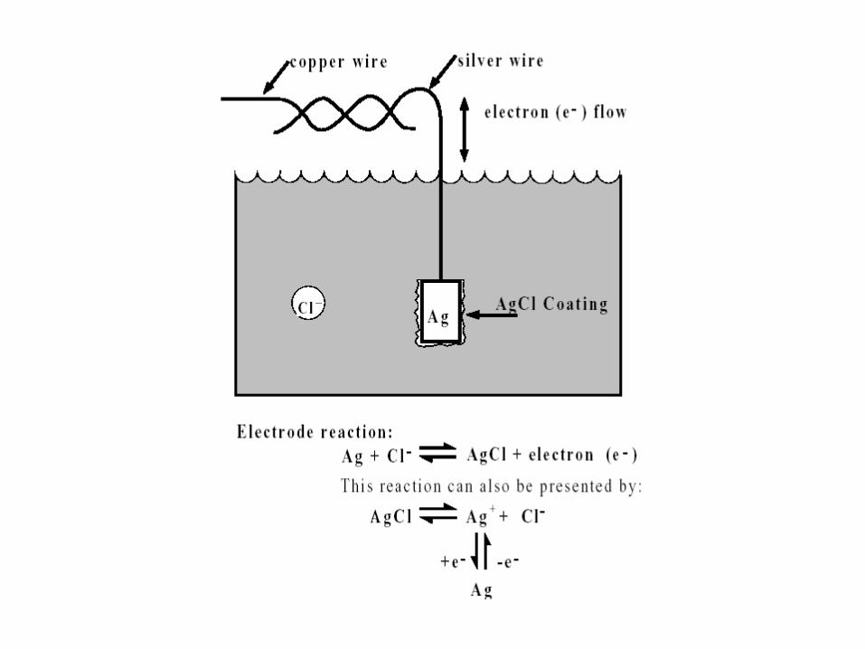
Slowly inactivating K current channel (Ram & Dagan, 1987)
1. Individual channels are either open or closed (no partial openings). Sometimes more than one channel is in a patch.
2. Each channel opening is only a brief event compared to the total duration of the whole cell current.
3. Channel opening and closing is variable in duration and latency.
4. The overall probability of channel opening is similar to the whole cell current
5. Second openings can happen if there’s no inactivation.
Similarly, individual potassium channels, calcium channels, and other channels
can be studied by patch clamping

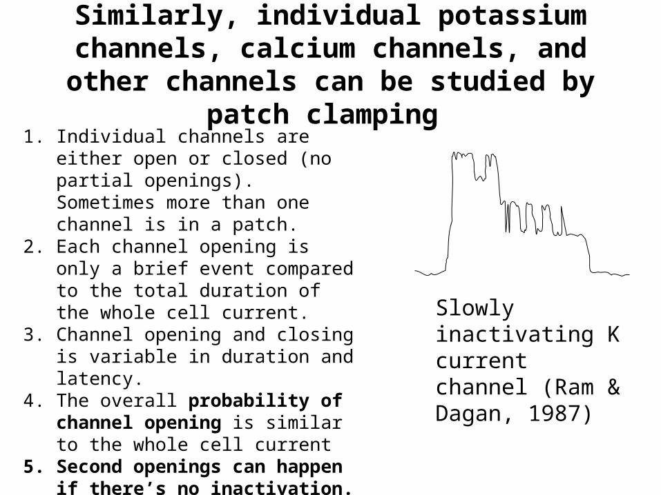
On-Cell
Cell-Attached
The configurations of the patch-clamp technique

On-Cell
Inside-out patch
The configurations of the patch-clamp technique

Whole Cell
On-Cell
The configurations of the patch-clamp technique

Whole Cell
The configurations of the patch-clamp technique

Whole Cell
outside-out patch
The configurations of the patch-clamp technique

Rs
CmRc
The whole-cell configuration

NaCl 144NaH2PO4 0.4
KCl 4
MgSO4 0.53
CaCl2 1.8
Glucose 5.5
HEPES 5
+
ICa blocker
Intracellukar solution (mM)(for K currents)
Extracellular solution (mM)(for K currents)
K-aspartate 100
KCl 25
K2HPO4 10,
K2EGTA 5
K2ATP 3
MgCl2 1
HEPES 10

Extracellular solution
Patch-clamp amplifier
IBM PC
Micropipette
+ __
++
+ ++
_
_
__ _ ++
__
++_
Cell
-40 mV
-20 mV ... +50 mV10 ms ... 5000 ms
Intracellular solution
The whole cell configuration


The “run-down“ effect
The ATP-sensitive potassium current

The L-type calcium current
The “run-down“

Whole Cell
Whole Cell, perforated patch
- amphotericin-B- nystatin
The configurations of the patch clamp technique

The “run-down”
The L-type calcium current

Cell isolation
- Ca2+ - free perfusion
- enzymatic digestion (collagenase)
- mechanical separation

200 pA
100 ms
0 mV400 ms
-40 mV
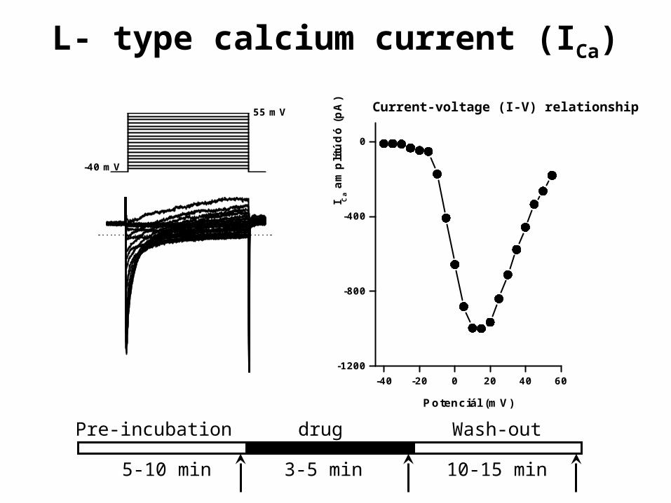
L- type calcium current (ICa)

-40 -20 0 20 40 60
-1200
-800
-400
0
I Ca
amp
lítú
dó
(p
A)
Potenciál (mV)
-35 mV-40 mV
L- type calcium current (ICa)

-40 -20 0 20 40 60
-1200
-800
-400
0
I Ca
amp
lítú
dó
(p
A)
Potenciál (mV)
-30 mV-40 mV
L- type calcium current (ICa)

-40 -20 0 20 40 60
-1200
-800
-400
0
I Ca
amp
lítú
dó
(p
A)
Potenciál (mV)
-25 mV-40 mV
L- type calcium current (ICa)

-40 -20 0 20 40 60
-1200
-800
-400
0
I Ca
amp
lítú
dó
(p
A)
Potenciál (mV)
-20 mV-40 mV
L- type calcium current (ICa)

-40 -20 0 20 40 60-1200
-800
-400
0
I Ca
amp
lítú
dó
(p
A)
Potenciál (mV)
-15 mV-40 mV
L- type calcium current (ICa)

-40 -20 0 20 40 60-1200
-800
-400
0
I Ca
amp
lítú
dó
(p
A)
Potenciál (mV)
-10 mV-40 mV
L- type calcium current (ICa)
-40 -20 0 20 40 60-1200
-800
-400
0
I Ca
amp
lítú
dó
(p
A)
Potenciál (mV)
55 mV
-40 mV
drug
5-10 min 3-5 min 10-15 min
Wash-outPre-incubation
Current-voltage (I-V) relationship
L- type calcium current (ICa)


![[Jürgen jost] postmodern_analysis](https://img.pdfslide.us/doc/110x75/5560c3acd8b42a033c8b5961/jurgen-jost-postmodernanalysis.jpg)