Embed Size (px)
Citation preview

RESEARCH REPORT
Cellular analysis of cleavage-stage chick embryos reveals hiddenconservation in vertebrate early developmentHiroki Nagai1, Maiko Sezaki1, Kisa Kakiguchi2, Yukiko Nakaya1, Hyung Chul Lee3, Raj Ladher4,Tomohiro Sasanami5, Jae Yong Han3, Shigenobu Yonemura2 and Guojun Sheng1,*
ABSTRACTBirds andmammals, phylogenetically close amniotes with similar post-gastrula development, exhibit little conservation in their post-fertilizationcleavage patterns. Data from the mouse suggest that cellularmorphogenesis and molecular signaling at the cleavage stage playimportant roles in lineage specification at later (blastula and gastrula)stages. Very little is known, however, about cleavage-stage chickembryos, owing to their poor accessibility. This period of chickdevelopment takes place before egg-laying and encompassesseveral fundamental processes of avian embryology, includingzygotic gene activation (ZGA) and blastoderm cell-layer increase. Wehave carried outmorphological and cellular analyses of cleavage-stagechick embryos covering the first half of pre-ovipositional development,from Eyal-Giladi and Kochav stage (EGK-) I to EGK-V. Scanningelectron microscopy revealed remarkable subcellular details ofblastomere cellularization and subgerminal cavity formation.Phosphorylated RNA polymerase II immunostaining showed thatZGA in the chick starts at early EGK-III during the 7th to 8th nucleardivision cycle, comparable with the time reported for other yolk-richvertebrates (e.g. zebrafishandXenopus). The increase in thenumberofcell layers after EGK-III is not a direct consequence of oriented celldivision. Finally, we present evidence that, as in the zebrafish embryo, ayolk syncytial layer is formed in the avian embryo after EGK-V. Our datasuggest that several fundamental features of cleavage-stagedevelopment in birds resemble those in yolk-rich anamniote species,revealing conservation in vertebrate early development. Whether thisconservation lends morphogenetic support to the anamniote-to-amniote transition in evolution or reflects developmental plasticity inconvergent evolution awaits further investigation.
KEY WORDS: Amniote, Cellularization, Chick, Cleavage, Yolksyncytium, Zygotic gene activation
INTRODUCTIONAmature avian oocyte is fertilized in the infundibulum shortly afterovulation. From fertilization to egg-laying, the period of pre-ovipositional development covers∼25 h in the chick, duringwhicha number of important early embryological events take place. These
events include the meroblastic cleavage, cellularization, zygoticgene activation, blastoderm layer increase and reduction, radialsymmetry breaking, and segregation of epiblast and hypoblastlineages (Sheng, 2014). Owing to poor accessibility, nocomprehensive molecular or cellular study on pre-ovipositionalembryos has been reported in the literature. General morphologicalcharacterization in the 1970s by Eyal-Giladi and Kochav providedus with a basic staging system, the EGK stages (Eyal-Giladi, 1991;Eyal-Giladi and Kochav, 1976; Kochav et al., 1980), and theultrastructure of embryos from the later half of this developmentalperiod was investigated in the 1990s with electron microscopy byEtches and colleagues (Watt et al., 1993).
According to the EGK staging system, pre-streak chickdevelopment is divided into 14 sub-stages, from EGK-I to EGK-XIV. The pre-ovipositional period includes the stages EGK-I toEGK-X. These 10 stages are further grouped into two developmentalphases. In the first, from EGK-I to EGK-VI (the cleavage stages),a chick embryo grows mainly by means of cell proliferation(1→10,000) and blastoderm cell-layer increase (1→6). In thesecond, from EGK-VII to EGK-X (the area pellucida formationstages), the center of the blastoderm above the subgerminal cavitythins out from six to two cell layers, leading to the formation of twocircular territories: the central area pellucida, which is detached fromthe yolk and contains the epiblast and hypoblast cell layers; and theperipheral area opaca, which varies in its thickness and maintainscellular interactions with the yolk. Radial symmetry breaking,one of the most important developmental events in vertebrateembryogenesis, also occurs during this phase.
With a paucity of molecular and cellular understanding, and withthe exception of primordial germ cells (PGCs) (Tsunekawa et al.,2000), chick embryos during the first phase of pre-ovipositionaldevelopment are generally regarded as being undifferentiated,containing equipotent cells whose morphological variations reflecttheir cellularization and proliferation heterogeneity rather than theirfate diversification. These embryos are also considered to beinactive in their preparation for lineage segregation (except for thePGCs) or radial symmetry breaking because embryonic axis can beefficiently re-specified with physical perturbation during the secondphase of pre-ovipositional development (Kochav and Eyal-Giladi,1971). Data from other vertebrate models, however, suggest thatmolecular and cellular events taking place during cleavage phase ofthe early development are crucial for dynamic signaling, lineagespecification and morphogenetic movements associated with thelate blastula/early gastrula development.
Among those crucial events are the initiation of zygotic geneactivation (ZGA), the separation of outside and inside blastomeres,and the formation of a yolk syncytial layer (YSL). In both mouseand zebrafish embryos, ZGA comes under direct control of keypluripotency regulators (Foygel et al., 2008; Lee et al., 2013b;Leichsenring et al., 2013) and is tightly associated with epigeneticReceived 8 October 2014; Accepted 3 February 2015
1Laboratory for Early Embryogenesis, RIKEN Center for Developmental Biology,Kobe, Hyogo 650-0047, Japan. 2ElectronMicroscopy Laboratory, RIKENCenter forDevelopmental Biology, Kobe, Hyogo 650-0047, Japan. 3Department ofAgricultural Biotechnology, Seoul National University, Seoul 151-921, Korea.4Laboratory for Sensory Development, RIKEN Center for Developmental Biology,Kobe, Hyogo 650-0047, Japan. 5Department of Applied Biological Chemistry,Faculty of Agriculture, Shizuoka University, 836 Ohya, Shizuoka 422-8529, Japan.
*Author for correspondence ([email protected])
This is an Open Access article distributed under the terms of the Creative Commons AttributionLicense (http://creativecommons.org/licenses/by/3.0), which permits unrestricted use,distribution and reproduction in any medium provided that the original work is properly attributed.
1279
© 2015. Published by The Company of Biologists Ltd | Development (2015) 142, 1279-1286 doi:10.1242/dev.118604
DEVELO
PM
ENT

reprogramming of parental genomes that, later in development,controls the specification of both somatic and germ cell lineages(Hackett and Surani, 2013). During mouse early development,8→16 and 16→32 divisions produce two types of blastomeres:those located outside, which are biased towards the trophectodermfate; and those located inside, which will become the inner cellmass (Rossant and Tam, 2009). This process is regulated bypolarized cell division and post-divisional cell rearrangement(Johnson and Ziomek, 1981; Parfitt and Zernicka-Goetz, 2010;Sasaki, 2010;Watanabe et al., 2014). The YSL is a special adaptivefeature of telolecithal development. Although eutherian eggs aredevoid of yolk, both birds and reptiles, two other extant amniotegroups, have telolecithal eggs. Studies in zebrafish, a geneticmodel for telolecithal embryonic development, suggest that theYSL is formed at the cleavage stage and that it undergoes highlycoordinated cytokinetic movements with the overlying blastodermcells (Carvalho and Heisenberg, 2010). It also regulatesmesendoderm induction by secreting Ndr1 and Ndr2 signals (twonodal relatedmolecules) under the transcriptional control ofMxtx2(Hong et al., 2011), and produces the ventralizing signal BMP2bunder the transcriptional control of MGA, MAX and Smad4proteins (Sun et al., 2014). To investigate how these events occur inthe chick and whether novel insights into vertebrate earlydevelopment can be gained by taking a comparative approach,we collected cleavage-stage chick embryos and performed in-depthmorphological and cell biological analyses.
RESULTS AND DISCUSSIONGeneral description of EGK-I to -V embryosPre-ovipositional eggs were retrieved from laying hens using anon-invasive method described previously (Eyal-Giladi andKochav, 1976; Lee et al., 2013a). Because the ultrastructure oflate cleavage-stage embryos and the area pellucida of formation-stage embryos had been described previously (Watt et al., 1993), wefocused our attention on the early and mid-cleavage stages, namelyfrom EGK-I to EGK-V. This period covers about 8 hours ofdevelopment. Together with time needed from the fertilization to thefirst cell division, it is considered here to represent the first half ofpre-ovipositional development (∼13 h out of a total of 25 h). Afteregg retrieval, shell and albumen were removed, and embryos werebriefly fixed in 4% PFA with intact yolk for morphologicalpreservation. They were then cut out together with a small amountof underlying yolk to retain embryo-yolk contacts, further fixed in4% PFA and stored at 4°C for later analysis. Out of a total of 138embryos collected, 73 were confirmed under a stereo-microscope torepresent stages between EGK-I and EGK-V (supplementarymaterial Fig. S1). Their general morphology is briefly describedhere. At EGK-I, all cells are connected to the yolk basally; whenviewed from the apical side, they are not yet enclosed by lateral cellmembrane, except for a few centrally located ones at late EGK-I. AtEGK-II, two types of cells, the central ones with closed lateralmembrane and peripheral ones which are open to the yolk laterally,can be easily distinguished. From EGK-III to EGK-V, the central,closed cells become progressively smaller as they divide, and thearea they occupy expands as more closed cells are produced from theperipheral open cells after division. In a majority of embryos at allEGK stages, the central cell cluster is positioned eccentrically in theblastoderm. Compared with those described by Eyal-Giladi andKochav (1976), our embryos had their morphology better preservedand, likely as a result of this, we did not notice anyobvious shrinkagein blastoderm size at EGK-III or large vacuoles at EGK-I and EGK-II, as described previously (Eyal-Giladi and Kochav,1976).
Scanning electron microscopy of EGK-I to -V embryosTo gain a better understanding of cellular morphogenesis duringearly cleavages, we performed scanning electron microscopy (SEM)analysis with EGK-I to EGK-V embryos (Fig. 1A; supplementarymaterial Figs S2-S6; summarized in Fig. 1B). Under SEM, cleavagefurrows and cell boundaries were easily visible. In an EGK-I embryowith eight open cells (Fig. 1A; supplementary material Fig. S2), theorder of three separate cytokinetic events could be estimated based onthe shape and extent of furrow progression. Fracture surface viewsindicated that transition from vertical furrow burrowing to horizontalfurrow burrowing took place ∼50-100 µm deep into the yolk(supplementary material Fig. S2C-E). The transition area hadelaborate membrane protrusions (supplementary material Fig. S2E),resembling the furrow based body (Bellairs et al., 1978; Gipson,1974). The apical surface was rich in microvilli, with small vesicularbodies at the cortex (supplementarymaterial Fig. S2F). Yolk granulesexhibited fine-graded distribution in their sizes. No obvious transitionzone could be distinguished between yolk granules to be includedin future blastoderm cells (<100 µm below the membrane) and thoseto be allocated to the yolk (supplementary material Fig. S2D,G).In an EGK-II embryo with 16 laterally closed cells (Fig. 1A;supplementary material Fig. S3), the center of closed cells waslocated clearly off the geometric center of the blastoderm, possibly asa consequence of eccentric localization of the zygotic nucleus. It isunclear whether this eccentricity has any predictive value with regardto where the future posterior side of the epiblast (dorsal side of theembryo) will be. The shape and size of central cells was variable(supplementary material Fig. S3B). Contrary to what had beensuggested based on light microscopy analysis (Eyal-Giladi andKochav, 1976), a few central cells at EGK-II had already finished thecellularization process with complete basal and lateral membranes(supplementary material Fig. S3C,D). Numerous small, intracellularvesicular bodies were present in all cells (supplementary materialFig. S3D,E), resembling those reported in other yolk-rich vertebrateembryos (Danilchik et al., 2003; Li et al., 2006). These vesicles arelikely to be involved in the cellularization and cell division processes,which require a constant supply of new plasma membrane. It is alsopossible, however, that they are related to the supply of surfaceproteins or to the secretion of extracellular matrix molecules.
At EGK-III (Fig. 1A; supplementary material Fig. S4), the territoryof laterally closed cells fromapical surface viewexpanded significantlyas more peripheral open cells gave rise to laterally closed daughtercells after division (supplementary material Fig. S4A-C). The apicalmembrane remained rich in microvilli (supplementary materialFig. S4D), but with reduced abundance at cleavage furrows. Asmall subgerminal cavity space appeared under some central cells(supplementary material Fig. S4E) and two-cell thick blastodermwasobserved occasionally (supplementary material Fig. S4F). This trendcontinued at EGK-IV (Fig. 1A; supplementary material Fig. S5). Atthis stage, the subgerminal cavity space became obvious under mostcentral cells and started to merge to form a bona fide cavity above theyolk cell surface (supplementary material Fig. S5C-E). A significantproportion of the blastoderm became two cells thick (supplementarymaterial Fig. S5C,D), with the very central part being three cells thick(supplementary material Fig. S5E). In slightly more peripheralregions, the subgerminal cavity under newly closed cells had justappeared as an isolated pocket between two closed cells and the yolkcell (supplementary material Fig. S5H). At the basal surface of thesecells, two closely apposed plasma membranes (supplementarymaterial Fig. S5H′, arrows) marked the completion of theircellularization. Interestingly, unlike the fine-graded size distributionbefore cellularization (supplementary material Fig. S2G), yolk
1280
RESEARCH REPORT Development (2015) 142, 1279-1286 doi:10.1242/dev.118604
DEVELO
PM
ENT

granules at the basal cortex of newly formed cells were much smallerthan those located in the adjacent yolk cells (supplementary materialFig. S5H′, arrowheads). At EGK-V (Fig. 1A; supplementary materialFig. S6), all non-edge cells of the blastoderm had completed thecellularization process. Many edge cells had also become fullycellularized (supplementary material Fig. S6C,E), although some ofthem remained open peripherally. A smooth transition in blastodermthickness, from one cell layer at the edge to four cell layers at thecenter, could be observed (supplementarymaterial Fig. S6C,D). Cellsbridging the one-cell and two-cell regions often exhibited wedge-shaped morphology (supplementary material Fig. S6C,F). Thesubgerminal cavity became a continuous, expanded space below themultilayered part of the blastoderm, but the one-cell layered partremained in close associationwith the underlying yolk cell membrane(supplementary material Fig. S6C-E). Cell-cell contacts amongblastomeres were prominent in both superficial and deep layers(supplementary material Fig. S6G-J).
Zygotic gene activation (ZGA) is initiated at late EGK-II/earlyEGK-IIIThe cellular morphogenetic process from EGK-I to EGK-V issummarized in Fig. 1B. During this period of chick development,cell (nucleus) number increases from 1 to ∼2000 (Park et al., 2006).ZGA in mammalian embryos (e.g. in the mouse and human) startsvery early, before the 3rd cell cycle (Vassena et al., 2011; Wang andDey, 2006; Xue et al., 2013; Yan et al., 2013). In the zebrafish, with atelolecithal egg, ZGAwas reported to start at the 128-cell stage (8thcell cycle) (Aanes et al., 2011; Harvey et al., 2013; Mathavan et al.,
2005). In other yolk-rich embryos, such asXenopus andDrosophila,ZGA can be detected at about 128- to 256-cell (nucleus) stage (8th to9th cell cycle) (Baroux et al., 2008; Tadros and Lipshitz, 2009). Ithas been hypothesized that instead of the absolute cell cycle number,the timing of ZGA in large, yolk-rich embryos is determined by thenucleocytoplasmic ratio and the maternal clock (Tadros andLipshitz, 2009). We stained early EGK stage chick embryos withan anti-phosphorylated-RNAPolymerase II CTD (p-PolII) antibodyto mark the zygotic gene transcription, DAPI to visualize the nucleiand E-cadherin to visualize cell boundaries. No specific p-PolIIimmunoreactive signals could be detected before late EGK-II stage(Fig. 2A, leftmost column panels). At late EGK-II/early EGK-III(Fig. 2A, 2nd and 3rd column panels), weak nuclear p-PolII signalswere observed in a few centrally located cells. By mid-/late EGK-IIIstages (Fig. 2A, 4th and 5th column panels), p-PolII signals becameprogressively stronger and could be readily detected in many centralcells. About half of p-PolII-positive cells had strong cytoplasmicsignals at late EGK-III, a phenomenon possibly related to thecytoplasmic-to-nuclear translocation of RNA PolII at the onset ofZGA, as observed in the mouse embryo (Bellier et al., 1997). AtEGK-IV (Fig. 2A, 6th and rightmost column panels; Fig. 2C,D),p-PolII signals became more robust and positive cells became moreabundant. During the entire transition period from mid-EGK-II tolate EGK-IV, p-PolII signals appeared in central, smaller cellssooner andmore strongly than in peripheral, larger cells. However, itis unclear whether this is causally related to changes in thenucleocytoplasmic ratio because many central cells were weak ornegative for p-PolII signals even when their similarly sized
Fig. 1. Scanning electron microscopy analysis of EGK-I to -V chick embryos. (A) Apical views of EGK-I to -V embryos under SEM. Scale bars: 500 µm(left); 200 µm (right). Details of the fracture surface are shown in supplementary material Figs S2-S6 and summarized in B. (B) Schematic view ofcellularization and cell proliferation processes during chick development from EGK-I to -V. Completely cellularized blastomeres are shown in blue. Numbersabove the blastoderm indicate the distribution of cell layer numbers.
1281
RESEARCH REPORT Development (2015) 142, 1279-1286 doi:10.1242/dev.118604
DEVELO
PM
ENT

neighbors were strongly positive (Fig. 2C). Supporting our ZGAobservation based on the p-PolII staining data, pre-ovipositionalembryos with GFP transgene insertion under the control of a CMVpromoter (Park andHan, 2012) showed noGFP signals atmid-EGK-II (Fig. 2B) and strong GFP signals at mid-EGK-IV (Fig. 2B). Takentogether, our data suggest that ZGA in the chick is initiated atlate EGK-II/early EGK-III (about 64-128 total cell stage; orapproximately during the 7th or 8th cell cycle) and becomesreadily detectable at mid- to late EGK-III (about 128-256 total cell-stage; 8th to 9th cell cycle). Chick embryos therefore initiate ZGAwith a developmental timing comparable with that in the zebrafishand Xenopus embryos, pointing to potential conservation inmolecular mechanisms controlling ZGA in yolk-rich embryos.
Separation of surface and inner cells is not caused byoriented cell divisionsFrom our scanning EM analysis, late EGK-II/early EGK-III is alsothe stage when the cellularization process becomes complete in afew centrally located cells and the increase in blastoderm cell-layernumber is initiated. This latter process is significant because itresults in the separation of two types of blastomeres: those locatedon the surface and exposed to the external environment; and thoselocated in the inside and shielded from external influences. Eyal-Giladi and colleagues hypothesized that oriented blastomeredivision led to this increase (Kochav et al., 1980). In eutherianembryos, this separation is directly correlated with the future fateof these cells: trophectoderm for the surface-located cells and inner
Fig. 2. ZGA in EGK-II to -IV chick embryos analyzed by anti-Ser5 phosphorylation of Pol II CTD (p-PolII) antibody staining and by GFP transgenesis.(A) Wild-type embryos ranging from mid-EGK-II to late EGK-IV are stained for p-PolII signals. Embryos are co-stained for E-cadherin (E-cad; marking cellboundaries) and DAPI (marking nuclei). Numbers (x/y) under embryo stage labels indicate approximate counts of laterally closed cells (x) and total cells (y).(Top) Whole-embryo views after DAPI staining. At higher magnification: DAPI (second row), E-cad (third row) and p-PolII (bottom row) staining. Signals for p-PolIIare negative at mid-EGK-II, very weak in late EGK-II, rapidly increase at EGK-III and become very strong at late EGK-IV. From late EGK-III to late EGK-IV,strong signals are detected both in the nucleus (in some central blastomeres) and in the cytoplasm (in adjacent blastomeres), although the proportion ofcytoplasmic-positive cells decreases gradually (C,D). p-PolII-negative, small blastomeres are also observed in central areas next to similarly sized positiveblastomeres (C). Signals in peripherally positioned blastomeres are much weaker or negative. (B) Embryos from GFP-transgenic male and wild-type femalecrosses show strong GFP signals at mid-EGK-IV but no GFP signals at mid-EGK-II. Scale bars: 500 µm.
1282
RESEARCH REPORT Development (2015) 142, 1279-1286 doi:10.1242/dev.118604
DEVELO
PM
ENT
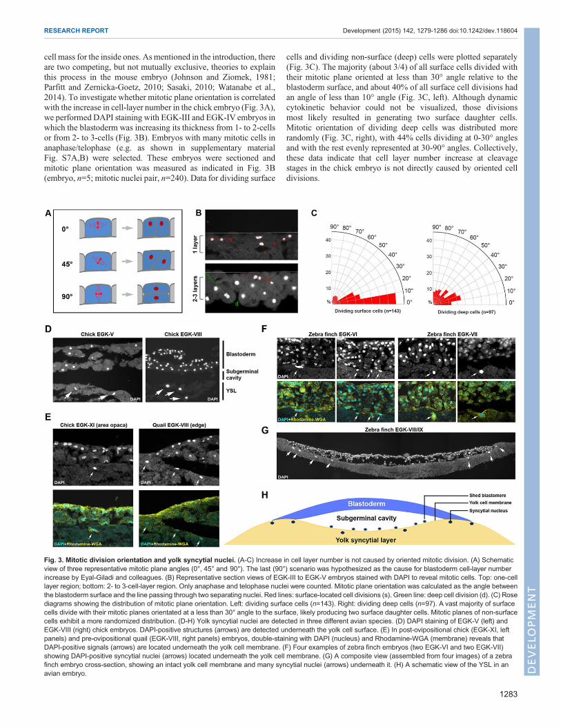
cell mass for the inside ones. As mentioned in the introduction, thereare two competing, but not mutually exclusive, theories to explainthis process in the mouse embryo (Johnson and Ziomek, 1981;Parfitt and Zernicka-Goetz, 2010; Sasaki, 2010; Watanabe et al.,2014). To investigate whether mitotic plane orientation is correlatedwith the increase in cell-layer number in the chick embryo (Fig. 3A),we performed DAPI staining with EGK-III and EGK-IV embryos inwhich the blastoderm was increasing its thickness from 1- to 2-cellsor from 2- to 3-cells (Fig. 3B). Embryos with many mitotic cells inanaphase/telophase (e.g. as shown in supplementary materialFig. S7A,B) were selected. These embryos were sectioned andmitotic plane orientation was measured as indicated in Fig. 3B(embryo, n=5; mitotic nuclei pair, n=240). Data for dividing surface
cells and dividing non-surface (deep) cells were plotted separately(Fig. 3C). The majority (about 3/4) of all surface cells divided withtheir mitotic plane oriented at less than 30° angle relative to theblastoderm surface, and about 40% of all surface cell divisions hadan angle of less than 10° angle (Fig. 3C, left). Although dynamiccytokinetic behavior could not be visualized, those divisionsmost likely resulted in generating two surface daughter cells.Mitotic orientation of dividing deep cells was distributed morerandomly (Fig. 3C, right), with 44% cells dividing at 0-30° anglesand with the rest evenly represented at 30-90° angles. Collectively,these data indicate that cell layer number increase at cleavagestages in the chick embryo is not directly caused by oriented celldivisions.
Fig. 3. Mitotic division orientation and yolk syncytial nuclei. (A-C) Increase in cell layer number is not caused by oriented mitotic division. (A) Schematicview of three representative mitotic plane angles (0°, 45° and 90°). The last (90°) scenario was hypothesized as the cause for blastoderm cell-layer numberincrease by Eyal-Giladi and colleagues. (B) Representative section views of EGK-III to EGK-V embryos stained with DAPI to reveal mitotic cells. Top: one-celllayer region; bottom: 2- to 3-cell-layer region. Only anaphase and telophase nuclei were counted. Mitotic plane orientation was calculated as the angle betweenthe blastoderm surface and the line passing through two separating nuclei. Red lines: surface-located cell divisions (s). Green line: deep cell division (d). (C) Rosediagrams showing the distribution of mitotic plane orientation. Left: dividing surface cells (n=143). Right: dividing deep cells (n=97). A vast majority of surfacecells divide with their mitotic planes orientated at a less than 30° angle to the surface, likely producing two surface daughter cells. Mitotic planes of non-surfacecells exhibit a more randomized distribution. (D-H) Yolk syncytial nuclei are detected in three different avian species. (D) DAPI staining of EGK-V (left) andEGK-VIII (right) chick embryos. DAPI-positive structures (arrows) are detected underneath the yolk cell surface. (E) In post-ovipositional chick (EGK-XI, leftpanels) and pre-ovipositional quail (EGK-VIII, right panels) embryos, double-staining with DAPI (nucleus) and Rhodamine-WGA (membrane) reveals thatDAPI-positive signals (arrows) are located underneath the yolk cell membrane. (F) Four examples of zebra finch embryos (two EGK-VI and two EGK-VII)showing DAPI-positive syncytial nuclei (arrows) located underneath the yolk cell membrane. (G) A composite view (assembled from four images) of a zebrafinch embryo cross-section, showing an intact yolk cell membrane and many syncytial nuclei (arrows) underneath it. (H) A schematic view of the YSL in anavian embryo.
1283
RESEARCH REPORT Development (2015) 142, 1279-1286 doi:10.1242/dev.118604
DEVELO
PM
ENT
A yolk syncytial layer (YSL) is formed during early aviandevelopmentAsmentioned in the introduction, the importance of the YSL in earlydevelopment has been demonstrated in the zebrafishmodel.However,whether a similar YSL exists in the avian embryo has not beeninvestigated. DAPI staining and section analysis suggested that nosyncytial nuclei could be found at stages EGK-I to -IV (not shown).At EGK-V, occasional syncytial nuclei were detected (Fig. 3D, left),which became brighter, larger and more abundant at later EGK stages(Fig. 3D, right, showing an EGK-VIII embryo) and persisted throughpost-ovipositional stages (Fig. 3E, left, showing an EGK-XI embryo).These nuclei are to be distinguished fromDAPI-positive cells locatedabove the surface of the yolk cell (e.g. as shown in Fig. 3G;supplementary material Fig. S7F), which are shed from theblastoderm cell mass (as described by Eyal-Giladi and Kochav) andare frequently observed during the blastoderm thinning process fromEGK-VII onwards. To confirm the existence of a YSL in the avianembryo, we performed similar analysis with quail (Fig. 3E, right) andzebra finch (Fig. 3F,G) embryos. Pre-ovipositional quail eggs wereretrieved andDAPI staining of these embryos revealed the presence ofsyncytial nuclei, especially underneath the area opaca and close to theblastoderm margin (Fig. 3E, right). Zebra finch eggs are laid at anearlier developmental stage than chicken or quail eggs, and zebrafinch embryos at stages EGK-VI to -IX can be collected easily fromfreshly laid eggs (S. S. Mak, C. Alev, H.N., A.Wrabel, Y. Matsuoka,A. Honda, G. Sheng and R. K. Ladher, unpublished). DAPI stainingand section analysis of EGK-VI to -IX zebra finch embryos supportedour chick and quail data (Fig. 3F,G). Syncytial nuclei were detectedmore abundantly in the zebra finch (Fig. 3G, arrows) and co-stainingwith a membrane marker (rhodamine-labeled WGA) revealedthat these DAPI-positive signals were located underneath the yolkcell membrane (Fig. 3F). Interestingly, many peripherally locatedsyncytial nuclei in the finch embryo could be found even in whole-mount views (supplementarymaterial Fig. S7C-E). There nuclei werelocated away from the blastoderm edge, as revealed by phalloidinco-staining (supplementary material Fig. S7D,D′,E,E′) and in section(supplementary material Fig. S7F). Taken together, these datastrongly support the hypothesis (Fig. 3H) that a YSL is present inthe avian embryo. Whether this YSL plays a role in patterning theoverlying blastoderm remains to be clarified.
SummaryThe first half of pre-ovipositional chick development, from EGK-Ito EGK-V, was investigated in this study (Table 1). During thisdevelopmental period, a chick embryo undergoes ∼11 rounds of
mitotic divisions. The cell number increases from one to ∼2000and the cell-layer number increases from one to four. Meroblasticcleavages continue from EGK-I, when all cells are open, to lateEGK-V, when all cells are fully cellularized. Blastomere-yolkseparation starts at late EGK-II and ZGA starts at late EGK-II/early EGK-III (∼7th to 8th nuclear division cycle). Separation ofoutside and inside blastomeres is not due to oriented cell division,as previously suggested. Syncytial nuclei located underneath theyolk cell membrane can be detected from EGK-V and becomemore abundant during the second half of pre-ovipositionaldevelopment. The presence of yolk syncytial nuclei is alsoobserved in quail and zebra finch embryos. Our data onblastoderm cell-layer increase support the hypothesis that thisprocess is controlled by cellular mechanisms other than orientedmitotic division. Collectively, our data suggest that many featuresof cleavage-stage development in the chick resemble those in thezebrafish. Whether this conservation is a result of convergentevolution or is indicative of hardwired molecular and cellularmechanisms regulating vertebrate early development awaits futureinvestigation.
MATERIALS AND METHODSEmbryo collectionEGK stage chick embryos were collected from laying white leghorn hensusing a non-invasive, manual retrieval (abdominal massage) method aspreviously described (Lee et al., 2013a). The retrieval was carried out in theafternoon or early evening and was timed to target eggs in the first half ofpre-ovipositional development. Each collected egg was placed in a Petridish. The eggshell was opened and removed, and the egg yolk was adjustedso that the embryo was positioned at the top. Excessive albumen coveringthe embryo was removed and a few drops of ice-cold 4% paraformaldehyde(PFA) fixative (Alev et al., 2013) were put on the vitelline membrane overthe embryo to preserve the morphology. After a few minutes of fixation atroom temperature, the embryo, together with a small amount of peripheraland underlying yolk, was excised from the bulk of the yolk and placed inPannett-Compton saline solution (Alev et al., 2013). The vitellinemembrane was then carefully removed and the embryo together withattached yolk was transferred to 4% PFA for further fixation and storage at4°C. Out of 138 embryos obtained this way, 85 embryos were analyzed forstage distribution under a stereo microscope, and 73 out of 85 were withinthe cleavage stages according to the EGK staging system (13 EGK-I, 16EGK-II, 15 EGK-III, 17 EGK-IV and 12 EGK-V). Pre-ovipositional GFPtransgenic chick embryos and pre-ovipositional quail embryos werecollected using the same abdominal massage method. Post-ovipositionalzebra finch eggs were collected daily from locally maintained colonies andzebra finch embryos were processed for staining using the same protocol asfor the chick embryos.
Table 1. Summary of EGK-I to -V embryos
EGK I II III IV V VI → X
Total number of nuclei or cells(median)
1-16 (∼8) 16-64 (∼32) 64-256 (∼128) 256-1k (∼512) 1k-4k (∼2k) 4k-16k (∼8k) → ∼55k
Nuclear division cycle 1st-4th 4th-6th 6th-8th 8th-10th 10th-12th 12th-14th → 15th-16thHours post-fertilization 5.5 7 8.5 10 13.5 15.5 → 25Laterally closed cells (median) 0 ∼16 ∼80-90 ∼250-300 ∼All All → AllCell layer number 1 1 2 3 4 5-6 → 1 or 2Basally closed cells − −/+ + + + + → +Zygotic gene activation − −/+ + + + + → +Yolk syncytium − − − − −/+ + → +Cellularization of edge cells − − − − −/+ + → +
Avian early development from fertilization at the beginning of EGK-I to complete cellularization by the end of EGK-V is viewed using the following criteria: totalnumber of cells or nuclei; nuclear division cycle; time post-fertilization; number of laterally closed cells; number of cell layers in central blastoderm; blastomerebasal closure; zygotic gene activation; yolk syncytium and edge cell cellularization. Of practical values among them are the total number of cells/nuclei andnumber of laterally closed cells. However, early development is a continuous process and these criteria should be used only as a general guide.−, negative; +, positive; −/+, weakly or partially positive.
1284
RESEARCH REPORT Development (2015) 142, 1279-1286 doi:10.1242/dev.118604
DEVELO
PM
ENT

Scanning electron microscopy (SEM)Samples for SEM analysis were prepared as described previously(Ikenouchi et al., 2013). Briefly, EGK stage chick embryos were fixedwith 2.5% glutaraldehyde and 2% formaldehyde in 0.1 M sodiumcacodylate buffer (pH 7.4) for 2 h at room temperature, followed by post-fixation with 1% OsO4 in the same buffer for 2 h on ice. Post-fixed embryoswere dehydrated in ethanol and then transferred to isoamyl acetate, followedby critical point drying (JCPD-5, JEOL). Specimen surface was coatedusing an osmium coater (Neoc-STB, Meiwafosis). For fracture surfaceimages after whole-mount SEM, embryo specimen was settled on a smallpiece of adhesive tape, and fractured using a fine tungsten needle. All SEMimages were acquired using scanning electron microscope JSM-5600LV(JEOL) with accelerating voltage set at 10 kV.
Immunofluorescence, nuclear staining, imaging and imageanalysisAll steps except for the imaging were carried out at 4°C. For phosphorylatedRNApolymerase II detection, fixed embryoswerewashed inPBTw(PBSwith0.1% Tween-20) for 3×20 min, followed by permeabilization in PBTr (PBSwith 0.1% Triton X-100) for 3×30 min and by blocking in blocking solution(PBS with 1% DMSO, 0.1% Triton X-100 and 5% skimmed milk). Blockedembryos were incubated overnight with anti-RNA polymerase II CTD pSer5(1:1000, Abcam Cat# ab5131), washed in PBTw, reblocked in blockingsolution and incubated in the secondary antibody (Alexa Fluor 594 goat anti-rabbit IgG(H+L) (1:300, Molecular Probes Cat# a11012) before imaging.DAPI (Molecular Probes, D1306) and 36/E-Cadherin antibody (BDTransduction Laboratories, 610182) were used for whole-mount nuclear andE-cadherin staining, respectively. Yolk cell membrane was marked usingrhodamine-labeled wheat germ agglutinin (WGA) (Vector Laboratories, Cat#RL-1022). Whole-mount fluorescence images were acquired using OlympusFV1000 microscope. Section fluorescence images were acquired usingOlympus BX51W1 or Olympus FV1000 microscope after processing for10 µmparaffin-embedded section or 12 µmcryosection followedbymountingin ProLong Gold antifade reagent with DAPI (Molecular Probes Cat#P36931). Mitotic division angles were measured using Fiji software and datawere plotted as rose diagrams using GeoRose software.
AcknowledgementsWe thank Anna Wrabel and Siu-Shan Mak for help with finch colony maintenanceand Kanako Ota and members of the J.Y.H. laboratory for help and coordinationduring the collection of pre-ovipositional stage chicken embryos.
Competing interestsThe authors declare no competing or financial interests.
Author contributionsG.S., H.N. and J.Y.H. designed the experiments; H.N., M.S., K.K., Y.N., H.C.L., R.L.,T.S., S.Y. and G.S. performed the experiments; all authors analyzed the data; G.S.and H.N. wrote the manuscript.
FundingThis work was supported by an internal grant to G.S. from RIKEN Center forDevelopmental Biology. Deposited in PMC for immediate release.
Supplementary materialSupplementary material available online athttp://dev.biologists.org/lookup/suppl/doi:10.1242/dev.118604/-/DC1
ReferencesAanes, H., Winata, C. L., Lin, C. H., Chen, J. P., Srinivasan, K. G., Lee, S. G. P.,Lim, A. Y. M., Hajan, H. S., Collas, P., Bourque, G. et al. (2011). ZebrafishmRNA sequencing deciphers novelties in transcriptome dynamics duringmaternal to zygotic transition. Genome Res. 21, 1328-1338.
Alev, C., Nakano, M., Wu, Y., Horiuchi, H. and Sheng, G. (2013). Manipulatingthe avian epiblast and epiblast-derived stem cells. Methods Mol. Biol. 1074,151-173.
Baroux, C., Autran, D., Gillmor, C. S., Grimanelli, D. and Grossniklaus, U.(2008). Thematernal to zygotic transition in animals and plants.Cold Spring Harb.Symp. Quant. Biol. 73, 89-100.
Bellairs, R., Lorenz, F. W. and Dunlap, T. (1978). Cleavage in the chick embryo.J. Embryol. Exp. Morphol. 43, 55-69.
Bellier, S., Chastant, S., Adenot, P., Vincent, M., Renard, J. P. and Bensaude, O.(1997). Nuclear translocation and carboxyl-terminal domain phosphorylation ofRNA polymerase II delineate the two phases of zygotic gene activation inmammalian embryos. EMBO J. 16, 6250-6262.
Carvalho, L. and Heisenberg, C.-P. (2010). The yolk syncytial layer in earlyzebrafish development. Trends Cell Biol. 20, 586-592.
Danilchik, M. V., Bedrick, S. D., Brown, E. E. and Ray, K. (2003). Furrowmicrotubules and localized exocytosis in cleaving Xenopus laevis embryos. J. CellSci. 116, 273-283.
Eyal-Giladi, H. (1991). The early embryonic development of the chick, as anepigenetic process. Crit. Rev. Poult. Biol. 3, 143-166.
Eyal-Giladi, H. and Kochav, S. (1976). From cleavage to primitive streak formation:a complementary normal table and a new look at the first stages of thedevelopment of the chick. I. General morphology. Dev. Biol. 49, 321-337.
Foygel,K., Choi, B., Jun, S., Leong,D. E., Lee, A.,Wong,C.C., Zuo,E., Eckart, M.,Reijo Pera, R. A., Wong, W. H. et al. (2008). A novel and critical role for Oct4 as aregulator of the maternal-embryonic transition. PLoS ONE 3, e4109.
Gipson, I. (1974). Electron microscopy of early cleavage furrows in the chickblastodisc. J. Ultrastruct. Res. 49, 331-347.
Hackett, J. A. and Surani, M. A. (2013). Beyond DNA: programming andinheritance of parental methylomes. Cell 153, 737-739.
Harvey, S. A., Sealy, I., Kettleborough, R., Fenyes, F.,White, R., Stemple, D. andSmith, J. C. (2013). Identification of the zebrafish maternal and paternaltranscriptomes. Development 140, 2703-2710.
Hong, S.-K., Jang, M. K., Brown, J. L., McBride, A. A. and Feldman, B. (2011).Embryonic mesoderm and endoderm induction requires the actions of non-embryonic Nodal-related ligands and Mxtx2. Development 138, 787-795.
Ikenouchi, J., Hirata, M., Yonemura, S. and Umeda, M. (2013). Sphingomyelinclustering is essential for the formation of microvilli. J. Cell Sci. 126,3585-3592.
Johnson, M. H. and Ziomek, C. A. (1981). The foundation of two distinct celllineages within the mouse morula. Cell 24, 71-80.
Kochav, S. and Eyal-Giladi, H. (1971). Bilateral symmetry in chick embryodetermination by gravity. Science 171, 1027-1029.
Kochav, S., Ginsburg, M. and Eyal-Giladi, H. (1980). From cleavage to primitivestreak formation: a complementary normal table and a new look at the first stagesof the development of the chick. II. Microscopic anatomy and cell populationdynamics. Dev. Biol. 79, 296-308.
Lee, H. C., Choi, H. J., Park, T. S., Lee, S. I., Kim, Y. M., Rengaraj, D., Nagai, H.,Sheng, G., Lim, J. M. and Han, J. Y. (2013a). Cleavage events and spermdynamics in chick intrauterine embryos. PLoS ONE 8, e80631.
Lee, M. T., Bonneau, A. R., Takacs, C. M., Bazzini, A. A., DiVito, K. R., Fleming,E. S. and Giraldez, A. J. (2013b). Nanog, Pou5f1 and SoxB1 activate zygoticgene expression during the maternal-to-zygotic transition. Nature 503, 360-364.
Leichsenring, M., Maes, J., Mossner, R., Driever, W. and Onichtchouk, D.(2013). Pou5f1 transcription factor controls zygotic gene activation in vertebrates.Science 341, 1005-1009.
Li, W. M., Webb, S. E., Lee, K. W. and Miller, A. L. (2006). Recruitment andSNARE-mediated fusion of vesicles in furrow membrane remodeling duringcytokinesis in zebrafish embryos. Exp. Cell Res. 312, 3260-3275.
Mathavan, S., Lee, S. G. P., Mak, A., Miller, L. D., Murthy, K. R. K., Govindarajan,K. R., Tong, Y., Wu, Y. L., Lam, S. H., Yang, H. et al. (2005). Transcriptomeanalysis of zebrafish embryogenesis using microarrays. PLoS Genet. 1,260-276.
Parfitt, D.-E. and Zernicka-Goetz, M. (2010). Epigenetic modification affectingexpression of cell polarity and cell fate genes to regulate lineage specification inthe early mouse embryo. Mol. Biol. Cell 21, 2649-2660.
Park, T. S. and Han, J. Y. (2012). piggyBac transposition into primordial germ cellsis an efficient tool for transgenesis in chickens. Proc. Natl. Acad. Sci. USA 109,9337-9341.
Park, H. J., Park, T. S., Kim, T. M., Kim, J. N., Shin, S. S., Lim, J. M. and Han, J. Y.(2006). Establishment of an in vitro culture system for chicken preblastodermalcells. Mol. Reprod. Dev. 73, 452-461.
Rossant, J. and Tam, P. P. L. (2009). Blastocyst lineage formation, early embryonicasymmetries and axis patterning in the mouse. Development 136, 701-713.
Sasaki, H. (2010). Mechanisms of trophectoderm fate specification inpreimplantation mouse development. Dev. Growth Differ. 52, 263-273.
Sheng, G. (2014). Day-1 chick development. Dev. Dyn. 243, 357-367.Sun, Y., Tseng, W.-C., Fan, X., Ball, R. and Dougan, S. T. (2014). Extraembryonic
signals under the control of MGA, Max, and Smad4 are required for dorsoventralpatterning. Dev. Cell 28, 322-334.
Tadros, W. and Lipshitz, H. D. (2009). The maternal-to-zygotic transition: a play intwo acts. Development 136, 3033-3042.
Tsunekawa, N., Naito, M., Sakai, Y., Nishida, T. and Noce, T. (2000). Isolation ofchicken vasa homolog gene and tracing the origin of primordial germ cells.Development 127, 2741-2750.
Vassena, R., Boue, S., Gonzalez-Roca, E., Aran, B., Auer, H., Veiga, A. andBelmonte, J. C. I. (2011). Waves of early transcriptional activation andpluripotency program initiation during human preimplantation development.Development 138, 3699-3709.
1285
RESEARCH REPORT Development (2015) 142, 1279-1286 doi:10.1242/dev.118604
DEVELO
PM
ENT

Wang, H. and Dey, S. K. (2006). Roadmap to embryo implantation: clues frommouse models. Nat. Rev. Genet. 7, 185-199.
Watanabe, T., Biggins, J. S., Tannan, N. B. and Srinivas, S. (2014). Limitedpredictive value of blastomere angle of division in trophectoderm and inner cellmass specification. Development 141, 2279-2288.
Watt, J. M., Petitte, J. N. and Etches, R. J. (1993). Early development of the chickembryo. J. Morphol. 215, 165-182.
Xue, Z., Huang, K., Cai, C., Cai, L., Jiang, C.-y., Feng, Y., Liu, Z., Zeng, Q.,Cheng, L., Sun, Y. E. et al. (2013). Genetic programs in human andmouse early embryos revealed by single-cell RNA sequencing. Nature 500,593-597.
Yan, L., Yang, M., Guo, H., Yang, L., Wu, J., Li, R., Liu, P., Lian, Y., Zheng, X.,Yan, J. et al. (2013). Single-cell RNA-Seq profiling of human preimplantationembryos and embryonic stem cells. Nat. Struct. Mol. Biol. 20, 1131-1139.
1286
RESEARCH REPORT Development (2015) 142, 1279-1286 doi:10.1242/dev.118604
DEVELO
PM
ENT


![Glycoinformatics approach for identifying target positions ... · host cell receptor and fusion of the cellular membrane, respectively [1,2,3]. S protein also contains furin cleavage](https://img.pdfslide.us/doc/110x75/5f3bae9d4a6a206937587eb4/glycoinformatics-approach-for-identifying-target-positions-host-cell-receptor.jpg)


![) [111] cleavage plane](https://img.pdfslide.us/doc/110x75/61c7329341512e61f73ea613/-111-cleavage-plane.jpg)