Embed Size (px)
Citation preview

Organs 8,273 (1980); d) M. F. Lim, A. M. Sun, Science 210,908 (1980); e) N. Sliwku, Angew. Chem. 87,556 (1975); Angew. Chem. Int. Ed. Engl. 14, 539 (1975).
[I771 G. Weissmunn, R. Cluiborne: Cell Membranes - Biochemistry, Cell Biology and Pathology. H. P. Publishing, New York 1975.
11781 H. Fischer, E. Rude, D. Sellin, Natunvissenschaften 57, 507 (1970). 11791 K. Dose, Th. Nuwroth, N . Wagner, private communication.
[tSO] U. Zimmermunn, J. Schultz, G. Pilwut, Biophys. J. 13, 1005 (1973). [181] P. Scheurich, U. Zimmermunn. M. Mischel, J Lamprecht, 2. Naturforsch.
11821 N. J. Harper, J. Med. Pharm. Chem. 1, 467 (1959). 11831 We are very much indebted to Professor H. uon Suulfeld (Fachbereich
Kunsterriehung der Universitat Mainz) who gave us the original draw- ing.
C 35, 1081 (1980)
Cells with Manipulated Functions: New Perspectives for Cell Biology, Medicine, and Technology
By Ulrich Zimmermann, Peter Scheurich, Gunter Pilwat, and Roland Benz"'
Exposure to electrical fields can reversibly increase the electrical conductivity and permeability of a cell membrane, which regulates and directs the exchange of materials and information be- tween the cell and its environment. If cell membranes (or artificial lipid membranes) are ex- posed to a field pulse of high intensity and short duration (ns to ps), local electrical break- down occurs in them. This electrical breakdown is associated with a large permeability change in the membrane, which is such that substances or particles (up to the size of genes) which cannot normally permeate through the membrane, are able to traverse the membrane into the cell. The original properties of the membrane are restored within ps to min, depending on the experimental conditions and the membrane properties.
Electrical breakdown in the zone of contact between the membranes of cells (or lipid vesi- cles), which have been made to adhere to each other by the action of weak inhomogeneous al- ternating electrical fields, leads to fusion of these cells with formation of a single cell having new functional characteristics. The electrical fusion method is very mild, and the yield of fused cells is high.
The electrically induced fusion and entrapment of membrane-impermeable substances and genes in cells provide a new tool for the production of a wide range of cells with manipulated functions, which could be used (or are being used) for the solution of a number of problems in cell biology, medicine and technology.
The application of electrical membrane breakdown to clinical diagnostics, the development of cellular carrier systems for the selective transport of drugs to a site of action within the or- ganism and the potential applications of electrically induced fusion for breeding salt-tolerant crop plants, for converting solar energy into ethanol, for synthesizing natural materials and manipulating genes, are described.
1. Introduction
Both the increasing demand for high-grade natural sub- stances and pharmaceuticals for medical and technical appli- cations, and the pressing problems of providing sufficient food and energy for the world population have led to the search for new technologies involving the use of biological systems. This interdisciplinary area of research, which com- prises the disciplines of chemistry, physics, biology and med- icine, is known as biotechnology. The use of microorganisms in industry for the production of medically and technically useful substances and pharmaceuticals has already proved
['I Prof. Dr. U. Zimmermann, Dr. P. Scheurich, Dr. G. Pilwat Arbeitsgruppe Membranforschung am Institut fur Medizin der Kernforschungsanlage Postfach 1913, D-5170 Jiilich 1 (Germany) Priv. Doz. Dr. R. Benz Fakultat fur Biologie der Universitat D-7750 Konstanz (Germany)
highly successful, although this area is still at an early stage"].
During the course of evolution, plant cells developed the ability to photosynthesize, i. e. of being capable to convert so- lar energy into chemical energy with a high degree of effi- ciency. This energy can, in turn, be converted by other cells and organisms into osmotic, mechanical and electrical work. Technologically, we are still far from being able to convert solar energy on a large scale and with a sufficient degree of efficiency.
Many organisms, such as algae and bacteria, have solved the problem of an optimum existence in a saline environ- ment, i. e. they are able to grow and multiply. The ability of crop plants to adapt to saline conditions, coupled with irriga- tion of the soil with salt or brackish water in the arid zones of the earth (which occupy more than 40% of the land mass) could thus make an important contribution to the solution of the world's food shortage[*].
An alternative approach to the solution of this problem is to breed crop plants that will give high yields even under un-
Angew. Chem. In l . Ed. Engl. 20, 325-344 (1981) @ Verlag Chemie GmbH, 6940 Weinheim, 1981 0570-0833/8l/0404-0325 $ 02.50/0 325

favorable conditions. Such high-yield varieties require a large amount of nitrogen fertilizer. Quite apart from the en- vironmental problems caused by fertilization, the energy used world-wide for the production of mineral nitrogen fer- tilizers is, even today, quite considerable. A possible solution would be fixation of nitrogen by biological means, i.e. via cells of crop plants which have been manipulated in such a way that they are capable of binding and utilizing atmos- pheric nitrogen directly like some bacterial3'.
The technological application of biological systems for the amelioration of food and energy shortages, as well as for the production of medically valuable substances in general, thus requires the manipulation and modification of the spectrum of functions at the cellular level. The cell membrane, the plasmalemma, plays a central role in any considerations of this nature. This lipid-protein membrane, which is only 10 nm thick, regulates the exchange of substances between the cell and its environment. Its selectivity, its capacity to act as a filter and valve, as well as the presence of specific carrier sys- tems for certain sugars, amino acids and ions, enables the cell to establish an intracellular environment which differs markedly from that of the external solution. In addition, in- tracellular compartmentalization with the aid of similarly constructed membranes (mitochondria, chloroplasts, nucleus etc.) fulfills the prerequisite of executing an enormous num- ber of biochemical and biophysical reactions in a minimum of ~paceI~*~1.
In principle, it is possible to manipulate the spectrum of cellular functions, provided some means can be found of in- troducing genes or non-toxic substances through the plasma membrane, which is normally impermeable to these sub- stances, without causing irreversible damage. Gene manipu- lation in microorganisms (procaryotes) has recently proved to be highly successful, although it has not been possible as yet to transfer the bacterial genes responsible for nitrogen fixation to higher plants''].
Fusion of cells, organelles or cell nuclei with different properties and of different origin, which results in the forma- tion of new cell systems, represents another type of gene ma- nipulation which can be used to produce cells with the de- sired propertie~l~.']. Even though there have been some quite remarkable results in this area using chemical and biological methods, we are certainly far from being able to satisfactorily manipulate cell function, particularly in more highly differ- entiated organisms (eucaryotes).
In this article a method is presented by means of which genes and substances which cannot normally permeate the membrane, appear in general able to cross the membrane of procaryotes and eucaryotes. In essence, this method entails subjecting the ceil membranes, in which electrical fields of high intensity (lo5-lo8 V/cm) occur naturally, to a short ex- ternal field pulse of comparable intensity[*? Under these conditions, the membrane breaks down locally, so that it be- comes permeable. This process is reversible, i. e. the mem- brane regenerates its original properties in time intervals which can be experimentally controlled.
The technique of electrical breakdown has been success- fully employed for the entrapment of drugs in erythrocytes
1') For a detailed discussion see Science 209, 1317-1438 (1980)
and lympho~ytes~~-'~1. Cells which have been modified in this manner can be used as carrier systems for organ- and tis- sue-specific transport of drugs or as storage systems in the blood stream, without eliciting any significant immunologi- cal reactions. With this type of carrier system (see also liZal) it is possible to achieve an optimum concentration of the drug at the target site, while at the same time avoiding undesirable toxic side effects at other sites within the organism caused by high dosages and repeated administration.
In recent years it has been demonstrated that the tech- nique of electrical breakdown can also be used for gene transfer and gene manipulation in eucaryotes. Furthermore, the fusion of cells by application of an electrical field puts a completely new perspective on the problems discussed above: problems which appear to be of a highly divergent nat~re[ '~l . In many areas we are still far removed from practi- cal applications, but this article is designed to point out po- tential applications of this electrical method in medicine (therapeutics and diagnostics), gene technology, cell biology and plant physiology (crop science), by describing the proc- esses which are induced in biological and artificial mem- branes in response to electrical fields, and by drawing atten- tion to the current applications of manipulated cells.
2. Electrical Breakdown of Membranes
The currently accepted model of the biological membrane is depicted in Figure 1. Phospholipids are arranged in a pla- nar bilayer into which peripheral or integral structural and carrier proteins are embedded in a mosaic-like l4].
The lateral fluidity of the phospholipids is very high, while movement vertical to the membrane surface is severely lim- ited. A 'flip-flop' movement of the lipids in this direction is therefore highly unlikely.
Fig. 1. Currently used model of cell membrane structure. Modified after 1141
In an equivalent electrical circuit, the membrane can be represented by a plate capacitor of specific capacitance C,, and a resistor of specific resistance R,, connected in parallel (Fig. 2). The aqueous external solution and the polar heads of the lipids represent the plates of the capacitor, while the membrane interior forms a dielectric with a relative dielec- tric constant of 2 to 3. The resistance of the aqueous external solution RE is in series with the membrane. The specific re- sistance of the cell membrane is in the order of lo2 to l o 4 R
326 Angew. Chem. Int. Ed. Engl. 20, 325-344 (19811

cm’, while that of artificial lipid bilayers is three to four or- ders of magnitude highedt5. 16]. The specific capacitances of artificial and biological membranes, on the other hand, are comparable having values of 0.3 to 0.7 pF cm-2 and 1 pF cm -*, respecti~ely[l~~’~].
Fig. 2. Diagrammatic representation of the charge pulse technique and a model circuit diagram representing a biological or artificial membrane. The membrane can be regarded as a plate capacitor of specific capacitance, C,,,, and a resistance connected in parallel (specific resistance, R,,,). In charge pulse experiments the membrane capacitance, C,, is connected to a voltage source (Vo between 1 and to0 V) for ns to )LS via a switch, S . After the pulse, the resistance of the outer cir- cuit is increased, so that the charge on the membrane capacitance can only decay by ion transport through the membrane with a time wnstant of T = R, . &.The resolution is about 50 ns. See text and [ZS] for further details.
It is well known that capacitors can only be charged to a certain maximum voltage. Above this critical voltage level, which is dependent on the separation of the plates and on the dielectric, electrical (dielectric) breakdown is observed in the capacitor. Electrical breakdown is associated with an ex- treme increase in the electrical conductivity of the capacitor which is usually irreversible, i. e. the capacitor is destroyed. In “self-regenerating” capacitors, on the other hand, the ori- ginal resistance and capacitance properties are restored. Un- der certain experimental conditions, biological membranes and artificial lipid bilayers behave in an analogous way to the electrical breakdown of the self-regenerating capacitors described abovei8, 19]. A prerequisite for reversible electrical breakdown in cell membranes is that the critical membrane voltage is reached in a matter of ns to p. Reversible electri- cal breakdown is observed at membrane voltages ranging be- tween 0.5 and 2.0 V, depending on temperature and charging time[t2~201. The increase in the electrical conductivity of the membrane and the associated increase in the permeability can be quite substantial. Proteins, deoxyribonucleic acids and even latex particles (diameter 0.2 pm) have been shown to permeate the membrane bamer of human red blood cells after field application[*‘-231. If the polarization times of the membrane are relatively short, the electrically induced changes in membrane structure and permeability are reversi- ble. The kinetics of the resealing process is a function of the temperature[241. At high temperatures (37 “C) the resealing processes are completed within a few seconds or minutes, while at lower temperatures (4 “C) the permeability of the
membrane remains high for many minutes to hours[’*J. The degree of reversibility of the membrane breakdown phenom- enon is largely dependent on the duration of the electrical field pulse. As far as biological membranes are concerned, the cells usually undergo irreversible changes if the field is applied for longer than 20 to 100 +s. When supercritical field pulses are applied, i. e. pulses whose intensity is 4-6 times higher than the intensity required to reach the breakdown voltage, the membrane and the cellular functions are irrever- sibly damaged, even after shorter application timed’’]. Cells in which the volume of the nucleus occupies about 70 to 80% of the total cell volume (e.g lymphocytes) are particularly sensitive to prolonged electrical field pulses of high intensity. In artificial planar lipid bilayer membranes, an irreversible mechanical destruction of the membrane is observed at pulse durations as short as 10 The latter phenomenon, which has been well known for artificial lipid bilayer mem- branes for more than 20 years, has been termed irreversible mechanical breakdown by Benz and Zimmermann (cf. [27]) in order to distinguish clearly the electrical and mechanical phenomenon in this pulse length range from that of reversi- ble electrical breakdown.
2.1. Artificial Lipid Bilayer Membranes
Over the past two years, electrical charge pulse experi- ments on artificial lipid bilayer membranes have provided important insights into the processes involved in electrical and mechanical breakdown. These studies, which will be dis- cussed in more detail, have paved the way both to the suc- cessful fusion of cells by using field pulses and to the entrap- ment of substances in cells to which the cell membrane is normally impermeable.
The schematic circuit diagram of the experimental set-up is shown in Figure 21281. The membrane is charged to a vol- tage, V,, at a low resistance in a short time (ns to ps) using an external voltage source of high output voltage Vo. After ap- plication of the pulse, the outer circuit has a high resistance and the charge on the membrane capacitance (or the induced voltage V,) can only decay by ion movement within the membrane.
From the exponentially decaying voltage it is possible to determine the relaxation time 7 = R, . C,,, of the membrane. Since the specific capacitance of the lipid bilayer membrane can be measured experimentally (see above), it is possible to determine the membrane resistance. The resolution of this charge pulse technique is in the order of 40 to lo0 ns, if the planar lipid bilayer membranes are in contact with a 1 M KCl solution on both side^^".^^]. Figure 3 shows a charge pulse experiment on a planar lipid bilayer consisting of oxidized cholesterol/n-decane. This membrane was polarized to a vol- tage of 100 mV (relaxation 1) with a charge pulse of approxi- mately 500 ns duration. Because of the high specific resist- ance, 107-10* R cmZ, of the unmodified lipid bilayer, the membrane discharges within seconds. The same membrane was subsequently charged to 400 mV. At this voltage, me- chanical breakdown of the membrane is observed, i.e. the membrane is irreversibly destroyed. Within about 300-400 ps, the membrane voltage drops to zero because of a widen- ing hole (increasing permeability) (relaxation 2). Chizmadz- hev et have developed a theory for the mechanism of
Angew. Chem. Inr. Ed. Engl. 20, 325-344 (1981) 327

200
Time (us)
Fig. 3. Mechanical breakdown of artificial planar lipid bilayer membranes con- sisting of oxidized cholesterol (dissolved in n-decane). In the first charge pulse experiment (1) the membrane was charged to a voltage of 100 mV (pulse not shown; pulse duration 500 ns). Because of the long RC-time of the membrane the discharging process is very slow (almost parallel to the axis representing time). In the second experiment (2) the membrane was charged to 400 mV with a charge pulse of higher amplitude. After c a 300 to 400 ps, the voltage declines to zero because of an irreversible mechanical breakdown 1271. Experimental conditions: solution 1 M KCI, temperature 17 “C.
I
Ol”’”.’-- 0 1 3 c
Time (psi
Fig. 4. Electrical (dielectric) breakdown of an artificial planar lipid bilayer mem- brane of oxidized cholesterol, The membrane was charged to 900 mV and 1.2 V with a charge pulse of high amplitude (pulse duration 500 ns) (experiment 1 and experiment 2, respectively). The rapid relaxation of the voltage after the end of the pulse is attributable to a large increase in the membrane conductance caused by reversible electrical breakdown. The voltage of about 1.2 V cannot be ex- ceeded even if the amplitude of the charge pulse is increased (experiment 3). With supercritical charge pulses, electrical breakdown already occurs during the charging process, so that the initial voltage is apparently lower due to the limited time resolution between pulse application and the voltage measurement. The re- sidual voltage across the membrane which remains after the electrical breakdown experiment, 11-31 is too low to induce mechanical, irreversible breakdown of the membrane. Experimental conditions: solution 1 M KCI, temperature 17 “C.
the mechanical breakdown in lipid bilayers based on pore fluctuation.
Figure 4 illustrates charge pulse experiments on an artifi- cial lipid membrane in which the membrane was charged to substantially higher voltages within 500 ns. In the first ex- periment (relaxation 1) the membrane was polarized to about 900 mV. The charging process was followed by a very rapid discharge of the membrane, which was attributable to its electrical (dielectric) breakdown. Since electrical break- down is reversible, the membrane remains mechanically sta- ble, so that it can be recharged. In the second experiment (re-
laxation 2) the membrane was polarized to a voltage of about 1.2 V. The subsequent relaxation of the membrane voltage is considerably faster than that observed under the conditions of the first experiment. The rapid discharging of the mem- brane, as a result of electrical breakdown, is attributable to a transient resistance change in the membrane, which may be eight orders of magnitude (from lo7-los R cm2 to 0.1 C l cm2).
Because of the electrical breakdown of the membrane, the voltage level of about 1 V cannot be exceeded, not even when the amplitude of the charge pulse is raised several f~ld[’~-~’]. Rather, the use of supercritical charge pulses re- sults in electrical breakdown during the charging process, so that the initial voltage drops as a result of the high conduc- tivity of the membrane (relaxation 3). By extrapolating the relaxation curve to the end of the charge pulse, it is possible to estimate from this and similar experiments that reversible electrical breakdown occurs within 10 ns. Since breakdown is a very rapid event, longer charging times lead to current flow through the membrane which, in turn, can lead to sec- ondary reactions. This result provides an explanation for the observation (see above) that, with longer pulse applications or higher field intensities, electrical breakdown causes in- creasingly irreversible changes in the cell and membrane. Studies on artificial lipid bilayers have shown that the cur- rents flowing through the membrane can take on quite sub- stantial value~[~’1.
The electrical breakdown of the membrane can be ex- plained in terms of local electro-mechanical compression. It can be shown t h e ~ r e t i c a l l y ~ ~ ~ - * ~ ~ that the membrane becomes locally unstable above a certain level of compression and that it then breaks down, possibly resulting in the formation of pores filled with electrolyte. This concept of electrical breakdown in lipid membranes has been supported by exten- sive studies on cell membrane~[” ,~~.~~1. It has been shown that pressure gradients across the membrane or hydrostatic pressure can lead to transient conductivity phenomena equi- valent to those observed during electrical field pulse applica- tions[30-321.
The number of pores created during electrical breakdown in lipid bilayer membranes is estimated to be about 107/cm2, and the pore radius is calculated to be 4 nm[331. During irre- versible mechanical breakdown, on the other hand, the bi- layer begins to tear from one point.
Compared to lipid protein membranes, the regeneration of lipid bilayer membranes is a very rapid process and is com- plete within a few ps. The regeneration of lipid bilayer mem- branes after electrical breakdown was examined in detail us- ing a double-pulse A current pulse of low ampli- tude is applied simultaneously with the pulse which is re- sponsible for eliciting electrical breakdown. By measuring the current and voltage, it is possible to monitor the closure of the electrically induced pores as a function of time (Fig. 5). The reduction of the specific conductivity (reciprocal of the specific resistance) follows an exponential curve (cf. Fig. 6). At room temperature the time constants for the regenera- tive process are in the order of 2 ps, while at lower tempera- tures they increase to 10
Under certain assumptions it is possible to calculate the lateral diffusion coefficients of the lipid molecules within the
328 Angew. Chem. Int. Ed. Engi. 20, 325-344 (19SI)

2.2. Cell Membranes
)r
> 4-
c .- 5j 10. -u c 0 u " .- t " 1-
a QI
v)
0 I I
Tune 0 1 s )
Fig. 5. Kinetics of the resealing process, after electrical breakdown, of an artificial lipid membrane consisting of oxidized cholesterol. Electrical breakdown is in- duced with a charge pulse of high amplitude and a duration of 500 ns (pulse not shown); a voltage pulse of low amplitude (48 mV) is simultaneously superim- posed. The upper trace shows the course of the membrane voltage, the lower one the course of the current through the membrane during the process of regenera- tion [33]. During the resealing process the voltage increases to 48 mV while the current decreases correspondingly. Experimental conditions: solution 1 M KCI, temperature 20°C.
1000 I
0 0.1 ! L 6 8 10 12 16 16
1 I I I I I 1 -
Time ()IS)
Fig. 6. Semi-logarithmic plot of the membrane conductivity during the resealing process, as a function of time. The corresponding current and voltage values are taken from Figure 5. The time constant of the exponential decline in conductivi- ty with time is about 2 ws; the initial conductivity was about 4 S cm-2.
membrane from the decline of the conductivity with time after electrical breakdown. The calculated value (lo-' cm2/ s) is in the same range as that determined by optical r n e a n ~ l ~ ~ ] . The time constants for the resealing processes in ar- tificial lipid bilayer membranes are considerably lower than those normally calculated for cell membranes (s to min, see above)[Iz1. One can thus conclude that the reversal of field- induced changes in the proteins is much slower. In addition, enzymatic processes seem to play some part in the regenera- tion of the membrane.
Studies of cross-linked polymeric phospholipid membranes, such as those carried out by Ringsdorf et al.[34,351, can be ex- pected to elucidate the mechanism of electrical breakdown and the subsequent resealing processes. The numerous varia- tion possibilities in the production of such artificial mem- brane systems represent a fascinating future area of re- search.
In cells, which are large enough for the introduction of mi- croelectrodesl2I, electrical breakdown can be measured by determining the current-voltage characteristics or by charge pulse techniques, by analogy with the experiments on the planar lipid bilayers. Extensive studies of breakdown have been carried out in large algal cells, in eggs of Fucus serratus and, more recently, in the squid giant axon[36-391. In marine algae, such as Valonia utricularis and HaIicystis parvula, which can reach a cell diameter of up to 1 cm, the break- down voltage exhibits the same dependence on pulse length126.381, within a certain range, as in the planar lipid bi- layers (Figs. 7 and 8). It is interesting to note that the range in which the breakdown voltage becomes dependent on the pulse length seems to be species-specific, an observation which is of great significance in the search for optimum con- ditions for electrically induced fusion and for the entrapment of impermeable substances in the cell.
t
0 I, I I I I I I I I I 05 1 2 5 10 20 50 100 200 500 1000
Charging time ( ~ s )
Fig. 7. Dependence of the electrical membrane breakdown voltage, V,, on the charging time, I, for a cell of the giant alga Valonia ufricularis (mean diameter: 0.5 cm). The electrical breakdown voltage is defined as the voltage whxch cannot be exceeded for a given charging time (pulse length). It should be noted that the dependence of the breakdown voltage on the pulse length over the range of0.8- 10 ws is different to that of Halicyxfisparuula (Fig. 8a). The inset shows the struc- ture of a plant cell. Taking into acwunt that there are two membranes in series in plant cells (tonoplast and plasmalemma membrane), the values for the electrical breakdown voltages are the same for very short and very long charging times, just as those measured in artificial lipid membranes (Fig. 8b); temperature 18 "C 1381.
In plant cells, there are two membranes in series; the tono- plast and the plasmalemma (see Fig. 7). The tonoplast mem- brane separates the vacuole, a salt-containing compartment, from the cytoplasm, while the plasmalemma membrane pro- tects the cytoplasm from the environment, as it does in ani- mal cells. In addition, plant cells are stabilized by a cell wall which consists of cellulose, hemicellulose and pectin. This means that osmotic processes can set up a high hydrostatic pressure difference between the cell interior and environ- ment under stationary conditions. The hydrostatic pressure difference is termed turgor pressure, and can assume values ranging between 1 and 40 bar[2~30],. If the plausible assump- tion that both membranes exhibit approximately the same breakdown voltage and RC-behavior is made, then the val- ues for the electrical breakdown voltage, determined for very
Angew. Chem. In[ . Ed. Engl. 20, 32s-344 (1981) 329
f z.ob 0 o o o o
0 0 0 0 0 0 0 0 a
0 0
0 0 0
6
> - 1.5
0
0 0
0
8 O o w
0
8
I I 1 I I 1 2 5 10 so 20
$ 0 000 8 0
0 - 100 200
stances across the membrane, or by measuring the resistance change in a particle analyzer, the latter being the more sensi- tive method.
Cell suspension
- Vacuum pump A Constant current source
Pulse height
Elec trades Operntional arnplifler
Fig. 9a. Diagrammatic representation of a panicle volume analyzer. See further details IS].
analysis
text for
Charging time ( p s )
The principle of the particle analyzer is illustrated in Fig- ure gal4*]. Two chambers, separated from each other by an electrically insulating wall, are filled with electrolyte. A small orifice in this wall forms a conducting connection be- tween the two chambers, each of which contains an electro- de. Because of the high conductivity of the electrolyte solu- tion the voltage between the two electrodes drops almost completely across the measuring orifice. The field intensity in the Orifice is thus in the range of lo3 to lo4 v/Cm. The di-
- y 4 ! 1.. I . . ; ,..~ , ~
' 19 .. 4 z 05 i - ; . 'y C l
m o 001 0 1 10 ( 0
Charging time lurl
Flg 8 Dependence of the breakdown voltage of the membrane on the charging time a) for a cell of the giant alga Halicystispannrla (mean diameter: 0.5 cm) and b) for artificial planar lipid membranes of oxidized cholesterol. In contrast to artifi- cial lipid membranes and Valonia urricularis. the dependence of the breakdown voltage on the pulse duration in H. paruula is observed over the range 5 to 100 )IS. Temperature 17OC 1251.
short or very long pulse durations, agree very well with those measured in artificial lipid bilayer membranes (Figs. 7 and
For small cells, such as erythrocytes, lymphocytes, bacteria and plant cells (algae) it is necessary to demonstrate that re- versible electrical breakdown has occurred by other means, since microelectrodes can no longer be used intracellularly without inflicting irreversible damage to the membrane. In general, the procedure with small cells is to either subject a cell suspension to an electrical field pulse of suitable intensi- ty and duration or to move cells through a locally limited electrical field. Due to an external electrical field the mem- brane is polarized to a voltage, V,, which is defined by the following equation (calculated for stationary
8).
where (I is the radius of the spherical cell (or the semi-axis parallel to the direction of the field, in the case of elliptical cells) and E is the field intensity. ff is the angle between the normal to the membrane surface and the field direction, i. e. with increasing field strength the breakdown voltage of the membrane is first reached at the poles (that is, in the direc- tion of the field (cos0" = 1)) before other areas of the mem- brane break down: f is the shape factor which takes into ac- count any distortions in the field brought about by the geom- etry of the cell. For spherical cells, f= 1.5, and for infinitely long cylinders, f = lI4l1.
The breakdown of the membranes of suspended cells can be demonstrated, either by studying the exchange of sub-
ameter of the orifice depends on the diameter of the cells to be investigated. It is usually 20 pm for bacteria and 40-60 pm for human red blood cells, tumor and other cells. The length of the orifice usually corresponds to the diameter. By creating a pressure difference across the measuring orifice, the cells, which are suspended in the electrolyte solution of one of the chambers, are drawn through the orifice. The non- conducting cells cause an increase in the electrical resistance of the orifice, which can be converted into a current or vol- tage signal by an operational amplifier.
The electrical field in the orifice is not homogeneous across its entire area. Furthermore, non-spherical cells may pass through the orifice with different orientations, The re- sistance change induced by a non-conducting particle (e. g. a cell surrounded by a membrane with a high electrical resist- ance) in the orifice, is thus not only dependent on the size of the particle in question but also on its orientation and path through the electrical field. After electrical amplification, the resistance change is analyzed by a pulse height analyzer. By calibrating the instrument with particles of known diameter, it is possible to determine the size distribution of the cells drawn through the orifice. Latex particles are normally used for calibration, since they exhibit a very distinct size distribu- tion and because their diameter is known accurately.
Because of the field inhomogeneity and the random orien- tation during passage through the orifice, the size distribu- tion of human red blood cells['] is skewed to the even though it is known from other experiments (see below) that the distribution of these cells is normal. Particle analyz- ers of this sort, which are still in general use in clinics, are quite unsuitable for studies of electrical breakdown.
['I Human red blood cells are biconcave and are deformed into ellipsoids be- cause of the hydrodynamic focussing conditions in the orifice 1441.
330 Angew. Chem. Int. Ed. Engl. 20, 325-344 (1981)
The inhomogeneity and the random orientation of cells passing through the orifice can be avoided if the cells are guided through the centre of the orifice by hydrodynamic fo- cussing (Fig. 9b)[*]. This is achieved using a jet capillary whose diameter corresponds to that of the measuring orifice and whose tip is directed towards the orifice. The distance of the capillary tip from the latter is of the same order as the di- ameter of the orifice (see Fig. 9b).
b Cell ruspewon
Electrolyte
Pump
- Pulse height analysts
Fig. 9b. Diagrammatic representation of a particle volume analyzer with hydro- dynamic focussing of the cells. Because of the focussing, the cells pass through the centre of the measuring orifice with the same orientations, an overestimation of the volume caused by peripheral disturbances (inhomogeneous field), such as occur in the apparatus illustrated in Figure 9a, can be avoided.
The hydrodynamic forces in the capillary tip (Bernoulli principle) force the cells to pass through the inhomogeneous field along the same pathway (i. e. along the central axis of the orifice). At the same time, non-spherical cells or cells which have been deformed by hydrodynamic forces are forced to enter the orifice under the same orientation, where- by the longitudinal axis of the particles is parallel to the cen- tral axis of the orifice. The cell-free electrolyte, which is drawn through the orifice along with the cell suspension, si- multaneously cools the orifice. Under these conditions, the temperature increase in the capillary is less than 1 "C, and hence thermal effects on the cell membranes can be ruled out. The voltage signal induced by the passage of a cell through the orifice is dependent only on the resistance change caused by the cell and is therefore governed by the size of the cell. The volume of the cell can thus be accurately determined, provided that the deformation of the cells caused by the hydrodynamic focussing conditions in the orif- ice and, in turn, the shape factor have previously been deter- mined by optical
t v) 1000 d d
QI u 3000
2000
n E 1000
% 0
W
3 z 0 0 50 180 150
Volume oJm3)
Fig. 9c. Volume distributions of human erythrocytes measured at different field intensities in the measuring arifice. The volume distribution (curve 1 ) was deter- mined at subcritical field strength (0.6 kV/cm). The distribution is normal and exhibits a mean volume of 83 wm3. Curves 2 and 3 show the increasing underes- timation with increasing field strength in the orifice (2.5 or 3.5 kV/cm) due to electrical breakdown of the erythrocyte membrane 1191.
Figure 9c shows a typical volume distribution for human red blood cells measured using this type of equipment["l. Measurements were carried out at a field strength of 0.6 kV/ cm in the orifice (curve 1). The distribution is normal and yields a mean volume of 83 pm3. If the field strength in the orifice is raised (by a corresponding increase in the voltage between the two electrodes) a size distribution is obtained which, up to a certain critical value for the field strength (ap- proximately 2.1 kV/cm), is identical with the distribution measured at low field strengths (provided that conditions of compensation gain were
Above the critical field strength, which leads to the electri- cal breakdown of the cell membrane, the volumes are under- estimated, resulting in an apparent shift of the volume distri- bution towards lower values (curves 2 and 3). The critical field strength at which underestimation occurs can usually only be accurately determined if the size distribution is not skewed at low field strengths (see above). The reason for this apparent underestimation of the volume at field strengths high enough to induce electrical breakdown is that the cur- rent (or field) lines partly pass through the cell interior which is considerably more conductive than the intact cell with its electrically insulating membrane. The change of resistance in the cell is correspondingly smaller and the volume of the cell apparently reduced. The underestimation of the cell volume is dependent on the field strength, on the one hand (Fig. 9c), and on the intracellular conductivity on the other. The more conductive the cell interior, the larger the underestimation (for a more detailed analysis see [@I). The intracellular con- ductivity of a cell is determined by the conductivity of the cytoplasm and the number and size of the compartments (or- ganelles) in the cell. These compartments are also sur- rounded by an insulating membrane. Because of the smaller dimensions, the membranes of the compartments only break down at substantially higher field strengths [see eq. (I)]. In principle, the field strength can be raised to the level where the organelle membranes will also break down, so that the conductivity in these compartments and their breakdown voltage can be determined separately. Such studies yield in- formation on the biochemical activity of the cell, since this is generally reflected by a change in the cell's conductivity spectrum.
The method described here for measuring the volume, the breakdown voltage and the internal conductivity of cells, has a few disadvantages. The number of cells to be studied must be relatively high (lo5 for each distribution), and measure- ment of the distribution as a function of the field strength is time-consuming and requires considerable effort. Further- more, the values for the breakdown voltage and the internal conductivity cannot be assigned to the volume of an individ- ual cell. It is therefore impossible, for example, to pick out cells with altered membrane or metabolic properties in the presence of a large number of cells with different properties. This is however, a prerequisite for broad diagnostic use in the laboratory.
In the procedure developed by Pilwat and Zimmermann (see e.g. r46491) these disadvantages have been elimated by modifying the experimental set-up (Fig. 9d). In this newly developed particle analyzer, the volume of the cell is mea- sured as it enters the orifice. The cell is then subjected to a li- nearly increasing field during its passage along the central
Angew. Chem. Int. Ed. Engl. 20, 303-304 (1981) 331
axis of the orifice. The level of the field strength is such that electrical breakdown of the membrane occurs when the cell is half-way through the orifice (Fig. 9d).
Electrolyte
k-$- C d I rurpenaon
Odferentmi ampitt,er
Fig. 9d. Multiparameter particle analyzer for electrical breakdown experiments on individual cells. During its passage through the orifice, every cell is first ex- posed to a constant field and then to a linearly increasing field. The increase in the field strength is chosen such that the membrane of the cell breaks down elec- trically about half-way through the orifice. This means that the cell volume, breakdown voltage of the membrane and the conductivity of the cell interior can be determined in one step. The increase in the field intensity in the measuring onfice induces an interference signal which is superimposed on the signal of the cell. The interference signal can be eliminated using a reference orifice in combi- nation with a differential amplifier. See 146-491.
The increase in the field intensity is achieved by applying a saw-tooth voltage (range 10-40 V) to the orifice: the sig- nal of the cell, which is of the order of 10 to 30 mV, is super- imposed on the saw-tooth voltage. It is thus necessary to de- velop a method of registering this small signal. This can be achieved by arranging a second, geometrically almost identi- cal orifice in parallel with the measuring orifice through which the cells are drawn. Cell-free electrolyte is drawn through this reference orifice, but the time-dependent field distribution in the reference orifice is the same as in the measuring orifice. By subtracting the voltage signals from the two orifices, using a differential amplifier, the voltage signal of the particle is obtained and is then amplified and analyzed electrically. Since it is technically not possible to produce orifices with exactly the same dimensions, the electric cur- rents and resulting temperatures (however insignificantly small) are not identical in both. A temperature difference of only 0.01 "C can induce a voltage signal of the same order of magnitude as the voltage signal induced by the cell. This problem can be completely eliminated using the appropriate electric circuitry and experimental conditions (for a more de- tailed account see 146,471).
Fig. 10 shows a typical measurement on a Friend cell. Friend cells are mouse erythroblasts which have been trans- formed by the Friend These cells represent a perma- nent cell line in which synthesis of hemoglobin can be in- duced by treatment with dimethyl sulfoxide. The first section of the Figure 10 corresponds to the constant field range where the signal height is proportional to the size of the cell. With increasing field strength the voltage signal of the cell increases linearly (Ohm's law), provided that the resistance of the cell membrane remains constant, and then continues to rise at a slower rate when breakdown of the membrane has occurred. From the ratio of the slope before breakdown to that after breakdown it is possible to calculate the degree of underestimation and, in principle, to determine the conduc- tivity of the cell interior.
The histogram in Figure 11 summarizes more than 500 in- dividual measurements described in Figure 10. The break-
75 - > E & 50- 0 c c n - z
LW V i m 25 -
0 4 LOO 600 aoo 1000 izoa v.oo
Field strength [V lcm)
Fig. 10. The voltage signal of a Friend cell in the electrical field of the multi-pa- rameter analyzer shown in Fig. 9d. When the cell enters the measuring orifice the voltage signal is first determined in a constant external field strength range (400 V/cm). In this range the signal of the cell is proportional to the volume and can thus be used for volume determinations. During its continuing passage through the orifice the cell is exposed to a linearly increasing field (see Fig. 9d). Accord- ing to Ohm's law the signal of the cells thus initially increases linearly, because the membrane resistance and hence the resistance of the cell do not change in this field range. The volume can also be determined from the slope of this increase and used as a control. The membrane breakdown voltage is reached above a field strength of about 850 V/cm. Since the resistance of the cell decreases, the signal of the cell continues to rise at a slower rate after breakdown, with increasing ex- ternal field intensity, than it did before breakdown of the membrane. From the ratio of the slope before and after breakdown conclusions about the intracellular conductivity of the cell can be drawn 14-49)
0.6 0.0 1.0 1.2
Breakdown voltage (Vo l t )
Fig. 11. Distribution of the electrical breakdown voltage in a population of Friend cells. The height of the second smaller maximum can vary considerably ac- cording to the culture conditions. The histogram represents 518 individual meas- urements as shown in Figure 10.
down voltage varies between 0.7 and 1.25 V, with a peak val- ue at 0.8 V. The range of volume of the cells, which was tak- en from the stationary growth phase, was between 500 and 1500 pm'. In the stationary phase, one often observes (as in other permanent cell lines) a second, considerably smaller peak at a higher breakdown voltage, usually 1.1 V (see Fig. 11). The underestimation of the cell volumes after electrical breakdown of the membrane exhibits a distribution similar to that of the breakdown voltage. The main peak, with a breakdown voltage of 0.8 V, is correlated with an underesti- mation of 45%, while the subsidiary peak is correlated with an underestimation of only 30%.
Studies of the sort, illustrated in Figures 10 and 11, pro- vide information on the yield of electrically induced fusion
332 Angew. Chem. Int. Ed. Engl. 20, 305-325 (1981)
of cells. To achieve an optimum yield in the fusion of cells by means of the electrical breakdown technique (see Section 4), the cells need to have a well-defined distribution for the breakdown voltage and volume. Under these conditions practically all the cells present in the field will fuse. Other- wise, only a proportion of cells exposed to the electrical field will normally fuse, because the membranes are not simulta- neously polarized to the electrical breakdown voltage. A ho- mogeneous distribution of the breakdown voltage for the whole population can be achieved by first subjecting the cells, in a discharge chamber, to a field p u l ~ e ~ ' ~ . ~ ' ~ , the inten- sity of which is so high that some of the cells (about 20- 40%) are irreversibly damaged. Irreversible damage occurs particularly in those cells which either have a low breakdown voltage or a large diameter, so that breakdown voltage is reached at very low field strengths [see equation (l)]. Those cells whose membranes have resealed can be separated by centrifugation. It is also advisable to remove large cells be- fore application of a field pulse. This can be achieved by fil- tering the cell suspension using a filter of appropriate pore size. The discharge ~hamberI '~.~'1 also permits the experi- mental study of solute exchange across the cell membrane in- duced by electrical breakdown and the entrapment of other- wise membrane-impermeable substances in the cells.
3. Electrically Induced Permeability Changes in the Membrane
Figure 12 shows a diagrammatic representation of the dis- charge chamber. Two electrodes, which are connected to a high-voltage capacitor via a high voltage switch, are sub- merged in a cell suspension. The capacitor is charged to the desired voltage by a high voltage source. The discharging process follows an exponential curve and the duration of the applied field pulse can thus be characterized by the RC-time, i. e. by the resistance of the solution and by the capacitance of the capacitor. Since the resistance of the solution can usually only be varied within certain small limits, the pulse duration must be varied by changing the capacitance of the capacitor. Pulse durations between 200 ns and 100 ys, which would be adequate for reversible electrical breakdown ex- periments, can thus be generated in this fashion.
Limiting resistor Cell suspensm
t ler t roder
Fig. 12. Experimental set-up for the pulse apparatus and the discharge chamber. The capacitor is charged to the desired voltage by a high voltage source via a hm- iting resistance. By closing the switch the charge on the capacitor is discharged into the cell suspension uiu two platinum electrodes. The discharging of the capa- citor follows an exponential course.
Figure 13 shows the results of experiments in which Friend cells (suspended in electrolyte solution buffered with phosphate buffer) were exposed to field pulses of varying in- tensity and duration at 4°C. Following the application of the field, the cells were transferred to a nutrient medium and
Time ( d )
Fig. 13. The growth of Friend cells after pulse application in a discharge cham- ber (Fig. 12). The field intensity was 10 kV/cm. Up to a pulse length of 1 ~s no influence on growth relative to the control is observed, whereas above a pulse length of 5 ps at the given field strength no growth at all occurs. For further de- tails see text (G. Pilwut, U. Rdesf, W. Goebel and U. Zimrnerrnunn, unpublished results).
their growth followed at 37 "C. It is evident from Figure 13 that there is no observable influence on the growth of those cells exposed to an electrical field up to a pulse length of 1 ps and field intensity of ca. 10 kV/cm. On the other hand, above a pulse duration of 5 ps, or at higher field intensities, irreversible destruction of some cells (see above) and thus a reduced growth rate, or no growth at all occurs.
Cells without a nucleus, such as human red blood cells, are considerably less sensitive to high field intensities and long application times. These cells can be exposed to field intensi- ties of 12 kV/cm and pulse durations of 40 ys, without any observable effects on the resealing processes of the mem- brane at 37 0C[121.
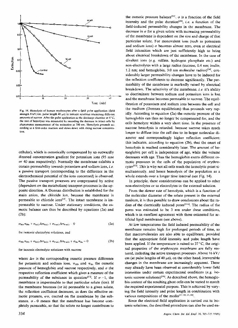
With a pulse duration of 40 ys and a field strength of 2 kV/cm, a reversible K+/Na' exchange is observed between the erythrocytes and the medium. This field intensity corre- sponds to a voltage of 1 V across the erythrocyte membrane, so that it may be concluded that reversible electrical break- down is associated with an increase in the transport of ions across the membrane. At higher field intensities, incipient hemolysis is observed in the erythrocytes. The rate of hemo- lysis, i. e. the rate at which hemoglobin is lost from the cells, depends not only on the applied field intensity and the pulse duration, but also on the composition of the isotonic external medium. In isotonic electrolyte solutions hemolysis sets in at field intensities of 3-4 kV/cm (pulse duration 40 ys). To a first approximation, the kinetics of hemolysis follow an ex- ponential course (first-order reaction), with a time constant of 1 minute. In isotonic solutions containing increasing con- centrations of sucrose, hemolysis is correspondingly slower. At a concentration of 40 mM sucrose, for example, the time constant is about 2-3 h (see Fig. 14). In a theoretical
based on the phenomenological equations of the thermodynamics of irreversible processesISz1, it has been pos- sible to draw conclusions from these experiments about the diameter of the pores generated by the electric field.
These theoretical considerations are based on the assump- tion that the electrically induced permeability changes in the cell membrane are followed by osmotic processes. In the ab- sence of an electric field there is an inwardly directed station- ary concentration gradient for sodium ions across the intact erythrocyte membrane (20 mM intracellular, 85 mM extra-
333 Angew. Chem. Inl. Ed. Engl. 20, 305-325 (1981)
the osmotic pressure balance['21. u is a function of the field 1
C .- ~
c " c c X W
.-
0.02 L I I I I 10 20 30 LO
0.01
Time Imin)
Fig. 14. Hemolysis of human erythrocytes after a field pulse application (field strength 8 kV/cm, pulse length 40 ps) in isotonic solutions containing different amounts of sucrose. After the pulse application in the discharge chamber at 4 "C. the rate of hemolysis was monitored by recording the decrease in intact cells by ohotometric measurement of the extinction at 700 nm. Hemolvsis oroceeds ac-
cellular), which is osmotically compensated by an outwardly directed concentration gradient for potassium ions (95 mM or 40 mM respectively). Normally the membrane exhibits a certain permeability towards potassium and sodium ions, i. e. a passive transport (corresponding to the difference in the electrochemical potential of the ions concerned) is observed. The passive transport processes are compensated by active (dependent on the metabolism) transport processes in the op- posite direction. A Donnan distribution is established for the main anion, the chloride ion, because the membrane is permeable to chloride The intact membrane is im- permeable to sucrose. Under stationary conditions, the os- motic balance can thus be described by equations (2a) and (2b):
for isotonic electrolyte solutions, and
for isotonic electrolyte solutions with sucrose
where Am is the corresponding osmotic pressure difference for potassium and sodium ions, nHb and msa the osmotic pressure of hemoglobin and sucrose respectively, and u the respective reflection coefficient which gives a measure of the permeability of the substance in question[52]. If u= 1, the membrane is impermeable to that particular solute (ion). If the membrane becomes (or is) permeable to a given solute, the reflection coefficient decreases, as does the effective os- motic pressure, un, exerted on the membrane by the sub- stance. a = O means that the membrane has become com- pletely permeable, so that the solute no longer contributes to
334
I - n intensity and the pulse duration[241, i.e. a function of the field-induced permeability changes in the membrane. The decrease in u for a given solute with increasing permeability of the membrane is dependent on the size and charge of that particular solute. For monovalent ions (such as potassium and sodium ions) u becomes almost zero, even at electrical field intensities which are just sufficiently high to bring about electrical breakdown of the membrane. In the case of divalent ions (e. g. sulfate, hydrogen phosphate etc.) and non-electrolytes with a large radius (sucrose, 0.4 nm; inulin, 1.2 nm; and hemoglobin, 3.0 nm molecular con- siderably larger permeability changes have to be induced for the reflection coefficients to decrease significantly. The per- meability of the membrane is markedly raised by electrical breakdown. The selectivity of the membrane, i. e. it's ability to discriminate between sodium and potassium ions is lost and the membrane becomes permeable to sucrose. The equil- ibration of potassium and sodium ions between the cell and the medium (Donnan equilibrium) thus proceeds very rap- idly. According to equation (2a) the osmotic pressure of the hemoglobin can thus no longer be compensated for, and the cells hemolyze within a very short time. In the presence of
-
, - cording to a firs-order reaction and slows down with rising sucrose concentra- tion.
sucrose hemolysis is retarded, because sucrose takes much longer to diffuse into the cell due to its larger molecular di- ameter and correspondingly higher reflection coefficient: this indicates, according to equation (2b), that the onset of hemolysis is reached considerably later. The amount of he- moglobin per cell is independent of age, while the volume decreases with age. Thus the hemoglobin exerts different os- motic pressures in the cells of the population of erythro- cyte~['~]. This is why not all cells reach the hemolytic point si- multaneously, and hence hemolysis of the population as a whole extends over a longer time interval (see Fig. 14).
In principle, these considerations can be applied to other non-electrolytes or to electrolytes in the external solution.
From the slower rate of hemolysis, which is a function of the molecular diameter of the solute present in the external medium, it is thus possible to draw conclusions about the ra- dius of the electrically induced The radius of the pores was estimated to be 3 nm under these conditions, which is in excellent agreement with those estimated for ar- tificial lipid membranes (see above).
At low temperatures the field induced permeability of the membrane remains high for prolonged periods of time, so that macromolecules are also able to equilibrate, provided that the appropriate field intensity and pulse length have been applied. If the temperature is raised to 37 "C, the origi- nal properties of the erythrocyte membrane are fully res- tored, including the active transport processes. Above 16 kV/ cm (at pulse lengths of 40 ps), on the other hand, irreversible changes in the membrane are increasingly apparent. These may already have been observed at considerably lower field intensities under certain experimental conditions (e. g. iso- tonic sucrose solution^)^^^^. As described above, the hemoglo- bin content of the resulting ghost cells can be varied to match the required experimental purpose. This is achieved by vary- ing the field intensity and pulse length in combination with various compositions of the media1'2.20.21.491.
Since the electrical field application is carried out in iso- tonic solutions, the described procedures can also be used en-
Angew. Chem. In[. Ed. Engl. 20, 305-32.5 (1981)
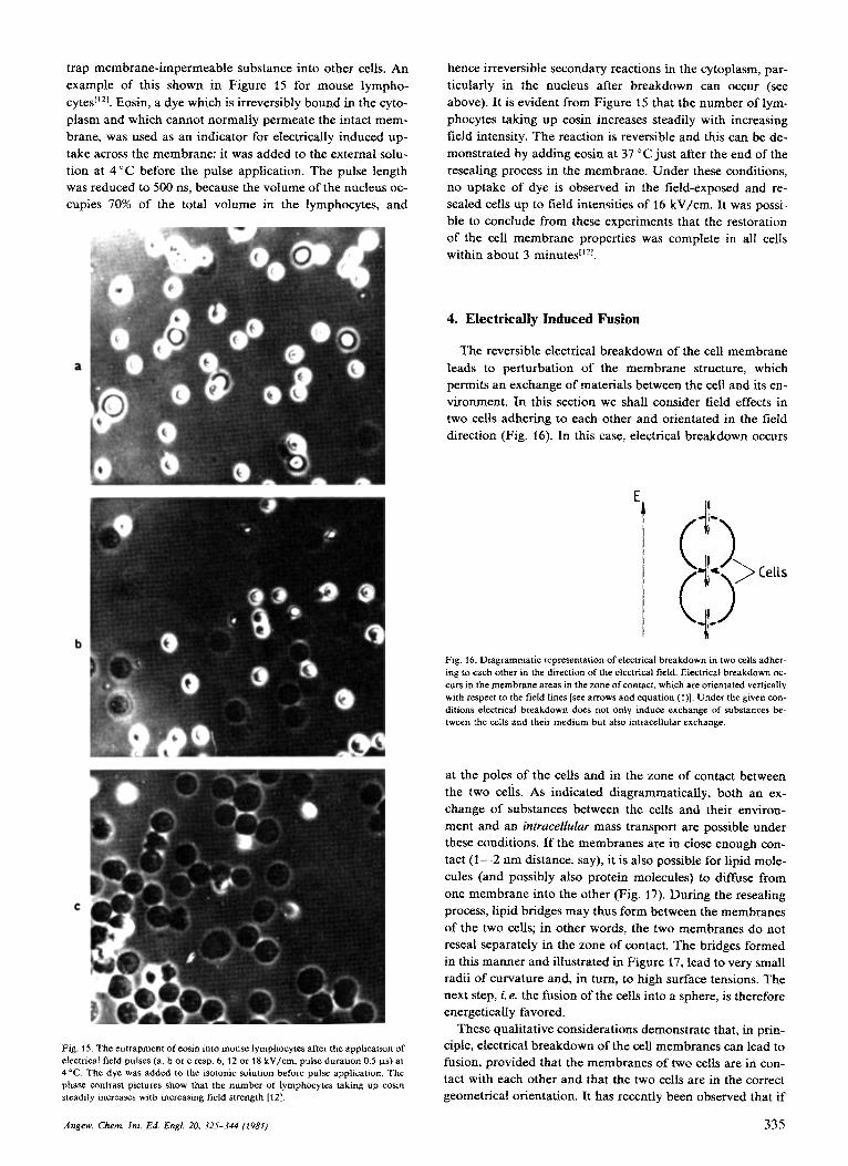
trap membrane-impermeable substance into other cells. An example of this shown in Figure 15 for mouse lympho- cytesI'Z1. Eosin, a dye which is irreversibly bound in the cyto- plasm and which cannot normally permeate the intact mem- brane, was used as an indicator for electrically induced up- take across the membrane: it was added to the external solu- tion at 4 "C before the pulse application. The pulse length was reduced to 500 ns, because the volume of the nucleus oc- cupies 70% of the total volume in the lymphocytes, and
Fig. IS. The entrapment of eosin into mouse lymphocytes after the application of electrical field pulses (a, b or c resp. 6. 12 or t 8 kV/cm, pulse duration 0.5 ps) at 4 "C. The dye was added to the isotonic solution before pulse application. The phase contrast pictures show that the number of lymphocytes taking up eosin steadily increases with increasing field strength [12].
hence irreversible secondary reactions in the cytoplasm, par- ticularly in the nucleus after breakdown can occur (see above). It is evident from Figure 15 that the number of lym- phocytes taking up eosin increases steadily with increasing field intensity. The reaction is reversible and this can be de- monstrated by adding eosin at 37 'Cjust after the end of the resealing process in the membrane. Under these conditions, no uptake of dye is observed in the field-exposed and re- sealed cells up to field intensities of 16 kV/cm. It was possi- ble to conclude from these experiments that the restoration of the cell membrane properties was complete in all cells within about 3
4. Electrically Induced Fusion
The reversible electrical breakdown of the cell membrane leads to perturbation of the membrane structure, which permits an exchange of materials between the cell and its en- vironment. In this section we shall consider field effects in two cells adhering to each other and orientated in the field direction (Fig. 16). In this case, electrical breakdown occurs
Fig. 16. Diagrammatic representation of electrical breakdown in two cells adher- ing to each other in the direction of the eleclrical field. Electrical breakdown oc- curs in the membrane areas in the zone of contact, which are orientated vertically with respect to the field lines [see arrows and equation (1)J. Under the given con- ditions electrical breakdown does not only induce exchange of substances be- tween the cells and their medium but also intracellular exchange.
at the poles of the cells and in the zone of contact between the two cells. As indicated diagrammatically, both an ex- change of substances between the cells and their environ- ment and an intracellular mass transport are possible under these conditions. If the membranes are in close enough con- tact (1-2 nm distance, say), it is also possible for lipid mole- cules (and possibly also protein molecules) to diffuse from one membrane into the other (Fig. 17). During the resealing process, lipid bridges may thus form between the membranes of the two cells; in other words, the two membranes do not reseal separately in the zone of contact. The bridges formed in this manner and illustrated in Figure 17, lead to very small radii of curvature and, in turn, to high surface tensions. The next step, i. e. the fusion of the cells into a sphere, is therefore energetically favored.
These qualitative considerations demonstrate that, in prin- ciple, electrical breakdown of the cell membranes can lead to fusion, provided that the membranes of two cells are in con- tact with each other and that the two cells are in the correct geometrical orientation. It has recently been observed that if
Angew. Chem. Ini. Ed. Engl. 20, 32s-344 (1981) 335

a
Function generator I -1
b
f d
Fig. 17. Model of the molecular processes which are thought to occur during electrically induced fusion. The lipid bilayers of the two membranes are in close contact vertical to the field lines (a), whereby 11 is assumed that protein-free areas are formed by lateral diffusion of proteins. Electrical breakdown leads to a dis- ruption of the membrane structure (b), so that bridges may be formed between the lipid bilayers of the two membranes during the reorganisation of the lipid molecules (c). The radius of curvature of the pores formed in this way is very small, so that formation of spherical “two-celled aggregates” is energetically fa- vored (d).
an erythrocyte cell suspension of high density is subjected to field pulses by discharging a high voltage capacitor[571, some fused cells are formed. However, the yield was very low, since the necessary formation of two-celled aggregates is nor- mally a rather improbable event. Brownian motion, the ne- gative surface charge of the cell membranes, and the asso- ciated repellent forces prevent the necessary contact between membranes. This is also the reason why cells usually exhibit only a weak tendency towards spontaneous fusion under nat- ural conditions. The current fusion methods are thus based on the use of “fusogenic” chemical substances or inactivated viruses having the same proper tie^^^^'^^^^^^^. Fusogenic chemi- cals or viruses bring about “agglutination” of the cells and si- multaneously disturb the membrane structure so that fusion is induced. However, as a rule, the cell suspensions have to be subjected to unphysiologically high or low pH-values or high calcium concentrations in order to bring about a suffi- ciently severe disturbance of the membrane16~7.s8.591. The best-known and most effective fusogenic substance is poly- ethylene glycol (molecular weight about 6000). This substance has been used with great success for the fusion of protoplasts. For the fusion of mammalian cells, on the other hand, the Sendai viruses, which have a strongly agglutinative action, have proved to be particularly effective. The viruses are inac- tivated by UV irradiation before use, so that their effect is not associated with an infection of the ell'^.^^. Viruses whose fusogenic action depends on their replication in the cell, have not achieved any importance for the in uitro fusion of cells.
A number of disadvantages have to be accepted with these classic fusion methods. The unphysiological conditions in- fluence the viability of the fused cells. The fusogenic sub- stances or viruses can also elicit irreversible changes in the
336
membranes, because they interact with the entire membrane surface for prolonged periods of time. Fusion is not syn- chronous and the yield is usually very low. Furthermore, the fusion methods are not understood mechanistically. They vary from one cell type to another and have to be optimized by trial and error. More recent fusion techniques suffer from similar In this case, the cells are made to adhere to each other by chemical or viral means and the dis- turbance of the membrane necessary for the initiation of fu- sion is brought about by a field pulse exceeding the break- down voltage of the membrane. Under these conditions, the yield is slightly higher, as demonstrated by Berg et al.[601 and Neumann et al.16‘1. These studies demonstrate that fusion can be achieved by means of the electrical breakdown of the cell membranes. This conclusion is also supported by the obser- vations of Senda et al.[621, who were able to show that two protoplasts held together with microcapillaries will fuse after the application of a field pulse. They, however, used dextran in the solution, which may have properties similar to polyethylene glycol. It is doubtful whether this method will find much practical application since the yield is a priori very low.
A generally applicable method with which different types of cells (or lipid vesicles) can be made to fuse with each other or with lipid vesicles has recently been developed by Zim- mermann and S c h e u r i ~ h ~ ‘ ~ . ~ ~ - ~ ~ ~ (Fig. 18). The electrically in-
Fusion chamber
Pulse generator
OsciNoscope
Fig. IK. Diagrammatic representation of the experimental set-up used for electri- cally induced fusion. Two electrodes, which can be observed under a microscope, are glued to a slide in parallel to each other. The frequency and amplitude of the dielectrophoretic voltage (see text) are controlled by a function generator; a pulse generator is connected in parallel and is used to elicit the breakdown pulse. The applied voltages are recorded on an oscilloscope. For further details see [lg.
duced fusion is based on the exclusive use of physical tech- niques and, in principle, consists of two stages. In the first stage, the membranes of the cells to be fused are brought into close contact by dielectrophoresis. In the second stage, fusion is induced in the resulting cell aggregates by electrical break- down.
Dielectrophoresis, which was first applied to living cells by Pohl and Cranei67,681 has been known for a long time. It is used to render the distribution of the field lines in an electro- static field visible with e. g. the aid of semolina particles sus- pended in oil. These particles orientate themselves along the field lines due to dielectric polarization.
Angew. Chem. Inf. Ed. Engi. 20, 325-344 (1981)

In principle, the same applies to a cell present in an electri- cal field (Figs. 18 and 19). In a homogeneous electrical field (Fig. 19a) a force is exerted on the cells because of their net
a b
C d
Fig. 19. Diagrammatic representation of electro- and dielectrophoresis. Because of the net charge on the outer surface of the membrane, cells or charged particles migrate in the direction of an electrode (electrophoresis) in a homogeneous elec- trical field (plate capacitor) (a). The direction of migration depends on the sign of the charge and on the direction of the external voltage. In general, cells have a negative surface charge. In addition, a dipole is induced in a dielectric particle or in a cell (regardless of whether these particles carry a net charge or not) (b). Since the electrical field intensity is equal on both sides of the particle or cell (b), this induced charge does not contribute to movement. Uncharged particles are thus unable to migrate in a homogeneous field. In an inhomogeneous electrical field, on the other hand, uncharged particles are also able to migrate, because the elec- trical field exerts a net force on the electrically induced dipole (c); the field inten- sity is not equal on both sides of the particle, resulting in a net force acting on the particle. This effect is known as dielectrophoresis. In contrast to the direction of migration during electrophoresis, the direction of the dielectrophoretic migration of the particles and cells is not reversed when the external electrical voltage be- tween the two electrodes IS reversed (d) (see [13, 671).
surface charge, and they begin to migrate towards the oppo- sitely charged electrode. This phenomenon is known as elec- trophoresis. Since the surface charge is usually negative, the cells migrate towards the anode. In addition, a dipole mo- ment is induced in the cells because of charge separation or because of orientation of dipoles in the membrane. However, this does not contribute to the movement of the cells since the field intensity in a homogeneous field acting on the gen- erated dipole is equal on both sides of the cell; in other words, a neutral particle will not migrate in a homogeneous field (Fig. 19b). In an inhomogeneous electrical field, on the other hand, electrophoresis is observed as before, but the neutral particles are also seen to migrate, usually in the direc- tion of greater field intensity (Fig. 19c). The reason for this migration is that the field intensities are different on the two sides of the dipole induced in the cell. The result is a net force which pulls the particle in the direction of greater field intensity. This effect is known as dielectroph~resis~~~~. The direction of the net force exerted on a neutral particle in an inhomogeneous field does not change (cf. Fig. 19d) when the direction of the field is reversed by inverting the external vol- tage. The electrophoretic migration of a cell, on the other hand, which is determined only by its surface charge, pro- ceeds in the opposite direction. In an inhomogeneous alter- nating electrical field, cells are no longer able to migrate be- cause of their net charge. Instead, the cells oscillate around a certain position in the field and the surface charge is “masked”. Only dielectrophoretically induced migration can occur, whereby the cells migrate towards the greater field in- tensity along the electrical field lines. The force exerted on
the cell under dielectrophoretic conditions, is dependent on the square of the field intensity, on the field gradient, the particle volume, the difference between the dielectric con- stants of the cell and its environment (as well as the corre- sponding difference between their conductivities). As a rule, these differences are positive, so that cells are observed to migrate to the region of greater field intensity. Under certain conditions, however, the difference between the dielectric constants can become negative, so that negative dielectro- phoresis is observed, i. e. migration into the region of lower field intensity. The reason for this is that the dielectric con- stant of a cell (or membrane) is a function of the frequency, since the membrane structure (including the boundary layers in contact with the solutions) is inhomogeneou~1~~.~~]. The boundary layers become polarized, which is reflected ma- croscopically by a change in the dielectric constant (Max- well-Wagner dispersion)[691. At certain frequencies the diel- ectric constant of the cell may thus become smaller than that of the surrounding solution so that negative dielectrophoresis results. As a rule, positive dielectrophoresis is observed in the frequency range between 1 kHz and 10 MHz (for exceptions see 167J).
Fig. 20. Diagrammatic representation of the formation of cell chains during diel- ectrophoresis. The cells adhere to each other and form chains at the electrodes along the electrical field lines, because the dipoles induced in the cells attract each other.
When cells approach each other during their migration along the field lines towards regions of greater field intensity, they attract each other because of their dipole moments (Fig. 20) so that they move towards the electrode in the form of two-, three- or four-celled chains (depending on the suspen- sion density), where they arrange themselves like “strings of pearls” along the field lines.
This configuration remains stable as long as the alternat- ing external field is applied. If the field is removed, the chains break up, since the cells repel each other due to their net charge and Brownian motion. Dielectrophoresis is thus a reversible process (provided that the field intensity is not too high, see below), which allows membranes to come into close contact.
A limiting factor in the dielectrophoretic aggregation of cells is the electrical conductivity of the medium. If this is too high, too much current flows in the solution leading to heat production and associated turbulences which prevent the formation of chains. It is thus necessary to use non-ionic so- lutions if possible (specific conductivity < S cm- ‘). The osmolarity[*l of the solution must be established by addi-
[‘I Where this term means the concentration of the osmotically active par- ticles.
Angew. Chem. Int. Ed. Engl. 20, 325-344 (1981) 337

Table 1: Electrically induced fusion [a].
Fusion between Medium Minimum Dielectrophoretic collection Electrical breakdown pulse Fusion Remarks Cell 1 Cell 2 electrode- Maximum time
distance field strength Frequency Field strength Duration [wml IV/cmI [a1 [ M W W/cmI la1 [PSI [min]
~~~~~~
Mesophyll- as 1 0.5 M 200 200 0 5 7 50 20 3-10 cf. 1631 protoplast, mannitol oats
Mesophyll- as 1 0.5 M 200 200 0.5 750 50 30-60 cf. 1131 protoplast, mannitol bean
~~ ~~~
Mesophyll- as 1 0.5 M 200 200 0.5 750 50 30-60 [c] protoplast, mannitol petunia
Guard cell- Mesophyll- 0.6 M 200 200 0.5 2000 50 20-40 cf. [64] protoplast, protoplast, mannitol bean bean
Mesophyll- as 1 0.3 M 300 70 1 protoplast, sorbitol Kalanchoe
~ ~ ~ ~ _ _
570 20 0.5 [d] cf. (651
Mesophyll- Vacuole, 0.3 M 300 70 1 protoplast, Kalanchoe sorbitol Kalanchoe
500 20 0.5 [dl cf. [65]
~ ~~~~~
Vacuole, as 1 0.4 M 300 50 1 500 20 0.1-0.2 [d] cf. [65] Kalanchoe sorbitol
Mesophyll- as 1 [b] 0.5 M 200 200 0 5 7 50 20 3-10 [d] cf. [65] protoplast, mannitol oats
Mesophyll- Mesophyll- 0.4 M 300 70 1 570 20 1 [d] cf. [65] protoplast, protoplast, sorbitol Kalanchoe oats
Human. as 1 0.3 M 100 1000 2 2200 4 2 4 cf. 1661
Friend- as 1 0.3 M 100 1000 2 2200 1-2 5-50 kl
erythrocytes [el glucose
cells [q glucose
Liposomes, as 1 0.075 M 300 330 2 diameter sucrose - 10 prn [h]
660 20 <O.l
[a] Plants: oats (Aueno sotiua), beans ( Viciafober), petunia (Petunio infato), Kalanchoe (Kolanchoe duigremontiuna). [b] The field strength was calculated assuming an ho- mogeneous electric field. [c] P. Scheurich, U. Tisoar, U. Zimmermonn, unpublished results. [d] The density of the solutions was increased by addition of&% Percoll and 5% Ficoll, resp. (pH 7.0). [el Pretreatment with pronase (1 mg/cm') or neuramidase (4 pgfcm'). [Q Pretreatment with pronase (1 mg/cm3); G. Pilwat, P. Scheurich, U. Zimmer- mann, unpublished results. [g] The fusion time depends on the composition of the medium (cf. text and Fig. 23). [h] Unilarnellar lipid vesicles; asolectin or egg-phosphati- dylcholin were taken as lipids; R. Benz, P. Scheurich, J. Yienken, U. Zimmermonn, unpublished results. [i] Protoplasts, which were grown under light and dark conditions, resp., were fused. Under light conditions chloroplasts are present, under dark conditions etioplasts, which are the precursors of the chloroplasts.
tion of non-electrolyte. Animal and plant cells have been shown to survive for long periods of time in solutions of mannitol, sucrose, glucose or neutral amino acids without any detectable irreversible deterioration of cell or membrane
In cells with high specific densities (e.g. mammalian cells with nuclei occupying up to 80% of the cell volume), the den- sity of the external medium must be raised with Percoll, be- cause otherwise the aggregated cells displace each other as a result of gravitational effects during and after the application of the electrical breakdown pulse.
Theory and e ~ p e r i m e n t [ ' ~ . ~ ~ - ~ ~ ] have shown that the field intensity for the breakdown of the membranes of the dielec- trophoretically aligned cells must be exceeded in order to in- duce fusion. The duration of the electrical field pulse, which leads to an optimum fusion of cells or cell organelles and li-
function[* 3.63-661
pid vesicles, depends on the properties of the respective bio- logical system to be fused. It is evident from Table 1 that the optimum range for the pulse length is between 15 and 50 )IS
for plant protoplasts and isolated vacuoles, whereas for mammalian cells (e. g. Friend cells and human erythrocytes) the optimum range is between 1 and 4 )IS. Outside these giv- en ranges, fusion is observed only in a few cases or not at all. A possible explanation for this observation may be a cell- or membrane-specific dependence of the breakdown voltage on the pulse length (see Figs. 7 and 8). In the optimum range for the pulse length nearly all cells, both plant and animal, which have been exposed to the dielectric breakdown vol- tage, will fuse. Electrically induced fusion is shown in Figure 21 for mesophyll cell protoplasts of Auena satiua (oats), in Figure 22 for a vacuole and a mesophyll protoplast of Kalan- choe daigremontiana, in Figure 23 for Friend cells and in Fig-
338 Angew. Chem. Int. Ed. Engl. 20. 32s-344 (1981)

Fig. 21. Electrically induced cell fusion of mesophyll cell protoplasts of Auena sa- tiva, some of which have been stained with neutral red, a vital dye which accu- mulates in the vacuole. The cells were suspended in a 0.5 M rnannitol solution and collected by dielectrophoresis at the electrodes of the fusion chamber (a). Fusion was induced by an electrical field pulse of 20 ( IS duration, the intensity of which was so high that electrical breakdown occurred in the contact zone of the membranes. The photographs show the fusion process 10 s (b), 300 s (c) and 600 s (d) after the breakdown pulse [63]. 1 crn=SO pm.
ure 24 for human erythrocytes. The relevant experimental conditions are given in Table 1. The time required for fusion following application of the pulse varies from seconds to 60 minutes (Table 1). The reason for this is not known but is probably related to different membrane fluidities in the var- ious cell types and to different properties of the cytoskeleton (fibrillar system) within the cells. A determining factor for the duration of the fusion process is the extent to which the cell wall in plant cell protoplasts and the glycocalix layer on the outer membrane surface of animal cells has been enzy- matically removed. The importance of the removal of these layers becomes apparent when erythrocytes, for example, are exposed to a critical alternating electrical field of long dura- tion (a few seconds), after dielectrophoretic alignment['*]. Under these conditions, irreversible membrane fusion is ob- served with the formation of long tubes (corresponding to the length of the cell chain). Actual cell fusion, i. e. the for- mation of a single spherical cell, does not take place. It is not known whether intracellular transport of materials occurs in these erythrocyte tubes which can reach lengths of up to 200
Fig. 22. Electrically induced fusion between a vacuole and a mesophyll cell pro- toplast of Kalanchoe daigremontiana (for conditions see Fig. 21). (a) Before the breakdown pulse, (b) 12 s, (c) 15 s and (d) 30 s after the breakdown pulse. For further details see text and Table I. 1 crn=35 pm [65].
pm or more and actually form bridges between the two elec- trodes.
For larger cells such as protoplasts and Friend cells it can be shown that the volume of the fused cells corresponds to the sum of the individual cell volumes, independent of the number of fused cells (see Figs. 21a, d and 22a, d). It has been observed that the tonoplast membranes, which sur- round the vacuoles, also fuse after fusion of the plasmalem- ma membranes. The fusion of the vacuoles was rendered visi- ble with neutral red, a vital dye which does not lead to any irreversible changes and is exclusively accumulated in the vacuole.
The fusion of human erythrocytes offers particularly fasci- nating applications if it is carried out under the conditions described in Table 1. At high suspension densities, a large number of cell chains are obtained in which the cells are in very close contact (Fig. 24a). The breakdown pulse leads to the formation of a giant erythrocyte (Fig. 24b) which may have a diameter of up to 200 pm (the normal diameter is 7- 8 pm). After fusion is complete, the glucose solution is re- placed by an electrolyte solution and the individual giant ery- throcytes may be isolated by a special technique using a pres- sure p r ~ b e [ ~ , ~ ' ] and subsequently examined. At first sight, the formation of giant erythrocytes seems to contradict the mod-
Angew. Chem. Int. Ed. Engl. 20, 325-344 (1981) 339

Fig. 23. Phase contrast photographs of electrically fused Friend cells. After brief pretreatment with pronase P (37"C, 1 mg/cm', 5 min) designed to remove the glycocalix, the cells were suspended in the fusion chamber in a 0.3 M glucose so- lution and orientated dielectrophoretically. l min after application of the break- down pulse (duration 2 ps), the cells were removed from the chamber with a spe- cially constructed micropipette. The photograph (a) was taken 2 min after induc- tion of electrical fusion. I t also shows, in addition to fused aggregates individual cells which were not exposed to the field pulse (a). When the cells are transferred to a nutrient medium, the fusion aggregates become spherical within 10 min (h). 1 cm=20 pm.
el developed for electrically induced fusion in Figure 17. Ac- cording to the theory, breakdown should only occur in the polar regions, i. e. in the membrane areas in the zone of con- tact along the field lines. However, since electrical break- down is an event lasting only 10 ns, as demonstrated by ex- periments on artificial lipid membranes, it does not seem im- probable that the breakdown voltage is also reached in those membrane areas vertical to the electrodes given that the field lines pass through the cells and that the electrical field is dis- torted by the high suspension density of the cells.
It is interesting to note that in permanent cell lines, such as the Friend cells shown in Figure 23, the cells become more rapidly spherical after the fusion process when transferred from the glucose solution to a nutrient medium.
Fig. 24. Electrically induced fusion of human erythrocytes. The glycocah layer was removed by treatlng the cells with neuramidase (37 "C, 4 pg/cm', 10 min). The cells were then suspended in 0.3 M glucose solution and collected dielectro- phoretically between the electrodes (a). The suspension density chosen was high, so that many erythrocyte chains with membrane contact were formed in parallel to one another. Application of two breakdown pulses (duration 4 ps) leads to the formation of giant erythrocytes (diameter up to 100 pm) (b). There is no signifi- cant hemolysis during formation of giant erythrocytes. Under phase contrast they appear optically homogeneous [66]. 1 cm = 30 pm.
Furthermore, it is worth pointing out that it is also possible to fuse organelles, such as chloroplasts (the site of photosyn- thesis) and mitochondria (the site of energy production) by electrical means, even though these organelles have two membrane envelopes arranged in series. Artificial lipid vesi- cles can also be fused by electrical breakdown into giant vesi- cles with diameters exceeding 100 pm.
Fusion between cells of different types and between lipid vesicles and cells have also been demonstrated (see Table l), so that one can conclude that electrical fusion is a generally applicable procedure for fusing different types of cells. Apart from the advantage of a high yield, fusion proceeds syn- chronously because it is triggered by a pulse. In future it should thus be possible to examine the molecular interme- diate stages (still unknown), which occur during the process of fusion, by electron microscopy. It is quite conceivable that changes in the field in the membrane may also be important, even when the cells are fused by chemical or viral means.
Since the electrically induced fusion method causes only the minimum of damage (only small membrane zones are in- fluenced for a very short time), the viability of the cells should be satisfactory. It has indeed been possible to demon- strate, for Friend cells, that growth continues after fusion and that hemoglobin synthesis can be induced by adding di- methyl sulfoxide to the suspension of fused cellslS01. There have been extensive studies on plant cell protoplasts of Peru-
Angew. Chem. Int. Ed. Engl. 20, 325-344 (t98t) 340

niu inflata, and various authors[31 were able to show that indi- vidual mesophyll cell protoplasts of this species exhibit “toti- potency”: with this plant it is thus possible to observe forma- tion of cell walls and subsequent cell division (and associated differentiati~n)~~’ resulting in the development of a whole plant. Fusion experiments on protoplasts derived from this species are thus particularly tempting, since the viability of the fusion aggregates can be tested relatively quickly. Opti- mum conditions for the formation of cell walls and cell divi- sion can be achieved by immobilizing the fused protoplasts. It was first demonstrated in our l ab~ra to ry [~ ’ - ’~~ that plant protoplasts and animal cells can be immobilized in a cross- linked alginate matrix maintained in the appropriate nu- trient medium. The alginate matrix is cross-linked by cal- cium ions. With the aid of this technique it has been possible to show that electrically fused protoplasts of P. inj7utu are capable of forming both cell walls and dividing. The immo- bilized cells can be released from the alginate matrix by add- ing a chelating agent (e.g. citrate) and then used for further breeding experiments. With citrate, nearly all the cells can be recovered without damage171-73]. Experiments on sea urchin eggs (performed in collaboration with H.-P. Richter, Max- Planck-Institut fur Biophysik, Frankfurt am Main) have re- vealed that fused eggs can still be fertilized by sperm.
5. Potential Applications of Electrical Membrane Breakdown
From the cell biological and medical point of view the breakdown voltage for the membrane can itself be used as a criterion for changes in membrane structure or composition. The variation in breakdown voltage during the growth cycle of a cell population (Fig. 11) indicates that there are changes in the electromechanical properties of the cell membrane during cell growth, which are reflected in changes in the breakdown voltage. Escherichiu coli cells also exhibit an in- crease in the average breakdown voltage during the transi- tion from logarithmic to stationary growth phase, as demon- strated in earlier studies with the particle analyzer as shown in Figure 9b[I9]. These observations may indicate that specif- ic growth phases can be characterized by the electrical break- down voltage, a result which is very attractive from the medi- cal and cell biological points of view, because the various stages of growth have until now normally only been identif- ied by staining the cells with fluorescent dyes and by exami- nation with an impulse cytoph~tometer[~~l. Changes in the breakdown voltage as a result of processes of differentiation are also known. Pilwat et were able to show that the breakdown voltage decreases significantly during the devel- opment of A uenu sutiuu (oats) chloroplasts from the etioplast stage. The development of chloroplasts is induced within 48 hours by illuminating plants that have been grown in dark- ness. The decrease in the breakdown voltage as a function of the illumination time was interpreted in terms of an increase in the number of transport proteins incorporated into the membranes of the chloroplasts. The fact that certain local anaesthetics can change the electrical breakdown voltage of erythrocyte membranes may also be of clinical relevance. In- creasing concentrations of benzyl alcohol decrease the break-
down voltage by more than 50%[76.771. However, it should be pointed out that the breakdown voltage of planar artificial li- pid membranes is apparently hardly influenced by the addi- tion of various agents. Extensive studies, taking into account the recent finding that the breakdown voltage is dependent on the pulse length, will certainly be necessary to arrive at a more clear-cut statement. Furthermore, it is possible that the breakdown properties of the membranes are more sensitive to chemical changes in artificial lipid vesicles than in planar membranes. The interpretation of electrical breakdown in terms of local electromechanical compression with the for- mation of pores, indicates that elastic and mechanical forces in the membrane play a role (see above). The small radius of curvature of both lipid vesicles and cells and the tension forces in the membrane could considerably influence the val- ue of the breakdown voltage, if the mechanical and elastic parameters of the membrane were influenced by the addition of certain substances. The development of a particle analyzer capable of making measurements on individual cells (Fig. 9d) will enable detailed studies of this nature to be carried out on phospholipid vesicles of varying chemical composi- tion, different charge and degree of cross-linkage (see also [ l 2 4 134.35.78-801
In general, the present trend in medical technology is to- wards the development of multi-parameter analyzers, i. e. de- vices which enable measurements of a number of character- istic membrane and cell parameters in individual cells to be made s i m ~ l t a n e o u s l y ~ ~ ~ - ~ ~ ~ . In the procedure described here (Fig. 9d), it is possible to measure the volume, breakdown voltage of the membrane and apparent underestimation of the cell volume after breakdown in individual cells, without any additional manipulation (e. g. staining etc.). As discussed above, the apparent underestimation of the cell volume is a direct measure of the intracellular conductivity pattern. The underestimation reflects the ratio of non-conducting to con- ducting space in the cell. This ratio depends, on the one hand, on the size and properties of the intracellular compart- ments within the cell, and, on the other hand, on the conduc- tivity of the cytoplasm which itself is sensitively regulated by the biochemical activity in the cell. Measurements on human erythrocytes, which had been loaded with non-electrolytes or electrolytes by electrical breakdown in the discharge cham- ber, have shown that the degree of equilibration between the cell interior and the external solution can be monitored di- rectly in the particle analyzer by measuring the apparent un- derestimation of the cell(lol. With appropriate calibration it is also possible to estimate the protein content of the cells. Studies of this type, which are of great clinical interest, are particularly simple to perform on human erythrocytes, since they have no nuclei or other intracellular compartments. Pil- wut and Zimmermunn (see e.g. [48]) were able to corroberate reports in the literature by showing that the hemoglobin con- tent of erythrocytes of various ages, which were separated in a density gradient (Percoll), is independent of volume and hence age. With the aid of the new particle analyzer (Fig. 9d) it should be possible, in the future, to routinely detect early stages of pathological changes in the hemoglobin content of individual cells in an erythrocyte population (caused by anaemia, for example).
Field induced entrapment of membrane-impermeable substances in the cell interior and the associated possibility
)
Angew. Chem. Inl. Ed. Engl. 20, 325-344 (1981) 341

of manipulating the spectrum of cellular functions, offer a number of interesting prospects from the cell biological point of view. Two possible applications, both of which are at this moment being explored in different laboratories, will be mentioned in order to emphasize this aspect:
Specific reaction sites in carrier systems within the mem- brane or intermediate stages in biochemical and biophysical reaction chains, which are asymmetrically arranged in the membrane, can be located and identified by the entrapment of appropriate substances with a specific action (e.g. inhibi- tors). Since proteins and enzymes can be reversibly entrap- ped in cells by electrical breakdown[211, experiments in which the luciferin-luciferase system can be entrapped in cells to determine the ATP turnover in the cell, have become realiza- ble. As a rule, since the turnover of ATP is determined using inhibitors and uncoupling agents, it is not always possible to exclude with any certainty side effects and non-specific inter- actions with the plasma membrane.
Cells whose functions have been manipulated, offer fasci- nating possibilities for clinical diagnostics and therapeutics. With a few exceptions, drugs (hormones, antibiotics, en- zymes, radionuclides etc.) do not act in a cell-, tissue- or or- gan-specific way. The development of carrier systems which direct drugs to specific target sites and release them in a con- trolled manner, or which circulate in the blood for long peri- ods of time acting as drug reservoirs, represents a very topical
So far, all work has concentrated on the development of artificial carrier systems (e. g. liposomes, microcapsules, binding to polymers, etc.). In spite of encouraging results, we are still far removed from the ultimate goal of clinical appli- cation. Compatibility with the organism, immunological reactions, lack of selectivity, and limitations in the life span of the carrier systems used at present are only some of the many problems which have still to be solved satisfactori-
trinsic advantages, in particular, the lack of significant im- munological reactions.
Experiments on mice, using erythrocytes which had been electrically loaded with a cytostaticum or other clinically rel- evant substances, confirm the satisfactory properties of the cellular carrier ~ y s t e m l ~ . ‘ ~ * ~ ~ * ~ ~ . ~ ~ ~ . A h’ igh percentage of the erythrocytes, which are loaded under the appropriate field conditions without any significant loss of hemoglobin and other cellular components, will circulate in the bloodstream after intravenous injection110.881. The life-span of these ma- nipulated cells is of the same order as that of intact mouse erythrocytes. If, on the other hand, field conditions are chos- en such that the hemoglobin content is very low after the re- sealing process (ghost cells) and the mean cell volume differs from that of the intact cells, the entrapped drug (e. g. metho- trexate, a cytostatic agent which blocks an important step in the synthesis of purine) is targeted specifically to the liv- er[97’2.841. Under these conditions, the manipulated erythro- cytes are recognized as foreign bodies by the spleen and liver and degraded accordingly, so that the drug is specifically rel- eased in these organs. By the simultaneous field-induced en- trapment of a drug and small magnetic particles (diameter about 10 nm) in erythrocytes, it is, in principle, possible to develop cellular carrier systems with other organ-specific properties. By external magnetic guidance, such “magnetic”
342
area of research in biomedical technology (see [10.79,84-871 ).
ly[10.79.84-86) . c ellular carrier systems have a number of in-
erythrocytes can be directed to many specific sites within the organism. Experiments with mice have shown[I0l that this procedure works in principle and that the magnetic particles can be eliminated by the body without any toxic reac- tionsI’O. 891.
The fusion of cells by electric breakdown opens up a whole new range of possibilities for the development of drug carrier systems. Ghost cells loaded with a drug can be fused with intact erythrocytes, lymphocytes and other cells of an organism. Since there is apparently no significant loss of in- tracellular components, carrier systems with a long life span in the body and possibly with completely new organ-specific properties can be developed.
Carrier systems consisting of fused erythrocytes and lipos- omes could be of particularly interest, since liposomes can be loaded with proteins or DNA (genes) by simple chemical methodsl8’1. By incorporating liposomes into the erythrocyte membrane it should be possible to modify the membrane properties of the erythrocyte locally, depending on the chem- ical composition of the liposome membrane, so that the ery- throcyte could release the entrapped substance in a con- trolled manner in space and time. The use of phospholipids, which can be polymerized under UV irradiation after the event of membrane fusion, is another promising tool for the development of an optimum drug carrier system134.35.901.
Electrically induced fusion in general offers new possibili- ties for applications in other fields, since contrary to tradi- tional methods it is universally applicable to all cells and ar- tificial lipid vesicles. Both the lack of damage to the cells and the high yield indicate that fused cell systems are highly via- ble.
Nevertheless, we are still very much at the beginning of this development, but given our present knowledge, we can point out a number of potential applications for this fusion method.
Fusing plant protoplasts of different origins to produce crop plants with new properties such as improved yield or better salt tolerance is quite conceivable. Salt-tolerant mu- tants of the soya bean are wel l -kn~wn~~’~. Several laborato- ries are thus attempting to fuse protoplasts of this mutant with protoplasts of other crop plants in order to produce a vi- able cell hybrid which can be used to breed plants having the combined properties of both.
The transfer of the gene responsible for nitrogen fixation from bacteria to plant protoplasts, with the aid of the electri- cal fusion method, is also coming into the realms of possibili- ty. The fusion of protoplasts from yeast cells with plant cells is another fascinating area which could find technological application. Yeast cells are able to convert sugar into alcohol. By immobilizing yeast cells in cross-linked polymer matrices it is possible to produce alcohol in a continuous process [see 192-951. Plant protoplasts on the other hand, are able to con- vert solar energy into sugar (and starch). A hybrid cell with the properties of both cells should thus be capable of con- verting solar energy directly into alcohol. If it were possible to operate such a cell system on a large scale, the implica- tions for the solution of our current energy problems would be enormous. Undoubtedly the difficulties that would need to be overcome to achieve these goals technologically, are immense. In particular, it would be necessary to examine the compatibility of the cytoplasmic properties, the genetic mate-
Angew. Chem. Int. Ed. Engl. 20, 325-344 (1981J

rial and other cellular factors when cells of different origins are fused. Even in the event of failure of the supposed tech- nological applications, studies of this nature should at least provide fresh impetus for cell biology and for research into membrane structure and function, which may well result in the discovery of other applications which are not immediate- ly obvious at present.
The results reviewed here were partly supported by grants from the Bundesministerium fur Forschung und Technologie and from the Deutsche Forschungsgemeinschaft. We are very grateful to Pro$ Dr. Gobel, University of Wiirzburg, for supplying us with Friend cells, and to Mrs. S. Alexowsky for ty- pewriting the manuscript.
Received: January 20, 1981 [A 360 IE] German version: Angew. Chem. 93,332 (1981)
[ I ] G. B. Brown, G. Manecke, L. B. Wingard, Jr.: Enzyme Engineering. Vol. 4.
[2] U . Zimmermann, E. Sfeudle, Adv. Bot. Res. 6, 45 (1978). I31 W. Klingmiiller: Genmanipulation und Gentherapie. Springer, Berlin 1976,
[4] M. H. Saier, Jr., C. D. Stiles: Molecular Dynamics in Biological Membra-
[5] P. J. Quinn: The Molecular Biology of Cell Membranes. Macmillan Press,
[6] G. Posfe, G. L. Nicholson: Cell Surface Reviews. Vol. 5. North Holland,
[7] N. R. Ringertz, R. E. Savage: Cell Hybrids. Academic Press, New York
[8] U. Zimmermann, J. Schulfz, G. Pilwat, Biophys. J . 13, 1005 (1973). [9] U. Zimmermann, G. Pilwaf, Z. Naturforsch. C 31, 732 (1976).
Plenum Press, New York 1978.
p. 193.
nes. Springer, Berlin 1975.
London 1976.
Amsterdam 1976.
1976.
[lo] U. Zimmermann in E. Goldberg. L. Donaruma, 0. Vogl: Targeted Drugs. Wiley, New York, in press.
1111 U. Zimmermann, Chem. Labor Betr. 28, 505 (1977). 1121 U. Zimmermann, J. Vienken, G. Pilwat, Bioelectrochem. Bioenerg. 7, 554
[12al L. Gros, H. Ringsdo@, H. Schupp, Angew. Chem. 93, 311 (1981); Angew.
[13] U. Zimmermann, P. Scheurich, Planta 151, 26 (1981). [I41 S. .I Singer, J. L. Nicolson, Science 175, 720 (1972). [I51 N. Higinbofham, Annu. Rev. Plant Physiol. 24, 25 (1973). (161 U. V. Lassen, 0. Sten-Knudsen, J . Physiol. (London) 195, 681 (1968). [17] G. Adam, P. Uuger, G. Stark: Physikalische Chemie und Biophysik.
Springer, Berlin 1977. [18] R. Benz: Kiinstliche Lipidmembranen: Modelle fur biologische Membra-
nen? Konstanzer Universitatsreden, Universitatsvcrlag Konstanz 1980. 1191 U. Zimmermann, G. Pilwat, F. Riemann, Biophys. J . f4, 881 (1974). [20] U. Zimmermann, G. Pilwaf. F. Beckers, F. Riemann, Bioelectrochem. Bio-
energ. 3, 58 (1976). [21] U. Zimmermann, F Riemann, G. Pilwaf, Biochim. Biophys. Acta 436, 460
(1976). 1221 J. Vienken. E. Jelfsch, U. Zimmermann. Cytobiologie Z. Exp. Zellforsch. 17,
182 (1978). [23] D. Auer, G. Brandner, W. Bodemer, Natunvissenschaften 63, 391 (1976). 1241 U. Zimmermann, G. Pilwaf, C. Holzapfel, K. Rosenheck, I. Membr. Biol. 30,
[25] R. Benz, U. Zimmermann, Biochim. Biophys. Acta 597, 637 (1980). [26] R. Benz, U. Zimmermann, Bioelectrochem. Bioenerg., 7, 724 (1980). [27] R. Benz, F. Beckers, U. Zimmermann, J . Membr. Biol. 48, 181 (1979). I281 R. Benz, P. fiuger, J . Membr. Biol. 27, 171 (1976). [29] J. Y. Abrdor, V. B. Arakelyan, L. Y. Chernomordik, Yu. A. Chizmadzheu, K
1301 U. Zimmermann, AMU. Rev. Plant Physiol. 29, 121 (1978). (311 U. Zimmermann. G. Pilwaf, A. Pegueux, R. Gilles, I . Membr. Biol. 54, 103
[32] A. Pegueux, R. Gilles, G. Pilwat, U. Zimmermann, Experientia 36, 565
[33] R. Benz, U. Zimmermann, Biochim. Biophys. Ada 640, 169 (1981). [34] H.-H. Hub, B. Hupfer, H. Koch, H. Ringsdorf, Angew. Chem. 92, 962
(1980); Angew. Chem. Int. Ed. Engl. 19, 938 (1980). [35] A. Akimofo, K. Dorn, L. Gros, H. Ringsdorf, H. Schupp, Angew. Chem., in
press. [36] B. Gauger, F. W. Benfrup, J . Membr. Biol. 48, 249 (1979). (371 U. Zirnmermann, F. Beckers, H. G. L. Cosfer, Biochim. Biophys. Acta 464,
(1980).
Chem. Int. Ed. Engl. 20, 305 (1981).
135 (1976).
F. Pastushenko, U. R. Tarasevich, Bioelectrochem. Bioenerg. 6, 63 (1979).
(1980).
(1980).
399 (1977).
[38] U. Zimmermann, R. Benz, J . Membr. Biol. 53, 33 (1980). 1391 R. Benz, F. Confi, Biochim. Biophys. Acta, in press. 1401 E. Jelfsch, 0. Zimmermann, Bioelectrochem. Bioenerg. 6. 349 (1979). I411 J. Hurley, Biophys. J . 10, 74 (1970). (421 H. E. Kubitschek in J. R. Noris. D. W. Ribbons: Methods in Microbiology.
[431 R. Thorn, Dtsch. Med. Wochenschr. 94, 617 (1969). 1441 R. Thom, A. Hampe, G. Sauerbrey, 2. Gesamte Exp. Med. 151, 331 (1969). [45] V. Kachef, J. Histochem. Cytochem. 24, 211 (1976). (461 G. Pilwat, U . Zimmermann, H. Schnabl in W. J . Lucas, J. Dainfy: Plant
Membrane Transport: Current Conceptual Issues. Elsevier/North Holland, Amsterdam 1980, p. 475.
[47] U. Zimmerrnann, M. Groves, H. Schnabl, G. Pilwat, J . Membr. Biol. 52, 37 (1980).
[48] U. Zimmermann, G. Pilwaf, M. R. Groves, Deutsch. Pat.-Anm. P 2828232.8 (1978); US-Pat. 4220916, Sept. 1980.
1491 U . Zrmmermann, G. Pilwat, J. Vienken, Recent Res. Cancer Res. 75, 265 (1980).
[SO] C. Friend, W. Scher, J. G. Holland, T. Solo. Proc. Natl. Acad. Sci. USA 68, 378 (1971).
[51] E. Neumann, K. Rosenheck, 1. Membr. Biol. 10, 279 (1972). Isla] G. Pilwat, U. Zimrnermann, unpublished results. [SZ] A. Kafchalsky, P. F. Curran: Nonequilibrium Thermodynamics in Biophy-
[53] H. Passow in C. Bishop, D. M. Surgenor: The Red Blood Cell. Academic
[54] W. D. Sfein: The Movement of Molecules Across Cell Membranes. Aca-
I551 D. Danon in R. Coldman. M. Rocksfern: The Physiology and Pathology of
1561 J. Teissie, T. Y. Tsong, J . Membr. Biol. 55, 133 (1980). [57] U. Zimmermann, G. Pilwat, Sixth International Biophysics Congress, Kyoto
[58] K. N. Kao, M. R. Michayluk, Planta 125, 355 (1974). 1591 Z. Toisfer, A. Loyter, J . Biol. Chem. 248, 422 (1973). [601 H:E. Jacob, W. Forsfer, H. Berg, Z . Allg. Mikrobiol., in press. 1611 E. Neumann, G. Gerisch, K. Opafz, Natunvissenschaften 67, 414 (1980). (621 M. Senda, 1. Takeda, S. Abe, T. Nakamura, Plant Cell Physiol. 20, 1441
[63] U. Zimmermann, P. Scheurich, Biochim. Biophys. Acta 641, 160 (1981). [64] P. Scheurich. U. Zimmermann, H. Schnabl, Plant Physiol., in press. [65] J. Vienken. M. Ganser, R. Hampp, U. Zimmermann, Physiol. Plant, in
1661 P. Scheurich, U. Zimmermann, Natunvissenschaften 68,45 (1981). [67] H. A. Pohl: Dielectrophoresis. Cambridge University Press, Cambridge
[68] J. S. Crane, H. A. Pohl. I. Electrochem. Soc. 115, 584 (1968). [69] H. P. Schwan. Adv. Biol. Med. Phys. 5, 147 (1957). [70] P. Scheurich, U. Zimmermann, M. Mischel, I. Lamprechf, Z . Naturforsch. C
1711 G. Pilwat, P. Washausen, J. Klein, U. Zimmermann, Z. Naturforsch. C 35,
[72] P. Scheurich, H. Schnabl, U. Zimmermann. Biochim. Biophys. Acfa 598,645
[73] H. Schnabl, P. Scheurich, U. Zimmermann, Planta 149, 280 (1980). [74] W Gohde, W. Ditfrich, Ada Histochem. 10, 429 (1971). I751 G. Pilwat, R. Hampp, U. Zimmermann, Planta 147, 396 (1980). [76] U. Zimmermann, G. Pilwat, F. Riemann in U. Zimmermann, J . Dainty:
[771 G. Pilwat, U. Zimmermann, F. Riemann, Biochim. Biophys. Acta 406, 424
I781 D. Day, H. H. Hub, H. Ringsdorf, Isr. J . Chem. 18, 325 (1979). (791 H. Ringsdo@ in R. J. Kostelnick: Midland Macromolecular Monographs.
[SO] H. Bader, H. Ringsdorf, J. Skura, Angew. Chem. 93, 109 (1981); Angew.
I811 R. C. Leif in G. L. Wed, G. F. Bahr: Automated Cell Identification and
1821 D. J. Arndf-Jovin, T. M. Jouin, J . Histochem. Cytochem. 22, 622 (1974). I831 V. Kachel, E. Glossner, E. Kordwig, G. Ruhenstrolh-Bauer, J . Histochem. Cy-
1841 U. Zimmermann, G. Pilwaf, B. Esser, J . Clin. Chem. Clin. Biochem. 16, 135
I851 G. Gregorindis, A. C. Allison: Liposomes in Biologxal Systems. Wiley, Chi-
1861 T. Ming Swi Chang: Biomedical Applications of Immobilized Enzymes and
(871 G. M. Ihler in G. Gregoriadis: Drug Camers in Biology and Medicine. Aca-
[88] K. Kinosifa, Jr., T. Y. Tsong, Nature 272, 258 (1978).
[89] T. Nakamura, K. Konno, T. Morone, N. Tsuya, M. Hatano, J . Appl. Phys.
Vol. 1. Academic Press, New York 1969, p. 593.
sin. Harvard University Press, Cambridge, Mass. 1965.
Press, New York 1964, p. 71.
demic Press, New York 1967.
Human Aging. Academic Press, New York 1975, p. 95.
1978, Abstr. IV-l9-(H), p. 140.
(1979).
press.
1978.
35, 1081 (1980).
352 (1980).
(1980).
Membrane Transport in Plants. Springer, Berlin 1974, p. 146.
(1975).
Vol. 5. Gordon and Breach, London 1978, p. 197.
Chem. Int. Ed. Engl. 20,91 (1981).
Sorting. Academic Press, New York 1970, p. 131.
tochem. 25, 804 (1977).
(1978).
Chester 1980.
Proteins. Vol. 1. Plenum Press, New York 1971, p. 147.
demic Press. London 1979, p. 129.
42, 1320 (1971).
Angew. Chem. Inf . Ed. Engl. 20,325-344 (1981) 343

1931 J. Klein, Kontakte (Merck) 3, 29 (1980). [941 P. Bradelius, B. Dens, K. Moshach, M. H. Zack, FEBS Lett. 103, 93
(951 M. Wada, J. Kato, I. Chibata, Eur. J. Appl. Microbial. Biotechnol. 10, 275
[90] D. S. Johnston, S. Soughera, M. Pons, D. Chapman, Biochim. Biophys. Acta 602, 57 (1980).
[91] H. Greenway, R. Munns, Annu. Rev. Plant Physiol. 31, 149 (1980). (1979).
335. (1 980). I921 I. Chihata, E Tosa, T. Sato, K. Yamarnoro, I. Takaro, F. Nishida in 111, p.
Half a Century of Polystyrene-A Survey of the Chemistry and Physics of a Pioneering Material
By Adolf Echte, Franz Haaf, and Jiirgen Hambrecht“’
Hermann Staudinger, the founder of polymer chemistry, would have been 100 years old this year. One of the key materials with which he elucidated the structure of high polymers was po- lystyrene.-Polystyrene, one of the most important thermoplastics, has now been manufac- tured industrially for some 50 years. Not only has it simplified our daily lives in a variety of ways, it has also been a model substance for the development and expansion of polymer chemi- stry and polymer physics, and it was the pioneering material for handling polymer solutions and melts. This article surveys the development and present state of knowledge of the polym- erization of styrene and of the relationships between structure and properties.
1. Introduction
This year Hermann Staudinger would have celebrated his 100th birthday. It was Staudinger who founded polymer chemistry as a discipline; it was he who elucidated the struc- ture of high molecular weight compounds and began to in- vestigate their properties systematically[‘l. For this funda- mental work he was awarded the Nobel Prize for Chemistry in 1953.
Staudinger’s anniversary almost coincides with another milestone in the history of macromolecular chemistry: poly- styrene had its 50th birthday in 1980. The pioneering materi- al of high polymers was first manufactured on an industrial scale in 1930. In the autumn of 1930 I.G. Farbenindustrie AG’s Ludwigshafen works-today BASF AG-became the first in the world to manufacture polystyrene industrially. Using this substance, Staudinger demonstrated that polymers are made up of fundamental units, the monomers, linked by primary valences.
only occasionally featured in the literature, as in a paper by B e r t h e l ~ t ‘ ~ ~ and, later, Stobbe and PosnjakfSI.
In 1920 Staudinger showed in his pioneering publication “Uber Polymerisation”‘61, that polystyrene is a high molecu- lar weight substance, in which monomer units (1) are linked by covalent bonds to form macromolecular chains (2). Stau- dinger also used polystyrene as a model substance in eluci- dating the radical chain mechanism of polymeri~ation[~’, and he resolved the long-standing controversy over the mecha- nism of formation of high polymers in favor of the radical chain concept, by hydrogenating polystyrene to polyvinylcy- clohexane without changing its molecular weight[*]. This dis- provedc’ll the low molecular weight synthesis principle of Hessl9] and the micelle theory of K. H. Meyer[”I.
n CH=CH, - CH,-CH,{CH-CH,)CH-CH, 6 8 6 “-’Q 1.1. Scientific Development
Polystyrene was first described by the Berlin pharmacist E. Simon in 1839Iz1. In 1945, J. Blyth and A. W. Hofmann rewg- nized that the polymerization of styrene was not accompa- nied by any change in chemical composition, and they called their glassy reaction product “Metastyrol”, i. e. “metasty- rene”‘31. This was followed by a period in which polystyrene
[‘I Dr. A. Echte, Dr. F. Haaf, and Dr. J. Hambrecht Plastics Laboratory, BASF Aktiengesellschaft D-6700 Ludwigshafen (Germany)
Macromolecular substances require special techniques of investigation, because the methods suitable for low molecu- lar weight substances are frequently inapplicable. As early as 1930, Staudinger first put forward a relationship between the viscosity of a polymer solution and the molecular weight of the and this was subsequently refined by Mark et al. and H o ~ w i n k “ ’ ~ ’ ~ ~ and put on a theoretical basis by W. Kuhn‘l’’. Styrene also served as the principal compound in elucidating the reaction kinetics of radical chain polymeriza- tion. ChaImerP”, FIory[171, and Breifenbach[lS1 established
344 0 Verlag Chemie GmhH, 6940 Weinheim, 1981 0570-0833/81/0404-0344 I 02.50/0 Angew. Chem. Int. Ed. Engl. 20, 344-361 (1981)