Embed Size (px)
Citation preview

Rev Physiol Biochem Pharmacol (2003) 148:1–80DOI 10.1007/s10254-003-0009-x
F. Wehner · H. Olsen · H. Tinel · E. Kinne-Saffran · R. K. H. Kinne
Cell volume regulation: osmolytes, osmolyte transport,and signal transduction
Published online: 4 April 2003� Springer-Verlag 2003
Abstract In recent years, it has become evident that the volume of a given cell is an im-portant factor not only in defining its intracellular osmolality and its shape, but also indefining other cellular functions, such as transepithelial transport, cell migration, cellgrowth, cell death, and the regulation of intracellular metabolism. In addition, besides in-organic osmolytes, the existence of organic osmolytes in cells has been discovered. Os-molyte transport systems—channels and carriers alike—have been identified and charac-terized at a molecular level and also, to a certain extent, the intracellular signals regulatingosmolyte movements across the plasma membrane. The current review reflects these de-velopments and focuses on the contributions of inorganic and organic osmolytes and theirtransport systems in regulatory volume increase (RVI) and regulatory volume decrease(RVD) in a variety of cells. Furthermore, the current knowledge on signal transduction involume regulation is compiled, revealing an astonishing diversity in transport systems, aswell as of regulatory signals. The information available indicates the existence of intricatespatial and temporal networks that control cell volume and that we are just beginning tobe able to investigate and to understand.
General introduction
For a number of years, cell volume regulation in mammalian cells has been considered ofminor physiological importance since overall osmolality of the plasma is one of the verytightly regulated parameters of the body. In the meantime, however, it has been realized
E. Kinne-Saffran deceased on December 6, 2002
F. Wehner ()) · H. Olsen · E. Kinne-Saffran · R. K. H. KinneMax-Planck-Institut fàr molekulare Physiologie, Otto-Hahn-Str. 11,44227 Dortmund, Germanye-mail: [email protected] · Tel.: +49-231-1332225
H. TinelInstitute of Cardiovascular Research II, Bayer AG Wuppertal, 42096 Wuppertal, Germany
that quite a variety of tissues such as renal medullary cells and chondrocytes are exposedto anisotonic extracellular media and thus require volume regulatory mechanisms. Aniso-tonic conditions can also arise in pathological conditions such as hypo- or hypernatraemiawhen homeostatic functions of the body are insufficient (Law 1999; Law 1998; Verbaliset al. 1989; Verbalis 1994). Similarly, cells encased in a rigid surrounding, such as braincells, depend on cell volume regulation for their proper function. Furthermore, even at nor-mal extracellular osmolalities cells can generate transmembrane osmotic differences dueto uptake of (organic) osmolytes or during transepithelial transport of solutes (McCartyand O’Neil 1991).
Cell volume changes also occur globally during maturation of erythrocytes, cell growth,differentiation, hypertrophy, and apoptosis. Locally, cell migration and shape changes re-quire volume adaptations (Lang et al. 1998b). Finally, cell volume has been identified as amechanism that regulates cell metabolism (Lang et al. 1989). This effect is particularly ev-ident in the liver, where cell swelling increases protein and glycogen synthesis and cellshrinkage increases protein and glycogen breakdown (HÉussinger 1996; HÉussinger 1998).Thus, cell volume and its importance during the whole life cycle of a cell has becomemore and more evident and this area of physiology and pathophysiology has been attract-ing an increasing number of investigators.
During recent years, it also became more and more accepted that not only inorganic os-molytes such as K+ and Cl` are employed to restore osmotic equilibrium across the cellmembranes but that also organic osmolytes play a significant role in cell volume homeo-stasis.
In most reviews, inorganic and organic osmolytes and their respective volume regulato-ry transporters are dealt with separately. The current review combines the two and discuss-es the question of a putative identity of some systems. This contribution also takes intoaccount the progress made in identifying the molecular entities of the transporters by mo-lecular biology techniques. Furthermore, the current state of knowledge with regard to theregulation and coordination of the various osmolyte transporters is reviewed in order toshed some light on the intriguing signal transduction networks used in cell volume regula-tion.
The osmometric behavior of cells
Any changes in cell volume of animal cells under anisotonic conditions are, in principle,based on a distinct permeability of the plasma membrane to water. In many instances, thispermeability is increased by specialized membrane proteins, which mediate the facilitateddiffusion of water, so-called water channels or aquaporins (Maunsbach et al. 1997;Marinelli and LaRusso 1997; Ma and Verkman 1999). Given a sizeable water permeabilityat its outer membrane, a cell then exhibits an “osmometric” behavior as its first passiveresponse to anisotonicity, i.e., a movement of water will occur, which changes cell volumeuntil the difference in osmotic pressure is equalized and a new chemical equilibrium ofwater across the plasma membrane is achieved. From a physicochemical point of view,however, a cell is not a perfect osmometer, which means that its volume will not changeto the same extent as is predicted by the law of Boyle/van’t Hoff. This means that appar-ently some 20%–40% of total cell volume usually comprises a phase that is not “cytosol-ic,” which means that it is not osmotically active (Lucke and McCutcheon 1932). In prac-tice, the osmotically active/inactive space of a living cell can be easily determined graphi-
2 Rev Physiol Biochem Pharmacol (2003) 148:1–80
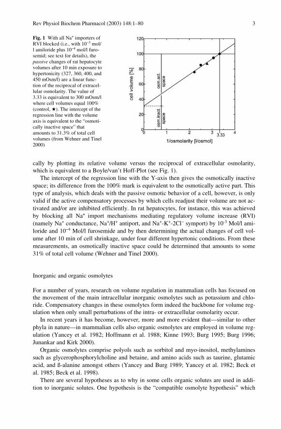
cally by plotting its relative volume versus the reciprocal of extracellular osmolarity,which is equivalent to a Boyle/van’t Hoff-Plot (see Fig. 1).
The intercept of the regression line with the Y-axis then gives the osmotically inactivespace; its difference from the 100% mark is equivalent to the osmotically active part. Thistype of analysis, which deals with the passive osmotic behavior of a cell, however, is onlyvalid if the active compensatory processes by which cells readjust their volume are not ac-tivated and/or are inhibited efficiently. In rat hepatocytes, for instance, this was achievedby blocking all Na+ import mechanisms mediating regulatory volume increase (RVI)(namely Na+ conductance, Na+/H+ antiport, and Na+-K+-2Cl` symport) by 10-3 Mol/l ami-loride and 10`4 Mol/l furosemide and by then determining the actual changes of cell vol-ume after 10 min of cell shrinkage, under four different hypertonic conditions. From thesemeasurements, an osmotically inactive space could be determined that amounts to some31% of total cell volume (Wehner and Tinel 2000).
Inorganic and organic osmolytes
For a number of years, research on volume regulation in mammalian cells has focused onthe movement of the main intracellular inorganic osmolytes such as potassium and chlo-ride. Compensatory changes in these osmolytes form indeed the backbone for volume reg-ulation when only small perturbations of the intra- or extracellular osmolarity occur.
In recent years it has become, however, more and more evident that—similar to otherphyla in nature—in mammalian cells also organic osmolytes are employed in volume reg-ulation (Yancey et al. 1982; Hoffmann et al. 1988; Kinne 1993; Burg 1995; Burg 1996;Junankar and Kirk 2000).
Organic osmolytes comprise polyols such as sorbitol and myo-inositol, methylaminessuch as glycerophosphorylcholine and betaine, and amino acids such as taurine, glutamicacid, and ß-alanine amongst others (Yancey and Burg 1989; Yancey et al. 1982; Beck etal. 1985; Beck et al. 1998).
There are several hypotheses as to why in some cells organic solutes are used in addi-tion to inorganic solutes. One hypothesis is the “compatible osmolyte hypothesis” which
Fig. 1 With all Na+ importers ofRVI blocked (i.e., with 10`3 mol/l amiloride plus 10`4 mol/l furo-semid; see text for details), thepassive changes of rat hepatocytevolumes after 10 min exposure tohypertonicity (327, 360, 400, and450 mOsm/l) are a linear func-tion of the reciprocal of extracel-lular osmolarity. The value of3.33 is equivalent to 300 mOsm/lwhere cell volumes equal 100%(control, H). The intercept of theregression line with the volumeaxis is equivalent to the “osmoti-cally inactive space” thatamounts to 31.3% of total cellvolumes (from Wehner and Tinel2000)
Rev Physiol Biochem Pharmacol (2003) 148:1–80 3

is based on the observation that high concentrations of inorganic salts such as NaCl orKCl perturb the function of enzymes or other proteins (Noulin et al. 1999), whereas organ-ic solutes do not (Yancey et al. 1982). Another hypothesis reflects the “counteracting os-molyte principle,” which refers to the finding that methylamines attenuate the destabiliz-ing effect of high concentrations of urea on protein function (Burg 1996). A third aspect isthat several organic osmolytes are electroneutral and can replace inorganic osmolyteswhich, when released across the cell membrane, may change the membrane potential andthereby neuronal excitability in the brain (Iwasa et al. 1980) or driving forces for electro-genic sodium-cotransport systems. Again, the brain is particularly interesting because ofthe existence of reuptake systems for excitatory amino acids that are transported by sodi-um-cotransport systems (Curtis and Johnston 1974).
One of the major places for organic osmolyte accumulation is the renal medulla, whereaccumulation occurs because of a broad range of extracellular osmolalities exceeding nor-mal osmolality (for review see Beck et al. 1985; Bagnasco et al. 1986; Yancey and Burg1989; Garcia-Perez and Burg 1991; Kinne et al. 1993; Kinne 1993; Kinne et al. 1995;Kinne 1998; Grunewald and Kinne 1999; Kinne et al. 2001) in particular in the directionof hyperosmolality (Grunewald et al. 1993a; Grunewald et al. 1994; Handler and Kwon2001). Also chondrocytes (de Angelis et al. 1999; Hall and Bush 2001) encounter hyperos-molality in the extracellular space because of the high concentration of fixed charges inthe mucopolysaccharides in which they are embedded. Limited expandability in the brain(Strange 1992; Pasantes-Morales et al. 1994a; Pasantes-Morales et al. 1994b; Pasantes-Morales et al. 2000b) and regulation of cell transparency in the lens (lens fiber cells; Burgand Kador 1988; Cammarata et al. 2002), also require organic osmolytes.
Intracellular accumulation of organic osmolytes involves mainly two processes. Thefirst represents uptake across the cell membrane by specific transport systems, the secondintracellular generation of the osmolyte by metabolic reactions. Examples of the formerare sodium-cotransport systems for myo-inositol, taurine, and betaine which use the sodi-um gradient across the plasma membrane as driving force. The general and specific prop-erties of these cotransport systems are summarized in the section entitled “Organic os-molytes in RVI.”
Metabolic reactions are primarily responsible for the generation of sorbitol from D-glu-cose and glycerophosphorylcholine (GPC). D-glucose taken up by the cells via uniport orsymport systems can be readily converted into sorbitol by the enzyme aldose reductase us-ing NADPH as cofactor (Gabbay 1973; Ohta et al. 1990). In an extensive study on renalinner medullary aldose reductase, the affinity for D-glucose was found to be about370 mMol/l, the affinity for NADPH was 7.5 �Mol/l. Thus, in the intact cell with aNADPH concentration of about 0.4 mMol/l the enzyme is saturated with NADPH, butonly to a small extent with D-glucose. Due to the high Km of the enzyme, in the intact cellsorbitol synthesis is characterized by the affinity of the cellular glucose uptake systems.For example, a value of about 60 mMol/l is obtained in intact IMCD cells, which reflectsthe Km usually observed for glucose uniporters GLUT 1 or GLUT 2 (Grunewald et al.1993b). Regulation of the synthesis occurs mainly by altering the Vmax of the enzyme; astrong increase is observed in a variety of cells when they are exposed to hypertonic con-ditions in vivo or in vitro (Bedford et al. 1987; Bagnasco et al. 1988; Grunewald et al.1998; Lang et al. 1998a). However, in disease states such as diabetes, the NADPH/NADPratio is another important regulator of the rate of sorbitol synthesis (Grunewald et al.1993b).
4 Rev Physiol Biochem Pharmacol (2003) 148:1–80
Sorbitol breakdown yielding fructose can occur via the sorbitol dehydrogenase pathwaywith NAD as the hydrogen acceptor. In some tissues that use sorbitol as organic osmolyte,these pathways are restricted to different, sometimes closely associated cells (Grunewaldet al. 1995; Kinne et al. 1997). In general, the activity of sorbitol dehydrogenase is muchlower than that of aldose reductase, thereby limiting the importance of this enzyme in os-motic adaptation (Grunewald et al. 1995; Kinne et al. 1997; Grunewald et al. 1998).
The major pathways involved in the metabolism of GPC in IMCD cells, for example,are the following. The precursor of GPC, choline, is taken up by sodium-independenttransport systems. Choline is phosphorylated and then incorporated into the phospholipidphosphatidylcholine (PC). By a stepwise removal of the fatty acid residues (involvingphospholipase A2), first lysophosphatidylcholine and then GPC are generated. Interesting-ly, this reaction seems to use a pool of PC different from that present in the plasma mem-brane. Breakdown of GPC is mediated by the GPC: choline diesterase reaction yieldingcholine and phosphoglycerol. The latter two reactions seem to be slow compared to thesynthetic pathway. The predominant “osmosensitive” enzyme is the GPC: choline di-esterase since studies in vivo and in vitro indicate that after longterm exposure of cells tohypertonic conditions, the rate of enzymatic breakdown is reduced and thus the overallconcentration of the organic osmolyte is increased (Zablocki et al. 1991; Bauernschmittand Kinne 1993).
The release of organic osmolytes from the cells involves channel-like proteins, in someinstances organic and inorganic osmolytes share the same transporter. Thus, a complexpicture emerges in which inorganic and organic osmolyte levels have to be controlled in awell-coordinated manner (Burg 1996). This coordination requires feedback systems thathave to be elucidated and taken into account when considering volume regulatory process-es in the cells.
Regulatory volume increase
Role of inorganic osmolytes in RVI
In order to achieve RVI, the intracellular osmolyte content has to be augmented rapidly.To this end, transport systems for inorganic osmolytes are activated as the first cellular re-sponse. The main osmolyte taken up by the cells is sodium since favorable driving forcesfor this cation exist across the plasma membrane. Sodium is subsequently exchangedagainst potassium by the action of the Na+,K+-ATPase, to restore the original sodium gra-dients and its electrochemical potential.
There are also mechanisms set in place that reduce loss of intracellular potassium andsometimes also chloride, depending on its electro-chemical equilibrium.
Na+/H+ antiport
Na+/H+ antiporters (NHEs) catalyze the secondary active and electroneutral exchange ofH+ against Na+. With one exception, they are most important for the regulation of cell pH,but some NHEs are effective mediators of RVI. Six NHE isoforms have been cloned todate (NHE1–NHE6) which exhibit a common molecular organization (see for review
Rev Physiol Biochem Pharmacol (2003) 148:1–80 5

Orlowski and Grinstein 1997; Counillon and Pouyssegur 2000; Ritter et al. 2001). Theyconsist of two main functional domains with amino acid identities of 45%–60% and 25%–35%, respectively. These are an N-terminal portion of 10–12 transmembrane regions (de-pending on the hydropathy algorithm used) and a large intracellular C-terminal part that isequipped with regulatory sites (see below). NHE1 to NHE5 are located in the plasmamembrane, while NHE6 is sorted to the mitochondrial inner membrane. NHE1 and NHE6are ubiquitously expressed whereas NHE2–NHE5 are restricted to specific tissues. In epi-thelia, NHE1 appears to be largely confined to the basolateral membrane (Coupaye-Gerardet al. 1996) but, in cell lines like OK (opossum kidney) or MDCK (Madin-Darby caninekidney), it may also occur apically (Noel et al. 1996). NHE2, NHE3, and NHE4 are foundpreferentially in the gastrointestinal tract and kidney where they appear to reside mainly inthe apical membrane (Soleimani et al. 1994; Dudeja et al. 1996; Noel et al. 1996; Hooger-werf et al. 1996; Sun et al. 1997; Bookstein et al. 1997). The presence of NHE2 in the kid-ney and NHE4 in the intestine, however, is discussed controversially (Bookstein et al.1997). NHE5 was detected in nonepithelial tissues, preferentially in the brain, but also inspleen, testis, and skeletal muscle (Klanke et al. 1995). In many cells, several NHE iso-forms are expressed and assigned to specific membrane domains so that (together withother transporters) they achieve a concerted cross-talk in the vectorial movement of ions;this especially holds for epithelia (Ritter et al. 2001).
Some NHEs are inhibited by submillimolar concentrations of amiloride and, more se-lectively, by its derivatives dimethyl-amiloride and ethyl-isopropyl-amiloride (Tse et al.1994; Counillon and Pouyssegur 2000). There are significant differences in sensitivity tothese blockers among the different isoforms, which (for amiloride) amount to almost fourorders of magnitude. The amiloride sensitivity generally follows the order NHE1 > NHE2>> NHE3 > NHE4 (Orlowski and Grinstein 1997; the latter two isoforms are thereforesometimes referred to as being amiloride-insensitive (Counillon and Pouyssegur 2000).The mitochondrial isoform NHE6 exhibits a rather low sensitivity to amiloride, but it isefficiently blocked by the analog, benzamil (Brierley et al. 1989).
With respect to their role in cell volume regulation, it was found that NHE1, NHE2,and NHE4 are activated under hypertonic conditions, whereas NHE3 is inhibited (Orlow-ski and Grinstein 1997; Hoffmann and Mills 1999; Ritter et al. 2001). NHE1 is clearly theNa+/H+ antiport most commonly employed in RVI, which is not surprising in regard to theubiquitous expression of the protein. As a matter of fact, the shrinkage-induced activationof Na+/H+ antiport that had been reported in a variety of preparations prior to the molecu-lar definition of NHE isoforms can now in many instances be attributed to NHE1 (Lang etal. 1998a; Lang et al. 1998b; Hoffmann and Mills 1999).
Concerning regulation, hypertonicity appears to shift the pHi sensitivity of NHE1 tomore alkaline values (Grinstein et al. 1985). Interestingly, although the C-terminal domainof NHE1 exhibits a number of phosphorylation sites (that are actually used for the activa-tion by hormones and mitogens), no direct phosphorylation of the protein occurs upon cellshrinkage (Sardet et al. 1990; Sardet et al. 1991; Grinstein et al. 1992). Rather, there ap-pears to be a significant hypertonicity-induced cross-talk between NHE1, stress fibers andthe small GTPase Rho so that osmotically-induced changes in the actin texture (also in-volving integrins) are very likely to be part of the signaling cascade-mediating activation(Watson et al. 1992; Grinstein et al. 1993; Vexler et al. 1996; Hooley et al. 1996; Tomi-naga et al. 1998; Tominaga and Barber 1998). In addition, Ca2+ may participate in thestimulation because the C-terminal domain of NHE1 contains two calmodulin bindingsites and deletion or mutation of the high affinity binding site significantly reduces the
6 Rev Physiol Biochem Pharmacol (2003) 148:1–80

osmotic sensitivity of the transporter (Bertrand et al. 1994). It is worth mentioning thatNHE1 is also subject to transcriptional osmotic regulation (Ritter et al. 2001).
The hypertonic activation patterns of NHE2 are less well-defined. Proline-rich regionsin the C-terminal portion of the protein appear to be involved which resemble SH3 bindingdomains and might reflect sites of interaction with the cytoskeleton and/or signaling mole-cules. Truncation experiments, however, revealed that these regions are probably involvedin the proper targeting of NHE2 rather than its regulation (Chow et al. 1999). Besides this,NHE2 is subject to transcriptional control (Soleimani et al. 1994; Bai et al. 1999).
NHE3 is inhibited under hypertonic conditions. This effect is due to a reduction of themaximal transport velocity rather than to changes in substrate affinities (Nath et al. 1996).NHE3 is highly sensitive to the actin texture of cells (Kurashima et al. 1999; Szaszi et al.2000). The protein associates with two regulatory factors, namely NHERF1 and NHERF2,and NHERF1 could be shown to bind to ezrin, an actin-binding protein of the ezrin-radix-in-moesin (ERM) family that links membrane proteins to the cytoskeleton. Based on dele-tion experiments, it could also be shown that the same C-terminal region of NHE3 medi-ates actin sensitivity and NHERF binding (Kurashima et al. 1999).
Moreover, among the small GTPases of the Rho family controlling actin assembly, theinhibitory form of RhoA (but not Rac1 and Cdc42) greatly depressed NHE3 activity andcomparable effects were observed upon a specific block of Rho kinase (ROK), a down-stream effector of RhoA; furthermore, inhibition of ROK reduced the phosphorylation ofmyosin light chain (MLC). These data strongly suggest that the RhoA-ROK signallingpathway is a mechanism for the control of NHE3 activity, which is, at least in part,achieved by controlling the phosphorylation of MLC and, consequently, the organizationof the actin cytoskeleton (Sz�szi et al. 2000).
Interestingly, considerable amounts of NHE3 are present in recycling endosomes (Rit-ter et al. 2001). This predominantly intracellular location may contribute to its paradoxicalbehavior during RVI.
NHE4 is clearly activated under hypertonic conditions; it exhibits a bell-shaped profilewith maximal functionality close to 500 mOsm/l (Bookstein et al. 1994). This relativelylow osmotic sensitivity is readily explained in terms of NHE4 tissue distribution withmaximal expression found in the renal medulla (Bookstein et al. 1994; Ritter et al. 2001)where a high extracellular osmolality prevails.
Na+-K+-2Cl` symport
NKCC1 and NKCC2 are mediators of electroneutral Na+, K+, Cl` cotransport across cellmembranes at a stoichiometry of 1:1:2 (with very few exceptions; see Haas and Forbush,III 2000; Russell 2000 for reviews). On the molecular level, they share many similaritieswith other members of Cl`-dependent cation transporters, namely the Na+-Cl` symporter(NCC) and the four isoforms of K+-Cl` symporters (KCC1 to KCC4) cloned so far. Thesesimilarities include molecular weights in the range of 110 kD to 130 kD (deglycosylated),12 predicted transmembrane regions, and large hydrophilic intracellular N- and C-terminaldomains. The most conserved regions of these transporters are the transmembrane do-mains, as well as the putative intracellular loops connecting them, particularly the one be-tween TM2 and TM3. NKCCs are blocked by the “loop” diuretics bumetanide and furose-mide at micromolar concentrations; KCCs exhibit a lower affinity to these compounds,whereas a possible inhibition of NCC remains ambiguous (Russell 2000). NKCC1 exhibits
Rev Physiol Biochem Pharmacol (2003) 148:1–80 7

a broad tissue distribution and is found in many secretory epithelia (where it resides in thebasolateral membrane), as well as in a variety of nonepithelial cells. In contrast, NKCC2is only present (apically) in the thick ascending limb of Henle’s loop and in the maculadensa of the kidney (Haas and Forbush, III 2000; Russell 2000).
NKCC1 is one of the most routinely employed transporters of cell volume regulation(Russell 2000) during RVI (Lang et al. 1998a; Lang et al. 1998b; Hoffmann and Mills1999; Haas and Forbush, III 2000). Activation of NKCC1 appears to involve phosphoryla-tion at serine/threonine residues and cell shrinkage results in NKCC1 phosphorylation in anumber of cells. In most instances, however, the actual kinase in charge remains to beidentified (Hoffmann and Mills 1999; Haas and Forbush, III 2000). Recently, the phos-phorylation of NKCC1 by c-Jun NH2-terminal kinase (JNK), a member of the mitogen-ac-tivated protein kinase (MAPK) family, was reported from bovine aortic endothelial cellsthat were shrunken under hypertonic or isotonic conditions (Klein et al. 1999). In rat hepa-tocytes, the hypertonic activation of Na+-K+-2Cl` symport (as well as that of Na+ conduc-tance) was inhibited by staurosporine, as well as by the PKC specific blocker bis-indolyl-maleimide I (BIM; Heinzinger et al. 2001). In human tracheal epithelial cells, the hyper-tonic activation of NKCC1 appeared to be mediated by PKC-d; this process is also likelyto involve the (extracellular signal-regulated) kinase ERK (Liedtke and Cole 2002). In ad-dition to phosphorylation and dephosphorylation, the activity of NKCC1 appears to be reg-ulated by the state of the actin network, as well as by accessory proteins that remain to becharacterized (Haas and Forbush, III 2000; Russell 2000).
Cation channels
Compared to the symport system discussed above, the rates of ion transport by channelsare some 4–5 orders of magnitude higher. Accordingly, any modulation of channel activityin response to changes of cell volume will serve as a fast and very efficient regulatorymechanism.
If one considers the electrochemical driving forces for Na+, K+, and Cl` transport acrossmost cell membranes, the activation of Na+-selective channels and conductive Na+ entrywould be a highly efficient mediator of RVI. The resultant depolarization of membranevoltage would favor a parallel conductive entry of Cl`. Na+ accumulating inside the cellwould then be extruded via Na+, K+-ATPase so that constant driving forces for Na+-cou-pled cotransporters are ensured. In sum, a net intracellular gain of K+ and Cl` and thus arapid increase of cell volume would be achieved by these mechanisms.
There is an increasing number of systems from which the hypertonic activation of cat-ion channels is reported. Two main classes of channels can be distinguished based on theirsensitivity to amiloride. In the following, first the amiloride sensitive channels will be dis-cussed.
The activation of conductive Na+ entry as a mechanism of RVI was originally proposedby Hoffmann (Hoffmann 1978) and Okada (Okada and Hazama 1989) for Ehrlich ascitestumor cells and the Intestine 407 cell line, respectively. In 1995, a hypertonic stimulationof cell membrane Na+ conductance was reported from current-clamp recordings on rat he-patocytes in confluent monolayer culture (Wehner et al. 1995; see Fig. 2).
Furthermore, in a quantitative study, it could be shown that the relative contribution ofNa+ conductance, Na+/H+ antiport, and Na+-K+-2Cl` symport to the initial uptake of Na+
under hypertonic stress was approximately 4:1:1 (Wehner and Tinel 1998). This clearly
8 Rev Physiol Biochem Pharmacol (2003) 148:1–80

renders Na+ conductance the prominent mechanism of RVI in this system. The Na+ con-ductance was inhibited by amiloride with an apparent Ki of 6 mMol/l and its overall sensi-tivity profile was EIPA > amiloride > benzamil (Wehner et al. 1997; BÛhmer et al. 2000).Hence, at first sight, the hypertonicity-induced Na+ conductance of rat hepatocytes mayreflect a low (amiloride) affinity type rather than an epithelial Na+ channel (ENaC); thelatter typically exhibits Ki values in the upper nanomolar range and an inverse pharmaco-logical profile (Garty and Palmer 1997; Fyfe et al. 1998). In voltage-clamp experimentson rat hepatocytes, a relatively low cation selectivity of the channel was detected with aPNa/PK of 1.4 (BÛhmer and Wehner 2001). In patch-clamp experiments in the cell-attachedconfiguration, hypertonicity-induced single channel events with a unitary conductance of6 pS were recorded (P. Lawonn and F. Wehner, unpublished observation). With respect tointracellular signaling events, the hypertonic activation of Na+ conductance (and Na+-K+-2Cl` symport, but not Na+/H+ antiport) is mediated by PKC (Heinzinger et al. 2001).
Fig. 2 A Cable analysis of specific membrane resistance, reflecting the hypertonic activation of Na+ con-ductance in rat hepatocytes. Experiments were carried out in the continuous presence of 0.5 mM quinine;extracellular osmolarity was increased from 300 mOsm/l to the osmolarities indicated; means€SEM; n=4–5. B Current-voltage relationships of hypertonicity-induced membrane currents in rat hepatocytes obtainedwith (two-channel microelectrode) voltage-clamp techniques. The differences between currents obtained at400 mOsm/l and 300 mOsm/l are depicted for cells injected with control-oligo-DNA (n) or anti-a-rENaColigo DNA (h) (from Wehner and Tinel 2000; BÛhmer and Wehner 2001)
Rev Physiol Biochem Pharmacol (2003) 148:1–80 9

In the human hepatoma cell-line HepG2, hypertonic stress led to the activation of anonselective cation conductance that was clearly sensitive to amiloride with 65% inhibi-tion at 10`5 Mol/l (Wehner et al. 2002a). Interestingly, this hypertonicity-induced conduc-tance was also partially inhibited by Gd3+ (10`4 Mol/l) and flufenamate (10`5 Mol/l) whichare typical blockers of nonselective but amiloride-insensitive cation channels in a varietyof systems (see below). Benzamil and EIPA (at 10`5 Mol/l each) had no effect on HepG2cation conductance (Wehner et al. 2002a). In Ehrlich-Lettre ascites tumor cells, hypertonicconditions activated a cation conductance that did not discriminate between Na+, K+, andLi+, but that was impermeable to NMDG and choline. In the range of 10-6 Mol/l to10`3 Mol/l this cation conductance was blocked by the ion channel inhibitors benzamil >Gd3+ > amiloride > EIPA (in order of potency; Lawonn et al. 2003). In immortalized hu-man nonpigmented ciliary epithelial cells, shrinkage upon return from hypo-osmotic tonormosmotic conditions was followed by a partial (post-RVD) RVI (Civan et al. 1996)that was mediated by the parallel operation of Na+/H+ antiport, Na+-Cl` symport, Na+-K+-2Cl` symport, as well as by conductive Na+ entry. In the range of 10`6 Mol/l and 10`5 Mol/l,this Na+ conductance was significantly inhibited by amiloride and—even more efficient-ly—by benzamil, the most effective blocker of the ENaC (Civan et al. 1997; Garty andPalmer 1997; Fyfe et al. 1998). In principal cells of rat cortical collecting duct hypertonicstress led to a distinct depolarization of membrane voltage coinciding with an increase ofcell Na+ (Schlatter et al. 1997). These effects were partially inhibited by 10`5 Mol/l amilo-ride or Gd3+ and the effects were additive. This result was interpreted in terms of a parallelactivation of Na+ channels and nonselective cation channels.
In human red blood cell ghosts, hypertonic stress induced a cation conductance that ap-peared to be equally permeable to Na+ and K+ (but impermeable to NMDG) and that waspartially inhibited by 10`4 Mol/l amiloride (Huber et al. 2001). These findings support ear-lier flux studies on lamprey erythrocytes in which hypertonic stress led to the activation ofan amiloride-sensitive Na+ transport that could not be attributed to Na+/H+ or Na+/Na+ an-tiport (Gusev and Sherstobitov 1996).
In U937 macrophages, hypertonic stress led to the activation of an inward current witha distinct selectivity for Na+ over K+ that was inhibited by amiloride with an apparent Ki
close to 1 mMol/l. At the single channel level, this cation conductance appeared to be relat-ed to a 6 pS channel. Interestingly, these channels are upregulated by pretreatment of thecells with glucocorticoids, which are known to regulate macrophage function (Gamper etal. 2000).
The shrinkage-activated amiloride sensitive cation channel appears to be related to(a)ENaC. In rat hepatocytes, antisense nucleotides attenuated the activation of the channel(BÛhmer and Wehner 2001; see Fig. 2B). In human ciliary body, a-ENaC mRNA wasidentified (Civan et al. 1997). Similarly, mRNA for a, b, and g subunits was detected inrat hepatocytes and these subunits could also be identified on the protein level (BÛhmerand Wehner 2001). Although the nonselectivity of hypertonicity-induced channels is quitein contrast to the high PNa/PK reported for a-, b-, g-rENaC (which is as high as 40; Fyfe etal. 1998) this is not contradictory per se to a possible contribution of ENaCs because theirbiophysical properties (also including single-channel conductance) strongly depend onsubunit composition (Garty and Palmer 1997) as well as their relative affinity to amilorideand its derivatives (Benos et al. 1997; Garty and Palmer 1997). Furthermore osmo-sensi-tivity of a-, b-, g-ENaC was reported when the channel subunits were expressed in oocytes(Awayda and Subramanyam 1998). Since some of the results are, however, conflicting,
10 Rev Physiol Biochem Pharmacol (2003) 148:1–80

further studies are necessary to clarify the relation between ENaC and the amiloride-sensi-tive cation channels.
The second group of hypertonicity-induced cation channels is clearly amiloride-insen-sitive up to concentrations of 10`4 Mol/l. Quite typically, however, these channels are in-hibited by the anti-inflammatory drug flufenamate (10`4 Mol/l), as well as by Gd3+ in therange of 10`5 to 10`3 Mol/l. They are expressed in human nasal epithelial cells (Chan andNelson 1992), in the human colon cell-lines CaCo-2 and HT29 (Nelson et al. 1996; Kochand Korbmacher 1999), in the mouse cortical collecting duct cell line M1 (Volk et al.1995), as well as in BSC-1 renal epithelial cells (derived from the African green monkey),A 10 vascular smooth muscle cells (established from rat embryonic aorta), and Neuro-2acells (derived from mouse neuroblastoma; Koch and Korbmacher 1999). In general, thesechannels are rather nonselective with respect to monovalent cations, although there arecertain peculiarities. In human nasal epithelium and in CaCo-2 cells, for instance, channelsdid not discriminate much between Na+, K+, and Cs+, but exhibited a significantly lowerpermeability to Li+ and, in the latter system, there was also a distinct permeability toNMDG (PNa/PNMDG=0.56; Chan and Nelson 1992; Nelson et al. 1996). In contrast, in theM1 cell-line the channel appeared to be equally permeable to Na+, K+, Cs+, Li+, and Rb+
but virtually impermeable to NMDG (Korbmacher et al. 1995; Volk et al. 1995). In addi-tion, there appear to be significant differences in the relative permeabilities of amiloride-insensitive channels to Cl` with PNa/PCl values in the range of 60 (Korbmacher et al. 1995)to 1.7 (Nelson et al. 1996). In some systems, the hypertonic activation of channels is Ca2+-independent (Chan and Nelson 1992) whereas, in others, channel activity appears to re-quire a minimum (Ca2+)i of 1 mMol/l (Korbmacher et al. 1995; Koch and Korbmacher1999). Nevertheless, these channels do not appear to conduct significant amounts of diva-lent cations (Korbmacher et al. 1995; Koch and Korbmacher 2000). The unitary conduc-tances of hypertonicity-induced but amiloride-insensitive channels generally is in therange of 15 pS to 27 pS (Korbmacher et al. 1995; Koch and Korbmacher 1999).
Most interestingly, at least some of these amiloride-insensitive nonselective cationchannels are typically inhibited by cytosolic ATP concentrations in the millimolar range(Koch and Korbmacher 1999). This raises some concerns as to their actual role in RVI be-cause, under most physiological conditions these channels will remain silent. Also of notein this respect, in a recent study on isolated rat colonic crypt cells, there was no detectableactivation of nonselective cation channels under hypertonic stress (Weyand et al. 1998),whereas these cells are known to express Ca2+-activated channels which become de-tectable in excized inside-out patches and which are very similar to those observed inHT29 cells (Bleich et al. 1996). Clearly, whereas nonselective and amiloride-insensitivecation channels appear to be ubiquitously expressed, their actual contribution to cell vol-ume regulation remains to be elucidated.
In principle, the activation of cation conductances—either sensitive or insensitive toamiloride—that do not discriminate much between Na+ and K+ will lead to both Na+ in-flux as well as K+ efflux. Because of the inside negative membrane voltage, however,overall cation gain will significantly exceed overall cation loss equivalent to an increaseof the overall intracellular osmotic activity.
Rev Physiol Biochem Pharmacol (2003) 148:1–80 11

K+ channels
In most systems, the continuous channel-mediated K+ leakage out of cells is equivalent toa significant loss of cellular osmolytes. This K+ loss is commonly compensated for by theactivation of Na+, K+-ATPase. Nevertheless, it is rather obvious that any inhibition of K+
channels under hypertonic conditions will per se facilitate RVI and, in some cells, thismechanism appears to be an important mediator of volume regulation.
An inhibition of K+ conductance was originally reported for the basolateral membraneof toad and rabbit urinary bladder (Lewis et al. 1985; Donaldson et al. 1989), as well as offrog skin (Costa et al. 1987; Leibowich et al. 1988) where cells were shrunken by extracel-lular Cl` removal (leading to a loss of Cl` because of the lower membrane permeability ofthe Cl` substitute used when compared to Cl` itself) and/or by increasing extracellular os-molarity. In addition, hypertonic stress reduced basolateral K+ conductance in rabbit prox-imal tubule (Lapointe et al. 1990; Macri et al. 1997) and human nasal epithelium (Willum-sen et al. 1994) and it decreased conductive K+ loss in MDCK cells (Ritter et al. 1991). Inmouse liver, a reduction of cell membrane K+ conductance appeared to be the main mech-anism of RVI (Graf et al. 1988; Wang and Wondergem 1991) and a transient inhibition ofK+ channels may contribute to the volume response of rat hepatocytes (Wehner et al.1995; Wehner and Tinel 1998). Interestingly, whole-cell recordings on freshly isolatedhippocampal neurons revealed a decrease of voltage-gated K+ currents under hypertonicconditions (Huang and Somjen 1997). In dissociated rabbit corneal epithelial cells, a largeconductance K+ channel (167 pS in symmetrical 150 mMol/l KCl) could be identified thatappeared to mediate the decrease in whole-cell K+ conductance to 44% of control uponchange to 130% extracellular osmolarity (Farrugia and Rae 1993). In a recent study on iso-lated rat colonic crypts, it was found that hypertonic stress (+50 mOsm/l and +100 mOsm/l)led to membrane depolarizations by 12 mV and 22 mV, respectively, coinciding with de-creases in whole-cell conductance to 70% and 50% of control (Weyand et al. 1998). Onthe molecular level, these effects appeared to be correlated to a 16 pS K+ channel that ex-perienced decreases in activity (N · Po) to 47% and 44% when compared to isotonic condi-tions. Of note, this reduction of channel activity was most likely triggered by a hypertonic-ity-induced decrease of (Ca2+)i. The channel was inhibited by (10`4 Mol/l) Ba2+ and(10`9 Mol/l) charybdotoxin (Bleich et al. 1996).
Anion channels
Inhibition of anion channels during RVI might directly contribute to the gain in osmolytesif the intracellular anion (Cl` or HCO3
`) activity is above electrochemical equilibrium. In-directly, it restricts the movement of the counterion for K+ flux across the membrane, thusimpeding K+ losses.
Decreases of Cl` conductance under hypertonic conditions have been reported onlyfrom a limited number of preparations. In cultured human nasal epithelium, hypertonicstress reduced apical Cl` conductance (Willumsen et al. 1994) whereas, in rabbit collapsedproximal convoluted tubules, the partial Cl` conductance of the basolateral membrane wasinhibited (Macri et al. 1997). In some instances, the decrease of Cl` conductance underhypertonic conditions may reflect an inhibition of hypotonicity-induced Cl` channel acti-vation. In human vas deferens cells in primary culture, for example, with 290 mOsm/l so-lutions in the experimental bath, as well as in the patch pipette, there was a slowly devel-
12 Rev Physiol Biochem Pharmacol (2003) 148:1–80

oping increase of Cl` conductance once the whole-cell configuration was achieved; thiseffect could be reversed by an increase of extracellular osmolarity (Winpenny et al. 1996).The increase of Cl` conductance was interpreted to be due to the additional osmotic activ-ity of intracellular macromolecules and the resultant swelling of cells.
Organic osmolytes in RVI
In most cells investigated, RVI after exposure to a hypertonic extracellular medium occurswithin minutes and involves the net uptake of inorganic osmolytes. During this short-termregulation of cell volume, changes in organic osmolytes transport are generally not in-volved, although it has been observed that the plasma membrane permeability for organicosmolytes such as sorbitol (e.g., in IMCD cells grown at 600 mOsm/l and exposed to900 mOsm/l medium) decreases (Bagnasco et al. 1988; Grunewald and Kinne 1989).Thus, leak pathways for organic osmolytes are downregulated to make the intracellular ac-cumulation of organic osmolytes—by transporters or metabolic synthesis—more effective.Whether these leak pathways are identical to the ones activated during RVD remains to beinvestigated.
If exposure of cells to hypertonicity is extended to periods of hours or days, adaptivechanges take place aimed to replace the augmented intracellular inorganic electrolytes byorganic osmolytes. To this end, intracellular synthesis is increased (sorbitol and GPC) orintracellular breakdown is decreased (GPC; for review see Burg 1995; Burg et al. 1997).For taurine, betaine, and myo-inositol, the predominant effect is on the rate of uptake,which increases severalfold in a matter of hours (see Fig. 3).
Thus, strictly speaking, organic osmolytes are not directly involved in RVI; they serve,however, in the long run to maintain cellular electrolyte homeostasis. Therefore, the cellu-lar uptake systems for organic osmolytes and their regulation are described in the follow-ing paragraphs.
Na+-Cl`-taurine symport
Taurine, a b-amino acid with a sulfonic acid instead of a carboxylic acid as head groupoccurs in plasma at concentrations of 40 �Mol/l. It has several major functions: as partnerin the formation of taurocholate, as neurotransmitter (Jacobsen and Smith 1968; Curtisand Johnston 1974), and as an organic osmolyte. Although several tissues, including liverand astrocytes, can synthesize taurine from cysteine, the high intracellular concentration(e.g., 20 mMol/l in IMCD cells) can only be achieved by an active uptake into the cell(Ruhfus et al. 1998). The transporter mediating this uptake is a secondary active sodium-chloride taurine cotransport system in which up to three sodium ions are translocated withone chloride ion and one taurine molecule. Taurine transport with these characteristics hasbeen found in renal (Chesney et al. 1985a; Kinne et al. 1998; Wolff and Kinne 1988;Zelikovic et al. 1989; Zelikovic and Budreau-Patters 1999) and intestinal brush border(O’Flaherty et al. 1997), in liver cells (Warskulat et al. 1997a, Warskulat et al. 1997b;Peters-Regehr et al. 1999), in H4IIE hepatoma cells (Warskulat et al. 1997b), Ehrlich asci-tes tumor cells (Hoffmann et al. 1988), astrocytes (Beetsch and Olson 1996), the retinalpigment epithelium (Handler and Kwon 2001), bovine aortic endothelial cells (Qian et al.2000), renal IMCD cells, and MDCK cells (Uchida et al. 1991). The apparent affinity
Rev Physiol Biochem Pharmacol (2003) 148:1–80 13

(Km) for taurine is in the range of 10 �M to 100 �M (Uchida et al. 1991). The transporteralso transports b-alanine and, with a lower affinity, g-amino butyric acid (GABA). Molec-ular cloning from MDCK cells revealed that the transporter corresponds to a protein with655 amino acids with a relative molecular mass of 74 kDa and probably has twelve trans-membrane helices (Uchida et al. 1992). It shows significant amino acid sequence similari-ty to the MDCK cell betaine/GABA transporter and other Na+, Cl`-dependent neurotrans-mitter transporters (Guastella et al. 1990). In dog tissues, the order of mRNA abundancefor this transporter (dTAUT) was kidney cortex � kidney medulla > ileal mucosa > brain> liver > heart > epididymis. Taurine transporters cloned from other sources such as LLC-PK1 cells from pig kidney, human retinal pigment epithelium, thyroid, placenta, and bo-vine aortic endothelial cells (Qian et al. 2000) were of similar size (621 and 620 aminoacids, respectively) and have a high amino acid sequence similarity.
In a variety of cells and tissues such as MDCK cells (Uchida et al. 1991), human andbovine lens epithelial cells (Cammarata et al. 2002), bovine aortic endothelial cells (Qianet al. 2000), rat liver macrophages (Warskulat et al. 1997a), H4IIE hepatoma cells(Warskulat et al. 1997b), primary cultures of rat hepatocytes (Warskulat et al. 1997a), ratastrocyte cultures (Beetsch and Olson 1996), and human Caco-2 cells (Satsu et al. 1999),exposure to hyperosmotic media results in an increase in Vmax of taurine transport and aconcomitant increase in transporter mRNA (Burg et al. 1997; Handler and Kwon 2001;Cammarata et al. 2002).
High affinity taurine transport is inhibited by phosphorylation via PKC by decreasingaffinity for taurine and sodium and reducing maximal velocity (Kulanthaivel et al. 1991;
Fig. 3 Time course of basolater-al myo-inositol (A) and betaine(B) uptake into MDCK cellsswitched into hypertonic medi-um. On day 0, cells cultured onfilters in defined medium with10% FBS were switched to samemedium made hypertonic(500 mOsm/l) by addition of raf-finose. Isotonic cells were main-tained in isotonic defined medi-um with 10% FBS. Uptake wasperformed at 37�C for 30 minwith 10 �Mol/l myo-inositol and10 �Mol/l betaine. Results aremeans€SD to triplicate samples(from Yamauchi et al. 1991)
14 Rev Physiol Biochem Pharmacol (2003) 148:1–80

Brandsch et al. 1993; Nakamura et al. 1996; Mollerup and Lambert 1998; Lima et al.2000; Qian et al. 2000). Furthermore, taurine uptake adaptively decreases when taurineavailability is augmented (Chesney et al. 1985b; Han et al. 1998). Although these regula-tory processes appear not to be linked to the response to hypertonicity, they complicatethe study of the molecular mechanisms of the latter. However, it can be assumed that themechanisms involved in osmotic adaptation are similar to the ones described below for thebetaine transport system, since both systems respond in a very comparable manner.
Na+-Cl`-betaine symport
In medullary kidney cells and chondrocytes, which use betaine in volume regulation, intra-cellular concentrations of up to 50 mMol/l are found. Although cells usually contain cho-line dehydrogenase that catalyses the synthesis of betaine from choline, most of the beta-ine is taken up from the extracellular medium. The same holds for MDCK (Yamauchi etal. 1991) and PAP-HT25 cells (Ferraris et al. 1996). The uptake of betaine is dependent onthe presence of both sodium and chloride and the transport system (Yamauchi et al. 1992;Moeckel et al. 1997) has been identified to belong to the family of brain GABA and nor-adrenaline transporters (Guastella et al. 1990).
Initial cloning of the transporter from MDCK cells revealed a protein of 614 amino ac-ids with a molecular weight of 69 kDa. Its proposed membrane topology predicts 12 trans-membrane helices and an intracellular NH2 and COOH terminus (Yamauchi et al. 1992).When expressed in oocytes, the protein showed both betaine and g-amino-n-butyric acid(GABA) transport activities that were chloride- as well as sodium-dependent (Matskevitchet al. 1999; Forlani et al. 2001). Because of these transport properties and the 43%–49%identity in the amino acid sequence (Rasola et al. 1995) with the rat brain GABA trans-porter and the human brain noradrenaline transporter, it was named betaine-GABA-trans-porter (BGT1).
Consecutive studies showed that the gene extends over 28 kilobases and consists of18 exons. In addition, the 50 end of the gene has three different first exons, thus a complexmixture of mRNAs exists. Three main types of mRNA (A, B, and C) can be distinguishedthat differ considerably in their 50 untranslated sequences. Each type is expressed in a tis-sue-specific manner: kidney medulla (and MDCK cells) contain all three types, brain andkidney cortex, type B and C, and liver only type A (Takenaka et al. 1995; Burg et al.1997).
Betaine transport shows apparent affinities (Km) of 0.3 mMol/l–0.5 mMol/l in MDCKcells, and in oocytes that express BGT1, the affinity for GABA is at or below 0.1 mMol/l.At 1 mMol/l substrate, the current induced in BGT 1-expressing oocytes decreased in thefollowing order: betaine > GABA > diaminobutyric acid = b-alanine > proline. At1 mMol/l betaine, the affinity for sodium was 93.3 mMol/l and the affinity for chloride76.1 mMol/l (Yamauchi et al. 1992).
In electrophysiological studies, a coupling ratio of Na/Cl/betaine of 3:2:1 was found.Furthermore, these studies revealed significant kinetic differences compared to the neuro-nal GABA-transporter GAT-1 (Matskevitch et al. 1999).
For the symport process, a transport model of ordered binding was proposed (Matske-vitch et al. 1999; Forlani et al. 2001) in which betaine binds first to the extracellular sideof a transporter. Sodium binding occurs thereafter, followed by chloride binding. Translo-cation of betaine and sodium is already observed in the absence of chloride, but chloride
Rev Physiol Biochem Pharmacol (2003) 148:1–80 15

augments the translocation. As for other cotransport systems, this model would predict thatbetaine facilitates sodium and chloride binding to the transporter; corresponding increasesin the transporter affinities at higher betaine concentrations have been observed. The exactmechanism of chloride (and sodium and betaine) translocation are unknown to date. Itshould be mentioned, however, that in squid motor neurons betaine activates a large Cl`
selective current which is sodium-dependent (Petty and Lucero 1999). This observationraises the possibility that the betaine transporter might posses chloride channel properties.
Augmented extracellular osmolality increases betaine uptake into MDCK cells (Naka-nishi et al. 1990; Yamauchi et al. 1991; Kempson 1998), rabbit PAP-HT 25 cells (renalpapillary epithelium; Ferraris et al. 1996), and porcine chondrocytes (de Angelis et al.1999) at the level of the transporter by increasing Vmax but not Km of betaine uptake (seeFig. 3). The increase in uptake is preceded by an enhanced transcription of BGT1 inMDCK cells followed by increases in BGT1-mRNA. As in the case of the sodium-myo-inositol transporter a “tonicity-responsive enhancer element” (designated TonE) of thegene is involved that spans `69/`50 of the sequence (Burg et al. 1997; Handler and Kwon2001). In transgenic mice, the 50-flanking region which contains TonE also mediates os-motic regulation of transcription in vivo (Handler and Kwon 2001). Osmoregulation ofBGT1 mRNA has also been observed in renal medulla in the intact rat (Miyai et al. 1996;Moeckel et al. 1997), and in rat hepatic stellate cells (Peters-Regehr et al. 1999). Althoughthe time course and extent of regulation of BGT-1 and SMIT by hypertonicity are quitesimilar, the regulatory pathways may be different at one point or another (Atta et al. 1999)
In addition to the relatively slow adaptation of betaine transport in vitro, more rapidchanges have also been reported. For example, in mouse isolated perfused straight proxi-mal tubules, basolateral betaine uptake (measured as substrate-dependent depolarization)is enhanced when the extracellular osmolality is increased (VÛlkl and Lang 2001). Thespeed of regulation suggests nongenomical processes. A similar rapid activation was ob-served in squid motor neurons (Petty and Lucero 1999). Whether these responses involveprotein phosphatases that, potentially by dephosphorylation, could stimulate betaine trans-port is not clear.
Hypertonic activation of betaine uptake shows some more peculiarities. Under isotonicconditions betaine uptake into MDCK cells proceeds only across the basolateral mem-brane; after exposure to hypertonicity basal-lateral uptake is increased, but in addition, sig-nificant uptake across the apical membrane is observed (Yamauchi et al. 1991). This find-ing might be related to the existence of various isoforms of BGT1 (see above) that mightdiffer in their cellular location.
Na+-Myo-inositol symport
Physiological plasma concentrations of myo-inositol range in mammals from 4.5 mMol/lto 6.6 mMol/l, whereas intracellular concentrations up to 133 mMol/l can be found in ratglial (Strange et al. 1991) and renal medullary cells (Bagnasco et al. 1986; Nakanishi et al.1988; Wirthensohn et al. 1989; Yancey and Burg 1989; Garcia-Perez and Burg 1991; Size-land et al. 1993; Grunewald et al. 1995; Grunewald and Kinne 1999; Handler and Kwon2001). This large concentration difference suggests active uptake of myo-inositol intothese cells. Similarly, active myo-inositol uptake is observed in rat pancreatic islets, bo-vine lens epithelial cells, hamster small intestine, rat mesanglial cells, rat hepatocytes,crystalline lense, rabbit peripheral nerve, retinal pigment epithelial cells, rabbit ciliary
16 Rev Physiol Biochem Pharmacol (2003) 148:1–80

body, isolated rat Schwann cells, and endothelial cells (for references see Porcellati et al.1999). This uptake process involves a sodium/myo-inositol symport system (SMIT) whichas secondary active transport system employs the transmembrane electrochemical poten-tial difference of sodium for intracellular accumulation of myo-inositol. Apparent Km val-ues for myo-inositol range from 30 �Mol/l in MDCK cells (Kwon et al. 1992) to~94 �Mol/l in rabbit renal brush border (Hammerman et al. 1980) and 104 �Mol/l in rabbitTALH cells (Yorek et al. 1999). Interestingly, also D-glucose is a substrate of this trans-porter (Hammerman et al. 1980; Hager et al. 1995). Phlorizin, the well-known inhibitorof the sodium-D-glucose cotransporter, also inhibits the sodium-myo-inositol transporter(Ki <60 �Mol/l; Strange et al. 1991; Hager et al. 1995). Similarities between the two trans-porters were also detected at the DNA and protein level. There is 46% amino acid identityoverall and apparently similar membrane topology of the two transporters; SMIT is a poly-peptide of 718 amino acids with a relative molecular mass of 79.5 kDa (Kwon et al.1992).Tissue abundance of mRNA in dog tissues is kidney medulla > kidney cortex >brain (Kwon et al. 1992). The SMIT cloned recently from bovine lens epithelial cellsshows a 92% identity with the MDCK cell transporter (Zhou et al. 1994).
The sodium to myo-inositol stoichiometry of the transporter is 2:1 (Hager et al. 1995).The stoichiometry and electrogenicity of the transporter forms the basis for the extensiveintracellular accumulation of myo-inositol. Kinetic and biochemical characteristics varyfrom tissue to tissue and cell to cell. These variations have recently been explained by theexistence of alternate splicing, which generates isoforms that differ in their intracellularprotein kinase A and protein kinase C phosphorylation sites at the carboxy terminus (Por-cellati et al. 1999). In epithelial cells, the cellular location can also differ; thus, in the renalproximal tubule the Na+/myo-inositol symport is present in the luminal brush border mem-brane, whereas in more distal renal cells the basolateral location prevails (see Grunewaldand Kinne 1999).
SMIT is increased by augmentation of the extracellular osmolality in MDCK cells(Nakanishi et al. 1989; Yamauchi et al. 1991), glial cells (Strange et al. 1991; Kwon et al.1992), neuronal cells (Yamashita et al. 1999), mesothelial cells (Matsuoka et al. 1999),bovine lens epithelial cells (Cammarata et al. 2002), and human retinal epithelial cells(Handler and Kwon 2001). In MDCK cells grown on filters, a 25-fold increase in uptakerate was observed within 24 h after exposure of the cells to hypertonic media (Yamauchiet al. 1991).
After restoring isotonicity, the transport rate returns to normal levels within 1 d. In bothinstances only the Vmax of the transport is affected and Km remains constant, suggesting achange in the number of transporters (Nakanishi et al. 1989). In similar studies in rat C6glioma cells, myo-inositol uptake also increased after exposure of the cells to 440 mOsm/lsolutions; however, the time course was slower and maximum uptake was obtained after48 h (Strange et al. 1991). The increased uptake in response to hypertonicity is precededby an increased abundance of mRNA for the transporter, which is the direct result of in-creased transcription of the gene (Yamauchi et al. 1993). Activation of transcription de-pends on an enhancer element named tonicity responsive enhancer (TonE). The SMIT-gene is regulated by multiple TonEs in its 50-flanking region (Burg et al. 1997; Zhou andCammarata 1999; Handler and Kwon 2001). An increase in SMIT m-RNA is also found ina TALH cell line derived from rabbit renal medulla (Yorek et al. 1999) and in mesothelialcells (Matsuoka et al. 1999). In the latter cell line, the first significant increase in mRNAcould be observed already 1 h after exposure of the cells to 490 mOsm. The increase intransporter mRNA has also been observed under in vivo conditions, for example, in rat in-
Rev Physiol Biochem Pharmacol (2003) 148:1–80 17

ner medullary collecting duct cells when d DAVP was administered to chronically diureticrats, a maneuver that rapidly increases the extracellular osmolality in the medulla (Burger-Kentischer et al. 1999). Injection of NaCl into rats increases SMIT-mRNA significantlywithin 1 h. Comparing the experiments in intact rats with cultured cells, the transportermRNA seems to respond more rapidly in vivo than in vitro (Yamauchi et al. 1995).
Amino acid transport system A
Amino acid transport system A (System A), which mediates sodium-dependent uptake ofneutral amino acids into mammalian cells, appears to be also subject to regulation by ex-tracellular osmolality. In MDCK cells, as well as chondrocytes, an upregulation within 4–6 h of hypertonic exposure was observed, well before any change in BGT1 activity (Chenet al. 1996a; de Angelis et al. 1999; Horio et al. 1997; Kempson 1998). The increase inSystem A is blocked by inhibitors of RNA and protein synthesis, suggesting that an in-crease in the number of transporters is part of the mechanism (Kempson 1998). The adap-tation of this system appears to be a relatively early response of cells exposed to hyperto-nicity; therefore, it deserves further investigation.
Regulatory volume decrease
Inorganic osmolytes in RVD
After cell swelling, transport systems are immediately activated that mediate the release ofthe major intracellular inorganic osmolytes potassium and chloride. Their transmembranemovement occurs either via separate pathways or directly coupled to each other.
K+ channels
Due to the outwardly directed K+ gradient in most animal cells, any increase of K+ channelactivity will augment the conductive exit of K+. In addition, the increase of cell membraneK+ conductance will hyperpolarize the cell membrane and (even if basal Cl` conductanceis not changing) this hyperpolarization will favor conductive Cl` efflux. Likewise, if thereis an initial increase of Cl` conductance and if cell Cl` is above electrochemical equilibri-um (as it is in most systems) this will augment Cl` exit and depolarize the cell membrane.Membrane depolarization in turn will facilitate conductive K+ efflux. The most effectivemechanism of RVD will, of course, be the parallel activation of K+ and Cl` channels. Be-cause of the pronounced voltage-mediated coupling between both pathways conductive K+
and Cl` release may result in a quasielectroneutral mode of KCl export.An increase of cell membrane K+ conductance under hypotonic conditions and its sig-
nificance for RVD have been reported from a variety of preparations (see Lang et al.1998a; Lang et al. 1998b for comprehensive reviews). In many studies, these mechanismswere analyzed by means of 86Rb+ (or 42K+) fluxes, intracellular microelectrode recordingsand rapid ion-substitutions, whole-cell patch-clamp measurements, and by use of specificK+ channel blockers. Here, we will focus on data at the molecular level that were obtained
18 Rev Physiol Biochem Pharmacol (2003) 148:1–80

by means of single-channel recordings and by the cloning and expression of some of thevolume-activated K+ channels.
BKCa or maxi-K+ channels exhibit large (big) unitary conductances in the range ofsome 100 pS to 250 pS and show under symmetrical high K+ solutions a linear current-to-voltage relation (see Vergara et al. 1998 for review). In most instances, BKCa channels areinhibited by Ba++, quinine, and TEA (tetraethylammonium), as well as by (the scorpionpeptide) charybdotoxin. They are selectively blocked by (the scorpion toxin) iberiotoxin.BKCa channels are found in neurons, skeletal muscle, smooth muscle, and in epithelialcells, where they reside in the apical membrane. BKCa channels are activated by mem-brane depolarization and micromolar concentrations of cell Ca2+. It is this Ca2+ sensitivitythat might function as a link to a swelling-induced increase in intracellular calcium. Like-wise, the voltage-dependence of BKCa channels may reflect the coupling mechanism to ahypotonicity-induced activation of Cl` channels (see above).
BKCa channels consist of hetero-oligomeric complexes of the pore-forming a-subunits(first cloned from Drosophila as dSlo (Atkinson et al. 1991; Adelman et al. 1992) and theauxiliary b-subunits (Garcia-Calvo et al. 1994; Behrens et al. 2002). The a-subunits areexpressed in multiple splice variants generating functional diversity among different cells,but they exhibit a high degree of homology among species. Their membrane topology isrelated to that of voltage-gated K+ channels (see below; Toro et al. 1998; Vergara et al.1998; Jensen et al. 2001). They contain seven transmembrane segments (so that the N-ter-minus is most likely extracellular), a positively charged TM4-segment (that is probablypart of the voltage sensor), and four additional hydrophobic segments forming a uniquesecondary structure at the C-terminal cytosolic tail that appears to mediate Ca2+ binding(Schreiber and Salkoff 1997).
The b-subunit is likely to function as an additional modulator of channel characteris-tics, e.g., with respect to Ca2+ sensitivity, but it does not appear to be an obligatory compo-nent of all BKCa channels. Four b-subunits have been cloned that are mainly expressed insmooth muscle, endocrine cells, epithelia, and the central nervous system (Behrens et al.2002). b-subunits have a proposed topology of spanning the membrane twice, with N- andC-termini inside the cell (Toro et al. 1998; Vergara et al. 1998).
The activation of BKCa channels under hypotonic conditions could be demonstrated inNecturus and rabbit proximal tubule cells (Dub¹ et al. 1990; Filipovic and Sackin 1991;Kawahara et al. 1991), in principal cells of rabbit and rat cortical collecting tubule (Linget al. 1992; Schlatter 1993; Stoner and Morley 1995; Hirsch and Schlatter 1997), in clonalkidney (Vero) cells derived from African green monkey (Hafting and Sand 2000), in aci-nar cells of rat lacrimal gland (Park et al. 1994), in embryonic chick hepatocytes (Pon andHill 1997), and in guinea pig jejunal villus enterocytes (MacLeod and Hamilton 1999b).In the A3 cell line derived from rabbit medullary thick ascending limb, hypotonic stress, aswell as negative pressure applied to the patch pipette (suction), significantly increased theopen probability of BKCa channels (Taniguchi and Guggino 1989). Comparable effectswere observed in intercalated cells of rat cortical collecting tubule (P�cha et al. 1991), inthe human colonic cell line CaCo-2 (Bear 1991), and in vascular smooth muscle cells (Kir-ber et al. 1992).
These results suggest a direct effect of membrane stretch on the channel itself or on aclosely associated component as a mode of channel regulation. Also of note is the distinctsensitivity of BKCa channels to extracellular ATP as it was observed in Vero cells (Haftingand Sand 2000). Thus, ATP release may well function as an autocrine (or paracrine) mech-
Rev Physiol Biochem Pharmacol (2003) 148:1–80 19

anism of hypotonic channel activation. In addition, a variety of protein kinases and phos-phatases appear to have an impact on BKCa channel activity (Levitan 1994; Vergara et al.1998; Hafting and Sand 2000) but their precise role in channel regulation remains to beelucidated.
IKCa channels are activated by cytosolic Ca2+ activities in the upper nanomolar rangeand exhibit an intermediate conductance that (at 0 mV) equals 20 pS–80 pS. They arevoltage-independent but weakly inwardly rectifying (under symmetrical high K+ condi-tions), (see Vergara et al. 1998; Jensen et al. 2001 for review). The apparently large rangeof unitary conductances reported for these channels is in part due to differences in experi-mental design and the very pronounced dependence of these channels on the actual (extra-cellular) K+ activity. IKCa channels are weakly inhibited by quinine but efficiently blockedby charybdotoxin and (more selectively) by clotrimazole. The latter compound was foundto have a high affinity to the “Gard×s channel” (this channel is the IKCa of erythrocytesand actually the first Ca2+-dependent K+ transport described; Gardos 2002) and was there-fore considered to be of potential use for the treatment of sickle cell anemia (Jensen et al.2001). IKCa channels are virtually insensitive to the bee venom toxin apamin, which dis-criminates them from most isoforms of SKCa channels (see below; Jensen et al. 2001).Interestingly, IKCa channels are selectively activated by the channel modulator 1-EBIO(1-ethyl-benzimidazolinone; Jensen et al. 2001).
The first IKCa channel was cloned from human tissues (KCNN4; Ishii et al. 1997; Joineret al. 1997; Logsdon et al. 1997; Jensen et al. 1998) and shortly thereafter the orthologuesof mouse and rat followed (Warth et al. 1999; Vandorpe et al. 2002). The human IKCa
channel is 88% and 90% identical to the mouse and rat channel, respectively, and 40% to42% identical to SKCa channels. The short cytosolic N-terminus is followed by six trans-membrane segments and a long intracellular C-terminus (Vergara et al. 1998; Jensen et al.2001). The channel pore is most probably located in a hydrophobic pocket between TM5and TM6 and the channel is likely to function as a homotetramer. The Ca2+ sensitivity ofIKCa channels appears to be mediated by calmodulin binding to a C-terminal domain. Us-ing Northern blot analyses, IKCa channels were mainly detected in tissues rich in epitheliaand endothelia (Jensen et al. 2001). This is of note because of the known high rates of vec-torial transport in such systems and the resultant need for an effective osmotic cell homo-iostasis.
There are several reports which identify this type of channel as the mediator of conduc-tive hypotonicity-induced K+ release. In the human epithelial cell line Intestine 407, hypo-tonic swelling led to a significant increase of cell Ca2+, thereby activating IKCa channels(Okada et al. 2001). Human T lymphocytes express a Ca2+-activated K+ conductance thatwas activated by hypotonic conditions (Khanna et al. 1999). The K+ conductance wasslightly inwardly rectifying and blocked by charybdotoxin as well as clotrimazole. Intransformed Madin-Darby canine kidney (MDCK-F) cells, hypotonic stress increased cellCa2+ and activated a 53 pS K+ channel that was inwardly rectifying (Schwab et al. 1993)and blocked by charybdotoxin, Ba2+, and TEA (Schwab and Oberleithner 1996; Schwab etal. 1999; Schneider et al. 2000). Because MDCK-F cells employ cell volume regulatorymechanisms for locomotion, cell migration could be shown to be inhibited with an identi-cal pharmacological profile (Schwab and Oberleithner 1996). Moreover, the channel ap-peared to be activated by 1-EBIO (Schwab et al. 1999). In the distal nephron cell line A6derived from Xenopus kidney, cell swelling activated a K+ channel that (in symmetrical130 mMol/l K+) was slightly inwardly rectifying with a unitary conductance of 29 pS (at
20 Rev Physiol Biochem Pharmacol (2003) 148:1–80
0 mV) and inhibited by quinine (Nilius et al. 1995). It is tempting to speculate that thechannel may be Ca2+-sensitive because, in A6 cells, hypotonic stress was shown to elicitmarked increases of cell Ca2+ that paralleled RVD and the activation of a TEA-sensitiveK+ conductance (Yu and Sokabe 1997; Urbach et al. 1999).
In some systems, the putative role of IKCa channels in the RVD process is not yet con-clusively defined. In MDCK cells for instance, hypotonic stress was reported to transientlyactivate K+ channels that were Ca2+-sensitive and in cell-attached patches (with 150 mMol/lK+ in the pipette); these channels were inwardly rectifying with a unitary conductance of29 pS at 0 mV (Weiss and Lang 1992). From the same system, Banderali and Roy (Ban-derali and Roy 1992a) reported a Ca2+-sensitive channel that showed a significant increaseof open probability upon hypotonic cell swelling as well and that was blocked by quinine(Roy and Banderali 1994). In symmetrical 145 mMol/l K+, however, the channel clearlyexhibited a linear current-to-voltage relation with a unitary conductance of 24 pS (Bander-ali and Roy 1992a; Roy and Banderali 1994). In an early report on Ehrlich ascites tumorcells, RVD was found to involve the activation of an inwardly rectifying K+ channel that(under symmetrical high K+ conditions) exhibited unitary conductances of 40 pS and15 pS in the negative and positive voltage range, respectively. The channel appeared to beCa2+-activated and its open probability did not depend on membrane voltage (Christensenand Hoffmann 1992). In a more recent study on the same preparation, however, Ca2+ sig-naling did not seem to play any significant role in RVD at all, and hypotonicity-inducedK+ release was found to be inhibited by Ba2+ but clearly not by charybdotoxin or clotrima-zole (Jorgensen et al. 1997). In some other systems, cell swelling activates K+ channels ofintermediate conductance that apparently are not related to IKCa. In Necturus proximal tu-bule for instance, cell-attached patches on the basolateral membrane revealed the presenceof a K+ channel that could be activated by negative pressure as well as by a reduction inbath osmolarity (Sackin 1989). There was no detectable voltage dependence of channelgating and the channel did not appear to be Ca2+-sensitive. Under symmetrical high K+
conditions (70 mMol/l), the channel exhibited a linear current-to-voltage relation and aconductance of 36 pS (Filipovic and Sackin 1992). In cell-attached patches on Xenopusproximal tubule, a basolateral K+ channel was observed that was activated by negativepressure and blocked by Ba2+ and was supposed to be involved in RVD (Kawahara 1990).With 100 mMol/l K+ in the pipette, the channel showed little if any inward rectificationwith a unitary conductance of 28 pS and it did not appear to be Ca2+-sensitive.
SKCa channels under symmetrical high K+ conditions exhibit a small conductance ofsome 4 pS–18 pS (at 0 mV) and are activated by nanomolar concentrations of Ca2+. Theyare inwardly rectifying and voltage-independent (see Vergara et al. 1998 for review). Inelectrically excitable cells, SKCa channels mediate the slow after-hyperpolarization follow-ing action potentials. SKCa channels have been recently cloned from—or identified in—ratbrain and colon (rSK1–3 and rSK4, respectively), as well as human brain (hSK1) and pla-centa (hSK4; KÛhler et al. 1996; Warth et al. 1999; Okada 1997; Joiner et al. 1997). Thetopology of SKCa channels is similar to that of voltage-gated K+ channels with six trans-membrane helices, an intracellular C- and N-terminus, positively charged residues in theTM4 segment, and the pore-forming region between TM5 and TM6. Primary sequences,however, are significantly different. The channels are likely to function as homotetramers,and gating of SKCa channels involves an interaction of calmodulin with C-terminal do-mains. SKCa isoforms differ markedly with respect to their sensitivity to apamin: SK2 ishighly sensitive to the bee venom toxin (with a Kd of 60 pMol/l for rSK2) whereas SK1
Rev Physiol Biochem Pharmacol (2003) 148:1–80 21

and SK4 are not significantly inhibited in concentrations up to 100 nMol/l; SK3 exhibitsan intermediate sensitivity (Vergara et al. 1998). The sensitivity to apamin is defined by adistinct group of amino acids in the deep pore of the channels, which also determine chan-nel sensitivity to D-tubocurarine, an additional, quite selective blocker. In a recent study, afull-length 2.1 k-bp cDNA (hSK2) highly homologous to rat brain rSK2 was isolated froma human liver cDNA library and identical cDNAs were obtained from human primary he-patocytes, human HuH-7 hepatoma cells, and human Mz-ChA-1 cholangiocarcinoma cells(Roman et al. 2002). Stable transfection of CHO cells with hSK2 resulted in the expres-sion of an apamin-sensitive K+ conductance as revealed by whole-cell patch-clamp record-ings. Thus far, there is only limited evidence for the involvement of SKCa in volume regu-lation. Although in Mz-ChA-1 cells hypotonic stress led to a prominent increase of K+
conductance, and this effect as well as RVD was partially inhibited by apamin (at 50 nMol/land 100 nMol/l, respectively), Ba2+ (at 5 mMol/l) exhibited a complete block of RVD,suggesting that additional K+ channels contribute to this process (Roman et al. 2002).
Two members of voltage-gated K+ channels could be defined as mediators of RVD,namely Kv1.3 and Kv1.5. They belong to the group of delayed rectifier channels that werefirst cloned from (electrically) excitable tissues (Stàhmer et al. 1989; Tamkun et al. 1991)where they stabilize resting potential and mediate the rapid repolarization of action poten-tials. K+ channels of the Kv group typically exhibit six transmembrane helices, an intracel-lular C- and N-terminus, and a positively charged TM4 segment that most likely is part ofthe gating machinery. Kv channels were found to be also expressed in electrically nonex-citable cells such as T-lymphocytes (Lewis and Cahalan 1995). There is increasing evi-dence suggesting that they play a role in cell volume regulation (Felipe et al. 1993; Lewisand Cahalan 1995). It was found, for instance, that a mouse T-lymphocyte cell-line(CTLL-2) that does not express voltage-dependent K+ channels did not exhibit any signifi-cant RVD under hypotonic conditions. Transient transfection of these cells with Kv1.3,however, reconstituted their capability of an almost complete volume regulatory responseand led to the generation of significant voltage-activated K+ currents (Deutsch and Chen1993). Moreover, the RVD was blunted in the presence of 50 nMol/l charybdotoxin. In astudy on human T-lymphocytes both hSK2 (see above) and Kv1.3 were found to con-tribute to K+ conductance and RVD (Khanna et al. 1999). In this investigation, the contri-bution of Kv1.3 was quantified in a differential approach by use of the specific blocker(scorpion toxin) margatoxin (applied at 5 nMol/l). Stable transfection of a mouse fibro-blast cell line (Ltk`) with Kv1.5 prevented isotonic cell swelling (that was elicited by theapplication of dexamethasone) and elicited sizeable voltage-activated K+ currents (Felipeet al. 1993). Both effects of channel expression were blocked by 60 mMol/l quinine.
The IsK protein was first cloned from rat kidney by use of the Xenopus oocyte expres-sion system (Takumi et al. 1989). Its most remarkable feature is its small size of 126–130amino acids (depending on the species) with a single transmembrane a–helical domain(Busch and Maylie 1993; Busch and Suessbrich 1997). In various systems, expression ofIsK elicited a K+ conductance upon membrane depolarization that was very slowly activat-ing with time constants of 10 s and more (Tai et al. 1997). IsK functions as a regulatorysubunit of KvLQT1 (which exhibits the typical topology of voltage-dependent K+ channelsand a tissue distribution that is very similar to that of IsK) thus forming the functional“minK” channels ( Barhanin et al. 1996; Tai et al. 1997). Alternative partners for bothKvLQT1, as well as IsK, however, may also exist.
22 Rev Physiol Biochem Pharmacol (2003) 148:1–80

In Xenopus oocytes that were injected with IsK cRNA (and that were later found to en-dogenously express a KvLQT1 analog; Sanguinetti et al. 1996), slowly developing volt-age-activated K+ currents were detected (Busch et al. 1992). Hypotonic stress led to a pro-nounced increase of these currents, to an accelerated activation, and to a shift in voltagedependence to more negative membrane voltages. The above effects were completelyabolished in Ca2+-free solutions (Busch et al. 1992). A significant contribution of minKchannels to the RVD process was also reported from vestibular dark cells of gerbil innerear (Shiga and Wangemann 1995; Wangemann et al. 1995) and from mouse tracheal epi-thelium (Lock and Valverde 2000). In the latter system, RVD was insensitive to Ba2+ andapamin and only weakly inhibited by TEA, which is to be expected for IsK. In contrast,clofilium (a quaternary ammonium blocker, used at 100 mMol/l), which is a rather selec-tive blocker of the KvLQT/IsK complex, potently inhibited RVD. Moreover, in trachealepithelial cells, the IsK (`/`) knockout mouse, RVD was no more detectable (Lock andValverde 2000).
In a recent study, TASK-2 (KCNK5) was identified as a mediator of hypotonicity-in-duced conductive K+ release in Ehrlich ascites tumor cells (Niemeyer et al. 2001). TASK,for TWIK (Tandem of P domains in Weak Inward rectifier K+ channels)-related acid-sensi-tive K+ channels, belong to the group of two pore-domain K+ channels (K2P; Lesage andLazdunski 2000). These channels have four transmembrane segments and they operate asdimers. There is a widespread distribution of K2P channels in both (electrically) excitableand nonexcitable tissues. K2P channels appear to define the background K+ conductiveproperties of many cell membranes. They are insensitive to most classical K+ channelblockers (Lesage and Lazdunski 2000). When expressed in HEK293 cells, TASK-2 elicit-ed hypotonicity-induced currents that are very similar to those in Ehrlich cells, e.g., withrespect to ion sensitivity (K+ > Rb+ >> Cs+ > NH4
+ > Na+ ffi Li+) and inhibition by clofili-um, with an IC50 of 25 mMol/l. In addition, mTASK-2 (but not mTASK-1 and mTASK-3)could actually be identified in Ehrlich cells (Niemeyer et al. 2001).
In conclusion, hypotonicity-induced K+ channels represent an effective mechanism forthe selective release of K+ during RVD. They differ considerably with respect to unitaryconductance, gating mechanism, and molecular organization. On the other hand, the highdegree of variability among the numerous proteins actually recruited for conductive K+ re-lease underlines the general importance of this mechanism for RVD (Wehner 1998).
Cl` channels
The apparently most abundant form of hypotonicity-induced Cl` channels belong to agroup that has been named VSOR (volume-sensitive outwardly-rectifying) Cl` channels(Okada 1997), VRAC (for “volume-regulated anion channels” (Nilius and Droogmans2001), or VSOAC (volume-sensitive organic osmolyte-anion channels; Jackson et al.1994). These channels are referred to as mediators of ICl,swell (Nilius et al. 1999) or ICl,vol
(Hoffmann and Mills 1999). They appear to be ubiquitously expressed (see Strange et al.1996; Okada 1997 for comprehensive reviews; see also Nilius and Droogmans 2001). Anexample for such a channel is given in Fig. 4.
Their significant outward rectification persists under symmetrical Cl` conditions. Aninteresting feature of VSOR Cl` channels is their pronounced inactivation when membranevoltage is stepped from very negative values to above +50 mV. The channels exhibit inter-mediate conductances in the range of 10 pS to 40 pS at negative and 40 pS to 80 pS at
Rev Physiol Biochem Pharmacol (2003) 148:1–80 23

positive membrane voltages. The activation of VSOR Cl` channels strictly depends on thenonhydrolytic binding of ATP at physiological intracellular concentrations (Jackson et al.1994; Jackson et al. 1996; Meyer and Korbmacher 1996). Extracellular ATP in contrast,induces an open-channel block and it does so in its Mg2+-free form (Nilius et al. 1994a;Jackson and Strange 1995a; Jackson et al. 1996; Tsumura et al. 1996). VSOR Cl` channelsexhibit the permeability sequence SCN` > I` > NO3
` > Br` > Cl` > HCO3` > F` > isethio-
nate > gluconate > glycine > taurine > aspartate, glutamate (Boese et al. 1996a; Strange etal. 1996; Okada 1997; Nilius and Droogmans 2001). The halide selectivity corresponds toEisenman’s sequence I, suggesting a relatively weak interaction of permeant ions with cat-ionic sites inside the channel when compared to their dehydration energy (Wright andDiamond 1977). The physiological significance of the permeability of the channels to cer-
Fig. 4 A Activation of anion conductance in rat inner medullary collecting duct (IMCD) cells under hypo-tonic conditions. Whole-cell patch-clamp recording with symmetrical CsCl solutions; the osmolarity of thepipette solution was 600 mOsm/l. At the time indicated, osmolarity of the superfusate was reduced from600 mOsm/l to 500 mOsm/l. Voltage ramps from `60 to +60 mV and 2 s duration were applied every 15 s.B Current-voltage relationship before (line a), at 6.5 min (line b), and at 11 min (line c) of hypotonicity;currents were referred to membrane capacitance. C Time-dependent inactivation of hypotonicity-inducedCl- conductance at depolarizing voltages. Membrane voltage was stepped from `80 mV to positive valuesin the range of +30 to +120 mV with 10 mV increment after each pulse. Pulse duration and interval betweenpulses were 10 s and 15 s, respectively (A and B from Boese et al. 1996b; C from Boese et al. 1996a)
24 Rev Physiol Biochem Pharmacol (2003) 148:1–80

tain amino acids that function as organic osmolytes will be discussed below (see the sec-tion entitled “Swelling activated release”).
VSOR Cl` channels are inhibited by submillimolar concentrations of SITS and DIDS.This inhibition is reversible and it is voltage-dependent so that outward currents are moresensitive than inward currents (which is supposed to reflect the preferred entrance of thesecompounds into the channel mouth at positive membrane voltages; Boese et al. 1996b;Okada 1997). In a voltage-independent fashion, the channel is also efficiently inhibited byNPPB and, in some preparations, by DPC and its derivatives flufenamate and niflumate.Interestingly in some systems, VSOR Cl` channels were found to be blocked by inhibitorsof p-glycoprotein (the product of the MDR1 gene), like tamoxifen, 1,9-dideoxy-forskolin,verapamil, and quinidine, which led to the hypothesis of a close molecular correlation be-tween these membrane proteins. This notion appeared initially to be strongly supported byexperiments in which p-glycoprotein was heterologously expressed in various cell systems(Gill et al. 1992; Valverde et al. 1992). Currently even a possible role of p-glycoprotein asa regulator of VSOR Cl` channels is questioned (Okada 1997; Nilius and Droogmans2001).
Despite the rather ubiquitous presence of VSOR Cl` channels, their molecular entitystill remains a mystery. One of the reasons for this is the absence of any specific inhibitorof VSOR Cl` channels. A possible molecular candidate is ICln, a protein that was originallycloned from a Madin Darby canine kidney (MDCK) cell library and that elicited a nucleo-tide-sensitive (“n”), outwardly rectifying chloride current (with rapid inactivation at posi-tive voltages) when expressed in Xenopus oocytes (Paulmichl et al. 1992). Because of thepresence of VSOR Cl` channels (or functionally related channels) in almost all cells, how-ever, it was always difficult to decide whether the ICln protein reflects a channel regulatoror the channel itself (Strange 1998; Fàrst et al. 2000b). A possible role of ICln as a regula-tor has been inferred from the observation that, in most instances, it appears to be localizedin the cytoplasm rather than in the plasma membrane (Krapivinsky et al. 1994; Buyse etal. 1997). On the other hand, a hypertonicity-induced translocation of the ICln protein fromthe cytosol to the plasma membrane has also been reported (Laich et al. 1997; Musch etal. 1997; Musch et al. 1998). This observation per se could, of course, equally well reflectthe insertion of a channel or the movement of a regulatory factor. Hydrophobicity analysisindicated that ICln lacks transmembrane helices that are believed to form the pore of mostvertebrate ion channels (Paulmichl et al. 1992). Accordingly, a putative model of an ICln
channel was proposed consisting of a homodimer with each monomer containing four b-strands so that an eight-stranded, antiparallel b-barrel transmembrane pore is formed(Paulmichl et al. 1992; Fàrst et al. 2000b). Interestingly, when purified ICln was reconsti-tuted in lipid bilayers, single channel events could be detected that exhibited a significantrectification; ion substitutions, however, revealed that the channels observed were rathercation- than anion-selective (Li et al. 1998; Fàrst et al. 2000a). In heart lipid bilayers, how-ever, chloride selectivity was found (Garavaglia et al. 2002).
ClC-2 and ClC-3 were also considered as molecular candidates for VSOR Cl` channels.The first member of this group of voltage-gated channels (ClC-0) was cloned from theelectric ray Torpedo (Jentsch et al. 1990). Typically, ClCs exhibit ten to twelve transmem-brane helices and the N- and C-termini are located in the cytosol. Most ClCs (except someprokaryotic isoforms) contain two hydrophobic domains close to the C-terminus that maybe involved in their cellular sorting (Schmidt-Rose and Jentsch 1997; Jentsch et al. 1999).ClCs were supposed to function as dimers, with each monomer showing a separate pore.
Rev Physiol Biochem Pharmacol (2003) 148:1–80 25

This structure could recently be proven by X-ray analysis on two bacterial ClC isoformswith 3 resolution (Dutzler et al. 2002). ClC-2 is a broadly expressed Cl` channel(Jentsch et al. 1999) that is activated by strong (nonphysiological) hyperpolarizations,acidic extracellular pH, and cell swelling (Grànder et al. 1992). However, it clearly differsfrom VSOR Cl` channels with respect to its voltage dependence (it is inwardly rectifying)and its anion selectivity (PCl > PI). An actual role of ClC-2 in cell volume regulation was amatter of debate (Jentsch et al. 1999), but recent evidence supports this notion. In Xenopusoocytes heterologously expressing ClC-2, for instance, hypotonic stress elicited a signifi-cant increase of cell membrane Cl` conductance (Stegen et al. 2000). Likewise in Sf9cells, expression of ClC-2 led to swelling-activated chloride currents that could be specifi-cally inhibited by anti-ClC-2 antibodies (Xiong et al. 1999). In mouse atrial and ventricu-lar myocytes, a Cl` current was observed that was activated by hyperpolarization of mem-brane voltage and current activation was increased by cell swelling (and inhibited by cellshrinkage). The selectivity sequence of currents was Cl` > I` >> aspartate, and RT-PCRand Northern blot analysis confirmed transcriptional expression of ClC-2 in both tissues(Duan et al. 2000). In rat HTC hepatoma cells, hypotonic stress elicited inwardly rectify-ing Cl` currents that could be inhibited by anti-ClC-2 antibodies. Moreover, a cDNAclosely homologous to rat brain ClC-2 was isolated and ClC-2 mRNA and membrane pro-tein expression was demonstrated by in situ hybridization, immuno-cytochemistry, andWestern blot analysis (Roman et al. 2001).
ClC-3 exhibits many similarities to VSOR Cl` channels including outward rectification,unitary conductance, anion selectivity, voltage-dependent inactivation, and inhibition byextracellular nucleotides, stilbene derivatives, and tamoxifen (Strange 1998; Jentsch et al.1999; Hume et al. 2000). There are some significant differences remaining, however.Among these is the enormous basal activity of ClC-3 when compared to VSOR Cl` chan-nels, the latter being undetectable under isotonic conditions. Moreover, ClC-3 appears tobe inhibited by PKC, whereas PKC stimulates VSOR Cl` channels in many preparations(Okada et al. 1998; Okada 1998; Strange 1998).
In most systems, hypotonic stress elicits a significant increase in cell Ca2+. This in turnmay lead to the activation of Ca2+-dependent Cl` channels (CaCCs) that may contributesignificantly to the overall volume response (Jentsch 1996; Hoffmann and Mills 1999;Nilius and Droogmans 2001). In Ehrlich ascites tumor cells and in the mouse inner medul-lary collecting duct cell line mIMCD-K2, for instance, the contribution of both VSOR Cl`
channels and CaCCs to the hypotonicity-induced Cl` release could be determined in aquantitative fashion (Pedersen et al. 1998; Boese et al. 2000). CaCCs activate slowly atpositive and inactivate rapidly at negative voltages. They exhibit strong outward rectifica-tion with unitary conductances in the range of 3 pS to 10 pS and the ion permeability se-quence is I` > Cl` > gluconate (see Nilius and Droogmans 2001 for review). Channel ac-tivity is high at positive and low at negative voltages. CaCC currents are blocked byDIDS, NPPB, niflumate, and tamoxifen. The molecular correlate of CaCCs have not yetbeen unequivocally identified. The first member of a gene family of Ca2+ dependent Cl`
channels was cloned from bovine trachea (Cunningham et al. 1995) and since then a num-ber of isoforms followed (Fuller and Benos 2000; Fuller et al. 2001). These “CLCA” areheterodimers of some 90 kDa and 35 kDa that are cleaved from a common precursor (atthe inner loop between TM3 and TM4) and the most likely topology are five transmem-brane regions with an extracellular N- and an intracellular C-terminus. There are a numberof similarities between CLCAs and CaCCs but also significant differences, including uni-
26 Rev Physiol Biochem Pharmacol (2003) 148:1–80
tary conductances which appear to be higher in CLCAs. An actual role of CLCAs in cellvolume regulation remains to be demonstrated (Nilius and Droogmans 2001; Papassotiriouet al. 2001).
BCl (or maxi Cl`) channels that are activated under hypotonic conditions have been re-ported from cardiac myocytes of new-born rats (Coulombe and Coraboeuf 1992), mouseN1E115 neuroblastoma cells (Falke and Misler 1989), rat cortical astrocytes in primaryculture (Jalonen 1993), the rabbit renal cortical collecting duct cell line RCCT-28A(Schwiebert et al. 1994), and primary cultures of bovine pigmented ciliary epithelium(Zhang and Jacob 1997). Typically, BCl channels exhibit unitary conductances in therange of 100 pS to 400 pS and a linear current-to-voltage relationship. In most instances,up to five identical subconductance levels were detected. BCl channels have a bell-shapedvoltage dependence with very low activity outside the range of –40 mV to +40 mV. Theanion-to-cation selectivity is higher than ten and the channels are inhibited by DIDS,NPPB, and DPC, but apparently not by tamoxifen. BCl channels may be activated bymembrane stretch or by patch excision from nonswollen cells. Based on the effects of cy-tochalasins, a direct regulatory interaction with the actin cytoskeleton is assumed (Strangeet al. 1996; Nilius et al. 1997). BCl channels may be related to the voltage-dependent an-ion channel (VDAC), a porin-like channel found in eukaryotic mitochondria or to thebrain-derived voltage-dependent anion channel 1 (BR1-VDAC), which is also expressedin lymphocytes, kidney, heart, and skeletal muscle (Dermietzel et al. 1994; Thinnes et al.2001).
Hypotonicity-induced SCl (or mini Cl`) channels with unitary conductances in therange of 2 pS to 10 pS have been reported from single-channel patch-clamp studies on theventricular membrane of choroid plexus (Christensen et al. 1989), Ehrlich ascites tumorcells (Christensen and Hoffmann 1992), and pigmented and nonpigmented bovine ciliaryepithelium (Zhang and Jacob 1997). It is because of these low conductances that the bio-physical properties and the actual contribution of SCl channels to macroscopic conduc-tances were sometimes hard to define. In the human colon cell-line HT29, for instance, theCl` channels activated by cell swelling appeared to be too small to be detected by use ofsingle-channel patch-clamp techniques (Kubitz et al. 1992). Stationary noise-analysis ofwhole-cell currents was considered a way out and unitary currents in the range of 0.26 pSto 6 pS were determined by this method in mouse and human T and B lymphocytes (Lewiset al. 1993; Schumacher et al. 1995), human neutrophils (Stoddard et al. 1993), bovinechromaffin cells (Doroshenko and Neher 1992), and various endothelial, epithelial, fibro-blast, and blood cells (Nilius et al. 1994b).
As was outlined in detail by Strange and coworkers (Jackson and Strange 1995b; Jack-son et al. 1996), however, this approach may lead to a marked underestimation of unitaryconductances (and to a considerable overestimation of the actual number of channel mole-cules involved) because some basic assumptions made—the number of channels remainsconstant and these channels respond to cell swelling with a gradual increase in open prob-ability—may not be valid (see also Nilius et al. 1997). Instead, nonstationary noise-analy-sis may be more appropriate as could be shown for (at least some) VSOR Cl` channelswhich respond to hypotonic stress with an abrupt shift of open probability from virtuallyzero to a value very close to unity so that the number of channels contributing to the over-all membrane response is increasing, i.e., it is not constant (Jackson and Strange 1995b;Boese et al. 1996a). Accordingly, some of the above data obtained by noise-analysis may
Rev Physiol Biochem Pharmacol (2003) 148:1–80 27

need reevaluation, and the apparently low unitary conductances determined may actuallybe considerably higher.
Taken together, despite the enormous efforts undertaken in recent years to define thebiophysical and molecular properties of hypotonicity-induced Cl` channels, the overall im-age is still elusive and, in some instances, rather conflicting results were obtained. One ofthe most important steps to be achieved will be towards the molecular understanding ofVSOR Cl` channels, which appear to be the most commonly used mechanism of conduc-tive Cl` release during RVD.
K+-Cl` symport
Electroneutral KCl release appears to be one of the major features during RVD in manysystems (Lang et al. 1998a; Lauf and Adragna 2000; Hoffmann and Mills 1999; Lang etal. 1998b).
K+-Cl` symport was first defined in erythrocytes as Cl` dependent and ouabain-insensi-tive K+ transport and, thereafter, detected in many other cell types (see Lauf and Adragna2000 for review). Four isoforms of K+-Cl` symporters (KCC1–KCC4) have been clonedto date which exhibit significant molecular similarities to the Na+-Cl` symporter (NCC)and to the known mediators of Na+-K+-2Cl` symport (NKCC1 and NKCC2; see the sec-tion entitled “Na+-K+-2Cl` symport”). All together, they form the superfamily of cation-Cl` cotransporters (CCCs) that appears to originate from an ancient short transport proteinfound in cyanobacteria (Gillen et al. 1996). CCCs contain twelve putative transmembranedomains with large N- and C-terminal regions in the cytosol. Typically, however, KCCshave a large extracellular loop between TM5 and TM6 that is variably N-glycosylated be-tween the different isoforms (Gillen et al. 1996; Lauf and Adragna 2000). TM2 probablyreflects the binding/transport site for K+, whereas TM4 and TM7 are likely to be involvedin Cl` transport (Lauf and Adragna 2000). KCC1 appears to be ubiquitously expressed(Haas and Forbush III 2000; Lauf and Adragna 2000), while KCC2 was found exclusivelyin the brain (Payne 1997; Jarolimek et al. 1999). KCC3 was most prominent in heart andkidney, but also detectable in skeletal muscle, placenta, liver, lung, pancreas, endothelialcells, and in the central nervous system (Hiki et al. 1999; Mount et al. 1999; Pearson et al.2001). KCC4 exhibited a rather restricted expression pattern with significant amounts oftranscript only in muscle, lung, heart, and kidney (Mount et al. 1999). The likely functionof KCC2 in the central nervous system is to decrease cell Cl` below its electrochemicalequilibrium and thus to facilitate the hyperpolarizing potency of GABA or glycine-gatedCl` channels.
KCC1 (Gillen et al. 1996; Holtzman et al. 1998; Mount et al. 1999; Su et al. 1999; Mer-cado et al. 2000; Lauf et al. 2001) and KCC3 (Mount et al. 1999; Race et al. 1999), as wellas KCC4 (Mercado et al. 2000), are clearly activated under hypotonic conditions (whenheterologously expressed in HEK-293 cells or Xenopus oocytes). KCC2 was found to beinsensitive to cell swelling (Payne 1997). With respect to the regulation of KCCs, dephos-phorylation by specific phosphatases (e.g., PP1A) appears to be the trigger for stimulation(Lauf and Adragna 2000). This is an intriguing difference to NKCCs which are activatedby phosphorylation and which are mediators of the opposite cellular volume response,namely RVI. Also certain protein kinases do participate in KCC regulation. In rat vascularsmooth muscle cells, it was found that protein kinase G (PKG)-1 posttranscriptionally ledto an acute upregulation of KCC3 mRNA (Di Fulvio et al. 2001b). Moreover, the NO/
28 Rev Physiol Biochem Pharmacol (2003) 148:1–80

cGMP signaling pathway participates in the mRNA regulation of KCC1, which is the ma-jor mediator of RVD in these cells (Di Fulvio et al. 2001a).
Cation channels
In some systems, hypotonic stress leads to the activation of nonselective cation channels.At first sight this may be surprising because, due to the steeper electrochemical Na+ thanK+ gradient across the cell membrane, nonselective cation channels are expected to medi-ate a net uptake rather than a release of osmolytes. It has to be considered, however, thatsome of these channels exhibit a slightly higher permeability for K+ when compared toNa+. Moreover, certain nonselective cation channels are permeable to divalent cations sothat they may play a role in Ca2+ signaling. Some nonselective cation channels are sensi-tive to membrane stretch.
A cation channel with a PK/PNa of 4.7 was reported from cultured rat mesangial cells(Craelius et al. 1993). In cell-attached patches, this channel could be activated by hypoton-ic stress and by negative pressure (suction) applied to the patch pipette. The channel ex-hibited unitary conductances of 76 pS and 40 pS with high K+ and high Na+ in the pipette,respectively. It was impermeable to Ca2+ and Ba2+ and its mean open time increased withmembrane depolarization. In the rat pheochromocytoma cell-line PC12, cell swelling, aswell as negative pressure, activated a cation channel of 46 pS with high K+ and 27 pS withhigh Na+ in the (cell-attached) pipette (Cornet et al. 1993). In ventricular myocytes of therabbit, hypotonicity induced a cation conductance with a PK/PNa of 5.9 that was blockedby 10 mMol/l Gd3+ (Clemo and Baumgarten 1997).
A second group of cation channels does not discriminate significantly among smallmonovalent cations, but these channels exhibit a sizeable permeability to Ca2+ (and/orBa2+). Channels of this type were found in Ehrlich ascites tumor cells (Christensen andHoffmann 1992), rat hepatocytes (Bear 1990), single proximal tubule cells of frog kidney(Robson and Hunter 1994), porcine cerebral capillaries (Popp et al. 1992; Popp et al.1993), rat atrial cells (Kim and Fu 1993; Kim 1993), guinea-pig gastric smooth musclecells (Yamamoto and Suzuki 1996), mouse N1E115 neuroblastoma cells (Falke and Misler1989), as well as in Rana and Xenopus oocytes (Taglietti and Toselli 1988; Yang andSachs 1989; Hurst and Hunter 1990; Schàtt and Sackin 1997). Unitary conductances werein the range of 15 pS to 40 pS and, in most instances, channels were blocked by micromo-lar concentrations of Gd3+.
Recently, first molecular correlates to hypotonicity-induced, nonselective, and Ca2+-permeable cation channels could be identified in human, rat, mouse, and chicken. Theseare the “osmosensitive, transient receptor potential channel 4” (OTRPC4; Strotmann et al.2000), TRP 12 (Wissenbach et al. 2000), and the “vanilloid receptor-related, osmoticallyactivated channel” (VR-OAC; Liedtke et al. 2000). They belong to a family of gene prod-ucts that are structurally related to the light-activated transient receptor potential (Trp) cat-ion channel from Drosophila melanogaster (Montell and Rubin 1989). Typically, theseosmo-sensitive channels have six putative membrane-spanning domains, a cytosolic N-and C-terminus, and a pore loop between TM5 and TM6; of note, there are three ankyrin-repeat domains close to the N-terminus (Liedtke et al. 2000). The channels appear to beabundantly expressed in kidney but also in lung, liver, spleen, fat tissue, heart, brain, testis,and endothelial cells. When expressed in HEK293 cells, OTRPC4 channels were activatedunder hypotonic conditions and exhibited outward rectification with unitary conductances
Rev Physiol Biochem Pharmacol (2003) 148:1–80 29

of 30 pS for inward and 90 pS for outward currents (Strotmann et al. 2000). PCa/PNa was0.8 and PK : PCs : PNa : PLi equaled 1.9:1.3:1:0:0.9 (Strotmann et al. 2000; Nilius et al.2001). The cation channel was insensitive to membrane stretch. It was inhibited by Gd3+
(70%) and La3+ (30 %) at 100 mMol/l each and completely blocked by 10 mMol/l of thepoly-cation ruthenium red, an effective inhibitor of the vanilloid receptor VR1 and thevanilloid receptor-like protein VRL-1 (Strotmann et al. 2000). Expression of VR-OAC inCHO cells led to hypotonicity-induced whole-cell currents with strong outward rectifica-tion (and with dual rectification in the absence of extracellular Ca2+; Liedtke et al. 2000).In cell-attached measurements (at +80 mV), unitary current events corresponding to a con-ductance of 310 pS became detectable. VR-OAC channels were blocked by 500 mMol/lGd3+. Transient transfection of CHO and HEK293 cells with TRP 12 led to increased cy-tosolic Ca2+ levels when compared to nontransfected cells and, upon hypotonic stimula-tion, cell Ca2+ further increased (Wissenbach et al. 2000).
Swelling and stretch-activated channels that are nonselective for small monovalent cat-ions and apparently impermeable to Ca2+ were found in the basolateral membrane of frogproximal tubule (Hunter 1990), in the mouse kidney cell line mIMCD-3 (Ono et al. 1994),and in human epididymal cells (Chan et al. 1994). Unitary conductances were in the rangeof 7 pS to 25 pS. In toad urinary bladder and in the A6 cell line derived from Xenopuskidney, cell swelling activated nonselective cation channels that were blocked by extracel-lular Ca2+ at millimolar concentrations (van Driessche and Erlij 1994; van Driessche et al.1994). The actual role of these channels in the RVD process is completely unclear and re-mains to be elucidated.
Organic osmolytes in RVD
After exposure to hypotonic media, cells begin almost immediately to release organic os-molytes in addition to inorganic osmolytes. This release is not the consequence of a gener-al increase of the membrane permeability to organic solutes, but shows a marked specific-ity (Grunewald and Kinne 1989; Furlong et al. 1991) and thus involves specific releasepathways. These pathways are only briefly activated as exemplified for sorbitol effluxfrom IMCD cells in Fig. 5 (Kinne 1998).
Some of them have quite low osmotic thresholds. In some cells, organic osmolyte re-lease accounts for about 50% of the total osmolytes released during RVD; thereby, its im-
Fig. 5 Time course of activationof sorbitol efflux from IMCDcells during exposure to hypoton-ic solution. IMCD cells isolatedat 600 mOsm/l were exposed attime 0 to a medium of300 mOsm/l. Membrane perme-ability was calculated based onthe difference between intracellu-lar and extracellular concentra-tion of sorbitol (from Czekay etal. 1994)
30 Rev Physiol Biochem Pharmacol (2003) 148:1–80
portance in overall cell volume regulation becomes evident. It also becomes necessary forthe cells to coordinate the various channel activities so that they can operate in concert atthe proper activity. How this coordination is achieved is one of the enigmas that remain tobe solved.
Swelling-activated taurine release
Taurine release induced by cell swelling is a well-studied phenomenon in Ehrlich Ascitestumor cells (Hoffmann et al. 1988), articular chondrocytes (Hall and Bush 2001), HeLacells (Hall et al. 1996; Stutzin et al. 1999), glial cells (Jackson and Strange 1993; Roy1995; Roy and Malo 1992), astrocytes (Pasantes-Morales et al. 1994b; Mongin et al. 1999;Olson 1999), liver cells (Warskulat et al. 1997a), endothelial cells (Weik et al. 1998;Nilius and Droogmans 2001), neurons (Pasantes-Morales et al. 1994a), and renal medul-lary cells (Banderali and Roy 1992b; Ruhfus and Kinne 1996; Sanchez-Olea et al. 1991).During RVD, 30% to 50% of the intracellular taurine leave the cell. Taurine efflux ispassive and directed only by the concentration difference (Sanchez et al. 1991; Roy andMalo 1992), lacks saturability and transstimulation. In addition, taurine flux in a variety ofcells is inhibited by a range of anion channel blockers (Goldstein and Davis 1994; Boeseet al. 1996b; Goldstein et al. 1996; Hall et al. 1996; Kinne et al. 1996; Ruhfus and Kinne1996; Junankar and Kirk 2000). These properties suggest that taurine efflux is mediatedby a transport system which is more similar to a channel than to a transporter (Kirk 1997;Kirk and Strange 1998; Perlman and Goldstein 1999; Shen et al. 2002). The search forsuch channels has revealed the following candidates thus far (see also Pasantes-Morales etal. 1994b).
VSOR-Cl` channel, VSOAC, or VRAC
As discussed in more detail above (see the section entitled “Cl` channels”) these channelsare outwardly rectifying (Boese et al. 1996a; Kirk and Strange 1998; Nilius and Droog-mans 2001) and have significant permeabilities to anionic amino acids, including thosewith anionic side chains (glutamate and aspartate), as well as the anionic form of zwitteri-onic amino acids such as taurine. Attempts to further investigate the substrate specificitywith regard to the organic osmolytes by measuring the effect of extracellular amino acidson RVD in glia cells (Pasantes-Morales et al. 1994b) or by electrophysiological studies re-vealed that the channel has a higher permeability to b-amino acids compared to a-aminoacids. The anionic amino acids glutamate and aspartate appear to have a lower permeabili-ty than electroneutral amino acids of similar size (Pasantes-Morales et al. 1994b; Kirk1997).
If compared to the chloride permeability of the channel, Ptaurine/PCl` ranges from 0.15to 0.25 in rat and human glial cells (Jackson and Strange 1993) and rat inner medullarycollecting duct cells (Boese et al. 1996a), and to 0.49 in MDCK cells (Banderali and Roy1992b). The size of the pore has been estimated to be between 5.6 and 11 (Jacksonand Strange 1993; Nilius and Droogmans 2001). The channel therefore, seems to selectthe substrates both according to size and charge of the molecule.
Some electrophysiological studies also suggest that myo-inositol and sorbitol interactwith the channel—since they are electroneutral molecules, no actual proof of translocationacross the channel could be provided (Jackson and Strange 1993).
Rev Physiol Biochem Pharmacol (2003) 148:1–80 31

In a variety of cells, VSOR-Cl` channel and the taurine efflux pathways have an almostidentical sensitivity to anion channel blockers. Figure 6 shows as an example the findingsin rat IMCD cells. Because the rate of activation is also similar, these data suggest thatchloride and taurine share the same release path.
Interestingly, VSOAC activity is regulated by the metabolic status of the cell. When in-tracellular ATP falls, the relative taurine efflux decreases. This has been observed in ratglial cells (Jackson et al. 1994), skate hepatocytes (Jackson et al. 1996), endothelial cells(Oike et al. 1994), and rat IMCD cells (Ruhfus and Kinne 1996). In patch clamp experi-ments, it was demonstrated that ATP or nonhydrolysable ATP derivatives have to be pres-ent at the cytoplasmic side of the membrane for activation of the channel by cell swelling(Jackson et al. 1994; Oiki et al. 1994; Oike et al. 1994; Jackson et al. 1996). These resultspoint against a direct role of phosphorylation in the activation process. However, bothphorbol esters and cAMP potentiate swelling induced organic osmolyte flux in C6 gliomacells, involving phosphorylation of VSOAC as a modifier of channel properties (Jacksonand Strange 1993).
In agreement with findings of Motais (Motais et al. 1991) in trout red blood cells and inskate erythrocytes (Wittels et al. 2000) on swelling-activated taurine efflux, the activationof VSOAC is suppressed by high intracellular chloride concentrations (Jackson et al.1996). The regulation by ATP and Cl` has been interpreted to indicate that the cell verysparingly uses the metabolically expensive organic osmolytes only in situations where suf-ficient intracellular energy remains for a rapid recovery (Jackson et al. 1994; Jackson et al.1996).
Fig. 6 Effects of various anion channel blockers on swelling-activated anion conductance in rat IMCDcells. Inhibition of volume-activated anion conductance under symmetrical taurine conditions (300 nM;pH 7.8; 31 nM charged; %inhibition ITau) plotted against inhibition of anion currents in symmetrical140 mM CsCl conditions (%inhibition ICl`) by various anion channel blockers (unpaired experiments); n=5for all experimental conditions. For most data points, SE values are smaller than the symbols used. NFAniflumic acid, DDFS 1,9-dideoxyforskolin, NPPB 5-nitro-2-(3-phenylpropylamino)benzoate, DIDS 4,40-di-isothiocyanostilbene-2,2disulfonic acid, SITS 4-acetamino-40-isothiocyanostilbene-2,2disulfonic acid (fromBoese et al. 1996b)
32 Rev Physiol Biochem Pharmacol (2003) 148:1–80

ICln protein
The ICln protein was cloned first from MDCK cells and exhibits a nucleotide-sensitiveoutwardly rectifying chloride current when expressed in oocytes (Krapivinsky et al. 1994;Fàrst et al. 2000a). These properties are similar to those of swelling-activated chloridechannels (see the section entitled “Cl` channels”). When reconstituted in lipid bilayers(Garavaglia et al. 2002) and in oocytes (Stegen et al. 2000), ICln shows a significant per-meability for taurine. Its proposed molecular structure composed of ß-barrels also bearssimilarities to porins invoked in organic solute transport in bacteria (Schirmer et al. 1995;Saier 2000). Furthermore, in neonatal myocytes, taurine efflux accompanies ICln transpo-sition from the cytoplasm to the plasma membrane with a similar time course, suggesting,but not proving, that ICln is a regulator of the taurine channel or the channel itself. Knockdown studies using antibodies or antisense oligonucleotides showed a significant reductionof RVD-related chloride currents (Gschwentner et al. 1995; Chen et al. 1999a); studies in-volving overexpression of ICln showed the reverse (Hubert et al. 2000). Unfortunately, nodata on organic osmolyte fluxes are available from these experiments. Thus, the role ofICln as one of the swelling-activated taurine channels remains to be determined. The po-tential role of the Cl`/HCO3
` antiporter and of phospholemman as taurine channel is dis-cussed in the section entitled “Cl`/HCO3
` antiport” and the section entitled “Phospholem-man,” respectively.
Finally, it should be noted that in several cells separate pathways for chloride and tau-rine have been described; thus, additional transporters seem to exist (Shennan et al. 1994;Lambert and Hoffmann 1994; Stutzin et al. 1999).
Myo-inositol release pathways
Flux studies in rat C6 glioma cells (Jackson and Strange 1993) and rat inner medullarycollecting duct cells (Ruhfus and Kinne 1996; Ruhfus et al. 1998) revealed striking simi-larities between swelling-activated myo-inositol and taurine fluxes. In glioma cells andIMCD cells, the rate of activation, the extent of release, and the sensitivity to anion chan-nel inhibitors were virtually identical (Jackson and Strange 1993; Ruhfus and Kinne1996). In addition, dependence on intracellular ATP levels could be demonstrated in thetwo cell types for both fluxes. In IMCD cells, the majority of the taurine and myo-inositoloccurs across the same membrane at the basal-lateral cell side (Kinne et al. 1996; Ruhfuset al. 1998). Furthermore, the activation pathways (Ca++-independent and G-protein inde-pendent) are identical (Ruhfus and Kinne 1996). Electrophysiological investigations onthe VSOAC in glia cells also showed that myo-inositol probably interacts with the channel(Jackson and Strange 1993).
Thus, it is very likely that myo-inositol efflux occurs via the same channel as taurine.Also, studies in skate erythrocyte suggest that taurine and myoinositol share a swelling-ac-tivated transport pathway (Goldstein and Davis 1994; Goldstein et al. 1996; Perlman andGoldstein 1999).
Sorbitol release pathways
Swelling-activated efflux of sorbitol has been observed in C6 glioma cells (Jackson andStrange 1993), a rabbit renal papillary cell line (Siebens and Spring 1989; Garty et al.
Rev Physiol Biochem Pharmacol (2003) 148:1–80 33

1991), HeLa Cells (Hall et al. 1996), rat IMCD cells (Grunewald and Kinne 1989; Ruhfuset al. 1998), rabbit kidney TALH cells (Kinne et al. 2000), and porcine urinary bladdercells (Mahler et al. 1998). In all instances tested, the flux, either measured as unidirection-al sorbitol uptake (Jackson and Strange 1993; Hall et al. 1996; Kinne et al. 2000) or assolute efflux (Garty et al. 1991), lacked saturation kinetics. Furthermore, anion channelblockers such as NPPB, DIDS, 1,9-dideoxyforskolin, and cation channel blockers such asquinidine, inhibited sorbitol efflux in most systems, suggesting the involvement of a chan-nel-like protein. With regard to the specificity towards the polyol, the length of the polyolchain and the orientation of the hydroxyl group at C6 appear to affect the permeability,although to a relatively small extent (Napathorn and Spring 1994).
In most cells, except IMCD cells, anions (chloride, iodide), potassium, choline, thym-ine, and taurine seem to be translocated via the same swelling-activated channel as sorbi-tol. This conclusion is based on the very similar inhibitory profile of various inhibitors onthe fluxes (Hall et al. 1996), a similar lag phase before opening, and a similar dependenceon the intracellular ATP level. In addition, in electrophysiological experiments in C6 cellssorbitol affected the volume-activated anion channel in such a way as to suggest an inter-action with the anion binding site (Jackson and Strange 1993).
In rat IMCD cells, however, several differences between taurine/myo-inositol effluxand sorbitol efflux have been found. DIDS and SITS stimulate rather than inhibit sorbitolefflux; time of activation and closure of the two efflux pathways differ, as does the osmot-ic threshold for activation (Kinne 1998). Furthermore, activation of the sorbitol effluxpathway is calcium-dependent, probably involving activation of G-proteins (Ruhfus et al.1996). Thus, for example, sorbitol efflux is reduced at low extracellular calcium and stim-ulated by arachidonic acid (Tinel et al. 1997; Kinne et al. 2000) and mastoparan under iso-tonic conditions (Ruhfus et al. 1996), whereas taurine (and myo-inositol) efflux is not af-fected by a change in overall calcium or by mastoparan, and inhibited by arachidonic acid(Kinne et al. 1996). However, local changes in the calcium concentration appear to affectswelling-induced Cl` channels (Lemonnier et al. 2002).
It has to be noted, however, that in IMCD cells a considerable (about 50%) fraction ofswelling-activated sorbitol efflux is not calcium dependent; thus, the existence of an addi-tional channel—similar to the one proposed for other cells—cannot be excluded.
On the other hand, there is recent evidence for a highly selective sorbitol channel. Stud-ies on malaria-induced pathways in chicken erythrocytes have shown that a solute channelis induced after infection with Plasmodium gallicaneum, which exhibits an about 17-foldhigher permeability to sorbitol than to taurine and is relatively insensitive to DIDS andtamoxifen (Staines et al. 2002).
Other systems involved in volume regulation
Cl`/HCO3` antiport
The superfamily of bicarbonate transporters (BTs) consists of two groups, namely, the AEfamily of anion exchangers (i.e., Cl`/HCO3
` antiporters) and the family of HCO3` trans-
porters that are coupled to Na+ (NBCs; see Boron 2001 for review). Whereas no directcontribution of NBCs to cell volume regulation has been reported so far, Cl`/HCO3
` anti-port is of considerable importance for, both RVI as well as RVD. Four isoforms have been
34 Rev Physiol Biochem Pharmacol (2003) 148:1–80

cloned to date (AE1–AE4) starting with murine AE1 (erythrocyte band 3) as early as 1985(Kopito and Lodish 1985). The erythrocyte AE1 is a 929 amino acid protein with twofunctional domains, a hydrophilic one that is related to its role as an anchor for the cyto-skeleton, and a hydrophopic one reflecting its function as an anion exchanger with 12transmembrane regions (Kopito and Lodish 1985). An N-terminal truncated isoform ofAE1 is present in the kidney (Brosius III et al. 1989). AE2 is widely expressed in nonery-throid tissues, including the kidney, and AE3 appears to be mainly expressed in electrical-ly excitable tissues. Recently, AE4 was cloned from kidney; it appears to represent theapical Cl`/HCO3
` antiporter in b-intercalated cells (Tsuganezawa et al. 2001).One of the prime functions of Cl`/HCO3
` antiport in cell volume regulation is theHCO3
`-dependent uptake of Cl` that is induced parallel to the hypertonic activation ofNa+/H+ antiport. Both transporters are functionally coupled via cell pH and the net resultof their activity is a quasi electroneutral uptake of Na+ and Cl`. Activation of Cl`/HCO3
`
antiport parallel to that of Na+/H+ antiport has been reported from a variety of system, in-cluding mammalian hepatocytes (Graf and HÉussinger 1996), human retinal pigment epi-thelial and nonpigmented ciliary epithelial cells (Civan et al. 1994b; Civan et al. 1996),C6 glioma cells (Mountian et al. 1996), flounder red blood cells (Weaver et al. 1999), aswell as from mouse medullary thick ascending limb (Hebert and Sun 1988). In addition, aparallel hypertonic stimulation of endogenous NHE1 and of heterologously expressedAE2 was reported from Xenopus oocytes (Jiang et al. 1997). A concerted activation ofNa+/H+ antiport and Cl`/HCO3
` antiport was also found in renal epithelial (MDCK-F) cellsthat employ these mechanisms of RVI for the purpose of locomotion (Klein et al. 2000).
Equally well, Cl`/HCO3` antiport may be activated under hypotonic conditions. In
skate and trout erythrocytes, and also when heterologously expressed in Xenopus oocytes,AE1 appears to mediate the hypotonicity-induced release of Cl` (see the section entitled“Swelling activated taurine release;” Musch et al. 1994; Fievet et al. 1995; Motais et al.1997; Guizouarn et al. 2001). Of note, it may do so in a channel-like fashion and, at thesame time, it also appears to increase cell membrane Na+ and K+ permeabilities (Gui-zouam et al. 2001).
AE 1 is one of the most clearly established molecular identities involved in taurinetransport from fish red blood cells that are nucleated (Motais et al. 1991; Motais et al.1997; Guizouarn et al. 2001). In particular, for AE 1 derived from trout, it has been con-vincingly demonstrated that expression of the protein in oocytes elicits both anion conduc-tance and taurine transport, which show the same inhibitor profile and are clearly distin-guishable from the endogenous ICl swell of the oocytes (Fievet et al. 1995; Fievet et al.1998; Koomoa et al. 2002).
The fact that in oocytes AE 1—contrary to intact trout red blood cells—cannot be acti-vated by hypotonic stress and is not DIDS-sensitive might reflect different conformationsof the protein in the two systems. Different conformations that change transport character-istics and sensitivity to modification have also been reported for ICln and phospholemman,and might be a general property of volume-regulated organic osmolyte channels. Anothercaveat remains: mouse AE 1 does not act as an osmolyte channel. In addition, in eel eryth-rocytes, significant differences between the AE 1-mediated SO4 transport and swelling-ac-tivated taurine transport were observed (Lewis et al. 1996).Thus, a role of AE 1 in taurinerelease from mammalian cells remains to be demonstrated.
Rev Physiol Biochem Pharmacol (2003) 148:1–80 35

Phospholemman
Phospholemman (PLM) is a small membrane protein of 15 kDa which was first purified,cloned, and sequenced from dog heart (Palmer et al. 1991). The mature protein consists of72 amino acids and has one single transmembrane domain. The name PLM denotes itshigh degree of phosphorylation, which (under most physiological conditions) appears tobe mediated by protein kinases A and C (Palmer et al. 1991; Walaas et al. 1994; Mounseyet al. 1999). When expressed in Xenopus oocytes, PLM induces anion currents that are ac-tivated by large hyperpolarizations of membrane voltage and most likely it does so by in-ducing endogenous oocyte channels (Moorman et al. 1992; Mounsey et al. 1999). On theother hand, when reconstituted in lipid bilayers, PLM could be shown to form functionalion channels itself which exhibited rather slow gating kinetics and an enormous unitaryconductance that is close to 700 pS (Moorman et al. 1995). It was also found that PLM(again in lipid bilayers) exhibits two different modes of operation, namely a cation- and ananion-selective one, and that the protein appears to spontaneously switch between thesemodes (Kowdley et al. 1997; Moorman and Jones 1998). PLM also exhibits a remarkablyhigh permeability for taurine with a Ptaurine/PCl as high as 70 (Moorman et al. 1995) and ahigh selectivity with regard to the NH2 group at the b-position (Moorman et al. 1995;Kowdley et al. 1997; Moorman and Jones 1998).
Overexpression of PLM in HEK293 cells significantly increased the membrane currentselicited by hypotonic stress (Moorman and Jones 1998) as well as the release of Cl` (I`)and taurine during RVD (Morales-Mulia et al. 2000); moreover, this osmolyte releasecould be markedly reduced by use of PLM-specific antisense oligonucleotides (Mor�n etal. 2001).
In solitary rat hepatocytes in primary culture, which could be shown to exhibit a distinctRVI (Kirschner et al. 1998) and which do express PLM, hypertonic conditions activate achannel that very much resembles PLM with respect to its unitary conductance, its gatingpattern, and its nonselectivity for Na+ over K+. Moreover, in Xenopus oocytes expressinghepatocyte PLM, hypertonic stress induces a nonselective cation conductance and noise-analysis reveals the activation of a channel with characteristics that appear to be similar tothose of PLM (Kirschner et al. 2003).
Taken together these results suggest a role of PLM in both RVD and RVI. This couldmean that PLM functions as a last line of defense so that a cell under strong anisotonicconditions opens a large osmotic shunt just to survive. As an alternative view, given theregulatory role PLM appears to play in some systems (Moorman et al. 1992; Mounsey etal. 1999) and its high degree of phosphorylation per se (Palmer et al. 1991; Walaas et al.1994; Mounsey et al. 1999) the protein may well be part of the signaling machinery thatactually tunes the ion transporters responsible for cell volume regulation.
Na+, K+-ATPase
Cell volume homeostasis depends on the balance between osmolyte entry into the cell andremoval of osmolytes from the cell interior. For cells that possess sodium entry pathways,such as sodium channels, Na+/H+ antiporter, Na+, K+, 2Cl` symporter, or other sodium co-transporters, sodium entering the cells is removed by the primary active Na+, K+-ATPase.The activity of this enzyme, thereby plays an important role in maintaining the cell vol-ume as indicated by the cell swelling observed in isotonic media when ouabain is present
36 Rev Physiol Biochem Pharmacol (2003) 148:1–80

in the incubation medium. This observation reflects the actual stoichiometry of Na+, K+-ATPase of 3 Na+/2 K+ which continuously leads to a net loss of cellular inorganic os-molytes. In this context, it is also of note that in some systems an inhibition of the enzymeappears to directly contribute to the RVI processes (see below).
Since the first description of the enzymatic activity in 1957 by Skou (Skou 1957), thetransport cycle has been described in great detail, the two subunits a and b have been iden-tified, and a cell-specific distribution of their isoforms has been described (for review seeBlanco and Mercer 1998).
The a-subunit has a molecular weight of 110 kDa, includes ten predicted transmem-brane domains, and is responsible for the catalytic activity of the enzyme. During thepump’s catalytic cycle the a-unit is transiently phosphorylated on a residue of the largecytoplasmic loop between transmembrane domains 4 and 5. This protein also containsphosphorylation sites for PKA and PKC. Residues of the fourth transmembrane segmentscontribute to the cation selectivity of the enzyme (for review see Dunbar and Caplan2000).
The b-subunit has a molecular mass of approximately 65 kDa and elicits only onetransmembrane segment with a cytosolic N-terminus. The extracellular C-terminus isheavily glycosylated (for review, see Dunbar and Caplan 2000). Assembly between the a-and b-subunits is required for the enzyme complex to exit the ER.
In addition, a g-subunit has been identified; there is still considerable debate as towhether this subunit is required for the enzyme activity and/or whether it represents a reg-ulatory membrane protein, which in some cells is closely associated with the enzyme (forreview see Therien and Blostein 2000). The variability of the expression of the differentsubunits, the composition of which changes the properties of the enzyme considerably, re-sults in a large variation of the response of the enzyme to changes in extracellular osmolal-ity. In the context of this chapter, therefore, only a few examples of typical responses willbe discussed.
During RVI following the exposure of cells to shrinkage in hypertonic media, the Na+,K+-ATPase is particularly important in cells that primarily and predominantly activate reg-ulatory osmolyte transport systems, which enhance the intracellular sodium content, suchas the Na+/H+ antiporter, the Na+, K+, 2Cl` symporter, or a sodium channel, as discussedabove. In a study in which osmolyte and Na+ transport balances of rat hepatocytes werequantified as a function of hypertonic stress (see Fig. 7), it could be demonstrated that oua-bain-sensitive 86Rb uptake, representing the transport activity of the Na+, K+-ATPase, in-creased almost fourfold in a saturable fashion with increasing extracellular osmolarity(Wehner and Tinel 2000).
This increase could be completely accounted for by the rise in intracellular sodium con-centration and the apparent Michaelis-Menten constant for sodium of the enzyme of12 mMol/l.
The regulation of transport activity by changes of intracellular sodium represents themost straightforward and well-known response of the Na+, K+-ATPase to an osmotic chal-lenge (Wojnowski and Oberleithner 1991). There are, however, also instances where eitherno increase in transport activity or even a decrease during RVI is observed. No change inouabain-sensitive 86Rb+ uptake was, for example, observed in the human hepatoma cellline Hep G2 (Wehner et al. 2002a). In view of the already relatively high Na+, K+-ATPasetransport activity in these cells under isotonic conditions, the authors argued that the en-zyme might normally function at Vmax level and therefore a further stimulation might not
Rev Physiol Biochem Pharmacol (2003) 148:1–80 37

be possible. In Malpighian tubule, an inhibition of Na+, K+-ATPase was observed duringRVI. Here, the increase in intracellular adenosine, which inhibits the enzyme activity, wassupposed to cause this effect (Caruso-Neves and Lopes 2000). In view of the fact that theNa+, K+-ATPase activity may also be altered by various kinases and phosphatases, as wellas eicosanoids, intracellular ATP levels, intracellular pH (Goldstein and Davis 1994;Therien and Blostein 2000), rate of K+ recycling across the membrane, and the membranepotential, the response of the enzyme during volume regulation can differ considerably de-pending on the cellular system being investigated.
It has to be noted that the alterations in Na+, K+-ATPase transport activity describedabove included regulation via phosphorylation, changes in transport rate, substrate, or in-hibitor concentrations, or modifications in driving forces without changes in mRNA or en-zyme content. Changes in the latter two parameters are observed when cells are exposedin vitro to hypertonic media for an extended time period. Thus, in mouse inner medullarycollecting duct cells (Ohtaka et al. 1996), MDCK cells (Bowen 1992), human proximal tu-bule cells (Yordy and Bowen 1993), and vascular smooth muscle cells (Muto et al. 1998),changes in a-mRNA and b-mRNA are observed as early as one h after exposure of cellsto a hypertonic medium. A maximum response is found between 8 h and 24 h of anisoton-ic incubation. The increase in mRNA leads to an augmentation of Na+, K+-ATPase abun-dance as demonstrated by a higher enzymatic activity in cell homogenates. An in vivomodulation of Na+, K+-ATPase expression was also observed in the renal papilla of water-deprived mice versus water-loaded mice, suggesting an osmoregulation of the enzyme inthe intact animal (Capasso et al. 2001).
The mechanisms involved in the upregulation are still unclear; changes in the intracel-lular sodium concentration combined with an increased synthesis of intermediate regulato-ry proteins have been discussed (Yordy and Bowen 1993; Muto et al. 1998).
Fig. 7 Balance between Na+ import and Na+ export by the Na+, K+-ATPase plus the actual increase of cellNa+. Rat hepatocytes in primary culture were exposed to increasing osmolarities. Note the saturability ofthe Na+, K+-ATPase pump rate (from Wehner and Tinel 2000)
38 Rev Physiol Biochem Pharmacol (2003) 148:1–80

The physiological rationale for this long term adaptation is probably the same as for theshort-term activation during RVI. In both instances, the cell aims to maintain a suitablesodium gradient across the plasma membrane. This mechanism assures the proper opera-tion of sodium cotransport systems that include sodium osmolyte cotransport systems (seethe section entitled “Organic osmolytes in RVI”) and also systems regulating important in-tracellular parameters, such as intracellular pH and intracellular calcium. Thus, the activa-tion of a net osmolyte export system during RVI, a reaction which at first sight appears tobe counterproductive, fulfills an important function for general cell activity.
The effect of cell swelling on the Na+, K+-ATPase is difficult to estimate since a varietyof stimulating and inhibiting parameters change simultaneously. Thus, cell swelling re-duces intracellular sodium concentration, but activates K+ channels (see the section enti-tled “K+ channels”) that facilitate K+ recycling and thus could increase Na+, K+-ATPaseactivity. The increase in membrane potential would tend to decrease the transport rate ofthe electrogenic pump. The net effect of these—and other—regulatory changes differsfrom cell to cell. Nevertheless, there is evidence from various tissues that ouabain inhibitsRVD, pointing to a significant role of the Na+, K+-ATPase in removing excess inorganicosmolytes from the cells (Fraser and Swanson 1994; Grunewald et al. 1994; Kanli andNorderhus 1998).
Signal transduction in volume regulation
Sensory systems and set points
Although the search for sensory mechanisms of cell volume changes has been carried outvery intensively during recent years, a conclusive model of an osmosensor in mammaliancells has not yet been presented. The main cause for this gap lies in the nature of cellularsignalling events. Usually, several signalling mechanisms are activated in parallel andsome of them do influence each other mutually. This integrative character of signallingnetworks makes the assignment of signalling events to distinct pathways difficult. Theconcept that not one single osmosensor but rather a complex sensory system exists, has tobe taken into account. Such a sensory system has to fulfill two major requirements. On theone hand, it has to be very sensitive to perceive even slight alterations of cell volume. Onthe other hand, the sensor must amplify and transfer the information to trigger signallingcascades leading to the activation of volume regulatory processes.
The mechanisms that have been reported to be implicated in sensing changes of cellhydration can be classified into three major groups, according to their cellular location.They can be (1) associated with the cytoskeleton perceiving changes in the interaction be-tween extracellular matrix and plasma membrane receptors; (2) intracellular solute sensorssensitive to changes in macromolecular crowding and/or ionic strength and (3) membrane-based sensors which record alterations of cell membrane stretch.
Extracellular matrix and cytoskeleton
Cells adhere to the ambient extracellular matrix (ECM) via cellular integrins which areclustered to focal adhesions upon binding of ECM-ligands. Proteins that are associatedwith these adhesion sites may serve to maintain structural integrity, and equally well, they
Rev Physiol Biochem Pharmacol (2003) 148:1–80 39

may be involved in signal transduction. For instance, the focal adhesion kinase FAK is atyrosine kinase that is involved in the transduction of integrin-mediated cellular responses(Burridge and Zhong 1997). The activity of both integrins and FAK is altered by focal ad-hesion clustering.
Focal adhesions and the intracellular actin cytoskeleton effect each other mutually intheir structures. Interestingly, evidence has been provided which indicates that integrinscould be part of a volume-sensing system. In rat skeletal muscle, for example, the ECM-integrin-mediated cell adhesion and the cytoskeleton are involved in mechanochemicaltransduction of cell volume changes to chemical signals modulating glutamine transport(Low and Taylor 1998). ECM-integrin binding also transduces changes in cell volumeand/or shape into changes in endothelial permeability of cultured bovine aortic endotheliaand frog mesenteric capillaries (Kajimura et al. 1997). At the frog neuromuscular junction,integrins contribute to the hypertonic enhancement of transmitter release (Kashani et al.2001).
The finding that integrins may be involved in sensing cell volume changes is coinciden-tal with the circumstance that in nearly all systems studied so far volume changes causealterations of the cytoskeletal organization. In the majority of the systems examined, hypo-tonic cell swelling evokes disorganization of the F-actin cytoskeleton. This has beenshown for Ehrlich Ascites tumor cells (Pedersen et al. 2001), rat IMCD cells (Czekay etal. 1994) shark rectal gland and C6 glial cells (reviewed in (Henson 1999; Papakonstantiet al. 2000; Pedersen et al. 2001). In contrast, hypotonic actin polymerization could be de-tected in rat hepatocytes (Theodoropoulos et al. 1992) and HL-60 cells (Hallows et al.1991). Increasing extracellular tonicity enhances the relative F-actin content in Ehrlich As-cites tumor cells (Pedersen et al. 2001) and lowers it in HL-60 cells (Hallows et al. 1991).These examples illustrate how diverse the response of the actin cytoskeleton to cell vol-ume changes may be.
The alteration of cell volume evokes changes of cell morphology which are paralleledby the development of mechanical tension. This tension impinges on both the cytoskeletonand the interaction between integrins and their ECM-ligands. In both cases, integrins arealtered in their activity by conformational changes. They may be activated and clusteredto focal adhesions or inactivated and dispersed from focal adhesions (Burridge andChrzanowska-Wodnicka 1996; Ingber 1997; Schoenwaelder and Burridge 1999).
The assembly of integrins to focal adhesions can influence the cytoskeleton by two syn-ergistic pathways, both of them involving activation of the small GTPase Rho. The contri-bution of Rho to the regulation of volume-sensitive transport mechanisms has been report-ed for several systems, (see Nilius et al. 1999; Okada 1997).
Activation of RhoA by integrin-mediated adhesion (Renshaw et al. 1996; Clark et al.1998; Ren et al. 1999) and/or by isometric tension of cytoskeletal components (Burridgeand Chrzanowska-Wodnicka 1996) induces the formation of stress fibers (Amano et al.1997). This change is caused by influencing the state of contractile structures or by effect-ing the conformation of actin-associated proteins (Schoenwaelder and Burridge 1999).Rho activates Rho-kinase, which phosphorylates and thereby inhibits myosin phosphatase.This results in elevated phosphorylation of myosin light chain (MLC; Kimura et al. 1996).The consequence is that myosin filament assembly and actin-activated myosin ATPase ac-tivity (Burridge and Chrzanowska-Wodnicka 1996) are enhanced. This leads to the bund-ling of actin filaments and the assembly of stress fibers. Rho may also phosphorylateMLC directly (Amano et al. 1996).
40 Rev Physiol Biochem Pharmacol (2003) 148:1–80

The alteration of stress fiber assembly by actin-associated proteins occurs via lipid de-rived messengers. The activation of PI-5 kinase by RhoA elevates the intracellular level ofphosphatidylinositol 4,5-bisphosphate (PIP2; Chong et al. 1994). PIP2 induces conforma-tional changes of the actin-associated proteins vinculin (Weekes et al. 1996), ezrin-radix-in-moesin (ERM; Niggli et al. 1995), and profilin. Thereby, binding sites for actin and oth-er proteins are exposed. The resulting changes in stress fiber tension are then transducedto the integrins, which thereby are altered in their activity. This bidirectional signalling be-tween the cytoskeleton and integrins allows the creation of a feedback-loop, by which theduration of volume-generated signalling can be determined (Burridge and Chrzanowska-Wodnicka 1996; Schoenwaelder and Burridge 1999).
In this context, another property of the sensor of volume changes, namely, the transduc-tion of the perceived information to effectors, may be realized by three different mecha-nisms. Firstly, alterations in the cytoskeletal structure may affect the activity of signallingmolecules by changing their intracellular localization. Secondly, the cytoskeleton may di-rectly interact with and act upon volume regulatory transporters and, thirdly, vesiculartransport, if involved in cell volume regulation, may be influenced, (see for example Cze-kay et al. 1994).
In many studies cited above, it is not yet clear whether the perturbations of the cyto-skeleton observed are the cause or the consequence of volume changes and/or regulatoryprocesses. Furthermore, a direct interaction with the target proteins and the consequencesof this interaction have not been recorded in the intact cell. Thus, a higher spatial and tem-poral resolution of these processes is necessary to be able to conclusively judge upon therole of the cytoskeleton and the ECM in volume sensing.
As was outlined above, the alteration of cell adhesion effects FAK, as well as integrinsignalling. For the activation of volume-sensitive Cl` currents, it has been proposed thatthe stimulation of a tyrosine kinase is one of the early events in osmosensing (Sadoshimaand Izumo 1993; Tilly et al. 1993). In most instances, however, tyrosine-phosphorylarionslast only for a short period of time, which is mainly due to the presence of tyrosine-specif-ic protein phosphatases. Accordingly, these tyrosine-phosphorylations, in most systems,are translated into longer lasting signalling events, namely, by the activation of serine/thre-onine phosphorylation cascades, such as the mitogen-activated protein kinase (MAP ki-nase) pathways. The sequential patterns by which MAP kinases are activated offer the op-portunity of an amplification, stabilization, and integration of intracellular signallingevents in response to extracellular stimuli at the same time. In fact, MAP kinases could beshown to be involved in the regulation of a variety of fundamental physiological processessuch as cytoskeletal organization, nuclear transcription, protein translation, cell growthand differentiation, as well as cell volume homoeostasis.
Activation of ERK1/2 MAP kinases by cell volume changes appears to be a quite com-mon phenomenon. A swelling-induced activation occurs in most mammalian cell types(van Der Wijk et al. 2000). In contrast, activation of ERK1/2 upon hypertonic cell shrink-age was observed, for instance, in MDCK cells (Itoh et al. 1994), H4IIE rat hepatoma cells(Schliess et al. 1996), and in bovine aortic endothelial cells (Duzgun et al. 2000). In thesesystems, hypertonic activation occurs within 10 min and the maximal induction is reachedafter 20 min–30 min.
The activity of p38 MAP kinase can be upregulated by hypertonicity as well as by hy-potonicity (see Shrode et al. 1998). Hypertonic activation occurs, for example in U 937and in IMCD-3 cells. In the latter system, the activation of p38 is achieved after approxi-
Rev Physiol Biochem Pharmacol (2003) 148:1–80 41

mately 10 min (Zhang and Cohen 1996; Shrode et al. 1997). Hypotonic activation of p38has been observed in the perfused rat liver (Vom Dahl et al. 2001), in HTC rat liver hepa-toma cells (Feranchak et al. 2001), and in intestine 407 cells (van Der Wijk et al. 1998;van Der Wijk et al. 2000). For the perfused rat liver and intestine 407 cells it could bedemonstrated that p38 is activated rapidly and without a detectable lag phase. In IMCD-3cells, the JNK MAP kinase is activated under hypertonic conditions (van Der Wijk et al.1998). The swelling-induced activation of JNK is a slow process, occurring with a delayof some 10 min–20 min (van Der Wijk et al. 2000).
Though MAP kinases are activated by the anisotonic alteration of cell volume in manysystems, a direct involvement in volume regulatory mechanisms has only been observedin a few systems. In human cervical cancer cells, for example, swelling-activated ERK1/2is linked to the activation of Cl` currents, K+ currents and taurine transport (van Der Wijket al. 2000; Shen et al. 2001). In contrast, even though Na+/H+ antiport can be activated inan ERK1/2-dependent manner, the osmotic activation of this antiporter does not involveERK1/2 (or p38 MAP kinase) in Chinese hamster lung fibroblasts (Aharonovitz and Gran-ot 1996; Bianchini et al. 1997; van Der Wijk et al. 2000). Moreover, Nilius et al. demon-strated that hypotonic activation of MAP kinases are not involved in the activation of vol-ume-sensitive Cl` currents (Nilius et al. 1997).
In most instances, the apparent absence of effects of MAP kinases on the (fast) regula-tory movements of inorganic osmolytes can be explained in terms of their slower timecourse of activation that instead reflects that of organic osmolyte transport and metabo-lism. Accordingly, osmosensitive MAP kinases may be related to cell volume regulationby activating transcription factors, since the required upregulation of the correspondingmetabolizing enzymes and organic osmolyte transporters is realized by enhancing theirlevels of transcription. An adjusted gene expression thus allows the cells to sustain cellvolume disturbances for a prolonged period of osmotic stress.
Macromolecular crowding and ionic strength
Macromolecules occur in a high intracellular concentration; they occupy typically 20%–30% of the total cell volume (Ellis 2001). This noncytosolic and not osmotically activespace (see the section entitled “The osmometric behavior of cells”) excludes molecules oflow molecular weight. This phenomenon, known as macromolecular crowding, has funda-mental effects on the rates and equilibria of biochemical reactions (Ellis 2001). Under con-ditions of increased macromolecular crowding, the reduction of excluded volume causes adecrease of total free energy of the solution. Therefore, the equilibrium constants of bind-ing and association reactions that exclude the least volume to all the other macromoleculespresent are increased. Macromolecular crowding alters biochemical reaction rates by influ-encing both molecular diffusion rates and thermodynamic activities. While molecular dif-fusion rates will be reduced by increased crowding (Luby-Phelps 2000), their thermody-namic activity will be enhanced. The net result of these opposing effects depends on thenature of the rate-limiting step. If a reaction rate is limited by the encounter rate of theinitial molecules, the rate will be decreased by crowding due to decreased diffusion rates.If the rate is limited by the activity of the transition complex, it will be increased bycrowding due to enhanced thermodynamic activities (Ellis 2001).
Macromolecular crowding is reduced by increasing ionic strength, because proteins in-teract with the ambient electrolytes. The volume regulatory set points are thereby shifted
42 Rev Physiol Biochem Pharmacol (2003) 148:1–80

to smaller volumes (Parker et al. 1995). Since even slight changes in macromolecularcrowding are sufficient to evoke large changes in enzymatic activity, macromolecularcrowding has been discussed as a candidate for a cell volume sensory system. Indeed, thevolume sensitive transporters Na+/H+ symporter (Parker et al. 1995), K+-Cl` symport(Minton et al. 1992; Minton 1994; Parker et al. 1995), VSOAC channel (Emma et al.1997), and Cl` channels (Hoffmann and Pedersen 1998; Nilius et al. 1998) are influencedby macromolecular crowding and ionic strength in various systems. A reduction of ionicstrength activates some transport systems involved in RVD. An increase in ionic strengthis supposed to mediate the induction of sodium organic osmolyte transporters and os-mosensitive enzymes during prolonged exposure of cells to hypertonic media (see Burg1995; and the section entitled “Organic osmolytes in RVI”).
Macromolecular crowding and ionic strength are quite global parameters of idealizedcells that can not yet be related to various cellular compartments and the place where sig-nal generation and transduction occurs. Thus, their importance in localized reactions re-mains to be determined.
Cell membrane stretch
Cell swelling causes stretching of the plasma membrane, whereas shrinkage leads to a de-crease in cell membrane stretch (Kinnunen 2000). These alterations of membrane tensionmay generate a signal perceived by stretch-sensitive channels. Among the stretch-sensitivechannels, both stretch-activated and stretch-inactivated channels can be found. Stretch-ac-tivated channels can either be nonselective cation channels permeable for K+, Na+ andCa2+, or channels selective for Ca2+, K+ or Cl` (Hoffmann and Dunham 1995; Lang et al.1998a).
The activation of nonselective stretch-activated cation channels upon cell swelling wasdemonstrated in several cells (Christensen 1987; Lansman et al. 1987; Falke and Misler1989; Bear 1990; Christensen and Hoffmann 1992). Activation of these channels con-tributes to RVD, probably mainly by the resulting Ca2+ current (Hoffmann and Dunham1995; Lang et al. 1998a). The resulting (local) increase in intracellular Ca2+ then activatesCa2+-sensitive K+ channels (Christensen 1987; Ubl et al. 1988). The loss of K+ is accom-panied by osmotic water movement, leading to volume decrease (see the sections entitled“K+ channels” and “Cation channels”).
The activation of selective stretch-activated ion channels contributes directly to cellvolume regulation. Stretch- and volume-sensitive K+ channels were demonstrated for Nec-turus proximal tubule cells (Filipovic and Sackin 1992), gall bladder cells (Vanoye andReuss 1999) and enterocytes (Dubinsky et al. 2000). Similarly, cell membrane stretch mayalso lead to the activation of stretch-sensitive maxi Cl` channels, which were reported tobe activated upon cell swelling in several systems (see the section entitled “Cl` chan-nels”).
Stretch-inhibited channels are not as common as stretch-activated channels, and theirrelevance for cell volume regulation is usually regarded as negligible (Morris 1990; Hoff-mann and Dunham 1995). Nevertheless, Suzuki et al. showed, that a cation channel clonedfrom rat kidney is inactivated by membrane stretch due to cell swelling and activated bycell shrinkage (Suzuki et al. 1999).
The mechanism by which stretch-sensitive channels are regulated by cell volumechanges has not yet been fully elucidated. Signals modulating stretch-sensitive channels
Rev Physiol Biochem Pharmacol (2003) 148:1–80 43

could be mechanical tension, conveyed by the membrane or the cytoskeleton (Sokabe etal. 1991; Ingber 1997), or the release of stretch-sensitive messengers, such as fatty acids(Kirber et al. 1992), or ATP (Wang et al. 1996).
Though a variety of ion channels were reported to respond to membrane stretch causedby cell volume alterations, the physiological significance of this signal remains question-able, since the activation of these channels requires considerable stretch (Okada 1997).For example, it could be demonstrated that significant cell membrane stretch only ariseswhen cell volume increases by 60%; volume regulatory mechanisms are, however, alreadyactivated when cell volume is altered by 5%–10% (Al Habori 2001). Thus, stretch-sensi-tive channels may only play a role in sensing cell volume changes when excessive cellswelling occurs and other signalling systems have failed (Lang et al. 1998a).
Lipid derived messengers
Phosphoinositides
Lipid signalling through phosphoinositides was reported to be involved in cell survival,cytoskeletal remodeling, metabolic control, and vesicular trafficking (Wymann and Pirola1998). Since all these processes are related to cell volume regulation (Lang et al. 1998a;Hoffmann and Mills 1999), it seems not surprising that there is ample evidence for the in-volvement of lipid-derived messengers in processes maintaining osmolyte balance. Phos-phoinositides can interfere at different levels with signalling cascades. As signalling inter-mediates, they can serve as substrate and cofactor for phospholipases or phosphatidylino-sitol kinases. They are also capable of regulating various enzymes like Rho GTPases andprofilin and modulating ion channels and transporters.
For Ehrlich Ascites tumor cells, it could be shown that the level of phosphatidylinositol4,5-bisphosphate [PI(4,5)P2] responds to cell volume alterations. A decrease and an in-crease in cellular PI(4,5)P2 levels occurs upon cell shrinkage and cell swelling, respective-ly (Hoffmann and Mills 1999). Similarly, a PI(4,5)P2 decrease could be shown for rat he-patocytes (Baquet et al. 1991; Vom Dahl et al. 1991) upon cell shrinkage. In Swiss 3T3mouse fibroblasts, hypertonic stress increases the levels of phosphatidylinositol 3,4-bis-phosphate [PI(3,4)P2] and phosphatidylinositol 3,4,5-trisphosphate [PI(3,4,5)P3; Van derKaay et al. 1999].
The effects of cell volume changes on PI(3,4,5)P3 levels, phosphatidylinositol 3-kinase(PI3 K) and phosphatidylinositol 5-phosphatase activity, and the initiated signalling path-ways are very variable. In rat skeletal muscle, hypotonicity as well as hypertonicity inducechanges of glutamine uptake that are dependent on the activity of PI3 K (Low and Taylor1998). The authors assume that PI3 K may serve to maintain an integrin-dependentmechanochemical transduction mechanism in an active state. The activity of PI3 K isalso required to initiate the hypertonic activation of Na+ conductance in rat hepatocytes(Wehner et al. 2002b).
In MDCK cells, hyperosmolality activates the serine-threonine-kinase Akt (also knownas protein kinase B) in a PI3K-dependent manner (Terada et al. 2001). The product ofPI3 K, PI(3,4,5)P3, interacts with Akt itself as well as with 3-phosphoinositide-dependentkinases (PDK1 and PDK2). The actual activation of Akt occurs via Thr and Ser phosphor-ylation by PDK1 and PDK2 (Alessi et al. 1997; Stephens et al. 1998).
44 Rev Physiol Biochem Pharmacol (2003) 148:1–80

It was reported that the activation of Akt by hypertonicity prevents hyperosmolality-in-duced apoptotic changes in MDCK cells (Terada et al. 2001). Together with the findingthat the hypotonic activation of PI3 K and Akt in HepG2 cells stimulates proliferation(Kim et al. 2001) ,this suggests that both enzymes influence the osmo-sensitive balancebetween cell survival and apoptosis.
In Swiss 3T3 mouse fibroblasts, hypertonicity-activated PI3 K elevates PI(3,4,5)P3 lev-els without enhancing Akt activity, due to negative regulation of Akt by osmotic stress(Van der Kaay et al. 1999). This is coincident with the hypertonic, PI(3,4,5)P3-dependentinhibition of PKBa in HEK-293 cells caused by inhibition of phosphorylation and activa-tion of dephosphorylation of Akt, probably via PP2A (Meier et al. 1998). The inhibition ofAkt seems to be triggered by PI(3,4,5)P3 levels, independent of whether these occur by ac-tivation of PI3 K (Van der Kaay et al. 1999) or by inhibition of phosphatidylinositol 5-phosphatase (Meier et al. 1998).
Hypertonic accumulation of PI(3,4,5)P3 activates p70 S6 kinase in Swiss 3T3 cells(Van der Kaay et al. 1999). This activation of p70 S6 kinase could be related to cell vol-ume regulation on the basis of alterations in glycogen metabolism, since p70 S6 kinase ac-tivity is required for osmotically triggered changes in glycogen synthesis in rat skeletalmuscle; during RVI, glycogen synthesis is inhibited, whereas it is stimulated during RVD(Low et al. 1996). The effect of cell volume changes on glycogen metabolism has alsobeen demonstrated for rat hepatocytes (HÉussinger et al. 1991). In this system, hypotonici-ty activates both PI3 K and p70 S6 kinase, but only PI3 K is involved in the hypotonicregulation of glycogen synthase (Krause et al. 1996).
The transcription of the serum and glucocorticoid dependent kinase (sgk) is clearly sen-sitive to cell volume changes. In the human hepatoma cell line HepG2, upregulation ofsgk occurs upon cell shrinkage, whereas swelling reduces sgk transcript levels (Waldeggeret al. 1997). Furthermore, sgk, like Akt and p70 S6, is a downstream target of PI3 K (Ko-bayashi and Cohen 1999). Most interestingly, stimulation of sgk results in the activationof ENaC in A6 cells (Chen et al. 1999b), rabbit cortical collecting duct cells (Naray-Fejes-Toth et al. 1999), and in Xenopus oocytes (BÛhmer et al. 2000). For the latter system, itcould also be demonstrated that sgk increases the abundance of the ENaC-protein in theplasma membrane (Alvarez et al. 1999; Wagner et al. 2001). In addition, sgk stimulatesthe activity of the neuronal K+ channel Kv 1.3 as well as that of Na+-K+-ATPase and maythereby contribute to cell volume regulation (Setiawan et al. 2002; WÉrntges et al. 2002).
The phosphorylation sites at which Akt and other protein kinases, such as p70 S6 ki-nase, PKCd, and SGK are activated exhibit high similarity. This suggests that PDK1 andPDK2 may contribute equally well to the PI3K-dependent activation of these enzymes(Kobayashi and Cohen 1999).
Moreover, in rat HTC hepatoma cells (Feranchak et al. 1998) and in Mz-Cha-1 humancholangiocarcinoma cells (Feranchak et al. 1999), PI3 K contributes to cell volume regula-tion by the activation of Cl` secretion via ATP release. The hypotonic activation of Na+/H+ exchange and HCO3
` absorption in rat renal medullary thick ascending limb (MTAL)requires PI3 K activity as well (Good et al. 2000). The fact, that PI3 K is activated by hy-potonicity as well as by hypertonicity in several systems reflects the general role of PI3 Kas a key enzyme in signalling cascades. The actual result of this activation is then definedby its downstream targets that may vary considerably between different cells.
Rev Physiol Biochem Pharmacol (2003) 148:1–80 45

Arachidonic acid and eicosanoids
Arachidonic acid (AA) can be directly released from phosphatidylcholine and phosphati-dylethanolamine by the activity of phospholipase A2 (PLA2). The cleavage of phosphati-dylinositol by phospholipase C yields arachidonic acid out of diacylglycerol (DAG). Phos-pholipase D produces phosphatic acid, which can be transferred to arachidonic acid by theaction of diglyceride lipase. Phosphatic acid may also be hydrolysed by PLA2 into AA andlysophosphatic acid (LPA).
An increase in the intracellular concentration of arachidonic acid and its derivates uponhypotonic cell swelling has been shown for rat IMCD cells (Tinel et al. 1997), human neu-roblastoma cells (Basavappa et al. 1998), and Ehrlich Ascites tumor cells (Thoroed et al.1997). This release of AA seems to be mainly caused by the activation of PLA2, as shownfor Ehrlich Ascites tumor cells (Thoroed et al. 1997), human platelets (Margalit et al.1993a), and human neuroblastoma cells (Basavappa et al. 1998).
In agreement with these findings, inhibition of PLA2 prevents the activation of hypo-tonically activated events like Cl` current in bovine pigmented ciliary epithelial cells(Mitchell et al. 1997) and iodide efflux in rat brain endothelial cells (von Weikersthal etal. 1997).
Influence on volume regulatory mechanism can be carried out by AA itself or by eico-sanoids made from AA. AA is able to directly inhibit volume-sensitive Cl` currents inguinea pig antral gastric myocytes (Xu et al. 1997), rat osteoblast-like cells (Gosling et al.1996), Ehrlich Ascites tumor cells (Hoffmann and Lambert 1994), human neuroblastomacells (Basavappa et al. 1998), and rat hepatocytes (Sakai et al. 1996). The activation ofC1C-2 Cl`channels by AA has been reported for the human lung epithelial cell lines Calu-3, A549, and BEAS-2B (Cuppoletti et al. 2001), and for HEK-293 cells (Tewari et al.2000). AA is capable of activating K+ channels in the colonic secretory cell line T84 (De-vor and Frizzell 1998), in rat neuronal cells (Kim et al. 1995), and in rabbit cortical col-lecting tubule (CCT) cells (Ling et al. 1992).
Via alteration of cytosolic Ca2+ (Tinel et al. 1997; Jorgensen et al. 1999), AA can influ-ence Ca2+-sensitive Cl` and K+ currents, as well as sorbitol efflux in IMCD cells (see thesection entitled “Sorbitol release pathways”).
The variety of the response of organic osmolyte release pathways in IMCD cells to AAis illustrated in Fig. 8. Here, isotonic taurine and myo-inositol efflux is inhibited, whereasAA can mimic the hypotonic response for sorbitol, betaine, and GPC.
AA can be metabolized in several pathways; two of them have been reported to be in-volved in mediating mechanisms of cell volume regulation. The lipoxygenase pathwayleads to formation of leukotrienes and prostanoids, whereas prostaglandins and thrombox-anes are formed in the cyclooxygenase pathway by the activity of cyclooxygenase and per-oxidase.
Products of the lipoxygenase pathway seem to be involved mainly in the activation ofRVD. The 5-LIP metabolite LTD4 is able to activate hypotonic Cl` and K+ currents in Ehr-lich Ascites tumor cells (Jorgensen et al. 1996); the isotonic cell shrinkage induced byLTD4 occurs by the activation of the same mechanisms (Lambert 1989). In addition,LTD4 acts as a second messenger for the taurine leak pathway and thereby contributes toRVD in this system (Lambert and Hoffmann 1993). An alteration of K+ channels by 5-LIPproduct has also been shown for mudpuppy red blood cells (Light et al. 1997). In humanskin fibroblasts, the activity of 5-LIP is required for the activation of swelling-induced Cl`
46 Rev Physiol Biochem Pharmacol (2003) 148:1–80

efflux (Mastrocola et al. 1993). The 12-LIP product hepoxilin A3 is involved in the hyper-tonic activation of K+ channels in human platelets (Margalit et al. 1993b).
In Ehrlich Ascites tumor cells, the stimulation of leukotriene synthesis is concomitantwith an inhibition of prostaglandin synthesis (Lambert et al. 1987). The acceleration ofRVD by LTD4 or AA and the deceleration by prostaglandin E2 (PGE2) in mouse coloniccrypts (Mignen et al. 1999) confirms that the reciprocal alteration of lipoxygenase and cy-clooxygenase pathway affect synergistically volume recovery in these systems. In EhrlichAscites tumor cells, PGE2 has no effect on the swelling-induced Cl` permeability or K+
permeability (Lambert et al. 1987). However, since PGE2 is able to activate Na+ channels,a decrease in PGE2 synthesis may contribute to volume decrease by a reduction in the pas-sive membrane conductance for Na+ (Hoffmann 1978). In MDCK cells (Steidl et al. 1991)and human erythrocytes (Li et al. 1996), PGE2 has been reported to activate K+ channelsresulting in a decrease of cell volume. Cl` channels can be activated by cyclooxygenase-dependent PGE2 synthesis in the hepatoma cell line HTC (Kilic and Fitz 2002). For humanamnion-derived WISH cells, it could be shown that COX-2 gene expression is upregulatedby hypotonic stress; as a consequence thereof, PGE2 release is increased (Lundgren et al.1997). Of note, there is no evidence for the involvement of PLA2-signalling in cell volumerestoration upon hypertonic stress.
An involvement of phospholipase C (PLC) in cell volume regulation has been reportedafter hypotonic cell swelling, as well as after hypertonic cell shrinkage. PLC can influence
Fig. 8 Effect of arachidonic acid (AA) and hypotonicity on osmolyte efflux in rat inner medullary collectingduct (IMCD) cells. Efflux of osmolytes (in % of total amount; €SE/10 min) was determined after 10 minexposure to 10 �M AA at isotonicity or after a decrease of extracellular osmolarity (600!300 mOsm/l) bymeans of HPLC; n=4 for all experimental conditions (B. Ruhfus and R.K. Kinne, unpublished data)
Rev Physiol Biochem Pharmacol (2003) 148:1–80 47

volume regulatory processes by different mechanisms, depending on the products of thereaction.
Ca2+ oscillations can be IP3` as AA-sensitive as well (Ling et al. 1992; Tinel et al.1997); both messengers result from PLC activity. Involvement of PLC in hypotonic Ca2+
oscillations has been reported for rat IMCD cells (Tinel et al. 1997), rat caudal artery en-dothelial cells (Shinozuka et al. 2001), rabbit proximale tubule cells (O’Neil and Leng1997), and bovine aortic endothelial cells (Oike et al. 2000). The relevance of Ca2+ sig-nalling for cell volume regulation is discussed in detail in the section entitled “Intracellularcalcium.”
In pigmented ciliary epithelial cells, volume-sensitive Cl` currents are dependent onthe activity of both PLA2 and PLC. The contribution of PLC may be the synthesis of AAor enhancement of PLA2 activity via a PKC-dependent phosphorylation of PLA2 (Mitchellet al. 1997).
Phosphatic acid and lysophosphatic acid
Phospholipase D (PLD) generates the lipid second messengers phosphatic acid (PA) andcholine by the cleavage of phosphatidylcholine. Only few examples exist for the involve-ment of PLD in volume-regulatory signalling. In skate erythrocytes, hypotonic treatmentincreases the turnover of phosphatidylcholine and the formation of PA due to the activa-tion of PLD (Musch and Goldstein 1990). Sorbitol-induced osmotic stress enhances activi-ty of PLD and PA release in rat adipocytes, leading to activation of aPKCs and transloca-tion of GLUT4 transporters (Sajan et al. 2002). The PIP2-synthesizing enzyme PIP 5-ki-nase can be regulated by PA; therefore, PLD may play a signalling role in PIP2 sensitiveevents (Cockcroft 2001). Since alterations of PIP2 levels are related to cell volume regula-tion, PLD may contribute to volume-dependent phosphoinositide signalling.
PA can be metabolized to AA by the action of PLD, but the relevance of PLD for vol-ume-induced AA metabolism seems to be minor. In Ehrlich Ascites tumor cells, the con-tribution of PLD to the hypotonic AA release could be excluded (Thoroed et al. 1997).
Enzymatic conversion of PA by extracellular PLA2 yields lysophosphatic acid (LPA);the initialization of intracellular signalling cascades occurs by binding of LPA to G proteincoupled receptors (Goetzl and An 1998). For Ehrlich Ascites tumor cells, it could be re-vealed that extracellular application of LPA caused cell shrinkage by evoking Ca2+ oscilla-tions and activation of Ca2+-sensitive K+ and Cl` currents. As a consequence of this vol-ume decrease, the Na+/H+ exchanger is activated (Pedersen et al. 2000). On the basis ofthe similarity between cell signalling after hypertonic cell shrinkage and after LPA treat-ment, the authors assume an interrelation in the regulation of both events.
Diacylglycerol
In erythrocytes, the intracellular level of diacylglycerol (DAG) is increased by hypotonici-ty (Musch and Goldstein 1990). DAG is a product of PLC and able to activate protein ki-nase C (PKC). PKC is a serine/threonine kinase of central importance in intracellular sig-nalling cascades; hence, it is not astonishing that PKC is activated upon hypotonic cellswelling as well as upon hypertonic cell shrinkage in various systems.
48 Rev Physiol Biochem Pharmacol (2003) 148:1–80

Contribution of PLC to RVI after hypertonic stress has been reported for NIH/3T3mouse fibroblasts (Zhuang et al. 2000) and Ehrlich Ascites tumor cells (Jensen et al.1993); in both systems, PLC acts via PKC. In NIH/3T3 cells, the hypertonic phosphoryla-tion of ERK 1/2 is due to DAG-dependent activation of classical and novel PKCs (Zhuanget al. 2000). Hypertonic activation of PLC enhances Na+/K+/2Cl` cotransport activity inEhrlich Ascites tumor cells via Ca2+-dependent PKC (Jensen et al. 1993).
In Ehrlich Ascites tumor cells, hypertonicity-activated PKC contributes to RVI by acti-vating NHE and NKCC, and by inhibiting volume-sensitive Cl` currents (Larsen et al.1994; Hoffmann and Dunham 1995; O’Donnell et al. 1995; Pedersen et al. 1996). Inhibi-tion of volume-sensitive Cl` currents by PKC has also been reported for human nonpig-mented ciliary epithelial cells (Coca-Prados et al. 1996), for cardiac ClC-3 expressed inNIH/3T3 cells (Duan et al. 1997), and HeLa cells (Hardy et al. 1995); however, in othersystems, PKC may either stimulate chloride currents or leave them unaffected (reviewedin Pasantes-Morales et al. 2000a). The contribution of PKC to shrinkage-induced activa-tion of NKCC is also cell-specific. While NKCC is downregulated by PKC in sweatglands (Toyomoto et al. 1997), NKCC remains unaffected in bovine endothelial cells(O’Donnell et al. 1995). PKC-dependent activation of NKCC has been demonstrated forrat hepatocytes; in this system, hypertonic Na+ currents are activated by PKC, while hy-pertonicity activated NHE is not regulated by PKC (Heinzinger et al. 2001). In NIH/3T3cells, hyperosmolality enhances the activity of c and n PKC, leading to activation ofERK1/2 (Zhuang et al. 2000).
After osmotic cell swelling, PKC is activated in the late phase of RVD in Ehrlich Asci-tes tumor cells; inhibition of VSCC by PKC terminates the volume response (Larsen et al.1994). In contrast, hypotonic Cl` membrane permeability is enhanced by PKC in HTC rathepatoma and Mz-ChA-1 human cholangiocarcinoma cells (Roman et al. 1998).
The swelling-induced release of organic osmolytes is also influenced by the hypertonicactivation of PKC. While inhibition of PKC prevents the hypertonic taurine release in ratcortical cup model (Estevez et al. 1999), the osmo-sensitive taurine release in cerebellargranule neurons is only prevented after chronic downregulation of PKC (Morales-Mulia etal. 2001). Thus, lipid-derived messengers are certainly involved in the cellular response toosmotic challenges. The complexity of the networks they are part of precludes, however,prediction of the outcome of the experiments aimed at elucidating their physiological role.Apparently, various cells have developed different responses; the reason for this phenome-non may lie in their differentiation into functionally diverse entities.
Cyclic nucleotides
The intracellular amounts of cAMP, as well as cAMP-dependent phosphorylation, havebeen demonstrated to be unaffected by osmotic cell shrinkage in duck and turkey erythro-cytes (Kregenow et al. 1976; Alper et al. 1980). Initiation of the cAMP pathway is morelikely to inhibit RVI, since cAMP-dependent activation of PKA can prevent the uptake ofthe organic osmolytes betaine and myo-inositol, as shown for MDCK cells (Preston et al.1995). The hypertonic increase of intracellular cAMP suppresses the activation of Erk andp38; both MAP-kinases are known to be activated after hypertonic cell shrinkage in vari-ous systems (Orlic et al. 2002). However, cAMP is able to evoke cell swelling in sweatglands (Ohtsuyama et al. 1993). Osmotic cell swelling has been found to increase intracel-
Rev Physiol Biochem Pharmacol (2003) 148:1–80 49

lular cAMP levels in various systems and cAMP leads to cell volume decrease (reviewedin Lang et al. 1998a).
In rectal gland tubular cells from Squalus acanthias, cAMP-mediated cell shrinkage oc-curs by stimulation of NaCl excretion. A PKA-dependent phosphorylation of CFTR-typeCl` channels is followed by activation of Na+-K+-2Cl` cotransporter (Greger 1996). In thehuman hematopoietic myeloblastic leukemia cell line ML-1, outward-rectified Cl` chan-nels are activated by PKA (Xu and Lu 1994). The activation of volume-sensitive Cl` cur-rents is inhibited by cAMP in myocardial cells, indicating that activation of these channelsrequires dephosphorylation of a cAMP-dependent phosphorylation site (Hall et al. 1995;Nagasaki et al. 2000).
Although hypotonicity increases intracellular cAMP levels in S49 mouse lymphomacells, cAMP is not involved in triggering volume regulatory processes (Watson et al.1991). As shown for the hypotonic myo-inositol efflux in human NT2-N neurons, PKAdoes not contribute to the osmotic regulation of this transporter (Novak et al. 2000). Theefflux of organic osmolytes can be inhibited by PKA, since inhibition of PKA enhancesthe speed of the hypotonic activation of anionic amino acid conductance in Leishmaniamajor promastigotes (Vieira et al. 1997).
Intracellular calcium
An involvement of calcium in volume regulation has been found in many cellular systems.In particular, the regulation of cell swelling seems to often be accompanied by calciumsignals. In the case of hypertonic stress and cell shrinkage, other intracellular signal trans-duction processes seem to be responsible for the recovery of the cell volume and thus thereports about an involvement of calcium in RVI are very rare (Marchenko and Sage 2000;Erickson et al. 2001). In this overview, we will focus, therefore, on the role of calciumduring RVD.
The core process of volume regulation after cell swelling is a release of osmolytes, re-sulting in reduction of cellular water content. Many different transport processes involvedin RVD have been found to be calcium-dependent. Calcium-regulated potassium conduc-tance via BKCa, IKCa, and SKCa have been found in many cells (see the section entitled“K+ channels”). Also chloride conductances induced by volume changes have been ob-served to be regulated by calcium/calmodulin-dependent processes (see the section enti-tled “Cl` channels”). In guinea pig jejunal villus epithelial cells, a calcium/calmodulinkinase II-mediated phosphorylation is a critical determinant of the volume regulation(MacLeod and Hamilton 1999a). In some cells, the calcium action on the same transportsystem seems to be transmitted via different effector proteins; for example, in human cer-vical cancer cells, myosin light-chain kinase and protein kinase C regulate a volume-sensi-tive chloride channel (Chou et al. 1998; Shen et al. 2002). In astrocytes, chloride conduc-tance as well as taurine release are processes regulated by calcium and calmodulin (Li etal. 2002). In rat IMCD cells, calcium is involved in the release of sorbitol (Ruhfus et al.1996). Recently, a coupling between arginine vasopressin-induced intracellular calciummobilization and apical exocytosis was investigated in isolated perfused rat IMCD seg-ments (Yip 2002). Vasopressin induced a rapid increase of the intracellular calcium fol-lowed by sustained calcium oscillations and changes in cell volume. Apical exocytosis ac-companied by an increase of osmotic water permeability could be prevented by an inhibi-tion of intracellular calcium release. The study shows that in IMCD cells vasopressin, via
50 Rev Physiol Biochem Pharmacol (2003) 148:1–80

the V2 receptor, triggers calcium oscillations which are required for exocytotic insertion ofaquaporin-2 (Yip 2002). Activation of the sorbitol channel in rabbit TALH cells involvesphosphorylation by a calcium/calmodulin kinase (Kinne-Saffran and Kinne 1997).
The source of the calcium involved in the volume regulation seems to be cell type-de-pendent. In several cells, RVD strictly depends on calcium entry across the plasma mem-brane. In these systems, the volume response can be inhibited by extracellular calcium re-moval or by addition of calcium channel blockers. A swelling-induced rise in intracellularcalcium in rabbit medullary thick ascending limb cells was absent in low-calcium solu-tions (Montrose-Rafizadeh et al. 1991; see Fig. 9).
Similarly, strict dependence on calcium entry was found in, e.g., osteosarcoma cells(Yamaguchi et al. 1989), rabbit proximal tubule cells (Suzuki et al. 1990), intestine 407cells (Hazama and Okada 1988), and cultured mouse primary sensory neurons (Viana etal. 2001). The diversity of calcium channels which are responsible for the calcium influxduring hypotonic stress in different cell types is very high. Cell swelling induces stretch ofthe plasma membrane; thus, it is possible that stretch-activated calcium channels are acti-vated by this process. Indeed, there are many reports showing that Gd3+, a nonspecific in-
Fig. 9 Changes in the intracellular calcium concentration of TALH cells (A, B) and IMCD cells (C, D). Attime indicated osmolarity was reduced from 600 mOsm/l to 300 mOsm/l by omission of sucrose. The ex-periments in calcium-containing solutions show a sustained calcium increase in both TALH (A) and IMCD(C) cells. In low-calcium solutions there was a calcium increase in the IMCD-cells due to a calcium releasefrom intracellular stores (D). In TALH cells (B) no changes of the calcium concentration could be observedin calcium-free solutions as a prevention of the calcium influx made the calcium-induced calcium releaseimpossible (A and B from Tinel et al. 2002; C and D from Tinel et al. 1994)
Rev Physiol Biochem Pharmacol (2003) 148:1–80 51

hibitor of stretch-activated channels, can block the calcium increase during hypotonicstress (Chen et al. 1996b; Urbach et al. 1999; Miyauchi et al. 2000; Viana et al. 2001).
Stretch-activated calcium influx might, in some cells, be mediated by voltage-sensitivecalcium pathways, as it has been shown in osteocytes (Miyauchi et al. 2000). In thesecells, antisense oligodeoxynucleotides against the a 1C subunit of voltage-operated L-typecalcium channels abolished the calcium increase during hypotonic stress. The expressionof the a 1C subunit of voltage-operated L-type calcium channels has also been shown inUMR-106 cells (Kizer et al. 1999). The existence of a voltage-activated calcium L-typechannel which may participate in the volume regulation has also been identified in the api-cal membrane of cultured rabbit proximal tubule cells using the patch-clamp technique(Zhang and O’Neil 1996). In many other cell types the calcium increase during hypotonicstress was sensitive to blockers of voltage-activated calcium channels (Montrose-Rafiza-deh and Guggino 1991; Bender et al. 1994; Mignen et al. 1999). In inner medullary col-lecting duct cells (IMCD), diltiazem, nifedipine, and verapamil partly reduced the calciumincrease (Tinel et al. 2000). However, the concentration of the blocker was quite high(40 �Mol/l). Thus, calcium influx seems to occur via calcium channels which possesspharmacological characteristics different from those of classical voltage-gated channel.The fact that an inhibitor of receptor-mediated channels (SKF 96365) also reduced the cal-cium influx in IMCD cells supports the idea (Tinel et al. 2000). Channels involved in hy-potonic calcium influx in thick ascending limb of Henle’s loop (TALH) cells could be in-hibited by nifedipine, but not diltiazem or verapamil (Tinel et al. 2002). These results canbe correlated with the existence of homologous genes for different types of subunits andseveral types of auxiliary subunits of voltage-dependent calcium channels (Isom et al.1994). It can also explain the difference compared to the characteristics of the calcium in-flux observed in TALH cells, which were not adapted to higher osmolarity (Montrose-Rafizadeh and Guggino 1991). In these cells, hypotonicity-induced calcium entry occurredvia a nifedipine- and verapamil-sensitive pathway.
A complete inhibition of the calcium increase by calcium channel blockers or lack ofthe calcium signal in calcium-free solution has often been interpreted as proof that the in-tracellular calcium stores are not involved in the calcium rise during hypotonic stress. Theassumption would be correct only if no ryanodine-sensitive calcium stores exist in the cell.Using caffeine, it has been shown that, e.g., TALH cells possess calcium stores that, al-though at first sight not involved in the volume regulation, can be mobilized experimental-ly in isotonic solution (Tinel et al. 2002). Caffeine causes calcium release from ryanodine-sensitive stores. The ryanodine receptor in the membrane of these stores is sensitive to cal-cium, which in micromolar concentration stimulates the receptor and triggers a calciumrelease (Tinel et al. 2000). This phenomenon is referred to as calcium-induced calcium re-lease (CICR), a mechanism responsible for, e.g., heart muscle contraction. In TALH cells,the existence of CICR during hypotonic stimulation has been shown using manganesequenching experiments (see Figs. 9, 10).
Calcium release from intracellular stores followed the calcium influx during hypotonicstress (Tinel et al. 2002). The involvement of CICR in volume regulation has already beendescribed in a human intestinal epithelial cell line (Intestine 407; Hazama and Okada1990). For the volume regulation of these cells, the calcium influx is necessary, but notsufficient, unless it elicits CICR. In rabbit corneal epithelial cell, hypotonic stress has beenfound to induce a calcium release from ryanodine-sensitive stores followed by activationof a calcium influx (Wu et al. 1997). The authors describe the mechanism as CICR; how-
52 Rev Physiol Biochem Pharmacol (2003) 148:1–80

ever, it corresponds more to the store-operated calcium influx. The RVD in these cellswas also strictly dependent on the calcium rise. In TALH cells, the volume regulation wasonly partly calcium-dependent; thus, the calcium release from intracellular stores probablyworks as an amplifier of the signal. CICR is very important for the generation of calciumoscillations and calcium waves (Tinel et al. 2000). In some cells, there are initiation siteswhere the signal appears first before spreading through the cell as a wave (Tinel et al.1999). Such local calcium events during the volume regulation of TALH cells could con-trol membrane recycling, as has been described to occur in IMCD cells (Czekay et al.1994). During exposure of IMCD cells to hypotonicity, the submembranal actin webrapidly disintegrates, and reserve vesicles, probably containing a sorbitol transporter,move to and fuse with the basolateral plasma membrane. The fusion causes a rapid in-crease in sorbitol permeability (see Fig. 4 above). These membrane areas are recovered byinternalization, and the transport systems for sorbitol are concomitantly retrieved (Czekayet al. 1994).
The majority of studies concerning the role of calcium in volume regulation describe abiphasic calcium rise consisting of a calcium influx followed by calcium release from in-tracellular stores which are not ryanodine-sensitive. Manoeuvres leading to depletion ofthe intracellular stores or inhibiting the calcium release abolish the calcium increase inthese cells during hypotonic stress (Hoffmann et al. 1984; McCarty and O’Neil 1991; Ben-der et al. 1994; Civan et al. 1994a; Mignen et al. 1999). Intracellular stores seem to partic-ipate in the shaping of the calcium signal when the changes of the cell volume occur inisotonic solution, e.g., in Ehrlich ascites tumor cells after stimulation with lysophosphatid-ic acid (Pedersen et al. 2000), or in pneumocytes after pretreatment with insulin (Maruna-ka et al. 1999). In some of the reports, the calcium stores are described as being inositol-trisphosphate (IP3)-sensitive. An increase of the intracellular concentration of IP3 has beenmeasured during isotonic swelling of hepatocytes (Baquet et al. 1991), and during volume
Fig. 10 Quench of fura-2 fluorescence by manganese under hypotonic conditions. The osmolarity was re-duced from 600 mOsm/l to 300 mOsm/l by omission of sucrose. A In TALH cells, the hypotonic stress incalcium-containing solutions induced a monoexponential decay of the fluorescence intensity at 360 nm, re-flecting a constant calcium influx. At 380 nm, the fluorescence decay was biphasic. During the first 40 s,both manganese and calcium entered the cell and induced a reduction of the fluorescence intensity. The ad-ditional fast decay was due to calcium-induced calcium release. B In IMCD cells hypotonic stress in calci-um-free solutions induced a fast decay of the fluorescence at 390 nm due to calcium release from intracellu-lar stores followed by an opening of a calcium influx pathway visible as a decrease in the fluorescence at360 nm.! start of increase in intracellular calcium,)start of calcium-entry from the extracellular medium(A from Tinel et al. 2002; B from Tinel et al. 1994)
Rev Physiol Biochem Pharmacol (2003) 148:1–80 53
regulation of rabbit proximal tubule cells (Suzuki et al. 1990) and astrocytes (Bender et al.1993).
Although in IMCD cells the calcium rise could be blocked by inhibitors of the calciumrelease from IP3-sensitive stores, the signal transduction pathways leading to the calciummobilization seem to be different (Tinel et al. 1994). In these cells, cell swelling in hypo-tonic solution is paralleled by an increase in intracellular calcium concentration typicallyappearing in two distinct phases. Whereas the first phase of the calcium response reflectspredominantly calcium release from intracellular stores, the second sustained componentis due to an increase in calcium entry from the extracellular compartment (Mooren andKinne 1994; Tinel et al. 1994; see Figs. 9 and 10). However, measurement of intracellularIP3 revealed no significant changes in the concentration of this second messenger (Tinel etal. 1997). Instead, inhibition of the arachidonic acid metabolism led to a considerable re-duction of calcium release, as well as RVD. Thus, in these cells the calcium mobilizationduring RVD may be initiated by arachidonic acid or by its metabolites. Under isotonicconditions, an application of arachidonic acid induces a considerable increase in the intra-cellular calcium concentration in IMCD cells, both in high and in low calcium solutions,indicating that IMCD cells posses arachidonic acid-sensitive calcium stores. ExtracellularATP induces in these cells an increase in the intracellular IP3 concentration and calciumrise, which reveals the existence of functionally active IP3-sensitive stores. Thus, IMCD-cells possess arachidonic acid-sensitive as well as IP3-sensitive calcium stores. Thesestores may be physically distinct; alternatively, the arachidonic acid-sensitive stores maybe a part of the IP3-sensitive pool. This is not contradictory to the finding that stimulationof calcium release by IP3 during volume regulation apparently does not occur in IMCDcells. Arachidonic acid could change the affinity of the IP3-receptor for IP3 so that the IP3
concentration already present in the cytosol of nonstimulated cells might induce a calciumrelease. It is possible that cooperation of different calcium stores control the volume regu-lation of these cells.
A combination of two different calcium-releasing mechanisms in the regulation of thecell volume has also been found in astrocytes (Bender and Norenberg 1994). In these cells,intracellular calcium release from CICR-stores and from IP3-sensitive intracellular calci-um stores play a role in the mechanism of potassium release under hyposmotic conditions.In submandibular gland acinar cells, the calcium increase is a result of a calcium releasefrom agonist-sensitive stores and from mitochondria (Park 2002). Mitochondria are knownto participate in modulating calcium signals in other cells (Hoffmann et al. 1984), and it ispossible that they fulfill this function also during volume regulation, but this question re-mains to be elucidated.
The signal transduction processes leading to generation of the second messengers andto the calcium release (IP3 or arachidonic acid) involves probably the action of phospholi-pases. Inhibitors of the phospholipase A2 impaired the volume regulation of, e.g., humanneuroblastoma cells (Basavappa et al. 1998) and human platelets (Margalit et al. 1993a).In cultured rabbit principal cells, apical application of the phospholipase A2 agonist in-creased single-channel open probability of a volume-sensitive potassium channel (Ling etal. 1992). In IMCD cells, the activation of the arachidonic acid cascade during RVD oc-curs via activation of phospholipase C and probably phospholipase A2 (Mooren and Kinne1994; see also the section entitled “Arachidonic acid and eicosanoids”).
Usually phospholipases are activated by G-proteins. In IMCD cells, G-proteins are in-volved in the generation of the calcium signal (Mooren and Kinne 1994). Involvement of
54 Rev Physiol Biochem Pharmacol (2003) 148:1–80

G-proteins in the regulation of RVD has also been reported to occur in human platelets(Margalit et al. 1993a), and inhibition of RVD by pertussis toxin has been described inproximal tubule cells (Suzuki et al. 1990).
How G-proteins are actually activated during osmotic swelling remains obscure. Amechanoreceptor which could serve as a volume sensor has only been described for neu-rons (Oliet and Bourque 1993). A mechanical-biochemical coupling involving G-proteinsand phospholipase A2 during cell swelling has been postulated for platelets (Margalit et al.1993a). OTRPC4, a nonselective calcium-permeable channel which belongs to the TRPchannel family, has been proposed to be a candidate for a molecular sensor that confersosmosensitivity in mammalian cells (Strotmann et al. 2000). It exhibits spontaneous activ-ity in isotonic media and is rapidly activated by a decrease (and is inhibited by an in-crease) of the extracellular osmolarity. Changes in osmolarity of as little as 10% result insignificant changes in intracellular calcium concentration in HEK293 cells expressing thechannel. This increase resulted exclusively from calcium influx from the extracellularspace, with no involvement of calcium release from intracellular stores. In contrast to themembers of the classical TRP channel family that are activated secondary to the activationof phospholipase C-coupled receptors, the OTRPCs have been proposed to be involved inthe transduction of physical and chemical stimuli. However, OTRPC4 activity was unaf-fected by pressure-induced stretching of the membrane. The authors propose the channelas a universal sensor in mammalian cells, but the results from other studies where calciumsignals could be abolished by inhibition of different steps of the signal transduction contra-dict this assumption.
In some cells, an autocrine (or paracrine) action of ATP has been found to initiate thecalcium signalling during the volume regulation. During osmotic swelling, intracellularATP is released to the extracellular space in a number of cells. In the immediate vicinityof the cell surface, released ATP has been shown to reach a concentration high enough tosimulate P2-purinergic receptors in a human epithelial cell line, intestine 407 (Dezaki et al.2000). RVD in these cells is facilitated by ATP by augmenting intracellular calcium risevia the stimulation of purinergic P2Y2 receptors. Similar observations were made in endo-thelial cells from the rat caudal artery (Shinozuka et al. 2001). In hepatocytes, the sig-nalling pathways seem to be more complicated. Ferenchak et al. have found a constitutiveand volume-dependent ATP release, which was a critical determinant of membrane chlo-ride permeability. P2 receptor antagonist suramin abolished the volume recovery after ex-posure of hepatocytes to hypotonic buffer (Feranchak et al. 2000). However, Roe et al.have shown that the swelling-induced calcium signal in hepatocytes was unaffected by ei-ther extracellular ATP depletion or blockade of P2 receptors (Roe et al. 2001). The swell-ing elicited an increase of the intracellular calcium, which was essential for ion channelactivation and volume recovery, but this increase did not stem from activation of volume-sensitive P2 receptors. Taken together, the results from both groups indicate that in hepa-tocytes two different pathways initiate the calcium signalling during hypotonic stress: apurinergic-dependent and a purinergic-independent pathway.
The mechanisms guaranteeing the interplay between the calcium release and calciuminflux are not very well understood. In IMCD cells, the calcium release from the stores isa prerequisite for the activation of the calcium influx (Tinel et al. 1994). The opening ofthe plasma membrane ion channels happens possibly to prolong the calcium response be-yond the point where intracellular stores are depleted and/or to support the refilling ofthese stores after calcium depletion. If the calcium release takes place in calcium-free iso-
Rev Physiol Biochem Pharmacol (2003) 148:1–80 55

tonic solutions, a readdition of calcium to the extracellular medium induces a sustained in-crease of the intracellular calcium concentration. This indicates that the IMCD cells pos-sess a capacitative calcium influx mechanism. The activation of this pathway is not depen-dent on an increase of the intracellular calcium concentration. Thus, there has to be anoth-er signal activating the calcium channels in the plasma membrane. In IMCD cells, the cy-toskeleton could forward the message from the empty stores to the plasma membrane asMooren and Kinne have observed a complete inhibition of the hypotonic calcium increaseby cytochalasin B (Mooren and Kinne 1994). The signal transduction from the stores tothe plasma membrane takes some 17 s, as could be estimated using the manganesequenching technique (Tinel et al. 1994; see Fig. 10).
In proximal straight tubule, a temporary dependence of the RVD on calcium has beenobserved (McCarty and O’Neil 1991). There is a short period of time (about 60 s), duringwhich extracellular calcium is required. Outside of this “calcium window,” RVD wouldinactivate and could not be reactivated by subsequent addition of calcium. It was foundthat the calcium permeability did not inactivate over several minutes, indicating that thetemporary dependence of RVD on extracellular calcium is not due to the transient activa-tion of calcium entry pathway (McCarty and O’Neil 1991).
A very interesting new aspect of the interaction between calcium stores and channelshas been found by Lemonnier et al. in human prostate cancer epithelial cells (Lemonnieret al. 2002). In these cells, although variations of both intracellular and extracellular calci-um concentrations had no visible effects on a volume-regulated anion channel, calcium in-flux via store-operated channels strongly modulates it. The authors suggest that interactionbetween the volume-regulated anion channel and calcium occurs in the confined compart-ments at the inner surface of the membrane that are not accessible to changes of globalintracellular calcium. These domains can be readily reached by calcium entering the cellvia plasma membrane, especially through the store-operated calcium channels. This pre-ferred access of store-operated calcium channels to the volume-regulated anion channelssuggests colocalization of these channels in the cell membrane (Lemonnier et al. 2002).
This finding belongs to the rare results illustrating the importance of the spatio-tempo-ral aspect of calcium signalling. The intensive work during the last few years showed that,e.g., in secretory cells, the global calcium rise observed in many studies is mainly due toexperimental manipulations and that local calcium events (sparks, oscillations, waves) re-flect what actually happens under physiological conditions (Tinel et al. 1999; Tinel et al2000). Future work concerning calcium signalling during volume regulation should con-centrate much more on the local calcium events. It would be very important to recognisethe mechanisms confining the signals in time and space to allow regulation of specific vol-ume-related processes without influencing other cellular processes influenced by calcium.
Conclusions and perspectives
The data presented above document once more the complexity of events that ultimatelyresult in the maintenance of cell volume and its restoration after disturbance by an osmoticdifference between intra- and extracellular space. This complexity, although difficult todecipher, provides different cells with various mechanisms that are adapted to protectthemselves and at the same time to fulfill their overall role in the organism. Within one
56 Rev Physiol Biochem Pharmacol (2003) 148:1–80

particular cell, the presence of multiple systems might open multiple possibilities toachieve a graded response to osmotic stimuli.
To obtain further insight into this complexity several steps at different levels of resolu-tion are necessary. At the level of the single molecules, proper molecular and pharmaco-logical identification, proper biophysical characterization, and proper analysis of biochem-ical alterations have to be continued and intensified. At the cellular level, the simultaneousmeasurement of the activities of the most important osmolyte transporters and the concen-tration of the various osmolytes at a higher temporal and spatial resolution have to be per-formed. The same holds for the determination of the localization and activity of the vari-ous elements of the signal transduction network.
Only then will it be possible to describe the dynamic nature of cell volume regulationand its underlying mechanisms in a quantitative way based on causal relationships.
Acknowledgements. We thank Leon Goldstein, Rene Notais, Bernd Nilius and Markus Paulmichlfor providing us with unpublished results of their current work. Stefan H. Boese was kind enough tocomment on the chapters relating to chloride channels.
The expert and speedy secretarial help of Petra Jurczynski, the outstanding editorial assis-tance of Natascha Kist, the knowledgeable advice of Rosemarie Hàbner and the professionalcreation of the artwork by Gesine Schulte are gratefully acknowledged.
Dedication: This chapter is dedicated to the many successful years of collaboration of theauthors and to the excellent Ph.D. students and Postdocs, who shared our excitement.
References
Adelman JP, Shen KZ, Kavanaugh MP, Warren RA, Wu YN, Lagrutta A, Bond CT, North RA (1992) Cal-cium-activated potassium channels expressed from cloned complementary DNAs. Neuron 9:209–216
Aharonovitz O, Granot Y (1996) Stimulation of mitogen-activated protein kinase and Na+/H+ exchanger inhuman platelets. Differential effect of phorbol ester and vasopressin. J Biol Chem 271:16494–16499
Al Habori M (2001) Macromolecular crowding and its role as intracellular signalling of cell volume regula-tion. Int J Biochem Cell Biol 33:844–864
Alessi DR, Deak M, Casamayor A, Caudwell FB, Morrice N, Norman DG, Gaffney P, Reese CB, Mac-Dougall CN, Harbison D, Ashworth A, Bownes M (1997) 3-Phosphoinositide-dependent protein ki-nase-1 (PDK1): structural and functional homology with the Drosophila DSTPK61 kinase. Curr Biol7:776–789
Alper SL, Palfrey HC, DeRiemer SA, Greengard P (1980) Hormonal control of protein phosphorylation inturkey erythrocytes. Phosphorylation by cAMP-dependent and Ca2+-dependent protein kinases of dis-tinct sites in goblin, a high molecular weight protein of the plasma membrane. J Biol Chem255:11029–11039
Alvarez de la Rosa D, Zhang P, Naray-Fejes-Toth A, Fejes-Toth G, Canessa CM (1999) The serum andglucocorticoid kinase sgk increases the abundance of epithelial sodium channels in the plasma mem-brane of Xenopus oocytes. J Biol Chem 274:37834–37839
Amano M, Chihara K, Kimura K, Fukata Y, Nakamura N, Matsuura Y, Kaibuchi K (1997) Formation ofactin stress fibers and focal adhesions enhanced by Rho-kinase. Science 275:1308–1311
Amano M, Ito M, Kimura K, Fukata Y, Chihara K, Nakano T, Matsuura Y, Kaibuchi K (1996) Phosphory-lation and activation of myosin by Rho-associated kinase (Rho- kinase) J Biol Chem 271:20246–20249
Atkinson NS, Robertson GA, Ganetzky B (1991) A component of calcium-activated potassium channelsencoded by the Drosophila slo locus. Science 253:551–555
Atta MG, Dahl SC, Kwon HM, Handler JS (1999) Tyrosine kinase inhibitors and immunosuppressants per-turb the myo-inositol but not the betaine cotransporter in isotonic and hypertonic MDCK cells. KidneyInt 55:956–962
Awayda MS, Subramanyam M (1998) Regulation of the epithelial Na+ channel by membrane tension. JGen Physiol 112:97–111
Rev Physiol Biochem Pharmacol (2003) 148:1–80 57

Bagnasco S, Balaban R, Fales HM, Yang YM, Burg M (1986) Predominant osmotically active organic so-lutes in rat and rabbit renal medullas. J Biol Chem 261:5872–5877
Bagnasco SM, Murphy HR, Bedford JJ, Burg MB (1988) Osmoregulation by slow changes in aldose reduc-tase and rapid changes in sorbitol flux. Am J Physiol 254:C788–C792
Bai L, Collins JF, Muller YL, Xu H, Kiela PR, Ghishan FK (1999) Characterization of cis-elements re-quired for osmotic response of rat Na+/H+ exchanger-2 (NHE-2) gene. Am J Physiol 277:R1112–R1119
Banderali U, Roy G (1992a) Activation of K+ and Cl` channels in MDCK cells during volume regulation inhypotonic media. J Membr Biol 126:219–234
Banderali U, Roy G (1992b) Anion channels for amino acids in MDCK cells. Am J Physiol 263:C1200–C1207.
Baquet A, Meijer AJ, Hue L (1991) Hepatocyte swelling increases inositol 1,4,5-trisphosphate, calcium andcyclic AMP concentration but antagonizes phosphorylase activation by Ca2+-dependent hormones.FEBS Lett 278:103–106
Barhanin J, Lesage F, Guillemare E, Fink M, Lazdunski M, Romey G (1996) KvLQT1 and IsK (minK) pro-teins associate to form the IKs cardiac potassium current. Nature 384:78–80
Basavappa S, Pedersen SF, Jorgensen NK, Ellory JC, Hoffmann EK (1998) Swelling-induced arachidonicacid release via the 85-kDa cPLA2 in human neuroblastoma cells. J Neurophysiol 79:1441–1449
Bauernschmitt HG, Kinne RKH (1993) Metabolism of the “organic osmolyte” glycerophosphorylcholine inisolated rat inner medullary collecting duct cells. II. Regulation by extracellular osmolality. BiochimBiophys Acta 1150:25–34
Bear CE (1990) A nonselective cation channel in rat liver cells is activated by membrane stretch. Am JPhysiol 258:C421–C428
Bear CE (1991) A K+-selective channel in the colonic carcinoma cell line: CaCo-2 is activated with mem-brane stretch. Biochim Biophys Acta 1069:267–272
Beck F, DÛrge A, Rick R, Thurau K (1985) Osmoregulation of renal papillary cells. Pflugers Arch 405Suppl 1, S28–S32
Beck FX, Burger-Kentischer A, Màller E (1998) Cellular response to osmotic stress in the renal medulla.Pflugers Arch 436:814–827
Bedford JJ, Bagnasco SM, Kador PF, Harris HW Jr., Burg MB (1987) Characterization and purification ofa mammalian osmoregulatory protein, aldose reductase, induced in renal medullary cells by high extra-cellular NaCl. J Biol Chem 262:14255–14259
Beetsch JW, Olson JE (1996) Hyperosmotic exposure alters total taurine quantity and cellular transport inrat astrocyte cultures. Biochim Biophys Acta 1290:141–148
Behrens R, Nolting A, Reimann F, Schwarz M, Waldschàtz R, Pongs O (2002) hKCNMB3 and hKCNMB4,cloning and characterization of two members of the large-conductance calcium-activated potassium bsubunit family. FEBS Lett 474:99–106
Bender AS, Norenberg MD (1994) Calcium dependence of hypo-osmotically induced potassium release incultured astrocytes. J Neurosci 14:4237–4243
Bender AS, Mantelle LL, Norenberg MD (1994) Stimulation of calcium uptake in cultured astrocytes byhypo-osmotic stress-effect of cyclic AMP. Brain Res 645:27–35
Bender AS, Neary JT, Norenberg MD (1993) Role of phosphoinositide hydrolysis in astrocyte volume reg-ulation. J Neurochem 61:1506–1514
Benos DJ, Fuller CM, Shlyonsky V Gh, Berdiev BK, Ismailov II (1997) Amiloride-sensitive Na+ channels:Insights and outlooks. News Physiol Sci 12:55–61
Bertrand B, Wakabayashi S, Ikeda T, Pouyss¹gur J, Shigekawa M (1994) The Na+/H+ exchanger isoform 1(NHE1) is a novel member of the calmodulin-binding proteins. J Biol Chem 269:13703–13709
Bianchini L, L’Allemain G, Pouyssegur J (1997) The p42/p44 mitogen-activated protein kinase cascade isdeterminant in mediating activation of the Na+/H+ exchanger (NHE1 isoform) in response to growthfactors. J Biol Chem 272:271–279
Blanco G, Mercer RW (1998) Isozymes of the Na-K-ATPase: heterogeneity in structure, diversity in func-tion. Am J Physiol 275:F633–F650
Bleich M, Riedemann N, Warth R, Kerstan D, Leipziger J, HÛr M, van Driessche W, Greger R (1996) Ca2+
regulated K+ and nonselective cation channels in the basolateral membrane of rat colonic crypt basecells. Pflugers Arch 432:1011–1022
Boese SH, Glanville M, Gray MA, Simmons NL (2000) The swelling-activated anion conductance in themouse renal inner medullary collecting duct cell line mIMCD-K2. J Membr Biol 177:51–64
Boese SH, Kinne RKH, Wehner F (1996a) Single-channel properties of swelling-activated anion conduc-tance in rat inner medullary collecting duct cells. Am J Physiol 271:F1224–F1233
58 Rev Physiol Biochem Pharmacol (2003) 148:1–80

Boese SH, Wehner F, Kinne RKH (1996b) Taurine permeation through swelling-activated anion conduc-tance in rat IMCD cells in primary culture. Am J Physiol 271:F498–F507
BÛhmer C, Wagner CA, Beck S, Moschen I, Melzig J, Werner A, Lin J-T, Lang F, Wehner F (2000) Theshrinkage-activated Na+ conductance of rat hepatocytes and its possible correlation to rENaC. CellPhysiol Biochem 10:187–194
BÛhmer C, Wehner F (2001) The epithelial Na+ channel (ENaC) is related to the hypertonicity-induced Na+
conductance in rat hepatocytes. FEBS Lett 494:125–128Bookstein C, Musch MW, DePaoli A, Xie Y, Villereal M, Rao MC, Chang EB (1994) A unique sodium-
hydrogen exchange isoform (NHE-4) of the inner medulla of the rat kidney is induced by hyperosmo-larity. J Biol Chem 269:29704-29709
Bookstein C, Xie Y, Rabenau K, Musch MW, McSwine RL, Rao MC, Chang EB (1997) Tissue distributionof Na+/H+ exchanger isoforms NHE2 and NHE4 in rat intestine and kidney. Am J Physiol 273:C1496–C1505
Boron WF (2001) Sodium-coupled bicarbonate transporters. JOP 2:176–181Bowen JW (1992) Regulation of Na+-K+-ATPase expression in cultured renal cells by incubation in hyper-
tonic medium. Am J Physiol 262:C845–C853Brandsch M, Miyamoto Y, Ganapathy V, Leibach FH (1993) Regulation of taurine transport in human co-
lon carcinoma cell lines (HT- 29 and Caco-2) by protein kinase C. Am J Physiol 264:G939–G946Brierley GP, Davis MH, Cragoe EJ Jr., Jung DW (1989) Kinetic properties of the Na+/H+ antiport of heart
mitochondria. Biochemistry (Mosc) 28:4347–4354Brosius FC III, Alper SL, Garcia AM, Lodish HF (1989) The major kidney band 3 gene transcript predicts
an amino-terminal truncated band 3 polypeptide. J Biol Chem 264:7784–7787Burg MB (1995) Molecular basis of osmotic regulation. Am J Physiol 268:F983–F996Burg MB (1996) Coordinate regulation of organic osmolytes in renal cells. Kidney Int 49:1684–1685Burg MB, Kador PF (1988) Sorbitol, osmoregulation, and the complications of diabetes. J Clin Invest
81:635–640Burg MB, Kwon ED, Kàltz D (1997) Regulation of gene expression by hypertonicity. Annu Rev Physiol
59:437–455Burger-Kentischer A, Màller E, Neuhofer W, MÉrz J, Thurau K, Beck F (1999) Expression of aldose reduc-
tase, sorbitol dehydrogenase and Na+/myo- inositol and Na+/Cl`-/betaine transporter mRNAs in indi-vidual cells of the kidney during changes in the diuretic state. Pflugers Arch 437:248–254
Burridge K, Chrzanowska-Wodnicka M (1996) Focal adhesions, contractility, and signaling. Annu Rev CellDev Biol 12:463–518
Burridge K, Zhong C (1997) Focal adhesion assembly. Trends Cell Biol 7:342–347Busch AE, Maylie J (1993) MinK channels: A minimal channel protein with a maximal impact. Cell Phy-
siol Biochem 3:270–276Busch AE, Suessbrich H (1997) Role of the ISK protein in the IminK channel complex. Trends Pharmacol
Sci 18:26–29Busch AE, Varnum M, Adelman JP, North RA (1992) Hypotonic solution increases the slowly activating
potassium current ISK expressed in Xenopus oocytes. Biochem Biophys Res Commun 184:804–810Buyse G, Voets T, Tytgat J, De Greef C, Droogmans G, Nilius B, Eggermont J (1997) Expression of human
pICln and ClC-6 in Xenopus oocytes induces an identical endogenous chloride conductance. J BiolChem 272:3615–3621
Cammarata PR, Schafer G, Chen SW, Guo Z, Reeves RE (2002) Osmoregulatory alterations in taurine up-take by cultured human and bovine lens epithelial cells. Invest Ophthalmol Vis Sci 43:425–433
Capasso JM, Rivard CJ, Berl T (2001) Long-term adaptation of renal cells to hypertonicity: role of MAPkinases and Na-K-ATPase. Am J Physiol Renal Physiol 280, F768-F776
Caruso-Neves C, Lopes AG (2000) Sodium pumps in the Malpighian tubule of Rhodnius sp. An Acad BrasCienc 72:407–412
Chan HC, Fu WO, Chung YW, Huang SJ, Chan PSF, Wong PYD (1994) Swelling-induced anion and cationconductances in human epididymal cells. J Physiol 478:449–460
Chan HC, Nelson DJ (1992) Chloride-dependent cation conductance activated during cellular shrinkage.Science 257:669–671
Chen JG, Coe M, McAteer JA, Kempson SA (1996a) Hypertonic activation and recovery of system A ami-no acid transport in renal MDCK cells. Am J Physiol 270:F419–F424
Chen Y, Simasko SM, Niggel J, Sigurdson WJ, Sachs F (1996b) Ca2+ uptake in GH3 cells during hypotonicswelling: the sensory role of stretch-activated ion channels. Am J Physiol 270:C1790–C1798
Chen L, Wang L, Jacob TJ (1999a) Association of intrinsic pICln with volume-activated Cl` current andvolume regulation in a native epithelial cell. Am J Physiol 276:C182–C192
Rev Physiol Biochem Pharmacol (2003) 148:1–80 59

Chen SY, Bhargava A, Mastroberardino L, Meijer OC, Wang J, Buse P, Firestone GL, Verrey F, Pearce D(1999b) Epithelial sodium channel regulated by aldosterone-induced protein sgk. Proc Natl Acad SciUSA 96:2514–2519
Chesney RW, Gusowski N, Dabbagh S (1985a) Renal cortex taurine content regulates renal adaptive re-sponse to altered dietary intake of sulfur amino acids. J Clin Invest 76:2213–2221
Chesney RW, Gusowski N, Dabbagh S, Theissen M, Padilla M, Diehl A (1985b) Factors affecting the trans-port of b-amino acids in rat renal brush-border membrane vesicles. The role of external chloride. Bio-chim Biophys Acta 812:702–712
Chong LD, Traynor-Kaplan A, Bokoch GM, Schwartz MA (1994) The small GTP-binding protein Rho reg-ulates a phosphatidylinositol 4-phosphate 5-kinase in mammalian cells. Cell 79:507–513
Chou CY, Shen MR, Hsu KS, Huang HY, Lin HC (1998) Involvement of PKC-a in regulatory volume de-crease responses and activation of volume-sensitive chloride channels in human cervical cancer HT-3cells. J Physiol 512:435–448
Chow CW, Woodside M, Demaurex N, Yu FH, Plant P, Rotin D, Grinstein S, Orlowski J (1999) Proline–rich motifs of the Na+/H+ exchanger 2 isoform. Binding of Src homology domain 3 and role in apicaltargeting in epithelia. J Biol Chem 274:10481–10488
Christensen O (1987) Mediation of cell volume regulation by Ca2+ influx through stretch-activated chan-nels. Nature 330:66–68
Christensen O, Hoffmann EK (1992) Cell swelling activates K+ and Cl` channels as well as nonselective,stretch-activated cation channels in Ehrlich ascites tumor cells. J Membr Biol 129:13–36
Christensen O, Simon M, Randlev T (1989) Anion channels in a leaky epithelium. A patch-clamp study ofchoroid plexus. Pflugers Arch 415:37–46
Civan MM, Coca-Prados M, Peterson-Yantorno K (1994a) Pathways signaling the regulatory volume de-crease of cultured nonpigmented ciliary epithelial cells. Invest Ophthalmol Vis Sci 35:2876–2886
Civan MM, Marano CW, Matschinsky FW, Peterson-Yantorno K (1994b) Prolonged incubation with ele-vated glucose inhibits the regulatory response to shrinkage of cultured human retinal pigment epithelialcells. J Membr Biol 139:1–13
Civan MM, Coca-Prados M, Peterson-Yantorno K (1996) Regulatory volume increase of human nonpig-mented ciliary epithelial cells. Exp Eye Res 62:627–639
Civan MM, Peterson-Yantorno K, S�nchez-Torres J, Coca-Prados M (1997) Potential contribution of epi-thelial Na+ channel to net secretion of aqueous humor. J Exp Zool 279:498–503
Clark EA, King WG, Brugge JS, Symons M, Hynes RO (1998) Integrin-mediated signals regulated bymembers of the rho family of GTPases. J Cell Biol 142:573–586
Clemo HF, Baumgarten CM (1997) Swelling-activated Gd3+-sensitive cation current and cell volume regu-lation in rabbit ventricular myocytes. J Gen Physiol 110:297–312
Coca-Prados M, Sanchez-Torres J, Peterson-Yantorno K, Civan MM (1996) Association of ClC-3 channelwith Cl` transport by human nonpigmented ciliary epithelial cells. J Membr Biol 150:197–208
Cockcroft S (2001) Signalling roles of mammalian phospholipase D1 and D2. Cell Mol Life Sci 58:1674–1687
Cornet M, Ubl J, Kolb H-A (1993) Cytoskeleton and ion movements during volume regulation in culturedPC12 cells. J Membr Biol 133:161–170
Costa PMF, Fernandes PL, Ferreira HG, Ferreira KTG, Giraldez F (1987) Effects of cell volume changeson membrane ionic permeabilities and sodium transport in frog skin (Rana ridibunda). J Physiol(Lond) 393:1–17
Coulombe A, Coraboeuf E (1992) Large-conductance chloride channels of new-born rat cardiac myocytesare activated by hypotonic media. Pflugers Arch 422:143–150
Counillon L, Pouyssegur J (2000) The expanding family of eukaryotic Na+/H+ exchangers. J Biol Chem275:1–4
Coupaye-Gerard B, Bookstein C, Duncan P, Chen XY, Smith PR, Musch M, Ernst SA, Chang EB, KleymanTR (1996) Biosynthesis and cell surface delivery of the NHE1 isoform of Na+/H+ exchanger in A6cells. Am J Physiol 271:C1639–C1645
Craelius W, Ross MJ, Harris DR, Chen VK, Palant CE (1993) Membrane currents controlled by physicalforces in cultured mesangial cells. Kidney Int 43:535–543
Cunningham SA, Awayda MS, Bubien JK, Ismailov II, Arrate MP, Berdiev BK, Benos DJ, Fuller CM(1995) Cloning of an epithelial chloride channel from bovine trachea. J Biol Chem 270:31016–31026
Cuppoletti J, Tewari KP, Sherry AM, Kupert EY, Malinowska DH (2001) ClC-2 Cl- channels in humanlung epithelia: activation by arachidonic acid, amidation, and acid-activated omeprazole. Am J PhysiolCell Physiol 281:C46–C54
Curtis DR, Johnston GA (1974) Amino acid transmitters in the mammalian central nervous system. ErgebPhysiol 69:97–188
60 Rev Physiol Biochem Pharmacol (2003) 148:1–80

Czekay RP, Kinne-Saffran E, Kinne RKH (1994) Membrane traffic and sorbitol release during osmo- andvolume regulation in isolated rat renal inner medullary collecting duct cells. Eur J Cell Biol 63:20–31
de Angelis E, Petronini PG, Borghetti P, Borghetti AF, Wheeler KP (1999) Induction of betaine-gamma-aminobutyric acid transport activity in porcine chondrocytes exposed to hypertonicity. J Physiol 518(Pt 1), 187–194
Dermietzel R, Hwang TK, Buettner R, Hofer A, Dotzler E, Kremer M, Deutzmann R, Thinnes FP, FishmanGI, Spray DC, (1994) Cloning and in situ localization of a brain-derived porin that constitutes a large-conductance anion channel in astrocytic plasma membranes. Proc Natl Acad Sci USA 91:499–503
Deutsch C, Chen L-Q (1993) Heterologous expression of specific K+ channels in T lymphocytes: Functionalconsequences for volume regulation. Proc Natl Acad Sci USA 90:10036–10040
Devor DC, Frizzell RA (1998) Modulation of K+ channels by arachidonic acid in T84 cells. II. Activationof a Ca2+-independent K+ channel. Am J Physiol 274:C149–C160
Dezaki K, Tsumura T, Maeno E, Okada Y (2000) Receptor-mediated facilitation of cell volume regulationby swelling-induced ATP release in human epithelial cells. Jpn J Physiol 50:235–241
Di Fulvio M, Lauf PK, Adragna NC (2001a) Nitric oxide signaling pathway regulates potassium chloridecotransporter-1 mRNA expression in vascular smooth muscle cells. J Biol Chem 276:44534–44540
Di Fulvio M, Lincoln TM, Lauf PK, Adragna NC (2001b) Protein kinase G regulates potassium chloridecotransporter-3 expression in primary cultures of rat vascular smooth muscle cells. J Biol Chem276:21046–21052
Donaldson PJ, Chen LK, Lewis SA (1989) Effects of serosal anion composition on the permeability proper-ties of rabbit urinary bladder. Am J Physiol 256:F1125–F1134
Doroshenko P, Neher E (1992) Volume-sensitive chloride conductance in bovine chromaffin cell mem-brane. J Physiol (Lond) 449:197–218
Duan D, Winter C, Cowley S, Hume JR, Horowitz B (1997) Molecular identification of a volume-regulatedchloride channel. Nature 390:417–421
Duan D, Ye L, Britton F, Horowitz B, Hume JR (2000) A novel anionic inward rectifier in native cardiacmyocytes. Circ Res 86:E63–E71
Dub¹ L, Parent L, Sauv¹ R (1990) Hypotonic shock activates a maxi K+ channel in primary cultured proxi-mal tubule cells. Am J Physiol 259:F348–F356
Dubinsky WP, Mayorga-Wark O, Schultz SG (2000) Potassium channels in basolateral membrane vesiclesfrom necturus enterocytes: stretch and ATP sensitivity. Am J Physiol Cell Physiol 279:C634–C638
Dudeja PK, Rao DD, Syed I, Joshi V, Dahdal RY, Gardner C, Risk MC, Schmidt L, Bavishi D, Kim KE,Harig JM, Goldstein JL, Layden TJ, Ramaswamy K (1996) Intestinal distribution of human Na+/H+ ex-changer isoforms NHE-1, NHE- 2, and NHE-3 mRNA. Am J Physiol 271:G483–G493
Dunbar LA, Caplan MJ (2000) The cell biology of ion pumps: sorting and regulation. Eur J Cell Biol79:557–563
Dutzler R, Campbell EB, Cadene M, Chait BT, MacKinnon R (2002) X-ray structure of a ClC chloridechannel at 3.0 A reveals the molecular basis of anion selectivity. Nature 415:287–294
Duzgun SA, Rasque H, Kito H, Azuma N, Li W, Basson MD, Gahtan V, Dudrick SJ, Sumpio BE (2000)Mitogen-activated protein phosphorylation in endothelial cells exposed to hyperosmolar conditions. JCell Biochem 76:567–571
Ellis RJ (2001) Macromolecular crowding: obvious but underappreciated. Trends Biochem Sci 26:597–604Emma F, McManus M, Strange K (1997) Intracellular electrolytes regulate the volume set point of the or-
ganic osmolyte/anion channel VSOAC. Am J Physiol 272:C1766–C1775Erickson GR, Alexopoulos LG, Guilak F (2001) Hyperosmotic stress induces volume change and calcium
transients in chondrocytes by transmembrane, phospholipid, and G-protein pathways. J Biomech34:1527–1535
Estevez AY, O’Regan MH, Song D, Phillis JW (1999) Hypo-osmotically induced amino acid release fromthe rat cerebral cortex: role of phospholipases and protein kinases. Brain Res 844:1–9
Falke LC, Misler S (1989) Activity of ion channels during volume regulation by clonal N1E115 neuroblas-toma cells. Proc Natl Acad Sci USA 86:3919–3923
Farrugia G, Rae J (1993) Effect of volume changes on a potassium current in rabbit corneal epithelial cells.Am J Physiol 264:C1238–C1245
Felipe A, Snyders DJ, Deal KK, Tamkun MM (1993) Influence of cloned voltage-gated K+ channel expres-sion on alanine transport, Rb+ uptake, and cell volume. Am J Physiol 265:C1230–C1238
Feranchak AP, Berl T, Capasso J, Wojtaszek PA, Han J, Fitz JG (2001) p38 MAP kinase modulates livercell volume through inhibition of membrane Na+ permeability. J Clin Invest 108:1495–1504
Feranchak AP, Fitz JG, Roman RM (2000) Volume-sensitive purinergic signaling in human hepatocytes. JHepatol 33:174–182
Rev Physiol Biochem Pharmacol (2003) 148:1–80 61
Feranchak AP, Roman RM, Doctor RB, Salter KD, Toker A, Fitz JG (1999) The lipid products of phospho-inositide 3-kinase contribute to regulation of cholangiocyte ATP and chloride transport. J Biol Chem274:30979–30986
Feranchak AP, Roman RM, Schwiebert EM, Fitz JG (1998) Phosphatidylinositol 3-kinase contributes tocell volume regulation through effects on ATP release. J Biol Chem 273:14906–14911
Ferraris JD, Burg MB, Williams CK, Peters EM, Garcia-Perez A (1996) Betaine transporter cDNA cloningand effect of osmolytes on its mRNA induction. Am J Physiol 270:C650–C654
Fievet B, Gabillat N, Borgese F, Motais R (1995) Expression of band 3 anion exchanger induces chloridecurrent and taurine transport: structure-function analysis. EMBO J 14:5158–5169
Fievet B, Perset F, Gabillat N, Guizouarn H, Borgese F, Ripoche P, Motais R (1998) Transport of un-charged organic solutes in Xenopus oocytes expressing red cell anion exchangers (AE1 s) Proc NatlAcad Sci USA 95:10996–11001
Filipovic D, Sackin H (1991) A calcium-permeable stretch-activated cation channel in renal proximal tu-bule. Am J Physiol 260:F119–F129
Filipovic D, Sackin H (1992) Stretch- and volume-activated channels in isolated proximal tubule cells. AmJ Physiol 262:F857–F870
Forlani G, Bossi E, Perego C, Giovannardi S, Peres A (2001) Three kinds of currents in the canine betaine-GABA transporter BGT-1 expressed in Xenopus laevis oocytes. Biochim Biophys Acta 1538:172–180
Fraser CL, Swanson RA (1994) Female sex hormones inhibit volume regulation in rat brain astrocyte cul-ture. Am J Physiol 267:C909–C914
Fuller CM, Benos DJ (2000) Ca2+-activated Cl` channels: a newly emerging anion transport family. NewsPhysiol Sci 15:165–171
Fuller CM, Ji HL, Tousson AM, Elble RC, Pauli BU, Benos DJ (2001) Ca2+-activated Cl` channels: a newlyemerging anion transport family. Pflugers Arch 443 Suppl 1, S107–S110
Furlong TJ, Moriyama T, Spring KR (1991) Activation of osmolyte efflux from cultured renal papillary epi-thelial cells. J Membr Biol 123:269–277
Fàrst J, Bazzini C, Jakab M, Meyer G, KÛnig M, Gschwentner M, Ritter M, Schmarda A, BottÅ G, Benz R,Deetjen P, Paulmichl M (2000a) Functional reconstitution of ICln in lipid bilayers. Pflugers Arch440:100–115
Fàrst J, Jakab M, Konig M, Ritter M, Gschwentner M, Rudzki J, Danzl J, Mayer M, Burtscher CM, Schir-mer J, Maier B, Nairz M, Chwatal S, Paulmichl M (2000b) Structure and function of the ion channelICln. Cell Physiol Biochem 10:329–334
Fyfe GK, Quinn A, Canessa CM (1998) Structure and function of the Mec-ENaC family of ion channels.Semin Nephrol 18:138–151
Gabbay KH (1973) The sorbitol pathway and the complications of diabetes. N Engl J Med 288:831–836Gamper N, Huber SM, Badawi K, Lang F (2000) Cell volume-sensitive sodium channels upregulated by
glucocorticoids in U937 macrophages. Pflugers Arch 441:281–286Garavaglia L, Rodighiero S, Bertocchi C, Manfredi R, Fàrst J, Gschwentner M, Ritter M, Bazzini C, Botta
G, Jakab M, Meyer G, Paulmichl M (2002) ICln channels reconstituted in heart-lipid bilayer are selec-tive to chloride. Pflugers Arch 443:748–753
Garcia-Calvo M, Knaus HG, McManus OB, Giangiacomo KM, Kaczorowski GJ, Garcia ML (1994) Purifi-cation and reconstitution of the high-conductance, calcium-activated potassium channel from trachealsmooth muscle. J Biol Chem 269:676–682
Garcia-Perez A, Burg MB (1991) Renal medullary organic osmolytes. Physiol Rev 71:1081–1115Gardos G (2002) The function of calcium in the potassium permeability of human erythrocytes. Biochim
Biophys Acta 30:653–654Garty H, Furlong TJ, Ellis DE, Spring KR (1991) Sorbitol permease: an apical membrane transporter in
cultured renal papillary epithelial cells. Am J Physiol 260:F650–F656Garty H, Palmer LG (1997) Epithelial sodium channels: Function, structure, and regulation. Physiol Rev
77:359–396Gill DR, Hyde SC, Higgins CF, Valverde MA, Mintenig GM, Sepffllveda FV (1992) Separation of drug
transport and chloride channel functions of the human multidrug resistance P-glycoprotein. Cell71:23–32
Gillen CM, Brill S, Payne JA, Forbush B, III (1996) Molecular cloning and functional expression of the K-Cl cotransporter from rabbit, rat, and human—A new member of the cation-chloride cotransporter fam-ily. J Biol Chem 271:16237–16244
Goetzl EJ, An S (1998) Diversity of cellular receptors and functions for the lysophospholipid growth factorslysophosphatidic acid and sphingosine 1-phosphate. FASEB J 12:1589–1598
Goldstein L, Davis EM (1994) Taurine, betaine, and inositol share a volume-sensitive transporter in skateerythrocyte cell membrane. Am J Physiol 267:R426–R431
62 Rev Physiol Biochem Pharmacol (2003) 148:1–80
Goldstein L, Davis-Amaral EM, Musch MW (1996) Organic osmolyte channels: transport characteristicsand regulation. Kidney Int 49:1690–1694
Good DW, Di Mari JF, Watts BA, III (2000) Hypo-osmolality stimulates Na+/H+ exchange and HCO3- ab-
sorption in thick ascending limb via PI 3-kinase. Am J Physiol Cell Physiol 279:C1443–C1454Gosling M, Poyner DR, Smith JW (1996) Effects of arachidonic acid upon the volume-sensitive chloride
current in rat osteoblast-like (ROS 17/2.8) cells. J Physiol 493:613–623Graf J, Haddad P, HÉussinger D, Lang F (1988) Cell volume regulation in liver. Renal Physiol Biochem
11:202–220Graf J, HÉussinger D (1996) Ion transport in hepatocytes: Mechanisms and correlations to cell volume, hor-
mone actions and metabolism. J Hepatol 24 Suppl. 1:53–77Greger R (1996) The membrane transporters regulating epithelial NaCl secretion. Pflugers Arch 432:579–
588Grinstein S, Rothstein A, Cohen S (1985) Mechanism of osmotic activation of Na+/H+ exchange in rat thy-
mic lymphocytes. J Gen Physiol 85:765–787Grinstein S, Woodside M, Sardet C, Pouyssegur J, Rotin D (1992) Activation of the Na+/H+ antiporter dur-
ing cell volume regulation. Evidence for a phosphorylation-independent mechanism. J Biol Chem267:23823–23828
Grinstein S, Woodside M, Waddell TK, Downey GP, Orlowski J, Pouyssegur J, Wong DC, Foskett JK(1993) Focal localization of the NHE-1 isoform of the Na+/H+ antiport: assessment of effects on intra-cellular pH. EMBO J 12:5209–5218
Grànder S, Thiemann A, Pusch M, Jentsch TJ (1992) Regions involved in the opening of CIC-2 chloridechannel by voltage and cell volume. Nature 360:759–762
Grunewald RW, Kinne RKH (1989) Intracellular sorbitol content in isolated rat inner medullary collectingduct cells. Regulation by extracellular osmolarity. Pflugers Arch 414:178–184
Grunewald RW, Kinne RKH (1999) Osmoregulation in the mammalian kidney: the role of organic os-molytes. J Exp Zool 283:708–724
Grunewald JM, Grunewald RW, Kinne RKH (1993a) Ion content and cell volume in isolated collectingduct cells: effect of hypotonicity. Kidney Int 44:509–517
Grunewald RW, Weber II, Kinne-Saffran E, Kinne RKH (1993b) Control of sorbitol metabolism in renalinner medulla of diabetic rats: regulation by substrate, cosubstrate and products of the aldose reductasereaction. Biochim Biophys Acta 1225:39–47
Grunewald JM, Grunewald RW, Kinne RKH (1994) Regulation of ion content and cell volume in isolatedrat renal IMCD cells under hypertonic conditions. Am J Physiol 267:F13–F19
Grunewald RW, Weber II, Kinne RKH (1995) Renal inner medullary sorbitol metabolism. Am J Physiol269:F696–F701
Grunewald RW, Wagner M, Schubert I, Franz HE, Muller GA, Steffgen J (1998) Rat renal expression ofmRNA coding for aldose reductase and sorbitol dehydrogenase and its osmotic regulation in innermedullary collecting duct cells. Cell Physiol Biochem 8:293–303
Gschwentner M, Nagl UO, Woll E, Schmarda A, Ritter M, Paulmichl M (1995) Antisense oligonucleotidessuppress cell-volume-induced activation of chloride channels. Pflugers Arch 430:464–470
Guastella J, Nelson N, Nelson H, Czyzyk L, Keynan S, Miedel MC, Davidson N, Lester HA, Kanner BI(1990) Cloning and expression of a rat brain GABA transporter. Science 249:1303–1306
Guizouarn H, Gabillat N, Motais R, Borgese F (2001) Multiple transport functions of a red blood cell anionexchanger, tAE1: its role in cell volume regulation. J Physiol (Lond) 535:497–506
Gusev GP, Sherstobitov AO (1996) An amiloride-sensitive, volume-dependent Na+ transport across theLamprey (Lampetra fluviatilis) erythrocyte membrane. Gen Physiol Biophys 15:129–143
Haas M, Forbush B, III (2000) The Na-K-Cl cotransporter of secretory epithelia. Annu Rev Physiol62:515–534
Hafting T, Sand O (2000) Purinergic activation of BK channels in clonal kidney cells (Vero cells) ActaPhysiol Scand 170:99–109
Hager K, Hazama A, Kwon HM, Loo DD, Handler JS, Wright EM (1995) Kinetics and specificity of therenal Na+/myo-inositol cotransporter expressed in Xenopus oocytes. J Membr Biol 143:103–113
Hall AC, Bush PG (2001) The role of a swelling-activated taurine transport pathway in the regulation ofarticular chondrocyte volume. Pflugers Arch 442:771–781
Hall JA, Kirk J, Potts JR, Rae C, Kirk K (1996) Anion channel blockers inhibit swelling-activated anion,cation, and nonelectrolyte transport in HeLa cells. Am J Physiol 271:C579–C588
Hall SK, Zhang J, Lieberman M (1995) Cyclic AMP prevents activation of a swelling-induced chloride-sensitive conductance in chick heart cells. J Physiol 488:359–369
Hallows KR, Packman CH, Knauf PA (1991) Acute cell volume changes in anisotonic media affect F-actincontent of HL-60 cells. Am J Physiol 261:C1154–C1161
Rev Physiol Biochem Pharmacol (2003) 148:1–80 63

Hammerman MR, Sacktor B, Daughaday WH (1980) myo-Inositol transport in renal brush border vesiclesand it inhibition by D-glucose. Am J Physiol 239:F113–F120
Han X, Budreau AM, Chesney RW (1998) Molecular cloning and functional expression of an LLC-PK1cell taurine transporter that is adaptively regulated by taurine. Adv Exp Med Biol 442:261–268
Handler JS, Kwon HM (2001) Cell and molecular biology of organic osmolyte accumulation in hypertonicrenal cells. Nephron 87:106–110
Hardy SP, Goodfellow HR, Valverde MA, Gill DR, Sepulveda V, Higgins CF (1995) Protein kinase C-me-diated phosphorylation of the human multidrug resistance P-glycoprotein regulates cell volume-acti-vated chloride channels. EMBO J 14:68–75
HÉussinger D (1996) The role of cellular hydration in the regulation of cell function. Biochem J 313:697–710
HÉussinger D (1998) Osmoregulation of liver cell function: signalling, osmolytes and cell heterogeneity.Contrib Nephrol 123:185–204
HÉussinger D, Hallbrucker C, Vom Dahl S, Decker S, Schweizer U, Lang F, Gerok W (1991) Cell volumeis a major determinant of proteolysis control in liver. FEBS Lett 283:70–72
Hazama A, Okada Y (1988) Ca2+ sensitivity of volume-regulatory K+ and Cl` channels in cultured humanepithelial cells. J Physiol 402:687–702
Hazama A, Okada Y (1990) Involvement of Ca2+-induced Ca2+ release in the volume regulation of humanepithelial cells exposed to a hypotonic medium. Biochem Biophys Res Commun 167:287–293
Hebert SC, Sun A (1988) Hypotonic cell volume regulation in mouse medullary thick ascending limb: ef-fects of ADH. Am J Physiol 255:F962–F969
Heinzinger H, van den Boom F, Tinel H, Wehner F (2001) In rat hepatocytes, the hypertonic activation ofNa+ conductance and Na+-K+-2Cl` symport—but not Na+-H+ antiport—is mediated by protein kinaseC. J Physiol (Lond) 536:703–715
Henson JH (1999) Relationships between the actin cytoskeleton and cell volume regulation. Microsc ResTech 47:155–162
Hiki K, D’Andrea RJ, Furze J, Crawford J, Woollatt E, Sutherland GR, Vadas MA, Gamble JR (1999)Cloning, characterization, and chromosomal location of a novel human K+-Cl` cotransporter. J BiolChem 274:10661–10667
Hirsch JR, Schlatter E (1997) Ca2+-dependent K+ channels in the cortical collecting duct of rat. Wien KlinWochenschr 109:485–488
Hoffmann EK (1978) Regulation of cell volume by selective changes in the leak permeabilities of Ehrlichascites tumor cells. In Jorgensen CB, Skadhauge E (eds) Osmotic and volume regulation. Alfred Ben-zon Symposium XI., Munksgaard, Copenhagen, pp. 397–417
Hoffmann EK, Dunham PB (1995) Membrane mechanisms and intracellular signalling in cell volume regu-lation. Int Rev Cytol 161:173–262
Hoffmann EK, Lambert IH (1994) On the similarity between the small Cl` channel and the taurine channelactivated after cell swelling in Ehrlich ascites tumor cells. Jpn J Physiol 44 Suppl 2:S49–S53
Hoffmann EK, Lambert IH, Simonsen LO (1988) Mechanisms in volume regulation in Ehrlich ascites tu-mor cells. Renal Physiol Biochem 11:221–247
Hoffmann EK, Mills JW (1999) Membrane events involved in volume regulation. Curr Top Membr48:123–196
Hoffmann EK, Pedersen SF (1998) Sensors and signal transduction in the activation of cell volume regula-tory ion transport systems. Contrib Nephrol 123:50–78
Hoffmann EK, Simonsen LO, Lambert IH (1984) Volume-induced increase of K+ and Cl` permeabilities inEhrlich ascites tumor cells. Role of internal Ca2+. J Membr Biol 78:211–222
Holtzman EJ, Kumar S, Faaland CA, Warner F, Logue PJ, Erickson SJ, Ricken G, Waldman J, Kumar S,Dunham PB (1998) Cloning, characterization, and gene organization of K-Cl cotransporter from pigand human kidney and C. elegans. Am J Physiol 275:F550–F564
Hoogerwerf WA, Tsao SC, Devuyst O, Levine SA, Yun CH, Yip JW, Cohen ME, Wilson PD, Lazenby AJ,Tse CM, Donowitz M (1996) NHE2 and NHE3 are human and rabbit intestinal brush-border proteins.Am J Physiol 270:G29–G41
Hooley R, Yu CY, Symons M, Barber DL (1996) G a 13 stimulates Na+-H+ exchange through distinctCdc42-dependent and RhoA-dependent pathways. J Biol Chem 271:6152–6158
Horio M, Yamauchi A, Moriyama T, Imai E, Orita Y (1997) Osmotic regulation of amino acids and systemA transport in Madin-Darby canine kidney cells. Am J Physiol 272:C804–C809
Huang R, Somjen GG (1997) Effects of hypertonia on voltage-gated ion currents in freshly isolated hippo-campal neurons, and on synaptic currents in neurons in hippocampal slices. Brain Res 748:157–167
Huber SM, Gamper N, Lang F (2001) Chloride conductance and volume-regulatory nonselective cationconductance in human red blood cell ghosts. Pflugers Arch 441:551–558
64 Rev Physiol Biochem Pharmacol (2003) 148:1–80

Hubert MD, Levitan I, Hoffman MM, Zraggen M, Hofreiter ME, Garber SS (2000) Modulation of volumeregulated anion current by I(Cln) Biochim Biophys Acta 1466:105–114
Hume JR, Duan D, Collier ML, Yamazaki J, Horowitz B (2000) Anion transport in heart. Physiol Rev80:31–81
Hunter M (1990) Stretch-activated channels in the basolateral membrane of single proximal cells of frogkidney. Pflugers Arch 416:448–453
Hurst AM, Hunter M (1990) Stretch-activated channels in single early distal tubule cells of the frog. J Phy-siol (Lond) 430:13–24
Ingber DE (1997) Tensegrity: the architectural basis of cellular mechanotransduction. Annu Rev Physiol59:575–599
Ishii TM, Silvia C, Hirschberg B, Bond CT, Adelman JP, Maylie J (1997) A human intermediate conduc-tance calcium-activated potassium channel. Proc Natl Acad Sci USA 94:11651–11656
Isom LL, De Jongh KS, Catterall WA (1994) Auxiliary subunits of voltage-gated ion channels. Neuron12:1183–1194
Itoh T, Yamauchi A, Miyai A, Yokoyama K, Kamada T, Ueda N, Fujiwara Y (1994) Mitogen-activatedprotein kinase and its activator are regulated by hypertonic stress in Madin-Darby canine kidney cells.J Clin Invest 93:2387–2392
Iwasa K, Tasaki I, Gibbons RC (1980) Swelling of nerve fibers associated with action potentials. Science210:338–339
Jackson PS, Churchwell K, Ballatori N, Boyer JL, Strange K (1996) Swelling-activated anion conductancein skate hepatocytes: Regulation by cell Cl` and ATP. Am J Physiol 270:C57–C66
Jackson PS, Morrison R, Strange K (1994) The volume-sensitive organic osmolyte-anion channel VSOACis regulated by nonhydrolytic ATP binding. Am J Physiol 267:C1203–C1209
Jackson PS, Strange K (1993) Volume-sensitive anion channels mediate swelling-activated inositol and tau-rine efflux. Am J Physiol 265:C1489–C1500
Jackson PS, Strange K (1995a) Characterization of the voltage-dependent properties of a volume-sensitiveanion conductance. J Gen Physiol 105:661–677
Jackson PS, Strange K (1995b) Single-channel properties of a volume-sensitive anion conductance. Currentactivation occurs by abrupt switching of closed channels to an open state. J Gen Physiol 105:643-660
Jacobsen JG, Smith LH (1968) Biochemistry and physiology of taurine and taurine derivatives. Physiol Rev48:424–511
Jalonen T (1993) Single-channel characteristics of the large-conductance anion channel in rat cortical astro-cytes in primary culture. Glia 9:227–237
Jarolimek W, Lewen A, Misgeld U (1999) A furosemide-sensitive K+-Cl` cotransporter counteracts intra-cellular Cl` accumulation and depletion in cultured rat midbrain neurons. J Neurosci 19:4695–4704
Jensen BS, Jessen F, Hoffmann EK (1993) Na+, K+, Cl` cotransport and its regulation in Ehrlich ascitestumor cells. Ca2+/calmodulin and protein kinase C dependent pathways. J Membr Biol 131:161–178
Jensen BS, Strobaek D, Christophersen P, Jorgensen TD, Hansen C, Silahtaroglu A, Olesen SP, Ahring PK(1998) Characterization of the cloned human intermediate-conductance Ca2+-activated K+ channel.Am J Physiol 275:C848–C856
Jensen BS, Strobaek D, Olesen SP, Christophersen P (2001) The Ca2+-activated K+ channel of intermediateconductance: a molecular target for novel treatments? Curr Drug Targets 2:401–422
Jentsch TJ (1996) Chloride channels: A molecular perspective. Curr Opin Neurobiol 6:303–310Jentsch TJ, Friedrich T, Schriever A, Yamada H (1999) The CLC chloride channel family. Pflugers Arch
437:783–795Jentsch TJ, Steinmeyer K, Schwarz G (1990) Primary structure of Torpeto marmorata chloride channel iso-
lated by expression cloning in Xenopus oocytes. Nature 348:510–514Jiang L, Chernova MN, Alper SL (1997) Secondary regulatory volume increase conferred on Xenopus oo-
cytes by expression of AE2 anion exchanger. Am J Physiol 272, C191-C202Joiner WJ, Wang LY, Tang MD, Kaczmarek LK (1997) hSK4 a member of a novel subfamily of calcium-
activated potassium channels. Proc Natl Acad Sci USA 94:11013–11018Jorgensen NK, Christensen S, Harbak H, Brown AM, Lambert IH, Hoffmann EK, Simonsen LO (1997) On
the role of calcium in the regulatory volume decrease (RVD) response in Ehrlich mouse ascites tumorcells. J Membr Biol 157:281–299
Jorgensen NK, Lambert IH, Hoffmann EK (1996) Role of LTD4 in the regulatory volume decrease re-sponse in Ehrlich ascites tumor cells. J Membr Biol 151:159–173
Jorgensen NK, Petersen SF, Hoffmann EK (1999) Thrombin-, bradykinin-, and arachidonic acid-inducedCa2+ signaling in Ehrlich ascites tumor cells. Am J Physiol 276:C26–C37
Junankar PR, Kirk K (2000) Organic osmolyte channels: a comparative view. Cell Physiol Biochem10:355–360
Rev Physiol Biochem Pharmacol (2003) 148:1–80 65

Kajimura M, O’Donnell ME, Curry FE (1997) Effect of cell shrinkage on permeability of cultured bovineaortic endothelia and frog mesenteric capillaries. J Physiol 503:413–425
Kanli H, Norderhus E (1998) Cell volume regulation in proximal renal tubules from trout (Salmo trutta) JExp Biol 201 (Pt 9):1405–1419
Kashani AH, Chen BM, Grinnell AD (2001) Hypertonic enhancement of transmitter release from frog mo-tor nerve terminals: Ca2+ independence and role of integrins. J Physiol 530:243–252
Kawahara K (1990) A stretch-activated K+ channel in the basolateral membrane of Xenopus kidney proxi-mal tubule cells. Pflugers Arch 415:624–629
Kawahara K, Ogawa A, Suzuki M (1991) Hypo-osmotic activation of Ca-activated K channels in culturedrabbit kidney proximal tubule cells. Am J Physiol 260:F27–F33
Kempson SA (1998) Differential activation of system A and betaine/GABA transport in MDCK cell mem-branes by hypertonic stress. Biochim Biophys Acta 1372:117–123
Khanna R, Chang MC, Joiner WJ, Kaczmarek LK, Schlichter LC (1999) hSK4/hIK1 a calmodulin-bindingKCa channel in human T lymphocytes. Roles in proliferation and volume regulation. J Biol Chem274:14838–14849
Kilic G, Fitz JG (2002) Heterotrimeric G-proteins activate Cl- channels through stimulation of a cyclooxy-genase-dependent pathway in a model liver cell line. J Biol Chem 277:11721–11727
Kim D (1993) Novel cation-selective mechanosensitive ion channel in the atrial cell membrane. Circ Res72:225–231
Kim D, Fu C (1993) Activation of a nonselective cation channel by swelling in atrial cells. J Membr Biol135:27–37
Kim D, Sladek CD, Aguado-Velasco C, Mathiasen JR (1995) Arachidonic acid activation of a new familyof K+ channels in cultured rat neuronal cells. J Physiol 484 (Pt 3):643–660
Kim RD, Roth TP, Darling CE, Ricciardi R, Schaffer BK, Chari RS (2001) Hypo-osmotic stress stimulatesgrowth in HepG2 cells via protein kinase B-dependent activation of activator protein-1. J GastrointestSurg 5:546–555
Kimura K, Ito M, Amano M, Chihara K, Fukata Y, Nakafuku M, Yamamori B, Feng J, Nakano T, OkawaK, Iwamatsu A, Kaibuchi K (1996) Regulation of myosin phosphatase by Rho and Rho-associated ki-nase (Rho-kinase) Science 273:245–248
Kinne RKH (1993) The role of organic osmolytes in osmoregulation: from bacteria to mammals. J ExpZool 265:346–355
Kinne RKH (1998) Mechanisms of osmolyte release. Contrib Nephrol 123:34–49Kinne RKH, Czekay RP, Grunewald JM, Mooren FC, Kinne-Saffran E (1993) Hypotonicity-evoked release
of organic osmolytes from distal renal cells: systems, signals, and sidedness. Renal Physiol Biochem16:66–78
Kinne RKH, Boese SH, Kinne-Saffran E, Ruhfus B, Tinel H, Wehner F (1996) Osmoregulation in the renalpapilla: membranes, messengers and molecules. Kidney Int 49:1686–1689
Kinne RKH, Grunewald RW, Ruhfus B, Kinne-Saffran E (1997) Biochemistry and physiology of carbohy-drates in the renal collecting duct. J Exp Zool 279:436–442
Kinne RKH, Tinel H, Kipp H, Kinne-Saffran E (2000) Regulation of sorbitol efflux in different renal med-ullary cells: similarities and diversities. Cell Physiol Biochem 10:371–378
Kinne RKH, Kipp H, Ruhfus B, Wehner F, Boese SH, Kinne-Saffran E (2001) Organic osmolyte channelsin the renal medulla: Their properties and regulation. Am Zool 41:728–733
Kinne RKH, Ruhfus B, Tinel H, Boese SH, Wehner F, Kinne–Saffran E (1995) Renal organic osmolytes:signal transduction pathways and release mechanisms. In de Santo NG, Capasso G (eds.) Acid-baseand electrolyte balance. Molecular, cellular and clinical aspects. Istituto Italiano per gli Studi Filosofi-ci, Cosenza, pp. 237–242
Kinne-Saffran E, Kinne RKH (1997) Sorbitol uptake in plasma membrane vesicles isolated from immortal-ized rabbit TALH cells: activation by a Ca2+/calmodulin-dependent protein kinase. J Membr Biol159:231–238
Kinnunen PK (2000) Lipid bilayers as osmotic response elements. Cell Physiol Biochem 10:243–250Kirber MT, Ordway RW, Clapp LH, Walsh Jr. JV, Singer JJ (1992) Both membrane stretch and fatty acids
directly activate large conductance Ca2+-activated K+ channels in vascular smooth muscle cells. FEBSLett 297:24–28
Kirk K (1997) Swelling-activated organic osmolyte channels. J. Membr. Biol. 158:1–16Kirk K, Strange K (1998) Functional properties and physiological roles of organic solute channels. Annu
Rev Physiol 60:719–739Kirschner U, Tinel H, Rosin-Steiner S, Giffey A, Kinne RKH, Wehner F (1998) Single rat hepatocytes in
primary culture as a model system for the study of regulatory volume increase (RVI) in liver. NovaActa Leopold 306:299–303
66 Rev Physiol Biochem Pharmacol (2003) 148:1–80

Kirschner U, Van Driessche W, Werner A, Wehner F (2003) Hypertonic activation of phospholemman insolitary rat hepatocytes in primary culture. FEBS Lett 537:151-156
Kizer N, Harter L, Hruska K, Alvarez U, Duncan R (1999) Volume regulatory decrease in UMR-106.01cells is mediated by specific a1 subunits of L-type calcium channels. Cell Biochem Biophys 31:65–79
Klanke CA, Su YR, Callen DF, Wang Z, Meneton P, Baird N, Kandasamy RA, Orlowski J, Otterud BE,Leppert M, (1995) Molecular cloning and physical and genetic mapping of a novel human Na+/H+ ex-changer (NHE5/SLC9A5) to chromosome 16q22.1. Genomics 25:615–622
Klein JD, Lamitina ST, O’Neill WC (1999) JNK is a volume-sensitive kinase that phosphorylates the Na-K-2Cl cotransporter in vitro. Am J Physiol 277:C425–C431
Klein M, Seeger P, Schuricht B, Alper SL, Schwab A (2000) Polarization of Na+/H+ and Cl`/HCO3` ex-
changers in migrating renal epithelial cells. J Gen Physiol 115:599–608Kobayashi T, Cohen P (1999) Activation of serum- and glucocorticoid-regulated protein kinase by agonists
that activate phosphatidylinositide 3-kinase is mediated by 3-phosphoinositide-dependent protein ki-nase-1 (PDK1) and PDK2. Biochem J 339:319–328
Koch JP, Korbmacher C (1999) Osmotic shrinkage activates nonselective cation (NSC) channels in variouscell types. J Membr Biol 168:131–139
Koch JP, Korbmacher C (2000) Mechanism of shrinkage activation of nonselective cation channels in M-1mouse cortical collecting duct cells. J Membr Biol 177:231–242
KÛhler M, Hirschberg B, Bond CT, Kinzie JM, Marrion NV, Maylie J, Adelman JP (1996) Small-conduc-tance, calcium-activated potassium channels from mammalian brain. Science 273:1709–1714
Koomoa DL, Musch MW, Goldstein L (2002) Comparison of the osmolyte transport properties induced bytrAE1 versus IClswell in Xenopus oocytes. J Membr Biol 185:57–63
Kopito RR, Lodish HF (1985) Primary structure and transmembrane orientation of the murine anion ex-change protein. Nature 316:234–238
Korbmacher C, Volk T, Segal AS, Boulpaep EL, FrÛmter E (1995) A calcium-activated and nucleotide-sen-sitive nonselective cation channel in M-1 mouse cortical collecting duct cells. J Membr Biol 146:29–45
Kowdley GC, Ackerman SJ, Chen Z, Szabo G, Jones LR, Moorman JR (1997) Anion, cation, and zwitterionselectivity of phospholemman channel molecules. Biophys J 72:141–145
Krapivinsky GB, Ackerman MJ, Gordon EA, Krapivinsky LD, Clapham DE (1994) Molecular characteriza-tion of a swelling-induced chloride conductance regulatory protein, plCln. Cell 76:439–448
Krause U, Rider MH, Hue L (1996) Protein kinase signaling pathway triggered by cell swelling and in-volved in the activation of glycogen synthase and acetyl-CoA carboxylase in isolated rat hepatocytes. JBiol Chem 271:16668–16673
Kregenow FM, Robbie DE, Orloff J (1976) Effect of norepinephrine and hypertonicity on K influx and cy-clic AMP in duck erythrocytes. Am J Physiol 231:306–311
Kubitz R, Warth R, Allert N, Kunzelmann K, Greger R (1992) Small-conductance chloride channels in-duced by cAMP, Ca2+, and hypotonicity in HT29 cells: ion selectivity, additivity and stilbene sensitiv-ity. Pflugers Arch 421:447–454
Kulanthaivel P, Cool DR, Ramamoorthy S, Mahesh VB, Leibach FH, Ganapathy V (1991) Transport of tau-rine and its regulation by protein kinase C in the JAR human placental choriocarcinoma cell line. Bio-chem J 277 (Pt 1), 53–58
Kurashima K, D’Souza S, Sz�szi K, Ramjeesingh R, Orlowski J, Grinstein S (1999) The apical Na+/H+ ex-changer isoform NHE3 is regulated by the actin cytoskeleton. J Biol Chem 274:29843–29849
Kwon HM, Yamauchi A, Uchida S, Preston AS, Garcia-Perez A, Burg MB, Handler JS (1992) Cloning ofthe cDNa for a Na+/myo-inositol cotransporter a hypertonicity stress protein. J Biol Chem 267:6297–6301
Laich A, Gschwentner M, Krick W, Nagl UO, Fàrst J, Hofer S, Susanna A, Schmarda A, Deetjen P, Burck-hardt G, Paulmichl M (1997) ICln a chloride channel cloned from kidney cells, is activated during regu-latory volume decrease. Kidney Int 51:477–478
Lambert IH (1989) Leukotriene-D4 induced cell shrinkage in Ehrlich ascites tumor cells. J Membr Biol108:165–176
Lambert IH, Hoffmann EK (1993) Regulation of taurine transport in Ehrlich ascites tumor cells. J MembrBiol 131:67–79
Lambert IH, Hoffmann EK (1994) Cell swelling activates separate taurine and chloride channels in Ehrlichmouse ascites tumor cells. J Membr Biol 142:289–298
Lambert IH, Hoffmann EK, Christensen P (1987) Role of prostaglandins and leukotrienes in volume regula-tion by Ehrlich ascites tumor cells. J Membr Biol 98:247–256
Lang F, Busch GL, Ritter M, VÛlkl H, Waldegger S, Gulbins E, HÉussinger D (1998a) Functional signifi-cance of cell volume regulatory mechanisms. Physiol Rev 78:247–306
Rev Physiol Biochem Pharmacol (2003) 148:1–80 67

Lang F, Busch GL, VÛlkl H (1998b) The diversity of volume regulatory mechanisms. Cell Physiol Biochem8:1–45
Lang F, Stehle T, HÉussinger D (1989) Water, K+, H+, lactate and glucose fluxes during cell volume regula-tion in perfused rat liver. Pflugers Arch 413:209–216
Lansman JB, Hallam TJ, Rink TJ (1987) Single stretch-activated ion channels in vascular endothelial cellsas mechanotransducers? Nature 325:811–813
Lapointe J-Y, Garneau L, Bell PD, Cardinal J (1990) Membrane crosstalk in the mammalian proximal tu-bule during alterations in transepithelial sodium transport. Am J Physiol 258:F339–F345
Larsen AK, Jensen BS, Hoffmann EK (1994) Activation of protein kinase C during cell volume regulationin Ehrlich mouse ascites tumor cells. Biochim Biophys Acta 1222:477–482
Lauf PK, Adragna NC (2000) K-Cl cotransport: properties and molecular mechanism. Cell Physiol Bio-chem 10:341–354
Lauf PK, Zhang J, Gagnon KB, Delpire E, Fyffe RE, Adragna NC (2001) K-Cl cotransport: immunohisto-chemical and ion flux studies in human embryonic kidney (HEK293) cells transfected with full-lengthand C- terminal-domain-truncated KCC1 cDNAs. Cell Physiol Biochem 11:143–160
Law RO (1998) The role of taurine in the regulation of brain cell volume in chronically hyponatraemic rats.Neurochem Int 33:467–472
Law RO (1999) Amino acid efflux and cell volume regulation in cerebrocortical minislices prepared fromchronically hyponatraemic and hypernatraemic rats. Neurochem Int 35:423–430
Lawonn P, Hoffmann EK, Hougaard C, Wehner F (2003) A cell shrinkage-induced cation conductance witha novel pharmacology in Ehrlich-Lettre-ascites tumour cells. FEBS Lett (in press)
Leibowich S, DeLong J, Civan MM (1988) Apical Na+ permeability of frog skin during serosal Cl` replace-ment. J Membr Biol 102:121–130
Lemonnier L, Prevarskaya N, Shuba Y, Vanden Abeele F, Nilius B, Mazurier J, Skryma R (2002) Ca2+
modulation of volume-regulated anion channels: evidence for colocalization with store-operated chan-nels. FASEB J 16:222–224
Lesage F, Lazdunski M (2000) Molecular and functional properties of two-pore-domain potassium chan-nels. Am J Physiol 279:F793–F801
Levitan IB (1994) Modulation of ion channels by protein phosphorylation and dephosphorylation. AnnuRev Physiol 56:193–212
Lewis RA, Bursell JD, Kirk K (1996) Anion-selectivity of the swelling-activated osmolyte channel in eelerythrocytes. J Membr Biol 149:103–111
Lewis RS, Cahalan MD (1995) Potassium and calcium channels in lymphocytes. Annu Rev Immunol13:623–653
Lewis RS, Ross PE, Cahalan MD (1993) Chloride channels activated by osmotic stress in T lymphocytes. JGen Physiol 101:801–826
Lewis SA, Butt AG, Bowler MJ, Leader JP, Macknight ADC (1985) Effects of anions on cellular volumeand transepithelial Na+ transport across toad urinary bladder. J Membr Biol 83:119–137
Li CH, Breton S, Morrison R, Cannon CL, Emma F, Sanchez-Olea R, Bear C, Strange K (1998) Recombi-nant pICln forms highly cation-selective channels when reconstituted into artificial and biological mem-branes. J Gen Physiol 112:727–736
Li G, Liu Y, Olson JE (2002) Calcium/calmodulin-modulated chloride and taurine conductances in culturedrat astrocytes. Brain Res 925:1–8
Li Q, Jungmann V, Kiyatkin A, Low PS (1996) Prostaglandin E2 stimulates a Ca2+-dependent K+ channelin human erythrocytes and alters cell volume and filterability. J Biol Chem 271:18651–18656
Liedtke CM, Cole TS (2002) Activation of NKCC1 by hyperosmotic stress in human tracheal epithelialcells involves PKC-delta and ERK. Biochim Biophys Acta 1589:77–88
Liedtke W, Choe Y, Marti-Renom MA, Bell AM, Denis CS, Sali A, Hudspeth AJ, Friedman JM, Heller S(2000) Vanilloid receptor-related osmotically activated channel (VR-OAC) a candidate vertebrate os-moreceptor. Cell 103:525–535
Light DB, Mertins TM, Belongia JA, Witt CA (1997) 5-Lipoxygenase metabolites of arachidonic acid regu-late volume decrease by mudpuppy red blood cells. J Membr Biol 158:229–239
Lima L, Cubillos S, Guerra A (2000) Regulation of high affinity taurine transport in goldfish and rat retinalcells. Adv Exp Med Biol 483:431–440
Ling BN, Webster CL, Eaton DC (1992) Eicosanoids modulate apical Ca2+-dependent K+ channels in cul-tured rabbit principal cells. Am J Physiol 263:F116–F126
Lock H, Valverde MA (2000) Contribution of the IsK (MinK) potassium channel subunit to regulatory vol-ume decrease in murine tracheal epithelial cells. J Biol Chem 275:34849–34852
Logsdon NJ, Kang J, Togo JA, Christian EP, Aiyar J (1997) A novel gene, hKCa4, encodes the calcium-activated potassium channel in human T lymphocytes. J Biol Chem 272:32723–32726
68 Rev Physiol Biochem Pharmacol (2003) 148:1–80

Low SY, Rennie MJ, Taylor PM (1996) Modulation of glycogen synthesis in rat skeletal muscle by changesin cell volume. J Physiol 495:299–303
Low SY, Taylor PM (1998) Integrin and cytoskeletal involvement in signalling cell volume changes to glu-tamine transport in rat skeletal muscle. J Physiol 512:481–485
Luby-Phelps K (2000) Cytoarchitecture and physical properties of cytoplasm: volume, viscosity, diffusion,intracellular surface area. Int Rev Cytol 192:189–221
Lucke B, McCutcheon M (1932) The living cell as an osmotic system and its permeability to water. PhysiolRev 12:68–139
Lundgren DW, Moore RM, Collins PL, Moore JJ (1997) Hypotonic stress increases cyclooxygenase-2 ex-pression and prostaglandin release from amnion-derived WISH cells. J Biol Chem 272:20118–20124
Ma TH, Verkman AS (1999) Aquaporin water channels in gastrointestinal physiology. J Physiol (Lond)517:317–326
MacLeod RJ, Hamilton JR (1999a) Ca2+/Calmodulin kinase II and decreases in intracellular pH are requiredto activate K+ channels after substantial swelling in villus epithelial cells. J Membr Biol 172:59–66
MacLeod RJ, Hamilton JR (1999b) Increases in intracellular pH and Ca2+ are essential for K+ channel acti-vation after modest “physiological” swelling in villus epithelial cells. J Membr Biol 172:47–58
Macri P, Breton S, Marsolais M, Lapointe JY, Laprade R (1997) Hypertonicity decreases basolateral K+
and Cl` conductances in rabbit proximal convoluted tubule. J Membr Biol 155:229–237MÉhler S, Kinne-Saffran E, Fujisue H, Kinne RKH, FÛllmann W (1998) Regulation of sorbitol content in
cultured porcine urinary bladder epithelial cells. Am J Physiol 274:F342–F347Marchenko SM, Sage SO (2000) Hyperosmotic but not hypo-osmotic stress evokes a rise in cytosolic Ca2+
concentration in endothelium of intact rat aorta. Exp Physiol 85:151–157Margalit A, Livne AA, Funder J, Granot Y (1993a) Initiation of RVD response in human platelets: mechan-
ical-biochemical transduction involves pertussis-toxin-sensitive G protein and phospholipase A2. JMembr Biol 136:303–311
Margalit A, Sofer Y, Grossman S, Reynaud D, Pace-Asciak CR, Livne AA (1993b) Hepoxilin A3 is theendogenous lipid mediator opposing hypotonic swelling of intact human platelets. Proc Natl Acad SciUSA 90:2589–2592
Marinelli RA, LaRusso NF (1997) Aquaporin water channels in liver: Their significance in bile formation.Hepatology 26:1081–1084
Marunaka Y, Niisato N, O’Brodovich H, Post M, Tanswell AK (1999) Roles of Ca2+ and protein tyrosinekinase in insulin action on cell volume via Na+ and K+ channels and Na+/K+/2Cl- cotransporter in fetalrat alveolar type II pneumocyte. J Membr Biol 168:91–101
Mastrocola T, Lambert IH, Kramhoft B, Rugolo M, Hoffmann EK (1993) Volume regulation in human fi-broblasts: role of Ca2+ and 5-lipoxygenase products in the activation of the Cl` efflux. J Membr Biol136:55–62
Matskevitch I, Wagner CA, Stegen C, Broer S, Noll B, Risler T, Kwon HM, Handler JS, Waldegger S,Busch AE, Lang F (1999) Functional characterization of the Betaine/gamma-aminobutyric acid trans-porter BGT-1 expressed in Xenopus oocytes. J Biol Chem 274:16709–16716
Matsuoka Y, Yamauchi A, Nakanishi T, Sugiura T, Kitamura H, Horio M, Takamitsu Y, Ando A, Imai E,Hori M (1999) Response to hypertonicity in mesothelial cells: role of Na+/myo-inositol cotransporter.Nephrol Dial Transplant 14:1217–1223
Maunsbach AB, Marples D, Chin E, Ning G, Bondy C, Agre P, Nielsen S (1997) Aquaporin-1 water chan-nel expression in human kidney. J Am Soc Nephrol 8:1–14
McCarty NA, O’Neil RG (1991) Calcium-dependent control of volume regulation in renal proximal tubulecells: I. Swelling-activated Ca2+ entry and release. J Membr Biol 123:149–160
Meier R, Thelen M, Hemmings BA (1998) Inactivation and dephosphorylation of protein kinase Balpha(PKBalpha) promoted by hyperosmotic stress. EMBO J 17:7294–7303
Mercado A, Song LY, V�zquez N, Mount DB, Gamba G (2000) Functional comparison of the K+-Cl` co-transporters KCC1 and KCC4. J Biol Chem 275:30326–30334
Meyer K, Korbmacher C (1996) Cell swelling activates ATP-dependent voltage-gated chloride channels inM-1 mouse cortical collecting duct cells. J Gen Physiol 108:177–193
Mignen O, Le Gall C, Harvey BJ, Thomas S (1999) Volume regulation following hypotonic shock in isolat-ed crypts of mouse distal colon. J Physiol 515:501–510
Minton AP (1994) Influence of macromolecular crowding on intracellular association reactions: possiblerole in volume regulation. In Strange K (ed) Cellular and molecular physiology of cell volume regula-tion. CRC Press, Boca Raton, pp 181–190
Minton AP, Colclasure GC, Parker JC (1992) Model for the role of macromolecular crowding in regulationof cellular volume. Proc Natl Acad Sci USA 89:10504–10506
Rev Physiol Biochem Pharmacol (2003) 148:1–80 69
Mitchell CH, Zhang JJ, Wang L, Jacob TJ (1997) Volume-sensitive chloride current in pigmented ciliaryepithelial cells: role of phospholipases. Am J Physiol 272:C212–C222
Miyai A, Yamauchi A, Moriyama T, Kaneko T, Takenaka M, Sugiura T, Kitamura H, Ando A, TohyamaM, Shimada S, Imai E, Kamada T (1996) Expression of betaine transporter mRNA: its unique localiza-tion and rapid regulation in rat kidney. Kidney Int 50:819–827
Miyauchi A, Notoya K, Mikuni-Takagaki Y, Takagi Y, Goto M, Miki Y, Takano-Yamamoto T, Jinnai K,Takahashi K, Kumegawa M, Chihara K, Fujita T (2000) Parathyroid hormone-activated volume-sensi-tive calcium influx pathways in mechanically loaded osteocytes. J Biol Chem 275:3335–3342
Moeckel GW, Lai LW, Guder WG, Kwon HM, Lien YH (1997) Kinetics and osmoregulation of Na+- andCl- -dependent betaine transporter in rat renal medulla. Am J Physiol 272:F100–F106
Mollerup J, Lambert IH (1998) Calyculin A modulates the kinetic constants for the Na+-coupled taurinetransport in Ehrlich ascites tumor cells. Biochim Biophys Acta 1371:335–344
Mongin AA, Cai Z, Kimelberg HK (1999) Volume-dependent taurine release from cultured astrocytes re-quires permissive [Ca2+]i and calmodulin. Am J Physiol 277:C823–C832
Montell C, Rubin GM (1989) Molecular characterization of the Drosophila trp locus: a putative integralmembrane protein required for phototransduction. Neuron 2:1313–1323
Montrose-Rafizadeh C, Guggino WB (1991) Role of intracellular calcium in volume regulation by rabbitmedullary thick ascending limb cells. Am J Physiol 260:F402–F409
Montrose-Rafizadeh C, Guggino WB, Montrose MH (1991) Cellular differentiation regulates expression ofCl` transport and cystic fibrosis transmembrane conductance regulator mRNA in human intestinalcells. J Biol Chem 266:4495–4499
Mooren FC, Kinne RKH (1994) Intracellular calcium in primary cultures of rat renal inner medullary col-lecting duct cells during variations of extracellular osmolality. Pflugers Arch 427:463–472
Moorman JR, Ackerman SJ, Kowdley GC, Griffin MP, Mounsey JP, Chen ZH, Cala SE, O’Brian JJ, SzaboG, Jones LR (1995) Unitary anion currents through phospholemman channel molecules. Nature377:737–740
Moorman JR, Jones LR (1998) Phospholemman: A cardiac taurine channel involved in regulation of cellvolume. Adv Exp Med Biol 442:219–228
Moorman JR, Palmer CJ, John III JE, Durieux ME Jones LR (1992) Phospholemman expression induces ahyperpolarization-activated chloride current in Xenopus oocytes. J Biol Chem 267:14551–14554
Morales-Mulia M, Pasantes-Morales H, Mor�n J (2000) Volume sensitive efflux of taurine in HEK293 cellsoverexpressing phospholemman. Biochim Biophys Acta 1496:252–260
Morales-Mulia S, Cardin V, Torres-Marquez ME Crevenna A, Pasantes-Morales H (2001) Influence of pro-tein kinases on the osmosensitive release of taurine from cerebellar granule neurons. Neurochem Int38:153–161
Mor�n J, Morales-Mulia M, Pasantes-Morales H (2001) Reduction of phospholemman expression decreasesosmosensitive taurine efflux in astrocytes. Biochim Biophys Acta 1538:313–320
Morris CE (1990) Mechanosensitive ion channels. J Membr Biol 113:93–107Motais R, Guizouarn H, Garcia-Romeu F (1991) Red cell volume regulation: the pivotal role of ionic
strength in controlling swelling-dependent transport systems. Biochim Biophys Acta 1075:169–180Motais R, Fievet B, Borgese F, Garcia-Romeu F (1997) Association of the band 3 protein with a volume-
activated, anion and amino acid channel: a molecular approach. J Exp Biol 200:361–367Mounsey JP, Lu KP, Patel MK, Chen ZH, Horne LT, John JE, III, Means AR, Jones LR, Moorman JR
(1999) Modulation of Xenopus oocyte-expressed phospholemman-induced ion currents by co-expres-sion of protein kinases. Biochim Biophys Acta 1451:305–318
Mount DB, Mercado A, Song L, Xu J, George AL, Jr., Delpire E, Gamba G (1999) Cloning and characteri-zation of KCC3 and KCC4, new members of the cation-chloride cotransporter gene family. J BiolChem 274:16355–16362
Mountian I, Chou KY, van Driessche W (1996) Electrolyte transport mechanisms involved in regulatoryvolume increase in C6 glioma cells. Am J Physiol 271:C1041–C1048
Musch MW, Goldstein L (1990) Hypotonicity stimulates phosphatidylcholine hydrolysis and generates di-acylglycerol in erythrocytes. J Biol Chem 265:13055–13059
Musch MW, Leffingwell TR, Goldstein L (1994) Band 3 modulation and hypotonic-stimulated taurine ef-flux in skate erythrocytes. Am J Physiol 266:R65–R74
Musch MW, Luer CA, Davis-Amaral EM, Goldstein L (1997) Hypotonic stress induces translocation of theosmolyte channel protein pICln in embryonic skate (Raja eglanteria) heart. J Exp Zool 277:460–463
Musch MW, Davis-Amaral EM, Vandenburgh HH, Goldstein L (1998) Hypotonicity stimulates transloca-tion of ICln in neonatal rat cardiac myocytes. Pflugers Arch 436:415–422
Muto S, Ohtaka A, Nemoto J, Kawakami K, Asano Y (1998) Effects of hyperosmolality on Na, K-ATPasegene expression in vascular smooth muscle cells. J Membr Biol 162:233–245
70 Rev Physiol Biochem Pharmacol (2003) 148:1–80

Nagasaki M, Ye L, Duan D, Horowitz B, Hume JR (2000) Intracellular cyclic AMP inhibits native and re-combinant volume-regulated chloride channels from mammalian heart. J Physiol 523:705–717
Nakamura H, Huang SH, Takakura K (1996) High-affinity taurine uptake and its regulation by protein ki-nase C in human glioma cells. Adv Exp Med Biol 403:377–384
Nakanishi T, Balaban RS, Burg MB (1988) Survey of osmolytes in renal cell lines. Am J Physiol255:C181–C191
Nakanishi T, Turner RJ, Burg MB (1989) Osmoregulatory changes in myo-inositol transport by renal cells.Proc Natl Acad Sci USA 86:6002–6006
Nakanishi T, Turner RJ, Burg MB (1990) Osmoregulation of betaine transport in mammalian renal medul-lary cells. Am J Physiol 258:F1061–F1067
Napathorn S, Spring KR (1994) Further characterization of the sorbitol permease in PAP-HT25 cells. Am JPhysiol 267:C514–C519
Naray-Fejes-Toth A, Canessa C, Cleaveland ES, Aldrich G, Fejes-Toth G (1999) sgk is an aldosterone-in-duced kinase in the renal collecting duct. Effects on epithelial Na+ channels. J Biol Chem 274:16973–16978
Nath SK, Hang CY, Levine SA, Yun CH, C., Montrose MH, Donowitz M, Tse CM (1996) Hyperosmolarityinhibits the Na+/H+ exchanger isoforms NHE2 and NHE3: An effect opposite to that on NHE1. Am JPhysiol 270:G431–G441
Nelson DJ, Tien XY, Xie WW, Brasitus TA, Kaetzel MA, Dedman JR (1996) Shrinkage activates a nonse-lective conductance: involvement of a Walker-motif protein and PKC. Am J Physiol 270:C179–C191
Niemeyer MI, Cid LP, Barros LF, Sepulveda FV (2001) Modulation of the two-pore domain acid-sensitiveK+ channel TASK-2 (KCNK5) by changes in cell volume. J Biol Chem 276:43166–43174
Niggli V, Andreoli C, Roy C, Mangeat P (1995) Identification of a phosphatidylinositol-4,5-bis-phosphate-binding domain in the N-terminal region of ezrin. FEBS Lett 376:172–176
Nilius B, Droogmans G (2001) Ion channels and their functional role in vascular endothelium. Physiol Rev81:1415–1459
Nilius B, Sehrer J, Droogmans G (1994a) Permeation properties and modulation of volume-activated Cl`-currents in human endothelial cells. Br J Pharmacol 112:1049–1056
Nilius B, Sehrer J, Viana F, De Greef C, Raeymaekers L, Eggermont J, Droogmans G (1994b) Volume-ac-tivated Cl` currents in different mammalian nonexcitable cell types. Pflugers Arch 428:364–371
Nilius B, Sehrer J, De Smet P, van Driessche W, Droogmans G (1995) Volume regulation in a toad epithe-lial cell line: Role of coactivation of K+ and Cl` channels. J Physiol (Lond) 487:367–378
Nilius B, Eggermont J, Voets T, Buyse G, Manolopoulos V, Droogmans G (1997) Properties of volume-regulated anion channels in mammalian cells. Prog Biophys Mol Biol 68:69–119
Nilius B, Prenen J, Voets T, Eggermont J, Droogmans G (1998) Activation of volume-regulated chloridecurrents by reduction of intracellular ionic strength in bovine endothelial cells. J Physiol 506:353–361
Nilius B, Voets T, Prenen J, Barth H, Aktories K, Kaibuchi K, Droogmans G, Eggermont J (1999) Role ofRho and Rho kinase in the activation of volume-regulated anion channels in bovine endothelial cells. JPhysiol (Lond) 516:67–74
Nilius B, Prenen J, Wissenbach U, Bodding M, Droogmans G (2001) Differential activation of the volume-sensitive cation channel TRP12 (OTRPC4) and volume-regulated anion currents in HEK-293 cells.Pflugers Arch 443:227–233
Noel J, Roux D, Pouyss¹gur J (1996) Differential localization of Na+/H+ exchanger isoforms (NHE1 andNHE3) in polarized epithelial cell lines. J Cell Sci 109:929–939
Noulin JF, Brochiero E, Lapointe JY, Laprade R (1999) Two types of K+ channels at the basolateral mem-brane of proximal tubule: inhibitory effect of taurine. Am J Physiol 277:F290–F297
Novak JE, Agranoff BW, Fisher SK (2000) Regulation of Myo-inositol homeostasis in differentiated humanNT2-N neurons. Neurochem Res 25:561–566
O’Donnell ME, Martinez A, Sun D (1995) Endothelial Na-K-Cl cotransport regulation by tonicity and hor-mones: phosphorylation of cotransport protein. Am J Physiol 269:C1513–C1523
O’Flaherty L, Stapleton PP, Redmond HP, Bouchier-Hayes DJ (1997) Intestinal taurine transport: a review.Eur J Clin Invest 27:873–880
O’Neil RG, Leng L (1997) Osmo-mechanically sensitive phosphatidylinositol signaling regulates a Ca2+ in-flux channel in renal epithelial cells. Am J Physiol 273:F120–F128
Ohta M, Tanimoto T, Tanaka A (1990) Characterization of aldose reductase and aldehyde reductase fromthe medulla of rat kidney. Chem Pharm Bull (Tokyo) 38:1639–1643
Ohtaka A, Muto S, Nemoto J, Kawakami K, Nagano K, Asano Y (1996) Hyperosmolality stimulates Na-K-ATPase gene expression in inner medullary collecting duct cells. Am J Physiol 270:F728–F738
Ohtsuyama M, Suzuki Y, Samman G, Sato F, Sato K (1993) Cell volume analysis of gramicidin-treated ec-crine clear cells to study regulation of Cl channels. Am J Physiol 265:C1090–C1099
Rev Physiol Biochem Pharmacol (2003) 148:1–80 71
Oike M, Droogmans G, Nilius B (1994) The volume-activated chloride current in human endothelial cellsdepends on intracellular ATP. Pflugers Arch 427:184–186
Oike M, Kimura C, Koyama T, Yoshikawa M, Ito Y (2000) Hypotonic stress-induced dual Ca2+ responsesin bovine aortic endothelial cells. Am J Physiol Heart Circ Physiol 279:H630–H638
Oiki S, Kubo M, Okada Y (1994) Mg2+ and ATP-dependence of volume-sensitive Cl` channels in humanepithelial cells. Jpn J Physiol 44: S77–S79
Okada Y (1997) Volume expansion-sensing outward-rectifier Cl` channel: fresh start to the molecular iden-tity and volume sensor. Am J Physiol 273:C755–C789
Okada Y (1998) Cell volume-sensitive chloride channels. Contrib Nephrol 123:21–33Okada Y, Hazama A (1989) Volume-regulatory ion channels in epithelial cells. News Physiol Sci 4:238–
242Okada Y, Oiki S, Hazama A, Morishima S (1998) Criteria for the molecular identification of the volume-
sensitive outwardly rectifying Cl` channel. J Gen Physiol 112:365–367Okada Y, Maeno E, Shimizu T, Dezaki K, Wang J, Morishima S (2001) Receptor-mediated control of regu-
latory volume decrease (RVD) and apoptotic volume decrease (AVD) J Physiol (Lond) 532:3–16Oliet SH, Bourque CW (1993) Mechanosensitive channels transduce osmosensitivity in supraoptic neurons.
Nature 364:341–343Olson JE (1999) Osmolyte contents of cultured astrocytes grown in hypo-osmotic medium. Biochim Bio-
phys Acta 1453:175–179Ono S, Mougouris T, DuBose TD, Jr., Sansom SC (1994) ATP and calcium modulation of nonselective cat-
ion channels in IMCD cells. Am J Physiol 267:F558–F565Orlic T, Loomis WH, Shreve A, Namiki S, Junger WG (2002) Hypertonicity increases cAMP in PMN and
blocks oxidative burst by PKA-dependent and -independent mechanisms. Am J Physiol Cell Physiol282:C1261–C1269
Orlowski J, Grinstein S (1997) Na+/H+ exchangers of mammalian cells. J Biol Chem 272:22373–22376P�cha J, Frindt G, Sackin H, Palmer LG (1991) Apical maxi K channels in intercalated cells of CCT. Am J
Physiol 261:F696–F705Palmer CJ, Scott BT, Jones LR (1991) Purification and complete sequence determination of the major plas-
ma membrane substrate for cAMP-dependent protein kinase and protein kinase C in myocardium. JBiol Chem 266:11126–11130
Papakonstanti EA, Vardaki EA, Stournaras C (2000) Actin cytoskeleton: a signaling sensor in cell volumeregulation. Cell Physiol Biochem 10:257–264
Papassotiriou J, Eggermont J, Droogmans G, Nilius B (2001) Ca2+-activated Cl` channels in Ehrlich ascitestumor cells are distinct from mCLCA1:2 and 3. Pflugers Arch 442:273–279
Park K, Lee S, Elliott AC, Kim JS, Lee JH (2002) Swelling-induced Ca2+ release from intracellular calciumstores in rat submandibular gland acinar cells. J Membr Biol 186:165–176
Park K-P, Beck JS, Douglas IJ, Brown PD (1994) Ca2+-activated K+ channels are involved in regulatoryvolume decrease in acinar cells isolated from the rat lacrimal gland. J Membr Biol 141:193–201
Parker JC, Dunham PB, Minton AP (1995) Effects of ionic strength on the regulation of Na/H exchangeand K-Cl cotransport in dog red blood cells. J Gen Physiol 105:677–699
Pasantes-Morales H, Chacon E, Sanchez-Olea R, Moran J (1994a) Volume regulation in cultured neurons:pivotal role of taurine. Adv Exp Med Biol 359:317–323
Pasantes-Morales H, Murray RA, Sanchez-Olea R, Moran J (1994b) Regulatory volume decrease in cul-tured astrocytes. II. Permeability pathway to amino acids and polyols. Am J Physiol 266:C172–C178
Pasantes-Morales H, Cardin V, Tuz K (2000a) Signaling events during swelling and regulatory volume de-crease. Neurochem Res 25:1301–1314
Pasantes-Morales H, Franco R, Torres-Marquez ME, Hernandez-Fonseca K, Ortega A (2000b) Amino acidosmolytes in regulatory volume decrease and isovolumetric regulation in brain cells: contribution andmechanisms. Cell Physiol Biochem 10:361–370
Paulmichl M, Li Y, Wickman K, Ackerman M, Peralta E, Clapham D (1992) New mammalian chloridechannel identified by expression cloning. Nature 356:238–241
Payne JA (1997) Functional characterization of the neuronal-specific K-Cl cotransporter: implications for[K+]o regulation. Am J Physiol 273:C1516–C1525
Pearson MM, Lu J, Mount DB, Delpire E (2001) Localization of the K+-Cl` cotransporter, KCC3, in thecentral and peripheral nervous systems: expression in the choroid plexus, large neurons and white mat-ter tracts. Neuroscience 103:481–491
Pedersen S, Hoffmann EK, Hougaard C, Lambert IH (2000) Cell shrinkage is essential in lysophosphatidicacid signaling in Ehrlich ascites tumor cells. J Membr Biol 173:19-29
Pedersen SF, Hoffmann EK, Mills JW (2001) The cytoskeleton and cell volume regulation. Comp BiochemPhysiol A Mol Integr Physiol 130:385–399
72 Rev Physiol Biochem Pharmacol (2003) 148:1–80

Pedersen SF, Kramhoft B, Jorgensen NK, Hoffmann EK (1996) Shrinkage-induced activation of the Na+/H+ exchanger in Ehrlich ascites tumor cells: mechanisms involved in the activation and a role for theexchanger in cell volume regulation. J Membr Biol 149:141–159
Pedersen SF, Prenen J, Droogmans G, Hoffmann EK, Nilius B (1998) Separate swelling- and Ca2+-activatedanion currents in Ehrlich ascites tumor cells. J Membr Biol 163:97–110
Perlman DF, Goldstein L (1999) Organic osmolyte channels in cell volume regulation in vertebrates. J ExpZool 283:725–733
Peters-Regehr T, Bode JG, Kubitz R, HÉussinger D (1999) Organic osmolyte transport in quiescent and ac-tivated rat hepatic stellate cells (Ito cells) Hepatology 29:173–180
Petty CN, Lucero MT (1999) Characterization of a Na+-dependent betaine transporter with Cl` channelproperties in squid motor neurons. J Neurophysiol 81:1567–1574
Pon DC, Hill CE (1997) Existence, properties, and functional expression of “Maxi- K”-type, Ca2+-activatedK+ channels in short-term cultured hepatocytes. J Cell Physiol 171:87–94
Popp R, Hoyer J, GÛgelein H (1993) Mechanosensitive nonselective cation channels in the antiluminalmembrane of cerebral capillaries (blood-brain barrier) EXS 66:101–105
Popp R, Hoyer J, Meyer J, Galla H-J, GÛgelein H (1992) Stretch-activated nonselective cation channels inthe antiluminal membrane of porcine cerebral capillaries. J Physiol (Lond) 454:435–449
Porcellati F, Hosaka Y, Hlaing T, Togawa M, Larkin DD, Karihaloo A, Stevens MJ, Killen PD, Greene DA(1999) Alternate splicing in human Na+-MI cotransporter gene yields differentially regulated transportisoforms. Am J Physiol 276:C1325–C1337
Preston AS, Yamauchi A, Kwon HM, Handler JS (1995) Activators of protein kinase A and of protein ki-nase C inhibit MDCK cell myo-inositol and betaine uptake. J Am Soc Nephrol 6:1559–1564
Qian X, Vinnakota S, Edwards C, Sarkar HK (2000) Molecular characterization of taurine transport in bo-vine aortic endothelial cells. Biochim Biophys Acta 1509:324–334
Race JE, Makhlouf FN, Logue PJ, Wilson FH, Dunham PB, Holtzman EJ (1999) Molecular cloning andfunctional characterization of KCC3 a new K-Cl cotransporter. Am J Physiol 277:C1210–C1219
Rasola A, Galietta LJ, Barone V, Romeo G, Bagnasco S (1995) Molecular cloning and functional character-ization of a GABA/betaine transporter from human kidney. FEBS Lett 373:229–233
Ren XD, Kiosses WB, Schwartz MA (1999) Regulation of the small GTP-binding protein Rho by cell adhe-sion and the cytoskeleton. EMBO J 18:578–585
Renshaw MW, Toksoz D, Schwartz MA (1996) Involvement of the small GTPase rho in integrin-mediatedactivation of mitogen-activated protein kinase. J Biol Chem 271:21691–21694
Ritter M, Fàrst J, WÛll E, Chwatal S, Gschwentner M, Lang F, Deetjen P, Paulmichl M (2001) Na+/H+
exchangers: Linking osmotic dysequilibrium to modified cell function. Cell Physiol Biochem 11:1–18Ritter M, Steidl M, Lang F (1991) Inhibition of ion conductances by osmotic shrinkage of Madin-Darby
canine kidney cells. Am J Physiol 261:C602–C607Robson L, Hunter M (1994) Volume-activated, gadolinium-sensitive whole-cell currents in single proximal
cells of frog kidney. Pflugers Arch 429:98–106Roe MW, Moore AL, Lidofsky SD (2001) Purinergic-independent calcium signaling mediates recovery
from hepatocellular swelling: implications for volume regulation. J Biol Chem 276:30871–30877Roman R, Feranchak AP, Troetsch M, Dunkelberg JC, Kilic G, Schlenker T, Schaack J, Fitz JG (2002) Mo-
lecular characterization of volume-sensitive SKCa channels in human liver cell lines. Am J Physiol282:G116–G122
Roman RM, Bodily KO, Wang Y, Raymond JR, Fitz JG (1998) Activation of protein kinase C alpha cou-ples cell volume to membrane Cl` permeability in HTC hepatoma and Mz-ChA-1 cholangiocarcinomacells. Hepatology 28:1073–1080
Roman RM, Smith RL, Feranchak AP, Clayton GH, Doctor RB, Fitz JG (2001) ClC-2 chloride channelscontribute to HTC cell volume homeostasis. Am J Physiol 280:G344–G353
Roy G (1995) Amino acid current through anion channels in cultured human glial cells. J Membr Biol147:35–44
Roy G, Banderali U (1994) Channels for ions and amino acids in kidney cultured cells (MDCK) during vol-ume regulation. J Exp Zool 268:121–126
Roy G, Malo C (1992) Activation of amino acid diffusion by a volume increase in cultured kidney (MDCK)cells. J Membr Biol 130:83–90
Ruhfus B, Kinne RKH (1996) Hypotonicity-activated efflux of taurine and myo-inositol in rat inner medul-lary collecting duct cells: evidence for a major common pathway. Kidney Blood Press Res 19:317–324
Ruhfus B, Tinel H, Kinne RKH (1996) Role of G-proteins in the regulation of organic osmolyte efflux fromisolated rat renal inner medullary collecting duct cells. Pflugers Arch 433:35–41
Ruhfus B, Bauernschmitt HG, Kinne RKH (1998) Properties of a polarized primary culture from rat renalinner medullary collecting duct (IMCD) cells. In Vitro Cell Dev Biol Anim 34:227–231
Rev Physiol Biochem Pharmacol (2003) 148:1–80 73

Russell JM (2000) Sodium-potassium-chloride cotransport. Physiol Rev 80:211–276Sackin H (1989) A stretch-activated K+ channel sensitive to cell volume. Proc Natl Acad Sci USA
86:1731–1735Sadoshima J, Izumo S (1993) Mechanical stretch rapidly activates multiple signal transduction pathways in
cardiac myocytes: potential involvement of an autocrine/paracrine mechanism. EMBO J 12:1681–1692
Saier MH, Jr (2000) Families of proteins forming transmembrane channels. J Membr Biol 175:165–180Sajan MP, Bandyopadhyay G, Kanoh Y, Standaert ML, Quon MJ, Reed BC, Dikic I, Farese RV (2002)
Sorbitol activates atypical protein kinase C and GLUT4 glucose transporter translocation/glucosetransport through proline-rich tyrosine kinase-2, the extracellular signal-regulated kinase pathway andphospholipase D. Biochem J 362:665–674
Sakai H, Kakinoki B, Diener M, Takeguchi N (1996) Endogenous arachidonic acid inhibits hypotonically-activated Cl- channels in isolated rat hepatocytes. Jpn J Physiol 46:311–318
Sanchez OR, Pasantes-Morales H, Lazaro A, Cereijido M (1991) Osmolarity-sensitive release of free aminoacids from cultured kidney cells (MDCK) J Membr Biol 121:1–9
Sanchez-Olea R, Moran J, Schousboe A, Pasantes-Morales H (1991) Hypo-osmolarity-activated fluxes oftaurine in astrocytes are mediated by diffusion. Neurosci Lett 130:233–236
Sanguinetti MC, Curran ME, Zou A, Shen J, Spector PS, Atkinson DL, Keating MT (1996) Coassembly ofKvLQT1 and minK (IsK) proteins to form cardiac IKs potassium channel. Nature 384:80–83
Sardet C, Counillon L, Franchi A, Pouyssegur J (1990) Growth factors induce phosphorylation of the Na+/H+ antiporter, glycoprotein of 110 kD. Science 247:723–726
Sardet C, Fafournoux P, Pouyssegur J (1991) Alpha-thrombin, epidermal growth factor, and okadaic acidactivate the Na+/H+ exchanger, NHE-1, by phosphorylating a set of common sites. J Biol Chem266:19166–19171
Satsu H, Miyamoto Y, Shimizu M (1999) Hypertonicity stimulates taurine uptake and transporter gene ex-pression in Caco-2 cells. Biochim Biophys Acta 1419:89–96
Schirmer T, Keller TA, Wang YF, Rosenbusch JP (1995) Structural basis for sugar translocation throughmaltoporin channels at 3.1 A resolution. Science 267:512–514
Schlatter E (1993) Regulation of ion channels in the cortical collecting duct. Renal Physiol Biochem16:21–36
Schlatter E, Ankorina-Stark I, Cermak R, Haxelmans S, Kleta R, Hirsch JR (1997) Cell shrinkage activatesa cation conductance in principal cells of rat cortical collecting duct. Cell Physiol Biochem 7:321-332
Schliess F, Sinning R, Fischer R, Schmalenbach C, HÉussinger D (1996) Calcium-dependent activation ofErk-1 and Erk-2 after hypo-osmotic astrocyte swelling. Biochem J 320:167–171
Schmidt-Rose T, Jentsch TJ (1997) Transmembrane topology of a CLC chloride channel. Proc Natl AcadSci USA 94:7633–7638
Schneider SW, Pagel P, Rotsch C, Danker T, Oberleithner H, Radmacher M, Schwab A (2000) Volume dy-namics in migrating epithelial cells measured with atomic force microscopy. Pflugers Arch 439:297–303
Schoenwaelder SM, Burridge K (1999) Bidirectional signaling between the cytoskeleton and integrins. CurrOpin Cell Biol 11:274–286
Schreiber M, Salkoff L (1997) A novel calcium-sensing domain in the BK channel. Biophys J 73:1355–1363
Schumacher PA, Sakellaropoulos G, Phipps DJ, Schlichter LC (1995) Small-conductance chloride channelsin human peripheral T lymphocytes. J Membr Biol 145:217–232
Schàtt W, Sackin H (1997) A new technique for evaluating volume sensitivity of ion channels. PflugersArch 433:368–375
Schwab A, Oberleithner H (1996) Plasticity of renal epithelial cells: The way a potassium channel supportsmigration. Pflugers Arch 432:R87-R93
Schwab A, Westphale H-J, Wojnowski L, Wànsch S, Oberleithner H (1993) Spontaneously oscillating K+
channel activity in transformed Madin-Darby canine kidney cells. J Clin Invest 92:218–223Schwab A, Schuricht B, Seeger P, Reinhardt J, Dartsch PC (1999) Migration of transformed renal epithelial
cells is regulated by K+ channel modulation of actin cytoskeleton and cell volume. Pflugers Arch438:330–337
Schwiebert EM, Mills JW, Stanton BA (1994) Actin-based cytoskeleton regulates a chloride channel andcell volume in a renal cortical collecting duct cell line. J Biol Chem 269:7081–7089
Setiawan I, Henke G, Feng Y, BÛhmer C, Vasilets LA, Schwarz W, Lang F (2002) Stimulation of Xenopusoocyte Na+,K+ ATPase by the serum and glucocorticoid-dependent kinase sgk1. Pflugers Arch444:426–431
74 Rev Physiol Biochem Pharmacol (2003) 148:1–80

Shen MR, Chou CY, Browning JA, Wilkins RJ, Ellory JC (2001) Human cervical cancer cells use Ca2+ sig-nalling, protein tyrosine phosphorylation and MAP kinase in regulatory volume decrease. J Physiol537:347–362
Shen MR, Furla P, Chou CY, Ellory JC (2002) Myosin light chain kinase modulates hypotonicity-inducedCa2+ entry and Cl` channel activity in human cervical cancer cells. Pflugers Arch 444:276–285
Shennan D, McNeillie S, Curran D (1994) The effect of a hypo-osmotic shock on amino acid efflux fromlactating rat mammary tissue: stimulation of taurine and glycine efflux via a pathway distinct from an-ion exchange and volume-activated anion channels. Exp Physiol 79:797–808.
Shiga N, Wangemann P (1995) Ion selectivity of volume regulatory mechanisms present during a hypo-os-motic challenge in vestibular dark cells. Biochim Biophys Acta 1240:48–54
Shinozuka K, Tanaka N, Kawasaki K, Mizuno H, Kubota Y, Nakamura K, Hashimoto M, Kunitomo M(2001) Participation of ATP in cell volume regulation in the endothelium after hypotonic stress. ClinExp Pharmacol Physiol 28:799–803
Shrode LD, Krump E, Grinstein S (1998) Activation of protein kinases upon volume changes: role in cellu-lar homeostasis. Contrib Nephrol 123:79–93
Shrode LD, Rubie EA, Woodgett JR, Grinstein S (1997) Cytosolic alkalinization increases stress-activatedprotein kinase/c-Jun NH2-terminal kinase (SAPK/JNK) activity and p38 mitogen-activated protein ki-nase activity by a calcium-independent mechanism. J Biol Chem 272:13653–13659
Siebens AW, Spring KR (1989) A novel sorbitol transport mechanism in cultured renal papillary epithelialcells. Am J Physiol 257:F937–F946
Sizeland PC, Chambers ST, Lever M, Bason LM, Robson RA (1993) Organic osmolytes in human and oth-er mammalian kidneys. Kidney Int 43:448–453
Skou JC (1957) The influence of some cations on an adenosine triphosphatase from peripheral nerves. Bio-chim Biophys Acta 23:394–401
Sokabe M, Sachs F, Jing ZQ (1991) Quantitative video microscopy of patch clamped membranes stress,strain, capacitance, and stretch channel activation. Biophys J 59:722–728
Soleimani M, Singh G, Bizal GL, Gullans SR, McAteer JA (1994) Na+/H+ exchanger isoforms NHE-2 andNHE-1 in inner medullary collecting duct cells. Expression, functional localization, and differentialregulation. J Biol Chem 269:27973–27978
Staines HM, Godfrey EM, Lapaix F, Egee S, Thomas S, Ellory JC (2002) Two functionally distinct organicosmolyte pathways in Plasmodium gallinaceum-infected chicken red blood cells. Biochim BiophysActa 1561:98–108
Stegen C, Matskevich I, Wagner CA, Paulmichl M, Lang F, Broer S (2000) Swelling-induced taurine re-lease without chloride channel activity in Xenopus laevis oocytes expressing anion channels and trans-porters. Biochim Biophys Acta 1467:91–100
Steidl M, Ritter M, Lang F (1991) Regulation of potassium conductance by prostaglandins in cultured renalepitheloid (Madin-Darby canine kidney) cells. Pflugers Arch 418:431–436
Stephens L, Anderson K, Stokoe D, Erdjument-Bromage H, Painter GF, Holmes AB, Gaffney PR, ReeseCB, McCormick F, Tempst P, Coadwell J, Hawkins PT (1998) Protein kinase B kinases that mediatephosphatidylinositol 3,4,5-trisphosphate-dependent activation of protein kinase B. Science 279:710–714
Stoddard JS, Steinbach JH, Simchowitz L (1993) Whole cell Cl` currents in human neutrophils induced bycell swelling. Am J Physiol 265:C156–C165
Stoner LC, Morley GE (1995) Effect of basolateral or apical hypo-osmolarity on apical maxi K channels ofeverted rat collecting tubule. Am J Physiol 268:F569–F580
Strange K (1992) Regulation of solute and water balance and cell volume in the central nervous system. JAm Soc Nephrol 3:12–27
Strange K (1998) Molecular identity of the outwardly rectifying, swelling-activated anion channel: Time toreevaluate pICln. J Gen Physiol 111:617–622
Strange K, Emma F, Jackson PS (1996) Cellular and molecular physiology of volume-sensitive anion chan-nels. Am J Physiol 270:C711–C730
Strange K, Morrison R, Heilig CW, DiPietro S, Gullans SR (1991) Upregulation of inositol transport medi-ates inositol accumulation in hyperosmolar brain cells. Am J Physiol 260:C784–C790
Strotmann R, Harteneck C, Nunnenmacher K, Schultz G, Plant TD (2000) OTRPC4 a nonselective cationchannel that confers sensitivity to extracellular osmolarity. Nat Cell Biol 2:695–702
Stàhmer W, Ruppersberg JP, Schroter KH, Sakmann B, Stocker M, Giese KP, Perschke A, Baumann A,Pongs O (1989) Molecular basis of functional diversity of voltage-gated potassium channels in mam-malian brain. EMBO J 8:3235–3244
Rev Physiol Biochem Pharmacol (2003) 148:1–80 75

Stutzin A, Torres R, Oporto M, Pacheco P, Eguiguren AL, Cid LP, Sepulveda FV (1999) Separate taurineand chloride efflux pathways activated during regulatory volume decrease. Am J Physiol 277:C392–C402
Su W, Shmukler BE, Chernova MN, Stuart-Tilley AK, De Franceschi L, Brugnara C, Alper SL (1999)Mouse K-Cl cotransporter KCC1: cloning, mapping, pathological expression, and functional regula-tion. Am J Physiol 277:C899–C912
Sun AM, Liu Y, Dworkin LD, Tse CM, Donowitz M, Yip KP (1997) Na+/H+ exchanger isoform 2 (NHE2)is expressed in the apical membrane of the medullary thick ascending limb. J Membr Biol 160:85–90
Suzuki M, Kawahara K, Ogawa A, Morita T, Kawaguchi Y, Kurihara S, Sakai O (1990) [Ca2+]i rises via Gprotein during regulatory volume decrease in rabbit proximal tubule cells. Am J Physiol 258:F690–F696
Suzuki M, Sato J, Kutsuwada K, Ooki G, Imai M (1999) Cloning of a stretch-inhibitable nonselective cationchannel. J Biol Chem 274:6330–6335
Szaszi K, Grinstein S, Orlowski J, Kapus A (2000) Regulation of the epithelial Na+ /H+ exchanger isoformby the cytoskeleton. Cell Physiol Biochem 10:265–272
Sz�szi K, Kurashima K, Kapus A, Paulsen A, Kaibuchi K, Grinstein S, Orlowski J (2000) RhoA and rhokinase regulate the epithelial Na+/H+ exchanger NHE3 - Role of myosin light chain phosphorylation. JBiol Chem 275:28599–28606
Taglietti V, Toselli M (1988) A study of stretch-activated channels in the membrane of frog oocytes: Inter-action with Ca2+ ions. J Physiol (Lond) 407:311–328
Tai KK, Wang KW, Goldstein SA, N (1997) MinK potassium channels are heteromultimeric complexes. JBiol Chem 272:1654–1658
Takenaka M, Bagnasco SM, Preston AS, Uchida S, Yamauchi A, Kwon HM, Handler JS (1995) The caninebetaine gamma-amino-n-butyric acid transporter gene: diverse mRNA isoforms are regulated by hyper-tonicity and are expressed in a tissue-specific manner. Proc Natl Acad Sci USA 92:1072–1076
Takumi T, Ohkubo H, Nakanishi S (1989) Cloning of a membrane protein that induces a slow voltage-gatedpotassium current. Science 242:1042–1045
Tamkun MM, Knoth KM, Walbridge JA, Kroemer H, Roden DM, Glover DM (1991) Molecular cloningand characterization of two voltage-gated K+ channel cDNAs from human ventricle. FASEB J 5:331–337
Taniguchi J, Guggino WB (1989) Membrane stretch: A physiological stimulator of Ca2+-activated K+ chan-nels in thick ascending limb. Am J Physiol 257:F347–F352
Terada Y, Inoshita S, Hanada S, Shimamura H, Kuwahara M, Ogawa W, Kasuga M, Sasaki S, Marumo F(2001) Hyperosmolality activates Akt and regulates apoptosis in renal tubular cells. Kidney Int60:553–567
Tewari KP, Malinowska DH, Sherry AM, Cuppoletti J (2000) PKA and arachidonic acid activation of hu-man recombinant ClC-2 chloride channels. Am J Physiol Cell Physiol 279:C40–C50
Theodoropoulos PA, Stournaras C, Stoll B, Markogiannakis E, Lang F, Gravanis A, HÉussinger D (1992)Hepatocyte swelling leads to rapid decrease of the G-/total actin ratio and increases actin mRNA lev-els. FEBS Lett 311:241–245
Therien AG, Blostein R (2000) Mechanisms of sodium pump regulation. Am J Physiol Cell Physiol279:C541–C566
Thinnes FP, Walter G, Hellmann KP, Hellmann T, Merker R, Kiafard Z, Eben-Brunnen J, Schwarzer C,Gotz H, Hilschmann N (2001) Gadolinium as an opener of the outwardly rectifying Cl` channel(ORCC) Is there relevance for cystic fibrosis therapy? Pflugers Arch 443:S111-S116
Thoroed SM, Lauritzen L, Lambert IH, Hansen HS, Hoffmann EK (1997) Cell swelling activates phospho-lipase A2 in Ehrlich ascites tumor cells. J Membr Biol 160:47–58
Tilly BC, van den Berghe N, Tertoolen LG, Edixhoven MJ, de Jonge HR (1993) Protein tyrosine phosphor-ylation is involved in osmoregulation of ionic conductances. J Biol Chem 268:19919–19922
Tinel H, Petersen OH (2000) Transport across intracellular organelles. In Seldin DW, Giebisch G (eds) Thekidney. Physiology and pathophysiology. Lippincott Williams and Wilkins, Philadelphia, pp 294–319
Tinel H, Wehner F, Sauer H (1994) Intracellular Ca2+ release and Ca2+ influx during regulatory volume de-crease in IMCD cells. Am J Physiol 267:F130–F138
Tinel H, Wehner F, Kinne RKH (1997) Arachidonic acid as a second messenger for hypotonicity-inducedcalcium transients in rat IMCD cells. Pflugers Arch 433:245–253
Tinel H, Cancela JM, Mogami H, Gerasimenko JV, Gerasimenko OV, Tepikin AV, Petersen OH (1999)Active mitochondria surrounding the pancreatic acinar granule region prevent spreading of inositoltrisphosphate-evoked local cytosolic Ca2+ signals. EMBO J 18:4999–5008
Tinel H, Kinne-Saffran E, Kinne RKH (2000) Calcium signalling during RVD of kidney cells. Cell PhysiolBiochem 10:297–302
76 Rev Physiol Biochem Pharmacol (2003) 148:1–80

Tinel H, Kinne-Saffran E, Kinne RKH (2002) Calcium-induced calcium release participates in cell volumeregulation of rabbit TALH cells. Pflugers Arch 443:754–761
Tominaga T, Barber DL (1998) Na-H exchange acts downstream of RhoA to regulate integrin-induced celladhesion and spreading. Mol Biol Cell 9:2287–2303
Tominaga T, Ishizaki T, Narumiya S, Barber DL (1998) p160ROCK mediates RhoA activation of Na-Hexchange. EMBO J 17:4712–4722
Toro L, Wallner M, Meera P, Tanaka Y (1998) Maxi-KCa a unique member of the voltage-gated K channelsuperfamily. News Physiol Sci 13:112–117
Toyomoto T, Knutsen D, Soos G, Sato K (1997) Na-K-2Cl cotransporters are present and regulated in simi-an eccrine clear cells. Am J Physiol 273:R270–R277
Tse C-M, Levine SA, Yun CHC, Brant SR, Nath S, Pouyss¹gur J, Donowitz M (1994) Molecular properties,kinetics and regulation of mammalian Na+/H+ exchangers. Cell Physiol Biochem 4:282–300
Tsuganezawa H, Kobayashi K, Iyori M, Araki T, Koizumi A, Watanabe S, Kaneko A, Fukao T, MonkawaT, Yoshida T, Kim DK, Kanai Y, Endou H, Hayashi M, Saruta T (2001) A new member of the HCO3
`
transporter superfamily is an apical anion exchanger of a-intercalated cells in the kidney. J Biol Chem276:8180–8189
Tsumura T, Oiki S, Ueda S, Okuma M, Okada Y (1996) Sensitivity of volume-sensitive Cl` conductance inhuman epithelial cells to extracellular nucleotides. Am J Physiol 271:C1872–C1878
Ubl J, Murer H, Kolb HA (1988) Hypotonic shock evokes opening of Ca2+-activated K channels in opos-sum kidney cells. Pflugers Arch 412:551–553
Uchida S, Nakanishi T, Kwon HM, Preston AS, Handler JS (1991) Taurine behaves as an osmolyte in Ma-din-Darby canine kidney cells. Protection by polarized, regulated transport of taurine. J Clin Invest88:656–662
Uchida S, Kwon HM, Yamauchi A, Preston AS, Marumo F, Handler JS (1992) Molecular cloning of thecDNA for an MDCK cell Na+- and Cl`-dependent taurine transporter that is regulated by hypertonicity.Proc Natl Acad Sci USA 89:8230–8234
Urbach V, Leguen I, O’Kelly I, Harvey BJ (1999) Mechanosensitive calcium entry and mobilization in re-nal A6 cells. J Membr Biol 168:29–37
Valverde MA, DÒaz M, Sepffllveda FV, Gill DR, Hyde SC, Higgins CF (1992) Volume-regulated chloridechannels associated with the human multidrug-resistance P-glycoprotein. Nature 355:830–833
Van der Kaay J, Beck M, Gray A, Downes CP (1999) Distinct phosphatidylinositol 3-kinase lipid productsaccumulate upon oxidative and osmotic stress and lead to different cellular responses. J Biol Chem274:35963–35968
van Der Wijk T, Dorrestijn J, Narumiya S, Maassen JA, de Jonge HR, Tilly BC (1998) Osmotic swelling-induced activation of the extracellular-signal-regulated protein kinases Erk-1 and Erk-2 in intestine407 cells involves the Ras/Raf-signalling pathway. Biochem J 331:863–869
van Der Wijk T, Tomassen SF, de Jonge HR, Tilly BC (2000) Signalling mechanisms involved in volumeregulation of intestinal epithelial cells. Cell Physiol Biochem 10:289–296
van Driessche W, Erlij D (1994) Cell swelling activates a poorly selective monovalent cation channel in theapical membrane of toad urinary bladder. Pflugers Arch 428:1–8
van Driessche W, De Smet P, De Smedt H (1994) Poorly selective cation channels in the apical membraneof A6 cells. Pflugers Arch 426:387–395
Vandorpe DH, Shmukler BE, Jiang L, Lim B, Maylie J, Adelman JP, De Franceschi L, Capellini MD, Brug-nara C, Alper SL (2002) cDNA cloning and functional characterization of the moese Ca2+-gated K+
channel, mIK1. J Biol Chem 273:21542–21553Vanoye CG, Reuss L (1999) Stretch-activated single K+ channels account for whole-cell currents elicited
by swelling. Proc Natl Acad Sci USA 96:6511–6516Verbalis JG (1994) Pathogenesis of hyponatremia in an experimental model of the syndrome of inappropri-
ate antidiuresis. Am J Physiol 267:R1617–R1625Verbalis JG, Drutarosky MD, Ertel RJ, Vollmer RR (1989) Adaptive responses to sustained volume expan-
sion in hyponatraemic rats. J Endocrinol 122:421–431Vergara C, Latorre R, Marrion NV, Adelman JP (1998) Calcium-activated potassium channels. Curr Opin
Neurobiol 8:321–329Vexler ZS, Symons M, Barber DL (1996) Activation of Na+-H+ exchange is necessary for RhoA-induced
stress fiber formation. J Biol Chem 271:22281–22284Viana F, de la PE, Pecson B, Schmidt RF, Belmonte C (2001) Swelling-activated calcium signalling in cul-
tured mouse primary sensory neurons. Eur J Neurosci 13:722–734Vieira LL, Lafuente E, Blum J, Cabantchik ZI (1997) Modulation of the swelling-activated amino acid
channel of Leishmania major promastigotes by protein kinases. Mol Biochem Parasitol 90:449–461
Rev Physiol Biochem Pharmacol (2003) 148:1–80 77

Volk T, FrÛmter E, Korbmacher C (1995) Hypertonicity activates nonselective cation channels in mousecortical collecting duct cells. Proc Natl Acad Sci USA 92:8478–8482
VÛlkl H, Lang F (2001) Electrophysiology of betaine transport in isolated perfused straight proximal tubule.Pflugers Arch 442:136–140
Vom Dahl S, Hallbrucker C, Lang F, HÉussinger D (1991) Role of eicosanoids, inositol phosphates and ex-tracellular Ca2+ in cell-volume regulation of rat liver. Eur J Biochem 198:73–83
Vom Dahl S, Dombrowski F, Schmitt M, Schliess F, Pfeifer U, HÉussinger D (2001) Cell hydration controlsautophagosome formation in rat liver in a microtubule-dependent way downstream from p38MAPKactivation. Biochem J 354:31–36
von Weikersthal SF, Hickman ME, Hladky SB, Barrand MA (1997) Hypotonicity-induced changes in anionpermeability of cultured rat brain endothelial cells. Biochim Biophys Acta 1325:99–107
Wagner CA, Ott M, Klingel K, Beck S, Melzig J, Friedrich B, Wild KN, BrÛer S, Moschen I, Albers A,Waldegger S, Tummler B, Egan ME, Geibel JP, Kandolf R, Lang F (2001) Effects of the serine/threo-nine kinase SGK1 on the epithelial Na+ channel (ENaC) and CFTR: implications for cystic fibrosis.Cell Physiol Biochem 11 209–218
Walaas SI, Czernik AJ, Olstad OK, Sletten K, Walaas O (1994) Protein kinase C and cyclic AMP-depen-dent protein kinase phosphorylate phospholemman, an insulin and adrenaline- regulated membranephosphoprotein, at specific sites in the carboxy terminal domain. Biochem J 304:635–640
Waldegger S, Barth P, Raber G, Lang F (1997) Cloning and characterization of a putative human serine/threonine protein kinase transcriptionally modified during anisotonic and isotonic alterations of cellvolume. Proc Natl Acad Sci USA 94:4440–4445
Wang K, Wondergem R (1991) Effects of hyperosmotic medium on hepatocyte volume, transmembranepotential and intracellular K+ activity. Biochim Biophys Acta 1069:187–196
Wang Y, Roman R, Lidofsky SD, Fitz JG (1996) Autocrine signaling through ATP release represents a nov-el mechanism for cell volume regulation. Proc Natl Acad Sci USA 93:12020–12025
Wangemann P, Liu J, Shen Z, Shipley A, Marcus DC (1995) Hypo-osmotic challenge stimulates transepi-thelial K+ secretion and activates apical IsK channel in vestibular dark cells. J Membr Biol 147:263–273
WÉrntges S, Friedrich B, Henke G, Duranton C, Lang PA, Waldegger S, Meyermann R, Kuhl D, Speck-mann EJ, Obermàller N, Witzgall R, Mack AF, Wagner HJ, Wagner A, BrÛer S, Lang F (2002) Cere-bral localization and regulation of the cell volume-sensitive serum- and glucocorticoid-dependent ki-nase SGK1. Pflugers Arch 443:617–624
Warskulat U, Wettstein M, HÉussinger D (1997a) Osmo-regulated taurine transport in H4IIE hepatomacells and perfused rat liver. Biochem J 321:83–690
Warskulat U, Zhang F, HÉussinger D (1997b) Taurine is an osmolyte in rat liver macrophages (Kupffercells) J Hepatol 26:1340–1347
Warth R, Hamm K, Bleich M, Kunzelmann E, Von Hahn T, Schreiber R, Ullrich E, Mengel M, TrautmannN, Kindle P, Schwab A, Greger R (1999) Molecular and functional characterization of the small Ca2+-regulated K+ channel (rSK4) of colonic crypts. Pflugers Arch 438:437–444
Watson AJ, Levine S, Donowitz M, Montrose MH (1992) Serum regulates Na+/H+ exchange in Caco-2 cellsby a mechanism which is dependent on F-actin. J Biol Chem 267:956–962
Watson PA, Giger KE, Frankenfield CM (1991) Activation of adenylate cyclase during swelling of S49cells in hypotonic medium is not involved in subsequent volume regulation. Mol Cell Biochem104:51–56
Weaver YR, Kiessling K, Cossins AR (1999) Responses of the Na+/H+ exchanger of European flounder redblood cells to hypertonic, b-adrenergic and acidotic stimuli. J Exp Biol 202:21–32
Weekes J, Barry ST, Critchley DR (1996) Acidic phospholipids inhibit the intramolecular association be-tween the N- and C-terminal regions of vinculin, exposing actin-binding and protein kinase C phos-phorylation sites. Biochem J 314:827–832
Wehner F (1998) Cell volume-regulated cation channels. Contrib Nephrol 123:8–20Wehner F, Tinel H (1998) Role of Na+ conductance, Na+-H+ exchange, and Na+- K+-2Cl` symport in the
regulatory volume increase of rat hepatocytes. J Physiol (Lond) 506:127–142Wehner F, Tinel H (2000) Osmolyte and Na+ transport balances of rat hepatocytes as a function of hyper-
tonic stress. Pflugers Arch 441:12–24Wehner F, Sauer H, Kinne RKH (1995) Hypertonic stress increases the Na+ conductance of rat hepatocytes
in primary culture. J Gen Physiol 105:507–535Wehner F, Tinel H, Kinne RKH (1997) Pharmacology of volume activated Na+ conductance in rat hepa-
tocytes. Physiologist 40:A-4Wehner F, Lawonn P, Tinel H (2002a) Ionic mechanisms of regulatory volume increase (RVI) in the human
hepatoma cell-line HepG2. Pflugers Arch 443:779–790
78 Rev Physiol Biochem Pharmacol (2003) 148:1–80

Wehner F, Olsen H, Bierhals K, Lin C-T, Waldmann H (2002b) Signal transduction in the hypertonic acti-vation of rat hepatocyte Na+ conductance. FASEB J A56
Weik C, Warskulat U, Bode J, Peters-Regehr T, HÉussinger D (1998) Compatible organic osmolytes in ratliver sinusoidal endothelial cells. Hepatology 27:569–575
Weiss H, Lang F (1992) Ion channels activated by swelling of Madin Darby canine kidney (MDCK) cells. JMembr Biol 126:109–114
Weyand B, Warth R, Bleich M, Kerstan D, Nitschke R, Greger R (1998) Hypertonic cell shrinkage reducesthe K+ conductance of rat colonic crypts. Pflugers Arch 436:227–232
Willumsen NJ, Davis CW, Boucher RC (1994) Selective response of human airway epithelia to luminal butnot serosal solution hypertonicity. Possible role for proximal airway epithelia as an osmolality trans-ducer. J Clin Invest 94:779–787
Winpenny JP, Mathews CJ, Verdon B, Wardle CJ, C., Chambers JA, Harris A, Argent BE, Gray MA(1996) Volume-sensitive chloride currents in primary cultures of human fetal vas deferens epithelialcells. Pflugers Arch 432:644–654
Wirthensohn G, Lefrank S, Schmolke M, Guder WG (1989) Regulation of organic osmolyte concentrationsin tubules from rat renal inner medulla. Am J Physiol 256:F128–F135
Wissenbach U, Bodding M, Freichel M, Flockerzi V (2000) Trp12 a novel Trp related protein from kidney.FEBS Lett 485:127–134
Wittels KA, Hubert EM, Musch MW, Goldstein L (2000) Osmolyte channel regulation by ionic strength inskate RBC. Am J Physiol Regul Integr Comp Physiol 279:R69–R76
Wojnowski L, Oberleithner H (1991) Hypertonicity in fused Madin-Darby canine kidney cells: transientrise in NaHCO3 followed by sustained KCl accumulation. Pflugers Arch 419:43–50
Wolff NA, Kinne R (1988) Taurine transport by rabbit kidney brush-border membranes: coupling to sodi-um, chloride, and the membrane potential. J Membr Biol 102:131–139
Wright EM, Diamond JM (1977) Anion selectivity in biological systems. Physiol Rev 57:109–156Wu X, Yang H, Iserovich P, Fischbarg J, Reinach PS (1997) Regulatory volume decrease by SV40-trans-
formed rabbit corneal epithelial cells requires ryanodine-sensitive Ca2+-induced Ca2+ release. J MembrBiol 158:127–136
Wymann MP, Pirola L (1998) Structure and function of phosphoinositide 3-kinases. Biochim Biophys Acta1436:127–150
Xiong H, Li C, Garami E, Wang Y, Ramjeesingh M, Galley K, Bear CE (1999) ClC-2 activation modulatesregulatory volume decrease. J Membr Biol 167:215–221
Xu B, Lu L (1994) Protein kinase A-regulated Cl` channel in ML-1 human hematopoietic myeloblasts. JMembr Biol 142:65–75
Xu WX, Kim SJ, So I, Kang TM, Rhee JC, Kim KW (1997) Volume-sensitive chloride current activated byhypo-osmotic swelling in antral gastric myocytes of the guinea-pig. Pflugers Arch 435:9–19
Yamaguchi DT, Green J, Kleeman CR, Muallem S (1989) Characterization of volume-sensitive, calcium-permeating pathways in the osteosarcoma cell line UMR-106–01. J Biol Chem 264:4383–4390
Yamamoto Y, Suzuki H (1996) Two types of stretch-activated channel activities in guinea-pig gastricsmooth muscle cells. Jpn J Physiol 46:337–345
Yamashita T, Yamauchi A, Miyai A, Taniguchi M, Yoshimine T, Tohyama M (1999) Neuroprotective roleof Na+/myo-inositol cotransporter against veratridine cytotoxicity. J Neurochem 72:1864–1870
Yamauchi A, Kwon HM, Uchida S, Preston AS, Handler JS (1991) Myo-inositol and betaine transportersregulated by tonicity are basolateral in MDCK cells. Am J Physiol 261:F197–F202
Yamauchi A, Uchida S, Kwon HM, Preston AS, Robey RB, Garcia-Perez A, Burg MB, Handler JS (1992)Cloning of a Na+- and Cl`-dependent betaine transporter that is regulated by hypertonicity. J BiolChem 267:649–652
Yamauchi A, Uchida S, Preston AS, Kwon HM, Handler JS (1993) Hypertonicity stimulates transcriptionof gene for Na+-myo-inositol cotransporter in MDCK cells. Am J Physiol 264:F20–F23
Yamauchi A, Miyai A, Shimada S, Minami Y, Tohyama M, Imai E, Kamada T, Ueda N (1995) Localiza-tion and rapid regulation of Na+/myo-inositol cotransporter in rat kidney. J Clin Invest 96:1195–1201
Yancey PH, Burg MB (1989) Distribution of major organic osmolytes in rabbit kidneys in diuresis and anti-diuresis. Am J Physiol 257:F602–F607
Yancey PH, Clark ME, Hand SC, Bowlus RD, Somero GN (1982) Living with water stress: evolution ofosmolyte systems. Science 217:1214–1222
Yang X-C, Sachs F (1989) Block of stretch-activated ion channels in Xenopus oocytes by gadolinium andcalcium ions. Science 243:1068–1071
Yip KP (2002) Coupling of vasopressin-induced intracellular Ca2+ mobilization and apical exocytosis inperfused rat kidney collecting duct. J Physiol 538:891–899
Rev Physiol Biochem Pharmacol (2003) 148:1–80 79

Yordy MR, Bowen JW (1993) Na,K-ATPase expression and cell volume during hypertonic stress in humanrenal cells. Kidney Int 43:940–948
Yorek MA, Dunlap JA, Lowe WL, Jr (1999) Osmotic regulation of the Na+/myo-inositol cotransporter andpostinduction normalization. Kidney Int 55:215–224
Yu W-G, Sokabe M (1997) Hypotonically induced whole-cell currents in A6 cells: relationship with cellvolume and cytoplasmic Ca2+. Jpn J Physiol 47:553–565
Zablocki K, Miller SP, Garcia-Perez A, Burg MB (1991) Accumulation of glycerophosphocholine (GPC)by renal cells: osmotic regulation of GPC:choline phosphodiesterase. Proc Natl Acad Sci USA88:7820–7824
Zelikovic I, Stejskal-Lorenz E, Lohstroh P, Budreau A, Chesney RW (1989) Anion dependence of taurinetransport by rat renal brush-border membrane vesicles. Am J Physiol 256:F646–F655
Zelikovic I, Budreau-Patters A (1999) Cl` and membrane potential dependence of amino acid transportacross the rat renal brush border membrane. Mol Genet Metab 67:236–247
Zhang JJ, Jacob TJ, C (1997) Three different Cl` channels in the bovine ciliary epithelium activated by hy-potonic stress. J Physiol (Lond) 499:379–389
Zhang MI, O’Neil RG (1996) An L-type calcium channel in renal epithelial cells. J Membr Biol 154:259–266
Zhang Z, Cohen DM (1996) NaCl but not urea activates p38 and jun kinase in mIMCD3 murine inner med-ullary cells. Am J Physiol 271:F1234–F1238
Zhou C, Agarwal N, Cammarata PR (1994) Osmoregulatory alterations in myo-inositol uptake by bovinelens epithelial cells. Part 2: Cloning of a 626 bp cDNA portion of a Na+/myo-inositol cotransporter, anosmotic shock protein. Invest Ophthalmol Vis Sci 35:1236–1242
Zhou C, Cammarata PR (1999) Characterization of the minimal osmotic response element of the bovineNa+/myo-inositol cotransporter gene. Exp Eye Res 68:137–142
Zhuang S, Hirai SI, Ohno S (2000) Hyperosmolality induces activation of cPKC and nPKC, a requirementfor ERK1/2 activation in NIH/3T3 cells. Am J Physiol Cell Physiol 278:C102–C109
80 Rev Physiol Biochem Pharmacol (2003) 148:1–80
![Osmolyte Depletion and Thirst Suppression Allow ... · Thirst is a powerful instinct that drives water-seeking behavior [28, 29], but squirrels do not leave the safety of the underground](https://img.pdfslide.us/doc/110x75/5f4b1d74e0885c2d042666de/osmolyte-depletion-and-thirst-suppression-allow-thirst-is-a-powerful-instinct.jpg)