Embed Size (px)
Citation preview

Cell, Vol. 79, 951-991, December 16, 1994, Copyright 0 1994 by Cell Press
Topographic O rganization of Sensory Projection to the O lfactory Bulb
Robert Vassar, Steve K. Chao, Raquel Sitcheran,” ennifer M. Nuiiez, Leslie B. Vosshall,
and Richard Axel Department of Biochemistry and Molecular Biophysics and Howard Hughes Medical Institute Columbia University College of Physicians and Surgeons
ew York, New York 10032
The detection of odorant receptor mRNAs within the axon terminals of sensory neurons has permitted us to ask whether neurons expressing a given receptor
reject their axons to common glomeruli within the olfactory bulb. In situ hybridization with five different receptor probes demonstrates that axons from neu- rons expressing a given receptor converge on one, or at most, a few glomeruli within the olfactory bulb. Moreover, the position of specific glomeruli is bilater- ally symmetric and is constant in different individuals within a species. These data support a model in which exposure to a given odorant may result in the stimula- tion of a spatially restricted set of glomeruli, such that the individual odorants would be associated with spe- cific topographic patterns of activity within the olfac- tory bulb.
Introduction
in vertebrate sensory systems, peripheral neurons receive information from the environment and transmit this infor- mation to the brain, where it is processed to provide an internal representation of the external world. Mammals possess an o!factory system of enormous discriminatory power. Humans, for example, are thought to be capable of distinguishing among thousands of discrete odors. This diversity of odor recognition is coupled with remarkable specificity, such that subtle alterations in the molecular structure of an odor can often lead to profound changes in perceived odor quality.
The initial step in olfactory discrimination requires the interaction of odorous ligands with a family of seven trans- membrane domain receptors on olfactory sensory neu- rons. The repertoire of mammalian olfactory receptors is extremely large and consists of about 1000 different genes (Buck and Axel, 1991; Levy et al., 1991; Parmentier et al., 1992; Ben-Arie et al., 1994). Discrimination among odorants requires that the brain determine which of the numerous receptors have been activated. In situ hybridiza- tion with olfactory receptor genes suggests that each cell expresses only one or a small number of receptor genes, such that individual olfactory neurons are functionally dis- tinct (Ngai et al., 1993a; Ressler et al., 1993). The problem
“Present address: Department of Physiology, University of California, San Francisco, San Francisco, California 94143.
of distinguishing which receptor has been activated can therefore be reduced to a problem of distinguishing which neurons have been activated.
How does the brain determine which of the functionally distinct sensory neurons have been activated? In other sensory systems, defined spatial patterns of sensory neu- rons or their projections are used to indicate the quality of the stimulus. In the somatosensorysystem, forexample, the different somatic sensorysubmodalit ies (touch, propri- oception, nociception, and thermoreception) result from the activation of distinct sensory cells that project to spe- cific regions of the brain via topographically segregated pathways (Mountcastle, 1957; Perl et al., 1962; Dykes et al., 1982; Berkley, 1985). The olfactory system, therefore, may also use spatial segregation of sensory input to en- code the identity of the odorant stimulus.
What features of the vertebrate olfactory apparatus might form the anatomic basis for a spatial map of olfactory information? Odorant stimuli are received from the envi- ronment by receptors on olfactory sensory neurons in the olfactory epithelium (Figure 1). Each olfactory neuron pro- jects a single unbranched axon. As the collection of axons emerge from ihe olfactory mucosa, they fasciculate to form the olfactory nerve. The axons of the olfactory neurons synapse with dendrites of the mitral and tufted cells in the olfactory bulb, the first relay station for olfacicry signaling in the brain. The mitral and tufted cells of the olfactory bulb in turn project axons to higher cortical centers via the olfactory tract. Thus, the anatomy of the olfactory system affords the opportunity for spatial segregation of afferent sensory input at all levels from the peripheral epithelium to the olfactory cortex (reviewed by Shepherd, 1991; Res- sler et al., 1994).
We have used receptor genes as molecular probes to map the position of individual sensory neurons in the epi- thelium as well as their projections to the olfactory bulb. In situ hybridization with specific receptor probes in fish demonstrates that neurons expressing individual recep- tors are randomly distributed in the epithelium (Ngai et al., 1993a). In mammals, neurons expressing a specific receptor segregate within one of four broad but circum- scribed zones within the epithelium (Ressler et ai., 1993; Vassar et al., 1993). However, within a given zone, neu- rons expressing a specific receptor appear to be randomly distributed. Despite the restriction of specific receptors to one of a small number of broad domains, the most im- portant feature of this sort of organization is the random distribution of receptor expression within a given domain.
These observations suggest that if spatial segregation is employed to encode odor quality, neurons expressing a given receptor, although randomly distributed throughout ,the epithelium, must project their axons to one or a subset of spatially defined glomeruli in the olfactory bulb. Such a model is consistent with anatomic considerations indi- cating that the number of glomeruli, lOOO-3000 in mam- mals (Meisami, 1979; Royet et ai., 1988) and 80 in fish (Baier and Korsching, 1994), approximates the number of

Olfactory mucosa
tjX;form
Olfactoly nerm layer
Glomerular layer
External plexiform layer
y&l cell
Granule cell layer
Figure 1. Cellular Organization of the Olfactory Bulb
Sensory neurons in the olfactory epithelium project axons to spherical regionsof neuropil in theolfactorybulbcalledglomeruli. Eachglomeru- Ius represents a discrete unit of synaptic contact comprising the termi- nals of approximately 3000 sensory axons and the dendrites of mitral, tufted, and periglomerular cells, The principal output neurons of the bulb are the mitral and tufted cells, which project axons to the cortexvia the olfactorytract. Granule and periglomerular cells are iocal inhibitory interneurons. (Adapted from Kandel et al., 1991.)
receptor genes in each organism (Buck and Axel, 1991; Ngai et al., 19936). This model is in accord with previous experiments demonstrating that different odorants elicit spatially defined patterns of glomerular activity in the olfac- tory bulb. Optical recordings (Kauer et al., 1987), meta- bolic labeling (Stewart et al., 1979; Lancet et al., 1982) and electrophysiological studies (Imamura at al., 1992; Katoh et al., 1993) demonstrate that individual glomeruli are differentially responsive to distinct odors. These obser- vations suggest that neurons responsive to a given odor- ant project to spatially defined glomeruli within the olfac- tory bulb.
In this study, we provide physical evidence that neurons expressing a given receptor project their axons to one or a small number of discrete glomeruli within the olfactory bulb. We have exploited the observation that receptor mRNA can be detected in the terminals of sensory axons in the olfactory bulb to map the projections of specific sensory neurons. We observe that neurons expressing a given receptor project their axons to one or a small number of topographically fixed glomeruli. The positions of specific glomeruli are bilaterally symmetric and are conserved in the brains of all animals within a species. These data sup-
port a model ofolfactorycoding in which the of odor quality would result from the detection of specific spatial patterns of glomerular activity within the olfactory bulb.
Results
The Establishment of a Topographic in the Olfactory Bulb Experiments were designed to ask whether neurons ex- pressing a given receptor converge upon a discrete subset of glomeruli within the olfactory bulb. Sensory neurons expressing a given receptor are randomly distributed within zones of the olfactory epithelium. To define a spatial map of the projections of the olfactory neurons within the bulb, it is necessary to identify markers that distinguish the axons of neurons expressing different receptors. The specific receptors or their respective mRNAs would pro- vide obvious markers if they were expressed in axons. mRNA in neurons is largely localized to the cell body; occa- sionally, specific RNAs are observed in dendrites, but mRNA is rarely found in axonal projections (Jirikowski et al., 1990; Brunet et al., 1991; Mohr et al., 1991; Land-y et al., 1992; reviewed by Steward and Banker, 1992). The mRNA encoding specific odorant receptors is abundant within the neuronal cell bodies in the sensory epithelium. Estimates from in situ hybridization analyses suggest that an olfactory sensory neuron expresses about 1000 copies of the mRNA for a single, specific receptor (data not shown). Each glomerulus in the bulb results from the con- vergence of axonal projections from about 3000 neurons (Meisami, 1979, 1989). If a receptor mRNA molecule is infrequently found at the axon terminal and neurons ex- pressing a given receptor indeed synapse on a single glo- merulus, this convergence should permit the detection of specific receptor mRNAs in individual glomeruli by in situ hybridization.
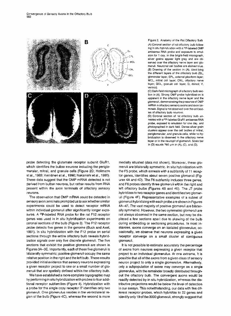
Control experiments were performed using the mRNA encoding the olfactory marker protein (OMP), which is ex- pressed in all olfactory sensory neurons (Farbman and Margolis, 1980) at a level ten times higher than that of receptor mRNA (R. V. and R. A., unpublished data). Previ- ous studies have detected OMP mRNA in the olfactory bulb by RNA blot hybridization (Rogers et al., 1987; Ehrlich et al., 1990). We therefore performed in situ hybridization experiments to ask whether OMP mRNA was detectable within axon terminals in the glomeruli of the olfactory bulb. Sensory axons from the olfactory epithelium enter the OS- factory nerve layer of the bulb and synapse with the den- drites of the mitral and tufted cells to form discrete spheri- cal glomeruli. The axons of the mitral and tufted cells form the major output tract of the bulb that projects to the olfac- tory cortex (Figures 1, 2A, and 25).
Coronal sections through the olfactory bulb were an- nealed with a33P-labeled OMP probe and exposed to emul- sion for 1 day. Strong hybridization signals are apparent throughout the olfactory nerve layer and in the vast major- ity of the glomeruli (Figure 2C). Hybridization is not ob- served in the mitral cell layer, nor in the granule cell layer. This pattern contrasts with in situ hybridization with a
Convergence of Sensory Axons in the Olfactory Bulb 963
probe detecting the glutamate receptor subunit GluRl, which identifies the bulbar neurons including the periglo- merular, mitral, and granule ceils (Figure 2D; Hollmann et al., 1989; Keinanen et al., 1990; Nakanishi et al., 1990). These data suggest that the OMP mRNA detected is not derived from bulbar neurons, but rather results from RNA present within the axon terminals of olfactory sensory neurons
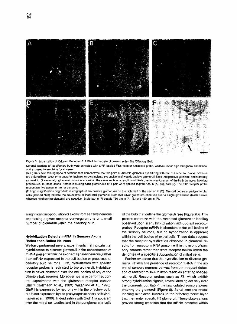
The observation that OMP mRNA could be detected in sensory axon terminals prompted us to ask whether similar experiments could be used to detect receptor mRNA within individual glomeruli after significantly longer expo- sures. A 33P-labeled RNA probe for the rat F12 receptor genes was used in in situ hybridization experiments on coronal sections of the bulb (Figure 3). The F12 receptor probe detects five genes in the genome (Buck and Axel, 1991). In situ hybridization with the F12 probe on serial sections through the entire olfactory bulb reveals hybrid- ization signals over only five discrete glomeruli. The five sections that exhibit the positive glomeruli are shown in Figures 3A-3E. Importantly, each of these five glomeruli is bilaterally symmetric; positive glomeruli occupy the same relative position in the right and the left bulb. These results provided initial evidence that sensory neurons expressing a given receptor project to one or a small number of glo- meruli that are spatially defined within the olfactory bulb.
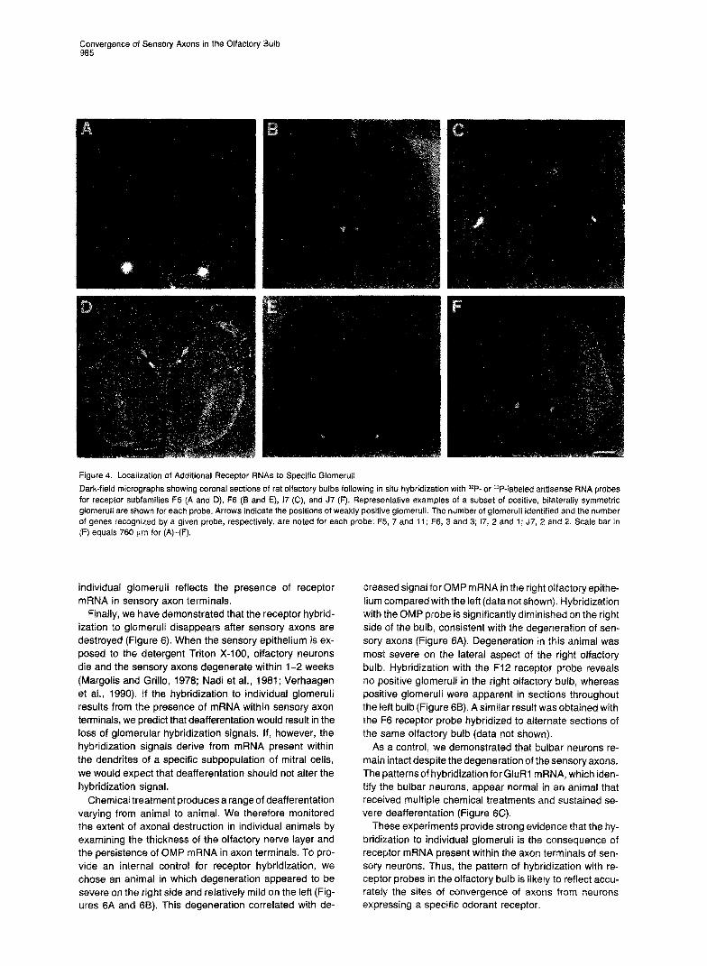
We have established a more complete topographic map by performing in situ hybridization with probes to four addi- tional receptor subfamilies (Figure 4). Hybridization with a probe for the single copy receptor I7 identifies only two giomeruli. One glomerulus resides in the ventrolateral re- gion of the bulb (Figure 4C), whereas the second is more
Figure 2. Anatomy of the Pat Olfactory Bulb
(A) Coronal section of rat olfactory bulb follow- ing in situ hybridization with a 33P-labeled OMP antisense RNA probe and exposure to emul- sion for 1 day. In this bright-field micrograph, silver grains appear light gray and are ob- served over the olfactory nerve layer and glo- meruli. Neuronal cell bodies are stained blue. (6) Drawing of the section in (A), identifying the different layers of the olfactory bulb (GL, glomerular layer; EPL, external plexiform layer; MCL, mitral cell layer; ONL, olfactory nerve layer; GCL, granule cell layer; D, dorsal; V, ventral). (C) Dark-field micrograph of oifactory bulb sec- tion in (A). Strong OMP probe hybridization is apparent in the olfactory nerve !ayer and the glomeruli, demonstrating the presence of OMP mRNA in olfactory sensory axons and axon ter- minals Signal is not observed over the cell bod- ies of olfactory bulb neurons. (D) Coronal section of rat olfactory bulb an- nealed with a33P-labeled GluAl antisense RNA probe, exposed to emuision for one day, and photographed in dark field. Dense silver grain clusters appear over the cell bodies of mitral, periglomerular, and granule celis, while no hy- bridization is observed in the olfactory nerve layer or in the neuropil of glomeruli. Scale bar in (D) equals 760 urn in (A), (C), and (D)
medially situated (data not shown). meruli are bilaterally symmetric. In situ hybridization with the F5 probe, which anneals with a subfamily of 11 recep- tor genes, identifies about seven positive glomeruli (Fig- ures 4A and 4D). The F6 subfamily includes three genes, and F6 probes identify three glomeruli within the right and left olfactory bulbs (Figures 48 and 4E). The J7 probe hybridizes to two receptor genes and identifies two glomer- uli (Figure 4F). Representative examples of a subset of glomeruli hybridizing with each probe are shown in Figures 4A-4F. The vast majority of positive glomeruli are bilater- ally symmetric. However, the two symmetric glomeruli are not always observed in the same section, but may be dis- placed a few sections apart due to skewing of the bulb during embedding or sectioning procedures. In most in- stances, axons converge on an isolated glomerulus; oc- casionally, we observe that neurons expressing a given receptor converge on a small cluster of contiguous glomeruli.
It is not possible to estimate accurately the percentage of axons from neurons expressing a given receptor that project to an individual glomerulus. At one extreme, it is possible that all of the axons from a given class of sensory neuron project to only a single glomerulus. Alternatively, only a subpopulation of axons may converge on a single glomerulus, with the remainder broadly distributed through- out the olfactory bulb. The convergent axons would be readily detected by in situ hybridization, whereas the dis- tributive projections would be below the level of detection in our assays. This notwithstanding, our data with five dif- ferent receptor probes, which hybridize to 22 genes and identify only 19ofthe3000glomeruli, stro~~~y~uggestt~at
Cell 984
Figure 3. Localization of Odorant Receptor F12 RNA lo Discrete Glomeruli within the Olfactory Bulb
Coronal sections of rat olfactory bulb were annealed with a 33P-labeled F12 receptor antisense probe, washed under high stringency conditions, and exposed to emulsion for 4 weeks. (A-E) Dark-field micrographs of sections that demonstrate the five pairs of discrete glomeruli hybridizing with the F1.2 receptor probe. Sections are ordered in an anterior-to-posterior fashion. Arrows indicate the positions of weakly positive glomeruli. Note that pasitive glomeruli are bilaterally symmetric. Occasionally, glomeruli did not occur within the same section, a result most likely due to misalignment of the bulb during embedding procedures. In these cases, frames including each glomerulus of a pair were spliced together as in (A), (D), and (E). The F12 receptor probe recognizes five genes in the rat genome. (F) High magnification bright-field micrograph of the positive glomerulus on the right half of the section in (C). The cell bodies of periglomerular cells (stained blue) indicate the boundaries of individual glomeruli. Note that silver grains are observed over a single glomerulus (black arrow), whereas neighboring glomeruli are negative. Scale bar in (F) equals 760 brn in (A)-(E) and 150 pm in (F).
a significant subpopulation of axons from sensory neurons expressing a given receptor converge on one or a small number of glomeruli within the olfactory bulb.
Hybridization Detects mRNA in Sensory Axons Rather than Bulbar Neurons We have performed several experiments that indicate that hybridization to discrete glomeruli is the consequence of mRNA present within the axons of sensory neurons, rather than mRNA expressed in the cell bodies or processes of olfactory bulb neurons. First, hybridization with specific receptor probes is restricted to the glomeruli. Hybridiza- tion is never observed over the cell bodies of any of the olfactory bulb neurons. Moreover, we have performed con- trol experiments with the glutamate receptor subunit GluRl (Hollmann et al., 1989; Nakanishi et al., 1990). GluFil is expressed by neurons within the olfactory bulb, but is not expressed by the presynaptic sensory cells (Kei- n&en et al., 1990). Hybridization with GluRl is apparent over the mitral cell bodies and in the periglomerular cells
of the bulb that outline the glomeruli (see Figure 2D). This pattern contrasts with the restricted glomerular labeling observed upon in situ hybridization with odorant receptor probes. Receptor mRNA is abundant in the cell bodies of the sensory neurons, but no hybridization is apparent within the cell bodies of mitral cells. These data suggest that the receptor hybridization observed in glomeruli re- sults from receptor mRNA present within the axons of sen- sory neurons rather than from receptor mRNA within the dendrites of a specific subpopulation of mitral cells.
Further evidence that the hybridization to discrete glo- meruli reflects the presence of receptor mRNA in the ax- ons of sensory neurons derives from the frequent detec- tion of receptor mRNA in axon fascicles entering specific glomeruli. Receptor probes such as F5, which exhibit strong hybridization signals, reveal labeling not only over the glomeruli, but also in the fasciculated sensory axons entering the glomeruli (Figure 5). Serial sections reveal labeling over axon bundles in the olfactory nerve layer that then enter specific F5 glomeruli. These observations provide strong evidence that the mRNA detected within
g;gnvergence of Sensory Axons in the Olfactory Bulb
Figure 4. Locaiization of Additional Receptor RNAs to Specific Glomeruli Dark-field micrographs showing coronal sections of rat olfactory bulbs following in situ hybridization with 32P-or =P-labeled antisense RNA probes for receptor subfamilies F5 (A and D), F6 (B and E), 17 (C), and J7 (F). Representative examples of a subset of positive, bilaterally symmetric giomeruli are shown for each probe. Arrows indicate the positions of weakly positive glomeruli. The number of glomeruli identified and the number of genes recognized by a given probe, respectively, are noted for each probe: F5, 7 and 11; F6, 3 and 3; 17, 2 and 1; J7, 2 and 2. Scale bar in (F) equals 760 w.m for (A)-(F).
individual giomeruli reflects the presence of receptor mRNA in sensory axon terminals.
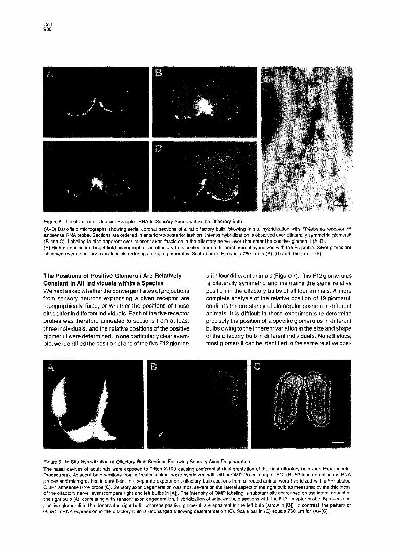
Finally, we have demonstrated that the receptor hybrid- ization to glomeruli disappears after sensory axons are destroyed (Figure 6). When the sensory epithelium is ex- posed to the detergent Triton X-100, olfactory neurons die and the sensory axons degenerate within 1-2 weeks (Margolis and Grillo, 1978; Nadi et al., 1981; Verhaagen et al., 1990). If the hybridization to individual glomeruli results from the presence of mRNA within sensory axon terminals, we predict that deafferentation would result in the loss of glomerular hybridization signals. If, however, the hybridization signals derive from mRNA present within the dendrites of a specific subpopulation of mitral cells, we would expect that deafferentation should not alter the hybridization signal.
Chemical treatment produces a range of deafferentation varying from animal to animal. We therefore monitored the extent of axonal destruction in individual animals by examining the thickness of the olfactory nerve layer and the persistence of OMP mRNA in axon terminals. To pro- vide an internal control for receptor hybridization, we chose an animal in which degeneration appeared to be severe on the right side and relatively mild on the left (Fig- ures SA and (33). This degeneration correlated with de-
creased signal for OMP mRNA in the right olfactory epithe- lium compared with the left (data not shown). Hybridization “with the OMP probe is significantly diminished on the right side of the bulb, consistent with the degeneration of sen- sory axons (Figure 6A). Degeneration in this animal was most severe on the lateral aspect of the right olfactory bulb. Hybridization with the F12 receptor probe reveals no positive glomeruli in the right olfactory bulb, whereas positive glomeruli were apparent in sections throughout the left bulb (Figure 6B). A similar result was obtained with the F6 receptor probe hybridized to alternate sections of the same olfactory bulb (data not shown).
As a control, we demonstrated that bulbar neurons re- main intact despite the degeneration of the sensory axons. The patternsof hybridization forGluR1 mRNA, which iden- tify the bulbar neurons, appear normal in an animal that received multipie chemical treatments and sustained se- vere deafferentation (Figure 6C).
These experiments provide strong evidence that the hy- bridization to individual glomeruli is the consequence of receptor mRNA present within the axon terminals of sen- sory neurons. Thus, the pattern of hybridization with re- ceptor probes in the olfactory bulb is likely to reflect accu- rately the sites of convergence of axons from neurons expressing a specific odorant receptor.
Cell 986
Figure 5. Localization of Cdorant Receptor RNA to Sensory Axons within the Olfactory Bulb
(A-D) Dark-field micrographs showing serial coronal sections of a rat olfactory bulb following in situ hybridization with 32P-iabeled receptor F5 antisense RNA probe. Sections are ordered in anterior-to-posterior fashion. Intense hybridization is observed over bilaterally symmetric glomeruli (6 and C). Labeling is also apparent over sensory axon fascicles in the olfactory nerve layer that enter the positive glomeruli (A-Q). (E) High magnification bright-field micrograph of an olfactory bulb section from a different animal hybridized with the F5 probe. Silver grains are observed over a sensory axon fascicle entering a single glomerulus. Scale bar in (E) equals 760 pm in (A)-(D) and 150 pm in (E).
The Positions of Positive Glomeruli Are Constant in All Individuals within a Species We next asked whether the convergent sites of projections from sensory neurons expressing a given receptor are topographically fixed, or whether the positions of these sites differ in different individuals. Each of the five receptor probes was therefore annealed to sections from at least three individuals, and the relative positions of the positive glomeruli were determined. In one particularly clear exam- ple, we identified the position of one of the five F-i 2 glomer-
uli in four different animals (Figure 7). This Ft.2 glomerulus is bilaterally symmetric and maintains the same relative position in the olfactory bulbs of all four animals. A more complete analysis of the relative position of 19 glomeruli confirms the constancy of glomerular position in different animals. It is difficult in these experiments to determine precisely the position of a specific glomerulus in different bulbs owing to the inherent variation in the size and shape of the olfactory bulb in different individuals. Nonetheless, most glomeruli can be identified in the same relative posi-
Figure 6. In Situ Hybridization of Olfactory Bulb Sections Following Sensory Axon Degeneration The nasal cavities of adult rats were exposed to Triton X-100 causing preferential deafferentation of the right otfactory bulb (see Experimental Procedures). Adjacent bulb sections from a treated animal were hybridized with either OMP (A) or receptor F12 (8) 32P-labeled antisense RNA probes and micrographed in dark field. In a separate experiment, olfactory bulb sections from a treated animal were hybridized with a33P-labeled GluRl antisense RNA probe (C). Sensory axon degeneration was most severe on the lateral aspect of the right bulb as measured by the thickness of the olfactory nerve layer (compare right and left bulbs in [A)). The intensity of OMP labeling is substantially decreased on the lateral aspect of the right bulb (A), correlating with sensory axon degeneration. Hybridization of adjacent bulb sections with the F12 receptor probe (8) reveals no positive glomeruli in the denervated right bulb, whereas positive glomeruli are apparent in the left bulb (arrow in IS]). In contrast, the pattern of GluRl mRNA expression in the olfactory bulb is unchanged following deafferentation (C). Scale bar in (C) equals 760 nm for (A)-(C).

Convergence of Sensory Axons in the Olfactory Butb 967
Figure 7. The Position of individual Glomeruli Is Constant in Different Animals
Dark-field micrographs of olfactory bulb sec- tions from four individuals following in situ hy- bridization with the F12 receptor probe. The four panels show the position of one of the five F12-positive glomeruli in four different individu- als. Note that the F12 glomerulus is bilaterally symmetric and maintains the same relative po- sition in all four olfactory bulbs. Scale bar in (D) equals 760 &rn in (A)-(D).
tion in all individuals analyzed. These observations dem- onstrate that neurons expressing a given receptor project to topographically distinct glomeruli. Moreover, the posi- tions of the specific glomeruli are not random; rather, they remain relatively constant in all individuals in a species.
oiscussion
opographic Map Encoding Odor Quality ?he initial event in the perception of odors involves the interaction of odorous ligands with receptors on sensory neurons. Discrimination among odors therefore requires a mechanism by which the brain can discern which of the numerous receptors have been activated. Individual olfactory sensory neurons are likely to express only one
a small number of receptor genes (Ngai et al., 1993a; ssler et al., 1993). The problem of distinguishing which
receptors have been activated therefore reduces to a prob- lem of distinguishing which neurons have been activated. A topographic map defining the positions of specific neu- rons in the olfactory epithelium, or the positions of their axons in the olfactory bulb, may be employed to determine which of the neurons have been activated. In this manner, the olfactory system may use spatial segregation of sen- sory input to encode the quality of an olfactory stimulus.
Previous studies have examined the spatial distribution of neurons expressing specific odorant receptors in the sensory epithelium (Nef et al., 1992; Strotmann et al., 1992; Ngai et al., 1993a; Raming et al., 1993; Ressler et al., 1993; Vassar et al., 1993). In mammals, neurons expressing a specific receptor are distributed within one of four broad but circumscribed zones within the epithelium. Within a given zone, however, neurons expressing a spe- cific receptor are not spatially segregated, but rather are
randomly dispersed within the epithelium (Ressler et al., 1993; Vassar et al., 1993). We have therefore asked whether neurons expressing a given receptor project their axons to common glomeruli within the olfactory bulb. The observation that individual neurons express about 1000 receptor mRNA molecules suggested an experimental ap- proach to map axonal projections based upon the pres- ence of mRNA at the axon terminal. A single glomerulus, about 200 pm in diameter, represents the site of conver- gence of about 3000 sensory axons. If the neurons that project to a single glomerulus each express the same re- ceptor mRNA and a small number of mRNA molecules are present at the axon terminal, then this convergence of axons might permit the identification of individual glo- meruli by in situ hybridization with specific receptor probes. In this study, we demonstrate that the neurons expressing a given receptor project to one, or at most, a few glomerular targets among the thousands of glomeruli within the olfactory bulb. Our data with five different recep- tor probes, which hybridize to 22 genes and identify only 19 of the 3000 glomeruli, strongly suggest that a significant subpopulation of axons from sensory neurons expressing a given receptor converge on one or a small number of glomeruli within the olfactory bulb. Moreover, these pro- jections define specific glomeruli that maintain a fixed po- sition in the brains of all animals within a species.
Preliminary analysis indicates that the axons of neurons from one topographic zone in the epithelium tend to con- verge on glomeruli that are localized to a discrete region of the olfactory bulb. For example, neurons expressing ?he F6, J7, and F5 receptor families are expressed in the same epithelial zone and project to glomeruii that segre- gate within a ventromedial strip of the bulb (Figure 8, right). These observations are consistent with i-etro

Cell 988
Figure 8. Summary of Specific Glomerular Positions within the Olfac- tory Bulb
Schematic representations of the lateral (left) and medial (right) sur- faces of the right olfactory bulb. The relative positions of 19 glomeruli identified with five different odorant receptor probes are summarized Glomeruli labeled with specific probes are color coded as follows: F5, green; F12, red; F6, blue; 17, black; J7, yellow. The majority of F5, F6, and J7 glomeruli segregate within an anterior-posterior strip on the ventromedial surface of the olfactory bulb (right; shaded gray). (A, anterior; P, posterior; D, dorsal; V, ventral.)
experiments demonstrating that neurons within defined regions of the epithelium project to broad but circum- scribed regions of the olfactory bulb (Astic and Saucier, 1986; Saucier and Astic, 1986; Astic et al., 1987; Schoen- feld et al., 1994). Thus, the zonal organization of receptor expression in the epithelium may be preserved in the pro- jection of sensory axons to the bulb. Compartmentaliza- tion of the epithelium and the bulb into anatomically and functionally discrete units of lesser complexity may reduce the problems inherent in regulating the expression of spe- cific receptors and may facilitate the segregation of axons within the olfactory bulb.
Our data are consistent with previous studies sug- gesting that glomeruli represent functional units, such that specific odorants elicit spatially defined patterns of glo- merular activity in the olfactory bulb. For example, after exposure to a single odorant, enhanced 2-deoxyglucose uptake is observed over a restricted group of glomeruli. Moreover, this pattern of P-deoxyglucose labeling of spe- cific glomeruli is distinct for different odors (Stewart et al., 1979; Jourdan et al., 1980; Lancet et al., 1982; Royet et al., 1987). Optical recordings of the olfactory bulb using voltage-sensitive dyes similarly reveal diffuse yet circum- scribed patterns of activity in response to single odors (Kauer et al., 1987). These patterns are distinct but over- lapping for different odors.
Electrophysiologicstudies provide independent support for a model of olfactory discrimination in which individual glomeruli are differentially responsive to different odors. Extracellular recording of spike responses from single mi- tral cells of a glomerulus indicate that individual glomeruli are “tuned” to discrete molecular structures (Mori et al., 1992; lmamuraetal., 1992; Katoh et al., 1993). The individ- ual mitral cells of a given glomerulus might all be maximally responsive to o&o-xylene, whereas the mitral cells of a topographically distinct glomerulus respond most strongly to para-xylene (Katoh et al., 1993).
These experimental approaches demonstrate the acti- vation of specific glomeruli with specific odors, but cannot relate the activation of specific sensory neurons with spe-
cific patterns of glomerular activation. Our data provide direct anatomic evidence that neurons expressing a given receptor, and therefore responsive to a given odorant, project with precision to one or a small number of glomeruli within the olfactory bulb. Since the positions of the individ- ual glomeruli are topographically defined, the olfactory bulb provides a two-dimensional map that identifies which of the numerous receptors have been activated within the sensory epithelium. One important implication of such a model is that exposure to a given odorant would result in the stimulation of a spatially restricted set of glomeruli, such that individual odorants would be associated with specifictopographicpatternsof activitywithin theolfactory bulb. The quality of an olfactory stimulus would therefore be encoded by the specific combination of glomeruli acti- vated by a given odorant.
implications for Sensory Processing How does the olfactory cortex decode this two-dimensional map within the bulb to allow the discrimination among a diverse array of odors? A given neuron is likely to express only one or a small number of receptors from the repertoire of 1000 genes (Ngai et al., 1993% Ressler et al., 1993). Since the organism can detect far greater than 1000 dis- crete odors, this implies that a given odorant will interact with multiple receptors, and a single receptor will interact with multiple odorous ligands. In this manner, an odorant, even one that consists of a single molecular species, will interact with a unique combination of receptors, which in turn results in the activation of specific combinations of glomeruli. The maintenance of a fixed map of glomeruli in the bulb therefore provides a mechanism to relay the information resulting from the activation of receptors in the periphery to the brain.
How is such a combinatorial code of glomerular activa- tion interpreted by the cortex’? In one model, the topo- graphic map in the olfactory bulb could project directly to olfactory cortex, providing a more central representation of the glomerular map. Cortical loci activated by a given odorant might then converge on higher centers to facilitate odor recognition. Alternatively, it is possible that each of the multiple glomeruli activated by a given odor would project fibers that converge on a single locus in the olfac- tory cortex. Each locus in the olfactory cortex would reflect the convergence of output fibers from a unique combina- tion of glomeruli, such that the activation of a cortical locus would encode a given odor quality. In this model, each odor would result in the activation of a unique combination of glomeruli, and each combination would be encoded in a specific locus in the cortex. This model is consistent with anterogradetracingexperiments indicating that mitral cells from a single glomerulus project to multiple loci in the olfactory cortex (Schwab and Price, 1978; Ojima et al., 1984; Buonviso et al., 1991). Moreover, retrograde tracing studies demonstrate that a cortical locus receives input from multiple glomeruli (Haberly and Price, 1977; Scott et al., 1980; Luskin and Price, 1982).
In either model, the ability of an odorant to activate a combination of glomeruli allows for the discrimination of adiversearrayofodorsfarexceeding the numberofrecep-

&wergence of Sensory Axons in the Olfactory Bulb
tom and their associated glomeruli. Despite the diversity afforded by combinatorial activation of glomeruli, the or- ganism is likely to be capable of discriminating only an extremely small subset of potential odors. As in other sen- sory systems, the olfactory system only perceives a mea- ger image of the sensory information in the environment. This restricted array of recognizable odors will be a func- tion of the repertoire of receptors expressed by the organ- ism as well as the nature of the glomerular combinations that are encoded in the cortex. Those odors that are per- ceived from the universe of potential odorants presumably reflect selection for that subset of odorants that are biologi- cally important for only the species. In this manner, individ- uals within a species will maintain a repertoire of receptor genes and a combination of cortical connections that per- mit the detection of those odors important for its survival and reproduction.
This model of olfactory perception shares several basic features with perception in other sensory systems. The brain analyzes avisual image by interpreting the individual components of the image: form, location, movement, color (Livingstone and Hubel, 1987; Zeki and Shipp, 1988). The unity of an image is accomplished by several parallel pro- cessing pathways, such thatthe image is initially dissected into tractable components and then reconstructed in higher visual centers of the cortex. A similar logic may be employed to discriminate olfactory stimuli. Olfactory processing initially requires that the odorant molecule is analyzed by “dissecting” its structural features, such that each of its structural elements will activate a unique subset of receptors, which in turn results in the activation of a unique subset of glomeruli. The odorous stimulus is then reconstructed by the olfactory cortex, which determines which of the numerous glomeruli have been activated.
The Development of a Topographic Map in the Olfactory Bulb The establishment of a topographic map of sensory projec- tions might initially involve the expression of receptors by sensory ceils. The choice of a given receptor would then be linked to the expression of guidance molecules, such that neurons expressing a given receptor project axons to a single glomerulus. Alternatively, it is possible that im- mature neurons, not yet expressing receptor, randomly project axons to any one of multiple glomeruli within the olfactory bulb. Contact with an individual glomerulus may then elicit a retrograde signal that directs the expression of specific receptor genes. In this manner, receptor gene expression would be regulated by the ultimate position of its axon terminal within the olfactory bulb. This model is unlikely, since we observe receptor expression in sensory neurons early in embryogenesis (rat embryonic day 14) prior to the birth of mitral cells and the presence of glomer- uli (C. Dulac and R. A., unpublished data). Moreover, anal- ysis of receptor gene expression in individual neurons sug- gests that the choice of receptor expressed by a neuron is not regulated, but rather is stochastic (Chess et al., 1994). Thus, it is likely that neurons stochastically express one receptor gene prior to synapse formation. Neurons ex-
pressing a given receptor will then form synapses with one or a small number of topographically fixed glomeruli.
How is this precise topographic map established? One attractive model combines guidance mechanisms during development with activity-dependent refinement to achieve the precision of connections between the sensory epithe- lium and olfactory bulb. It is possible, for example, that axonsof neuronsexpressing asubpopulation of the recep- tor repertoire are restricted in their projections to broad but defined regions of the olfactory bulb. Neuronal growth cones may, for example, be guided to their appropriate target by a gradient of recognition molecules. This guid- ance process could establish a coarse topographic map. Activity-dependent mechanisms, as a consequence of the synchronous firing of neurons responsive to a given odor- ant, could refine the pattern of synaptic connections within this restricted region of the bulb to establish a relatively precise topographic map. Such a model draws heavily on observations in the visual system indicating that retinal axons first use activity-independent mechanisms to form a coarse retinotopic map. Activity-dependent processes progressively refine this coarse map to provide a level of precision required to perceive the visual image (reviewed by ConstantinePaton et al., 1990; Goodman and Shatz, 4 993). Alternatively, mechanisms involving multiple guid- ance cues that provide each glomerulus with a unique identity, coupled with multiple receptors on the different sensory neurons, may provide the precision of bulbar con- nections.
The nature of the putative guidance cues in the target, as well as the recognition molecules on the sensory axons, remains elusive. One particularly attractive possibility is that the odorant receptors themselves may play a role in
atterning the projections of olfactory sensory axons. The presence of receptor mRNA in sensory axons may result in the functional expression of receptors on axonal projec- tions. These presynaptic receptors may be activated by guidance cues expressed by the postsynaptic mitral cells. in this manner, the odorant receptors may function in the dendrite in the recognition of odors and may exhibit a dis- tinct function in the axon in the establishment of a topo- graphic map that encodes odor quality In the olfactory bulb.
Experimental Procedures
In Situ Hybridization In situ hybridization was performed essentially as described (Schaeren-Wiemers and Gerfin-Moser, 1993), except that =P- or =P- iabeled RNA probes were used. Probes were synthesized in a 10 pl reaction with either 500 uCi of [32P]CTP or 330 t&i of [33P]LlTP (3000 Cilmmole and 1000 CVmmole, respectively, Amersham Life Sciences) using T3 or T7 RNA polymerases (Melton et al., 1984). Clones encod- ing full-length cDNAs (constructed in pSluescript II SK(+); Stratagene) were used as templates to synthesize probes (ranging from 1 .O-2.0 kb in size) from the following odorant receptor sequences: F5, F6, P12, and 17. The sequence encompassing TM4 through TM7 (approxi- mately 0.7 kb) of receptor J7 was isolated by the polymerase chain reaction (L. Suck and R. A., unpublished data). Sequences corre- sponding to the BamHI-Asp-718 fragment of OMP cDNA (Rogers et al., 1987) were used to synthesize a 1 kb 3’ OMP probe. Finally, a full-length GluFil cDNA clone (Hollmann et al., 1989; Nakanishi et al., 1990) was used to synthesize a 1.4 kb probe.
Cell 990
Fresh-frozen olfactory bulbs of adult female Sprague-Dawley rats (1 l-13 weeks old) were oriented for coronal sections and cut to 15- 30 urn thickness. Serial sections of entire bulbs were allowed to dry and then were fixed for 10 min in 4% paraformaldehyde in phosphate- buffered saline (PBS) at room temperature. After several rinses in PBS, sections were incubated in 0.25% acetic anhydride, 0.1 M trietha- nolamine (pH 8) washed again in PBS, and prehybridized for 2-4 hr at room temperature (Schaeren-Wiemers and Gerfin-Moser, 1993). Probes were diluted to a concentration of 1 x IO’-2 x lOa cpmlml in hybridization buffer (Wilkinson et al., 1987a, 1987b), and 100 ui was applied to each slide. Following a 14-20 hr incubation at 72OC, sections were washed in 0.2x SSC for I hr at 72OC, treated with 2 Kg/ml RNase A for 30 min at 37OC, and washed again in 0.2 x SSC at 72’C. After dehydration, slides were dipped in undiluted NTB-3 emulsion (Kodak) and allowed to expose at 4OC. Slides hybridized with OMP and GluRl probes were developed after 1 day, whereas slides hybridized with odorant receptor probes were exposed to the emulsion for 4 weeks. After development, sections were stained with toluidine blue 0, dehydrated, and mounted in Accumount 280 (Baxter). Sections were photographed using a Nikon Labophot-2 microscope with 2 x and 10 x objectives. Dark-field illumination was with a vertical Darklite attachment (MicroVideo Instruments).
Peripheral Deafferentation Chemical deafferentation of rat olfactory bulb was adapted from proce- dures described for the mouse (Margolis and Grillo, 1978; Nadi et al., 1981; Verhaagen et al., 1990; F. L. Margolis, personal communica- tion). FemaleSprague-Dawleyrats(ll-12weekold) werelightlyanes- thetized with an intraperitoneal injection of ketamine (40 mglkg). Ap proximately 0.5 ml of a 0.7% Triton X-100, 0.9% sodium chloride solution was injected into the right nostril of each animal using a blunted 23-gauge needle. Animals were sacrificed 2 weeks after this procedure, and the bulbs prepared for sectioning as described above.
In other experiments, we applied 1 ml of 1% Triton X-100, 0.9% sodium chloride, allowed the animals to recover for 2 days, and re- peated the procedure for a total of four or five treatments. Animals were sacrificed after a recovery period of 2 weeks following the last treatment.
In pilotexperiments, weaddeda bluedyetotheTriton X-lOOsolution and examined the extent of staining in the left and right nasal cavities 10 min after treatment. We found extensive staining of olfactory epithe- lium on both right and left sides in some animals and preferential accumulation on one side in others. This variability was apparent in the extent of sensory axon degeneration seen in the experimental animals. Some animals had uniformly denervated olfactory bulbs, whereas others had more extreme axonal degeneration on one side only. This appeared to be independent of the number of treatments, although deafferentation was usually more complete in animals receiv- ing multiple treatments.
Acknowledgments
We thank Tom Jessell, Eric Kandel, John Ngai, Andrew Tomlinson, and the members of the Axel Lab for helpful discussions and critical reading of the manuscript. We also thank Phyllis J. Kisloff for assis- tance in preparing the manuscript and David Rosenzweig for illustra- tions This work was funded by the Howard Hughes Medical Institute. R. V. is also a fellow of the Helen Hay Whitney Foundation and S. K. C. is supported by a grant from the National institutes of Health (T32 GM07367).
Received September 23, 1994; revised October 14, 1994.
References
Astic, L., and Saucier, D. (1986). Anatomical mapping of the neuroepi- thelial projection to the olfactory bulb in the rat. Brain Res. Bull. 76, 445-454. Astic, L., Saucier, D., and Holley, A. (1987). Topographical relation- ships between olfactory receptor cells and glomerular foci in the rat olfactory bulb. Brain Res. 424, 144-152.
Baier, H., and Korsching, S. (1994).Olfactorygfomeruli in therebrafish form an invariant pattern and are identifiable across animals. J. Neu- rosci. 74, 219-230.
Ben-Arie, N., Lancet, D., Taylor, C., Khen, M., Walker, N., Ledbetter, D. H., Carrozzo, R., Patel, K., Sheer, D., Lehrach, H., and North, M. A. (1994). Olfactory receptor gene cluster on human chromosome 17: possible duplication of an ancestral receptor repertoire. Hum. Mol. Gen. 3, 229-235.
Berkley, K. J. (1985). Multiple systems diverging from the dorsal col- umn nuclei in the cat. In Development, Organization, and Processing in Somatosensory Pathways, M. Rowe and W. D. Willis, eds. (New York: A. R. Liss), pp. 191-202.
Srunet, J.-F., Shapiro, E., Foster, S. A., Kandel, E. R., and line, Y. (1991). Identification of a peptide specific for Aplysia sensory neurons by PCR-based differential screening. Science 252, 856-859. Suck, L., and Axel, R. (1991). A novel multigene family may encode odorant receptors: a molecular basis for odor recognition. Cell 65, 175-I 87.
Buonviso, N., Revial, M. F., and Jourdan, F. (1991). The projections of mitral cells from small local regions of the olfactory bulb: an antero- grade tracing study using PHA-L (Phaseolus vulgaris Leucoaggluti- nin). Eur. J. Neurosci. 3, 493-500.
Chess, A., Simon, I., Cedar, H., and Axel, R. (1994). Allelic inactivation regulates olfactory receptor gene expression. Cell 78, 823-834.
Constantine-Paton, M., Cline, H. T., and Debski, E. (1990). Patterned activity, synaptic convergence, and the NMDA receptor in developing visual pathways. Annu. Rev. Neurosci. 73, 129-154.
Dykes, R. W., Rasmusson, D. D., Sretavan, D., and Rehman, N. B. (1982). Submodality segregation and receptive-field sequences in cu- neate, gracile, and external cuneate nuclei of the cat. J. Neurophysiol. 47, 389-416. Ehrlich, M. E., Grillo, M., Joh, T. H., Margolis, F. L., and Baker, H. (1990). Transneuronal regulation of neuronal specific gene expression in the mouse olfactory bulb. Mol. Brain Res. 7, 115-122.
Farbman, A. L., and Margolis, F. L. (1980). Olfactory marker protein ontogeny: immunohistochemical localization. Dev. Biol. 74, 205-215.
Goodman, C. S., and Shatz, C. J. (1993). Developmental mechanisms that generate precise patterns of neuronal connectivity. Cell 72iNeu- ron 70 (Suppl.), 77-98.
Haberly, L. B., and Price, J. L. (1977). The axonal projection patterns of the mitral and tufted cells of the olfactory bulb in the rat. Brain Res. 729, 152-I 57.
Hollmann, M., O’Shea-Greenfield, A., Rogers, S. W., and Heinemann, S. (1989). Cloning by functional expression of a member of the gluta- mate receptor family. Nature 342, 643-648. Imamura, K., Mataga, N., and Mori, K. (1992). Coding of odor mole- cules by mitral/tufted cells in rabbit olfactory bulb. I. Aliphatic com- pounds J. Neurophysiol. 68, 1986-2002.
Jirikowski, G. F., Sanna, P. P., and Bloom, F. E. (1990). mRNA coding for oxytocin is present in axons of the hypothalamo-neurohypophysial tract. Proc. Natl. Acad. Sci. USA 87, 7400-7404.
Jourdan, F., Duveau, A., Astic, L., and Halley, A. (1980). Spatial distri- bution of [“CjP-deoxyglucose uptake in the olfactory bulbs of rats stim- ulated with two different odours. Brain Res. 188, 139-154. Kandel, E. R., Schwartz, J. H., and Jessell, T. M. (1991). Principles of Neural Science, Third Edition (New York: Elsevier), p. 516. Katoh, K., Koshimoto, H., Tani, A., and Mori, K. (1993). Coding of odor molecules by mitral/tufted cells in rabbit olfactory bulb. II. Aromatic compounds. J. Neurophysiol. 70, 2161-2175.
Kauer, J. S., Senseman, D. M., and Cohen, L. B. (1987). Odor-elicited activity monitored simultaneously from 124 regions of the salamander olfactory bulb using a voltage-sensitive dye. Brain Res. 478, 255-261.
Keinlnen, K., Wisden, W., Sommer, B., Werner, P., Herb, A., Ver- doorn, T. A., Sakmann, B., and Seeburg, P. H. (1990). A family of AMPA-selective glutamate receptors. Science 249, 556-560.
Lancet, D., Greer, C. A., Kauer, J. S., and Shepherd, G. M. (1982). Mapping of odor-related neuronal activity in the olfactory bulb by high-
;;;vergence of Sensory Axons in the Olfactory Bulb
resolution P-deoxyglucose autoradiography. Proc. Natl. Acad. Sci. USA 79, 670-674.
Landry, C.. Crine, P., and DesGroseillers, L. (1992). Differential ex- pression of neuropeptide gene mRNA within the LUQ cells of Aplysia californica. J. Neurobiol. 23, 89-101.
Levy, N. S., Bakalyar, H. A., and Reed, R. R. (1991). Signal transduc- tion in oifactory neurons. J. Steroid Biochem. Mol. Biol. 39, 633-637.
Livingstone, M. S., and Hubel, D. H. (1987). Psychophysical evidence for separate channels for the perception of form, color, movement, and depth. J. Neurosci. 7, 3416-3468.
Luskin, M. B., and Price, J. 1.. (1982). The distribution of axon collater- als from the olfactory bulb and the nucleus of the horizontal limb of the diagonal band to the olfactory cortex, demonstrated by double retrograde labeling techniques. J. Comp. Neural. 209, 249-263.
Margolis, F. L., and Grille, M. (1978). An approach to chemical nerve section in the mouse olfactory pathway. Sot. Neurosci. Abstr. 4, 88.
Meisami, E. (1979). The developing rat olfactory bulb: prospects for a new model system in developmental neurobiology. In Neural Growth and Differentiation, E. Meisami and M. A. B. Brazier, eds. (New York: Raven Press). Meisami, E. (1989). A proposed relationship between increases in the number of olfactory receptor neurons, convergence ratio and sensitiv- ity in the developing rat. Dev. Brain Res. 46, Q-IQ.
Melton, D. A., Krieg, P. A., Rebagliati, M. R., Maniatis, T., Zinn, K., and Green, M. R. (1984). Efficient in vitro synthesis of biologically active RNA and DNA hybridization probes from plasmids containing a bacteriophage SP6 promoter. Nucl. Acids Res. 73, 7035-7056. Mohr, E., Fehr, S., and Richter, D. (1991). Axonal transport of neuro- peptide encoding mRNAs within the hypothalamo-hypophyseal tract of rats. EMBO J. 10, 2419-2424.
Mori, K., Mataga, N., and Imamura, K. (1992). Differential specificities of single mitral cells in rabbit olfactory bulb for a homologous series of fatty acid odor molecules. J. Neurophysiol. 67, 786-789.
Mountcastle, V. 8. (1957). Modality and topographic properties of sin- gle neurons of cat’s samatic sensory cortex. J. Neurophysiol. 20,408- 434.
Nadi, N. S., Head, R., Grille, M., Hempstead, J., Grannot-Reisfeld, N., and Margolis, F. L. (1981). Chemical deafferentation of the olfactory buib: plasticity of the levels of tyrosine hydroxylase, dopamine and norepinephrine. Brain Res. 273, 365-377.
Nakanishi, N., Shneider, N. A., and Axel, R. (1990). A family of gluta- mate receptor genes: evidence for the formation of heteromultimeric receptors wi?h distinct channel properties. Neuron 5, 569-581.
Nef, P., Hermans-Borgmeyer, I., Artieres-Pin, H., Beasley, L., Dionne, V. E., and Heinenann, S. F. (1992). Spatial pattern of receptor expres- sion in the olfactory epithelium. Proc. Natl. Acad. Sci. USA 89, 8948- 8952.
Ngai, J., Chess, A., Dowling, M. M., Necles, N., Macagno, E. R., and Axe& R. (1993a). Coding of olfactory information: topography of odor- ant receptor expression in the catfish olfactory epithelium. Cell 72, 667-680.
Ngai, J., Dowling, M. M., Buck, L., Axel, R., and Chess, A. (1993b). The family of genes encoding odorant receptors in the channel catfish. Cell 72, 657-666.
Ojima, H., Mori, K., and Kishi, K. (1984). The trajectory of mitral cell axons in the rabbit olfactory cortex revealed by intracellular HRP injec- tion. J. Comp. Neural. 230, 77-87.
Parmentier, M., Libert, F., Schurmans, S., Schiffmann, S., Lefort, A,, Eggerickx, D., Ledent, C., Mollereau, C., Gerard, C., Perret, J., Groote- goed, A., and Vassart, G. (1992). Expression of members of the puta- tive olfactory receptor gene family in mammalian germ cells. Nature 355,453-455.
Peri, E. Ft., Whitlock, D. G., and Gentry, J. R. (1962). Cutaneous projec- tion to second-order neurons of the dorsal column system, J. Neuro- physiol. 25, 337-358.
Raming, K,, Krieger, J., Strotmann, J., Boekhoff, I., Kubich, S., Baum- stark, C., and Breer, H. (1993). Cloning and expression of odorant receptors. Nature 367, 353-356.
Ressler, K., Sullivan, S.! and Buck, L. (1994). 4 molecular dissection of spatial patterning in the olfactory system. Curr. Opin. Neurobiol. 4, 588-596.
Ressler, K. J., Sullivan, S. L., and Buck, b. B. (1993). A zonai organiza- tion of odorant receptor gene expression in the olfactory epithelium. Cell 73, 597-609.
Rogers, R. E., Dasgupta, P., Gubler, U., Grille, M., Khew-Goodall, Y., and Margolis, F. L. (1987). Molecular cloning and sequencing of a cDNA for olfactory marker protein. Proc. Natl. Acad. Sci. USA 84, 1704-1708.
Royet, J. P., Sicard, G., Souchier, C., and Jourdan, F. (1987). Specific- ity of spatial patterns of glomerular activation in the mouse olfactory bulb: computer-assisted image analysis of 2-deoxyglucose autoradio- grams. Brain Res. 417, l-l 1.
Royet, J. P., Souchier, C., Jourdan, F., and Ploye, H. (1988). Morpho- metric study of the glomerular population in the mouse olfactory bulb: numerical density and size distribution along the rostrocaudal axis. J. Comp. Neurol. 270, 559-568.
Saucier, D., and Astic, L. (1986). Analysis of the topographical organi- zation of olfactory epithelium projections in the ret. Brain Res. Bull. 76,455-462.
Schaeren-Wiemers, N., and Gerfin-Moser, A. (1993). A single protocol to detect transcripts of various types and expression levels in neural tissue and cultured cells: in situ hybridization using digoxigenin- labeled cRNA probes. Histochemistry 700, 431-440.
Schoenfeld, T. A., Clancy, A. N., Forbes, W. B., and Macrides, F. (1994). The spatial organization of the peripheral olfactory system of the hamster. I. Receptor neuron projections to the main olfactory bulb. Brain Res. Bull. 34, 183-210.
Schwab, J. E., and Price, J. L. (1978). The cortical projection of the olfactory bulb: development in fetal and neonatal rats correlated with quantitative variations in adult rats. Brain Res. 157, 369-374.
Scott, J. W., McBride, R. L., and Schneider, S. P. (1980). Theorganiza- tion of projections from the olfactory bulb to the piriform cortex and olfactory tubercle in the rat. J. Comp. Neural. 794, 519-534.
Shepherd, G. M. (1991). Computational structure of the olfactory sys- tem. In Olfaction: A Model System for Computational Neuroscience, J. L. Davis, and H. Eichenbaum, eds. (Cambridge, MA: MIT Press).
Steward, O., and Banker, G. A. (1992). Getting the message from the gene to the synapse: sorting and intracelll;lar transport of RNA in neurons. Trends Neurosci. 75, 180-186.
Stewart, W. 5., Kauer, J. S., and Shepherd, G. M. (1979). Functional organization of rat olfactory bulb analyzed by the 2-deoxyglucose method. J. Comp. Neural. 785, 715-734.
Strotmann, J., Wanner, I., Krieger, J., Raming, K.: and Breer, H. (1992). Expression of odorant receptors in spatially restricted subsets of che- mosensory neurones. Neuroreport 3, 1053-1056.
Vassar, R., Ngai, J., and Axel, R. (1993). Spatial segregation of odorant receptor expression in the mammalian olfactory epithelium. Cell 74, 309-318.
Verhaagen, J., OestreichBr, A. B., Grille,. M., Khew-Goodall, Y.-S., Gispen, W. H., and Margolis, F. L. (1990). Neuroplasticity in the olfac- tory system: differential effects of central and peripheral lesions of the primary olfactory pathway on the expression of &5O/GAP43 and the olfactory marker protein. J. Neurosci. Res. 26, 31-34.
Wilkinson, D. G., Bailes, J. A., Champion, J. E., and McMahon, A. P. (1987a). A molecular analysis of mouse development from 8 to 10 days post-coitum detects changes only in eukaryotic globin expres- sion. Development 99, 493-500.
Wilkinson, D. G., Bailes, J. A., and McMahon, A. P. (‘i987b). Expres- sion of the proto-oncogene int-1 is restricted to specific neural cells in the developing mouse embryo. Cell 50, 79-88.
Zeki, S., and Shipp, S. (1988). The functional logic of cortical connec- tions. Nature 335, 311-317.


![991 fork[1]](https://img.pdfslide.us/doc/110x75/55d391fcbb61eb265d8b4658/991-fork1.jpg)