Embed Size (px)
Citation preview
Cell, Vol. 66, 649-659, September 6, 1991, Copyright 0 1991 by Cell Press
The Tissue-Specific Extinguisher Locus TSEI Encodes a Regulatory Subunit of CAMP-Dependent Protein Kinase Michael Boshart, Falk Weih, Mark Nichols, and Gunther Schiitz Institute of Cell and Tumor Biology German Cancer Research Center Im Neuenheimer Feld 280 D-6900 Heidelberg Federal Republic of Germany
Summary
The tissue-specific extinguisher locus TSEl, a domi- nant negative regulator of transcription in somatic cell hybrids, acts via a CAMP response element (CRE) to repress activity of a hepatocyte-specific enhancer. Guided by the antagonism between TSEl and CAMP- mediated signal transduction, we identified the regula- tory subunit Rla of protein kinase A (PKA) as the prod- uct of the TSEl locus. The evidence derives from concordant expression of Rla mRNA and TSEl genetic activity, high resolution mapping of the Rla gene and TSEl on human chromosome 17, and the ability of a transfected Rla cDNA to generate a phenocopy of TSEl-mediated extinction. The mechanism of TSEll Rla-mediated extinction involves repression of basal PKA activity, reduced phosphorylation of CREB at Ser-133, and a corresponding reduction of in vivo pro- tein binding at the target CRE.
Introduction
Fusion of distinctly differentiated somatic ceils usually re- sults in the loss of tissue-specific gene products of either parent, a phenomenon termed extinction. The concept that diffusible factors control cell type-specific gene ex- pression in trans was originally derived from such cell fu- sion experiments (for reviews see Davis and Adelberg, 1973; Davidson, 1974; Gourdeau and Fournier, 1990). These systems provide a genetic approach for identifying trans-dominant regulatory factors. To date, a genetic anal- ysis of extinction has been performed primarily in the hepa- toma x fibroblast hybrid cell system. Killary and Fournier (1984) identified a locus on mouse chromosome 11 and its human homolog, chromosome 17, the tissue-specific extinguisher 1 (TSEl), which coordinately represses hepa- tocyte-specific expression of several genes, including tyro- sine aminotransferase (TAT) and phosphoenolpyruvate carboxykinase (PEPCK) (Lem et al., 1988; Thayer and Fournier, 1989; Ruppert et al., 1990). All of the genes re- pressed by TSEl are normally inducible via the CAMP signal transduction pathway (Ruppert et al., 1990). Other extinguisher loci regulate expression of distinct sets of genes in hepatic cells, as all liver-specific traits are extin- guished in karyotypically complete hepatoma x fibroblast hybrids (Chin and Fournier, 1987). For example, Tse-2 on mouse chromosome 1 represses expression of the serum
albumin and alcohol dehydrogenase genes (Petit et al., 1986; Chin and Fournier, 1989).
Hepatoma microcell hybrids and deletion hybrids (Leach et al., 1989) that retain only human chromosome 17 or fragments thereof made it possible to analyze TSEl- specific extinction effects, for example, to identify the tar- get sequence for repression by TSEl in the TAT gene. A cyclic AMP response element (CRE), an essential compo- nent of a hepatocyte-specific enhancer located 3.6 kb up- stream of the TAT transcription start site, mediates repres- sion byTSE1 (Boshartet al., 1990). InductionviathecAMP pathway reverses TSEl -mediated repression of CRE ac- tivity and gene expression (Thayer and Fournier, 1989; Boshart et al., 1990; Ruppert et al., 1990). This functional antagonism between TSEl and the CAMP pathway is re- flected by corresponding changes in protein binding in vivo at the CRE of the TAT gene. Increased protein binding at the TATCRE upon CAMP induction is also observed in vitro using crude hepatoma extracts (Weih et al., 1990). We have suggested that TSEl inhibits posttranslational modification, e. g., phosphorylation of a CRE-binding pro- tein (CREB) by interfering in the CAMP signal transduction pathway (Boshart et al., 1990). Guided by this working hypothesis, we searched for the product of theTSE1 locus. As CAMP levels are not affected by TSEl (Thayer and Fournier, 1989; M. B., unpublished data), we could ex- clude a mechanism lowering CAMP levels and therefore acting upstream of protein kinase A (PKA) in the pathway. Consequently, we considered PKA inhibiting activities and protein phosphatases as promising candidates. The holo- enzyme of PKA is a tetramer consisting of two regulatory and two catalytic subunits and is inactive in the absence of CAMP. Activation occurs when two CAMP molecules bind to each regulatory subunit, eliciting a reversible con- formational change that releases active catalytic subunits (Taylor et al., 1990). Thus, regulatory subunits are condi- tional inhibitors of C. Four distinct regulatory subunit genes have been identified, and they are differentially ex- pressed (for review see McKnight et al., 1988a, 1988b). Here we provide evidence that the regulatory subunit Rla of PKA is the product of the TSEl locus.
Results
Basal PKA Activity Is Reduced in TSEl’ Hybrids Owing to the functional antagonism between TSEl- mediated extinction and CAMP signal transduction (Bos- hart et al., 1990) we tried to identify targets of TSEl action in the CAMP pathway. We measured PKA activity in ex- tracts from 7AD7 (TSEl-) and 7AE27 (TSEl+) hepatoma hybrid cells. These microcell hybrids retain part of human chromosome 17 with or without the chromosomal segment carrying human TSEl on a rat hepatoma background (Leach et al., 1989). Basal PKA activity without addition of CAMP was 7-fold lower in 7AE27 (TSEl+) cells (included in Table 1). Addition of 5 PM CAMP abolished this difference,
Cell 850
ECORI Hindlll
probe: human Rla
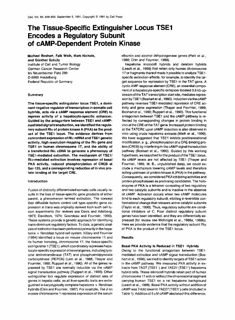
Figure 1. The Rla Gene Maps to Human Chromosome 17
Genomic DNAs were digested with EcoRl or Hindlll as indicated and 8 ng was analyzed by Southern blot hybridization with a 40-mer oligo- nucleotide probe complementary to nucleotides 23-62 of the human Rla coding region (Sandberg et al., 1987). This is within the least conserved part of the regulatory subunit sequence, so that the Rlf3 gene or RII genes were not detected. The human probe weakly cross- hybridized to a rat Rla restriction fragment, which provided an internal reference. The result was confirmed with Pstl- and Sstl-digested sam- ples (not shown).
indicating that expression of the catalytic subunit was simi- lar in both cell lines. This was confirmed by measuring catalytic subunit mRNA levels (data not shown). As one explanation, we considered the possibility that increased regulatory subunit expression led to reduced basal PKA activity in 7AE27 (TSEI +) cells. This might occur if TSEl either up-regulated a regulatory subunit encoded by the rat hepatoma genome or if TSEl itself encoded a human regulatory subunit. Therefore, we performed Southern blot hybridizations to test whether any of the four regulatory subunit genes mapped to human chromosome 17. With a mouse Rla probe (Clegg et al., 1987) we detected a human-specif ic restriction fragment in genomic DNA from 7AE27 (TSEl+) but not 7AD7 (TSEl-) ceils (data not shown). This result was confirmed with an oligonucleotide probe specific for human Rla (Figure 1). Thus, the human Rla gene and TSEl both map to a segment of chromo- some 17 retained in 7AE27 (TSEl+) but not in 7AD7 (TSEl-) cells. A previous report had assigned a sequence recognized by an Rla probe to human chromosome 7 (Scambler et al., 1987). It is now clear that this sequence is the closely related RI8 gene expressed specifically in neuronal t issues (Clegg et al., 1988 and G. S. McKnight, personal communication). RI18 has also been mapped to human chromosome 7 (Scambler et al., 1987).
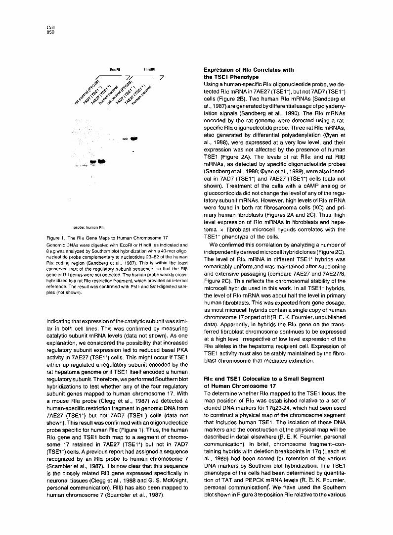
Expression of Rla Correlates with the TSEl Phenotype Using a human-specifio Rla oligonucleotide probe, we de- tected Rla mRNA in 7AE27 (TSEl+), but not 7AD7 (TSEl-) cells (Figure 2B). Two human Rla mRNAs (Sandberg et al., 1987) are generated by differential usage of polyadeny- lation signals (Sandberg et al., 1990). The Rla mRNAs encoded by the rat genome were detected using a rat- specific Rla oligonucleotide probe. Three rat Rla mRNAs, also generated by differential polyadenylation (Oyen et al., 1988) were expressed at a very low level, and their expression was not affected by the presence of human TSEl (Figure 2A). The levels of rat Rlla and rat RI18 mRNAs, as detected by specific oligonucleotide probes (Sandberg et al., 1988; Oyen et al., 1989) were also identi- cal in 7AD7 (TSEl-) and 7AE27 (TSEl+) cells (data not shown). Treatment of the cells with a CAMP analog or glucocorticoids did not change the level of any of the regu- latory subunit mRNAs. However, high levels of Rla mRNA were found in both rat f ibrosarcoma cells (XC) and pri- mary human fibroblasts (Figures 2A and 2C). Thus, high level expression of Rla mRNAs in fibroblasts and hepa- toma x fibroblast microcell hybrids correlates with the TSEl’ phenotype of the cells.
We confirmed this correlation by analyzing a number of independently derived microcell hybrid clones (Figure 2C). The level of Rla mRNA in different TSEl’ hybrids was remarkably uniform.and was maintained after subcloning and extensive passaging (compare 7AE27 and 7AE27/8, Figure 2C). This reflects the chromosomal stability of the microcell hybrids used in this work. In all TSEl+ hybrids, the level of Rla mRNA was about half the level in primary human fibroblasts. This was expected from gene dosage, as most microcell hybrids contain a single copy of human chromosome 17or part of it (R. E. K. Fournier, unpublished data). Apparently, in hybrids the Rla gene on the trans- ferred fibroblast chromosome continues to be expressed at a high level irrespective of low level expression of the Rla alleles in the hepatoma recipient cell. Expression of TSEl activity must also be stably maintained by the fibro- blast chromosome that mediates extinction.
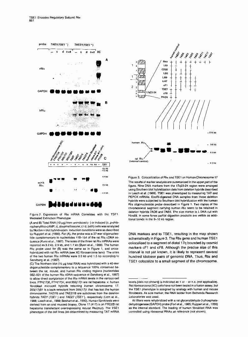
Rla and TSEl Colocalize to a Small Segment of Human Chromosome 17 To determine whether Rla mapped to the TSEl locus, the map position of Rla was established relative to a set of cloned DNA markers for 17q23-24, which had been used to construct a physical map of the chromosome segment that includes human TSEl. The isolation of these DNA markers and the construction okthe physical map will be described in detail elsewhere (B. E. K. Fournier, personal communication). In brief, chromosome fragment-con- taining hybrids with deletion breakpoints in 17q (Leach et al., 1989) had been scored for retention of the various DNA markers by Southern blot hybridization. The TSEl phenotype of the cells had been determined by quantita- tion of TAT and PEPCK mRNA levels (R. E,: K. Fournier, personal communicationf. WgRaGe used the Southern blot shown in Figure 3 @position Rla relative to the various
TSEl Encodes Regulatory Subunit Rla 851
probe 7AD7(TSEI -) 7AE27( TSEl +)
--II- - c - c d c+d XC
rRla
GAPDH @ @ m m
hRla
GAPDH wa4ouu
Figure 2. Expression of Rla mRNA Correlates with the TSEI- Mediated Extinction Phenotype
(A and B) Total RNA (10 ug) from uninduced (-) or induced (c, p-chlo- rophenylthio-CAMP; d, dexamethasone; c+d, both) cells was analyzed by Northern blot hybridization. Induction conditions were as described by Ruppert et al. (1990). For (A), the probe was a 37-mer oligonucleo- tide complementary to nucleotides 118-154 of the rat Rla cDNA se- quence (Kuno et al., 1987). The sizes of the three rat Rla mRNAs were reported as 3.2 kb, 2.9 kb, and 1.7 kb (@yen et al., 1988). The human Rla probe used for (B) was the same as in Figure 1. and cross- hybridized with rat Rla mRNA (see XC fibrosarcoma lane). The sizes of the two human Rla mRNAs were 3.0 kb and 1.5 kb according to Sandberg et al. (1987). (C)The Northern blot (15 pg total RNA) was hybridized with a 40-mer oligonucleotide complementary to a sequence 100% conserved be- tween the rat, mouse, and human Rla coding regions (nucleotides 562-601 of the human Rlu cDNA sequence of Sandberg et al., 1987) to allow direct comparison of the Rla mRNA levels in the various cell lines. FTH(I7)K, FTH(17)V, and 355(17)1 are rat hepatoma x human fibroblast microcell hybrids retaining human chromosome 17. 355(17)Bl is a back-selectant from 355(17)1 that has lost the human chromosome. 7AD7/5 and 7AE27/8 are subclones from the deletion hybrids 7AD7 (TSE7-) and 7AE27 (TSEI’), respectively (Lem et al., 1988; Leach et al., 1989; Boshartet al., 1990). Human fibroblasts were derived from an oral mucosa biopsy. Clone 17 (#17) is an FTOLB rat hepatoma transfectant overexpressing mouse Rla(mut). The TSEl phenotype of the cell lines (as determined by measuring TAT mRNA
N.20
L69
CD35
LD5
Ll90 Ll47
cF1
TSEI
CFB
PKCA
human RI
rat Rla (internal control)
- 4.3 kb
Figure 3. Colocalization of Rla and TSEi on Human Chromosome 17
The results of marker analysis are summarized in the upper part of the figure. Nine DNA markers from the 17q23-24 region were arranged using Southern blot hybridization data from deletion hybrids described in Leach et al. (1989). TSEl was phenotyped by measuring TAT and PEPCK mRNAs. EcoRCdigested DNA samples from these deletion hybrids were subjected to Southern blot hybridization with the human Rla oligonucleotide probe described in Figure 1. Two copies of the chromosomal segment carrying human Rla seem to be retained in deletion hybrids 7AD6 and 7AE6. The size marker is h DNA cut with Hindlll. In some lanes partial digestion products are visible as addi- tional bands in the 9-10 kb region.
DNA markers and to TSEl, resulting in the map shown schematically in Figure 3. The Flla gene and human TSEl colocalized to a segment of distal 17q bounded by cosmid markers cF1 and cF8. Although the precise size of this interval is not yet known, it is likely to represent several hundred kilobase pairs of genomic DNA. Thus, Rla and TSEl colocalize to a small segment of the chromosome.
levels [data not shown]) is indicated as + or - or n.a. (not applicable). Rat fibrosarcoma (XC) cells have not been tested in a fusion assay, but the TSEl phenotype is assigned by analogy with human and mouse fibroblasts. As size marker, the RNA ladder from Bethesda Research Laboratories was used.
All filters were rehybridized with a rat glyceraldehyde-3-phosphate- dehydrogenase (GAPDH) probe (Fort et al., 1985; Ruppert et al., 1990) as the internal standard. The loading of human fibroblast RNA was controlled using ribosomal RNAs as reference (not shown).
Cell 852
zinc
forskolin
CCI
ma mu,
TAT
PEPCK
GAPDH
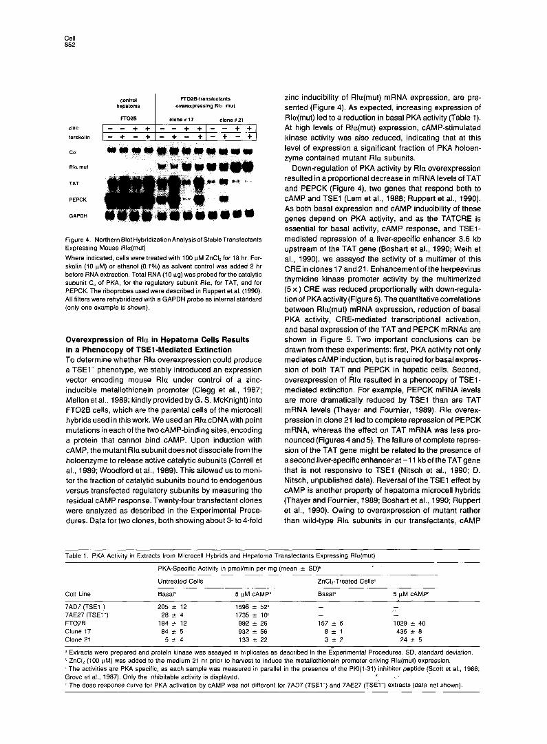
Figure 4. Northern Blot Hybridization Analysisof StableTransfectants Expressing Mouse Rla(mut)
Where indicated, cells were treated with 100 uM ZnCb for 18 hr. For- skolin (10 uM) or ethanol (0.1%) as solvent control was added 2 hr before RNA extraction. Total RNA (IO ug) was probed for the catalytic subunit C. of PKA, for the regulatory subunit Rlu, for TAT, and for PEPCK. The riboprobes used were described in Ruppert et al. (1990). All filters were rehybridized with a GAPDH probe as internal standard (only one example is shown).
Overexpression of Rla in Hepatoma Cells Results in a Phenocopy of TSEl-Mediated Extinction To determine whether Rla overexpression could produce a TSEl+ phenotype, we stably introduced an expression vector encoding mouse Rla under control of a zinc- inducible metallothionein promoter (Clegg et al., 1987; Mellon et al., 1989; kindly provided by G. S. McKnight) into FTOPB cells, which are the parental cells of the microcell hybrids used in this work. We used an Rla cDNA with point mutations in each of the two CAMP-binding sites, encoding a protein that cannot bind CAMP. Upon induction with CAMP, the mutant Rla subunit does not dissociate from the holoenzyme to release active catalytic subunits (Correll et al., 1989; Woodford et al., 1989). This allowed us to moni- tor the fraction of catalytic subunits bound to endogenous versus transfected regulatory subunits by measuring the residual CAMP response. Twenty-four transfectant clones were analyzed as described in the Experimental Proce- dures. Data for two clones, both showing about 3- to 4-fold
zinc inducibility of Rla(mut) mRNA expression, are pre- sented (Figure 4). As expected, increasing expression of Rla(mut) led to a reduction in basal PKA activity (Table 1). At high levels of Rla(mut) expression, cAMPstimulated kinase activity was also reduced, indicating that at this level of expression a significant fraction of PKA holoen- zyme contained mutant Rla subunits.
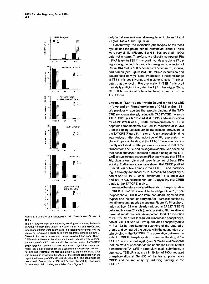
Down-regulation of PKA activity by Rla overexpression resulted in a proportional decrease in mRNA levels of TAT and PEPCK (Figure 4) two genes that respond both to CAMP and TSEl (Lem et al., 1988; Ruppert et al., 1990). As both basal expression and CAMP inducibility of these genes depend on PKA activity, and as the TATCRE is essential for basal activity, CAMP response, and TSEl- mediated repression of a liver-specific enhancer 3.6 kb upstream of the TAT gene (Boshart et al., 1990; Weih et al., 1990) we assayed the activity of a multimer of this CRE in clones 17 and 21. Enhancement of the herpesvirus thymidine kinase promoter activity by the multimerized (5 x) CRE was reduced proportionally with down-regula- tion of PKA activity(Figure 5). The quantitative correlations between Rla(mut) mRNA expression, reduction of basal PKA activity, CRE-mediated transcriptional activation, and basal expression of the TAT and PEPCK mRNAs are shown in Figure 5. Two important conclusions can be drawn from these experiments: first, PKA activity not only mediates CAMP induction, but is required for basal expres- sion of both TAT and PEPCK in hepatic cells. Second, overexpression of Rla resulted in a phenocopy of TSEl- mediated extinction. For example, PEPCK mRNA levels are more dramatically reduced by TSEl than are TAT mRNA levels (Thayer and Fournier, 1989). Rla overex- pression in clone 21 led to complete repression of PEPCK mRNA, whereas the effect on TAT mRNA was less pro- nounced (Figures 4 and 5). The failure of complete repres- sion of the TAT gene might be related to the presence of a second liver-specific enhancer at -11 kb of the TAT gene that is not responsive to TSEl (Nitsch et al., 1990; D. Nitsch, unpublished data). Reversal of the TSEl effect by CAMP is another property of hepatoma microcell hybrids (Thayerand Fournier, 1989; Boshartet al., 1990; Ruppert et al., 1990). Owing to overexpression of mutant rather than wild-type Rla subunits in our transfectants, CAMP
Table 1. PKA Activity in Extracts from Microcell Hybrids and Hepatoma Transfectants Expressing Rlu(mut)
PKA-Specific Activity in pmollmin per mg (mean + SD)
Cel! Line
7AD7 (TSEl ) 7AE27 (TSEl’) FTOZB Clone 17 Clone 21
Untreated Ceils
Basal’
205 + 12 28 f 4
184 e 12 84 f 5
5-t4
5 uM CAMP’
1598 f 52d 1735 * 10d
992 + 26 932 f 56 133 -c 22
ZnCI,-Treated Cells?
Basal’ 5 uM cAMPC
- - - - 157 f 6 1029 f 40
8+1 435 2 0 3&2 24 + 5
-
a Extracts were prepared and protein kinase was assayed in triplicates as described in the Experimental Procedures. SD, standard deviation. b ZnCI, (100 uM) was added to the medium 21 hr prior to harvest to induce the metallothionein promoter driving Rla(mut) expression. c The activities are PKA specific, as each sample was measured in parallel in the presence of the PKl(l-31) inhibitor peptide (Scott et al., 1986; Grove et al., 1987). Only the inhibitable activity is displayed. d The dose response curve for PKA activation by CAMP was not different for 7AD7 (TSEl-) and 7AE27 (TSEl’) extracts (data not shown).
TSEl Encodes Regulatory Subunit Rla 653
!I 1 mRNA RI a(mul)
FTOPS “17 “21
CRE-mediated transcription acIivation
mRNA PEPCK
mRNA TAT
Figure 5. Summary of Phenotypes in Ala Transfectant Clones 17 and 21
The mRNA levelswere quantitated by excising and counting the bands from the Northern blots shown in Figure 4. For TAT and PEPCK. two independent filters were quantitated (indicated by error bars), and the values for untreated FTOPB cells were arbitrarily defined as 100 U. PKA activities (mean f standard deviation) were taken from Table 1. CRE-mediated transcriptional stimulation was determined by transient transfection of a CAT construct with five tandem copies of a TATCRE oligonucleotide upstream of the herpesvirus thymidine kinase pro- moter (5 x Bl), as described in the Experimental Procedures. For each cell line and treatment, the fold stimulation by the multimerized CRE was calculated by setting the value for the control construct with the thymidine kinase promoter alone (pBLCAT5) to 1. The constructs are described in Boshart et al. (1990) and Ruppert et al. (1990). The values for relative protein binding were taken from Figure 6.
only partially reverses negative regulation in clones 17 and 21 (see Table 1 and Figure 4).
Quantitatively, the extinction phenotypes of microcell hybrids and the phenotype of transfectant clone 17 cells were very similar (Figures 4 and 5; Boshart et al., 1990; data not shown). Therefore, we directly compared Rla mRNA levels in TSEl’ microcell hybrids and clone 17 us- ing an oligonucleotide probe homologous to a region of Rla mRNA that is 100% conserved between rat, mouse, and human (see Figure 2C). Rla mRNA expression and basal kinase activity(Table 1) were both in the same range in TSEl+ microcell hybrids and in clone 17 cells. This indi- cates that the level of Rla expression in TSEl+ microcell hybrids is sufficient to confer the TSEl phenotype. Thus, Rla fulfills functional criteria for being a product of the TSEl locus.
Effects of TSEl/Rla on Protein Bound to the TATCRE In Vivo and on Phosphorylation of CREB at Ser-133 We previously reported that protein binding at the TAT- CRE in vivo was strongly reduced in 7AE27(TSEl +)vefsus 7AD7(TSEl-)cells(Boshartet al., 1990)and was inducible by CAMP (Weih et al., 1990). Overexpression of Rla in hepatoma transfectants also led to reduction of in vivo protein binding (as assayed by methylation protection) at the TATCRE (Figure 6). In clone 17, in vivo protein binding was reduced after zinc induction of Rla expression. In clone 21, protein binding at the TATCRE was almost com- pletely abolished and the pattern was similar to that of the f ibrosarcoma cells used as negative control. We conclude that basal and CAMP-induced protein binding at the TAT- CRE in vivo are dependent on PKA activity and that TSEl/ Rla plays a key role in cell-specific control of basal PKA activity. Furthermore, we have shown that CREB purified from rat liver or brain binds to the TATCRE, and that bind- ing is strongly enhanced by PKA-mediated phosphoryla- tion at Ser-133 (M. N. et al., submitted). Thus, the in vivo and in vitro results are concordant, suggesting that CREB binds to the TATCRE in vivo.
We have therefore analyzed the state of phosphorylation of CREB at Ser-133 in vivo. After labeling cells with [“‘PIor- thophosphate, CREB was immunopurified, digested with trypsin, and the peptide carrying Ser-133 was identified by two-dimensional peptide mapping (Figure 7). Phosphory- lation at Ser-133 was clearly reduced in 7AE27 (TSEl+) cells and in clone 21 cells (overexpressing Rla) relative to parental hepatoma cells. As expected, forskolin induction of 7AE27 (TSEl+) cells resulted in increased phosphoryla- tion of CREB at Ser-133. We quantitated phosphorylation at Ser-133 by densitometric scanning of the autoradio- grams and compared the values with the quantitated pro- tein binding at the TATCRE. The correlation between the extent of CREB phosphorylation in vivo and binding at the TATCRE in vivo is striking (Figure 7). We have also shown that the state of phosphorylation of purified CREB affects binding to the TATCRE in vitro (M. N. et al., submitted). In summary, TSEllRla acts by inhibition of PKA-mediated phosphorylation at Ser-133 of the transcription factor CREB and consequently by reducing binding to the TATCRE.

Cell 854
control FTO2B-transfectants hepatoma overexpressing Rln mut FTOPB
I clone X21
Figure 6. The In Vivo Footprint at the TATCRE Disappears upon Down-Regulation of PKA Activity
FTO26, clone 17, and clone 21 cells were cultured for 18 hr in serum- free medium with or without 25 PM ZnCI,, then induced with 20 FM forskolin for 30 min or treated with 0.1% ethanol as solvent control. Cells were reacted with DMS and prepared for genomic footprinting as described in Becker and Schlitz (1988). Thirty micrograms of Styl cut genomic DNA per lane was electrophoresed on a 6% denaturing polyacrylamide gel. After electroblotting and covalent cross-linking to a nylon membrane, hybridization was performed with a single-stranded cDNA probe (Weih et al., 1990) complementary to the sequence from -3516to3643oftheupperstrand.Guanosine(G)residuesfrom-3619 to -3675 of the upper strand are visible on the autoradiogram. The pattern of purified genomic DNA reacted in vitro with DMS was indistin- guishable from the XC rat fibrosarcoma (fibro) control lanes (not shown). Altered DMS reactivity of guanosines is marked with closed squares for enhancements and with open squares for protections. Numbers indicate the positions in base pairs relative to the start site of transcription. The part of the sequence homologous to the CRE consensus is boxed. Relative protein binding was quantitated bydensi- tometric scanning and calculated as the ratio between the intensities of the enhanced G at -3649 and the protected G at -3647. The ratio for XC fibrosarcoma cells (fibro, negative control) was 0.74 (average of six XC lanes). The ratio for uninduced FTOPB cells was 8.1 and defined as one relative protein binding unit.
Discussion
Here we demonstrate that the tissue-specific extinguisher locus TSEl encodes the regulatory subunit Rla of CAMP- dependent protein kinase. In an independent effort to clone the TSEl locus, Fournier and collaborators have isolated several cDNAs of genes expressed in 7AE27 (TSEl+) cells but not in 7AD7 (TSEl-) cells. One of these clones maps concordantly with TSEl on a map of human chromosome 17 and turned out to encode Rla (Ft. E. K. Fournier, pdrsonal communication). Both approaches led to identification of Rla as the product of the TSEl locus and reveal for the first time the molecular nature of an
7AE27- 7AE27+ forskolin
FTO 27- L
27+ #21
relative phosphotylation at Ser’”
relative protein binding
Figure 7. TSEllRla Affects In Vivo Phosphorylation of CREB at Ser-133
Cells were labeled in vivo with (32P]orthophosphate as described in the Experimental Proceduhs and induced with 10 W M forskolin for 10 min where indicated. lmmunopurification of CREB, trypsin digestion, and two-dimensional phosphopeptide mapping were performed as out- lined in the Experimental Procedures. The identity of the spot repre- senting the tryptic peptide RPSYR containing Ser-133 (arrowhead) was verified by mixing experiments with purified CREB protein and a synthetic peptide (amino acids 124-136; Gonzales and Montminy, 1989) both labeled with the purified catalytic subunit of PKA and then trypsinized(M. N. etal., submitted).CREBseemsto bephosphorylated in vivo at additional Ser positions by other protein kinases (Lee et al., 1990). The tryptic peptides containing these phosphoamino acids are insoluble under our conditions and could not be analyzed with this method. Traces of this material are visible at the site of sample applica- tion in the lower left corner. The spots representing phosphorylation at Ser-133 were quantitated for each sample by densitometric scan- ning, and relative values were calculated by arbitrarily setting CREB phosphorylation in FTOPB cells to 1. For comparison, the diagram includesquantitative results from in vivofootprintingexperiments(rela- tive protein binding values taken from Figure 6 and from Boshart et al. 119901 for cell line 7AE27 [TSEl+]).
extinguisher locus. This’exemplifies the usefulness of so- matic cell hybrids for identification of trans-dominant regu- latory factors. In mammalian cells, regulatory mutants can- not be readily obtained. Therdore, somatic cell hybrids are valuable tools for the anaPysi8 of tissue-specific gene expression, allowing the identification of dominant regula- tory factors irrespective of their mode of action.
The Mecha&sm of TSEl-Mediated Extinction The extinction phenotype in microcell hybrids results from differential expression of /3la/T?F$in hepaioma cells ver- sus fibroblasts, and from.stabje epigenetic maintenance of the high fibroblast&ecifc level of Rla expression in
TSEl Encodes Regulatory Subunit Rla 055
hybrids. High expression of Rla does not necessarily lead to an increase of the steady-state level of Rla protein, as rapid degradation of free RI regulatory subunits seems to adjust the level of regulatory to that of catalytic subunits of PKA (Steinberg and Agard, 1981; Uhler and McKnight, 1987; Otten and McKnight, 1989). However, an excess of regulatory over catalytic subunit synthesis lowers the amount of free, catalytically active catalytic subunits of PKA. Therefore, basal PKA activity is low in cells with high expression of RlaITSEl (see Table 1). In these cells, phos- phorylation of Ser-133 of the transcription factor CREB is reduced (Figure 7). CREB purified from rat brain (Yama- moto et al., 1988) and from rat liver (M. N. et al., submitted) is a substrate for PKA, and phosphorylation at Ser-133 strongly increases the binding affinity for certain CRE se- quences, including the TATCRE (M. N. et al., submitted). In addition, phosphorylation at Ser-133 is required for tran- scriptional activation by CREB (Gonzalez and Montminy, 1989). Down-regulation of basal PKA activity by high ex- pression of Rla/TSEl reduces protein binding at the TAT- CRE in vivo and lowers basal transcriptional activation mediated by the TATCRE, which is the target for repres- sion by TSEl. The perfect correspondence between in vitro and in vivo results (Boshart et al., 1990; Weih et al., 1990; this work; M. N. et al., submitted) indicates that CREB does bind the TATCRE in vivo. The TATCRE is essential for the function of a liver-specific enhancer at -3.6 kb of the TAT gene (Boshart et al., 1990), and TAT gene expression thus depends on basal PKA activity. TSEl -mediated extinction results from reduced PKA activ- ity and consequently reduced CREB phosphorylation. This mechanism explains why TSEl-mediated repression is reversible by CAMP (Thayer and Fournier, 1989; Boshart et al., 1990; Ruppert et al., 1990). The observation that all TSEl-responsive genes identified so far are inducible by CAMP (Ruppert et al., 1990) suggests that TSEl may repress transcription of these genes by the same mecha- nism, i.e., inhibition of CREB phosphorylation. We antici- pate that other transcription factors that require phosphor- ylation by PKA for their activity may also respond to Rlal TSEl.
The mechanism of extinction hasalso been investigated in hybrids between fibroblasts and pituitary cells, lymphoid cells, or hepatoma cells expressing growth hormone, im- munoglobulin genes, or albumin, respectively. In those hybrids the transcription rate or mRNA level of a tissue- specific transcription factor required for expression of these genes (GHF-1, OCT2, or HNFl, respectively) was down-regulated (McCormick et al., 1988; Junker et al., 1988; Bergman et al., 1990; Junker et al., 1990; Cereghini et al., 1990). Extinction can also be mediated by activation of silencer-like target sequences (Triputti et al., 1988; Zaller et al., 1988; Yu et al., 1989). Extinguisher loci have not yet been genetically defined in these systems, nor is it known how many intermediate regulatory steps are involved.
In the case of Rla/TSEl, the cascade of events leading to extinction is short: inhibition of a kinase (PKA) precludes phosphorylation of a transcription factor (CREB). It has been shown that inhibition of PKA can affect basal tran-
scriptional activity of several CAMP-regulated promoters (Day et al., 1989; Grove et al., 1989; Mellon et al., 1989). Here we show that the activity of a CRE can be either constitutive or conditional depending on the level of basal PKA activity. This suggests that the same factor (CREB) is required both for basal CRE-mediated activation and CAMP inducibility.
The Physiological Role of RlalTSEl The mechanism by which Rla/TSEl controls gene expres- sion in cell hybrids is likely to be part of the mechanism normally determining cell type-specific gene expression. Modulation of basal PKA activity by differential expression of Rla/TSEl may contribute to tissue-specific control of expression of genes like TAT and PEPCK, which are de- pendent on PKA. Indeed, Rla mRNA levels in adult mouse liver seem lower than in most other organs (M. 6. and S. Ruppert, unpublished data). Tissue-specific control of gene expression usually results from the combined action of several factors whose distribution is often not strictly cell type specific (Dynan, 1989). For example, in the liver- specific enhancer at -3.6 kb of the TAT gene, the mutual dependence of a CRE and a second essential and cell- specific element forms the basis of specificity for highly differentiated hepatoma cells (Boshart et al., 1990).
Another possible function of Rla/TSEl is suggested by a number of observations regarding developmental activa- tion of the TAT gene. TAT expression is not detectable before birth, but increases rapidly within the first hours postnatally (Greengard, 1970). This is accompanied by an increase in the newborn of hormones acting via the CAMP pathway, owing to postnatal hypoglycemia. These hor- mones seem to play a role in triggering the developmen- tally programmed onset of expression, as premature acti- vation of the TAT gene can be elicited by administration of CAMP in utero (Greengard, 1970; Ruiz-Bravo and Ernest, 1982). TSEl activity, as determined by the fusion assay (Peterson and Fournier, 1989) and Rla expression (data not shown) are high in mouse hepatoma ceils expressing fetal liver markers but low in adult hepatocytes (Gourdeau et al., 1989). Thus, RlaITSEl may play an important role in repressing the TAT gene in fetal liver before birth. The burst of hormones acting via CAMP could be sufficient to induce the gene shortly after birth. Mechanistically, this is analogous to extinction and reversal of extinction by CAMP in cell hybrids. A decrease of RlalTSEl expression around birth might then establish a basal level of PKA activity sufficient for adult TAT gene expression. Increased cata- lytic subunit expression, decreased expression of other regulatory subunits, or elevated levels of CAMP in adult liver (Wittmaack et al., 1983) might also contribute to es- tablishment of higher basal PKA activity after birth. We are currently investigating the developmental profile of regula- tory and catalytic subunit expression in the liver by in situ hybridization. The same mechanism might also control developmental activation of the PEPCK gene shortly after birth. McGrane et al. (1990) have shown with transgenic mice that a -460/+73 PEPCK promoter fragment is suffi- cient to confer a 200-fold increase in transgene mRNA after birth. PEPCK promoter fragments as small as -2001
Cdl 856
+73 seem to be responsive to TSEl in microcell hybrids (Thayer et al., 1990; Ft. E. K. Fournier, unpublished data). Clearly, the role for TSEl response and developmental activation of the CRE at -90 in the PEPCK promoter has to be investigated, as PEPCK expression is dependent on PKA activity (see Figure 3).
The genetic evidence for a specific role of Rla/TSEl in cell differentiation highlights the versatility of PKA in regulating cellular processes. The evolution and conserva- tion of four regulatory subunits (Rla, RIP, Rlla, RI@), each encoded by a separate gene, may have been necessary to accomplish such diverse functions as regulation of inter- mediary metabolism (Krebs and Beavo, 1979) and neu- ronal functions including learning (Drain et al., 1991) and gene transcription in the nucleus. Distinct tissue-specific expression patterns of each of the four isoforms have been found (McKnight et al., 1988a, 1988b; Cadd and McKnight, 1989). Available evidence suggests that cells can prefer- entially assemble Ftll holoenzyme and may only form Rla holoenzyme when catalytic levels exceed RII subunit lev- els (Otten and McKnight, 1989). The Rla subunit is more susceptible to degradation when not complexed with the catalytic subunit (Steinberg and Agard, 1981). Thus, Rla may have a compensatory role, adjusting the total amount of regulatory subunits in the cell to that of catalytic sub- units. Our results suggest that tissue-specific regulation of the level of Rla expression, by determining the stringency of this adjustment, can control the basal activity of PKA and thereby affect gene expression.
How Is RlalTSEl Expression Regulated? The transcriptional activity of the Rla/lSEl gene on hu- man fibroblast chromosome 17 is preserved when this chromosome is transferred via microcell fusion into the nucleus of a hepatoma cell that expresses Rla/TSEl at a very low level. Thus, two mitotically stable, epigenetic states of gene expression coexist and are inherited in cis. This unusual regulatory behavior is required for any extin- guisher locus to be detectable in the microcell hybrid assay system. Such a locus must be marked in some way that facilitates assembly of the locus in the appropriate tran- scriptional state following replication. This imprinting could be a covalent modification of the DNA, or a protein complex that is semiconservatively segregated to each strand during replication and serves as a template for its own reassembly(further possibilities are discussed by Pil- Ius and Rine, 1989). As Rla has a very GC-rich promoter region characteristic of “housekeeping” genes (Nowak et al., 1987; McKnight et al., 1988b), we considered methyla- tion of the weakly expressed Rla gene in hepatoma cells a possibility of marking the locus. However, when hepatoma cells were treated with 5azacytidine to demethylate CpG dinucleotides, we could not observe any increase in Rla expression (data not shown). Thus, the nature of the epige- netic change that characterizes Rla/TSEl gene activity remains to be defined.
Experimental,Procedures
Cell Culture and Transfections The rat hepatoma line FTOPB was described by Killary and Fournier
(1984) and the 7A series of deletion hybrids (including 7AD7 and 7AE27) by Leach et al. (1989). The microcell hybrids FTH(17)K, FTH(17)V, and 355(17)1 were constructed as outlined by Leach et al. (1989). 355(17)Bl was backselected from 355(17)1 as described (Killaty and Fournier, 1984). Hepatoma cells were cultured in I:1 (v/v) Dulbecco’s modified Eagle’s medium (DMEM):Ham’s F12. For the rat fibrosarcoma line XC (Svoboda, 1960) and human fibroblasts, DMEM was used. Media were supplemented with 10% fetal bovine serum, 100 U/ml penicillin, 100 wglml streptomycin, 10 mM HEPES (pH 7.4), and 2 mM glutamine. The medium for microcell hybrids also contained 800 pglml G418 (GIBCO, Bethesda Research Laboratories). Cells were grown at 37°C in 5% CO,. For transient transfections, we used the lipofection procedure described by Boshart et al. (1990). Where indicated, ZnCl, was added to 25 PM with the DNAlLipofectin mix. After 18 hr, fresh serum-free medium (with orwithout 25 KM ZnCI,) was added. Cell extracts were prepared after 48 hr, and CAT activity and protein were quantitated as described (Boshart et al., 1990).
Stable Clones Overexpressing Rla The construct MT-REV(AB)neo, kindly provided by G. S. McKnight, contains a mouse Rla cDNA with point mutations in each CAMP- binding site (Clegg et al., 1987; Mellon et al., 1989) driven by the mouse metallothionein I promoter plus an SV40 promoter/enhancer-driven neo gene. This construct was linearized at a unique Seal site and introduced into FTO2B rat hepatoma cells by electroporation as de- scribed in Boshart et al. (1990). Cells were plated at a density of lo6 per 150 mm dish and 3 days later subjected to selection with 800 Kg/ ml G418. Individual colonies were picked, expanded, and 24 clones were assayed by transient transfection (with and without i!nClp induc- tion) for CRE-mediated transcriptional activation. For this purpose the lipofection procedure was scaled down and performed in 24-well cell culture plates (3 x IO5 cells per well). A multimerized CRE upstream of the herpesvirus thymidine kinase promoter (6 x BI, Boshart et al., 1990) was linked to the luciferase gene and used as reporter. Cells were lysed in situ in thB wells with 100 mM potassium phosphate (pH 7.8), 1 mM DTT, 1% Triton X-100 and assayed directly for luciferase activityasdescribed (de Wet et al., 1987). Most clonesshowed reduced luciferase activity when compared with the parental FTOPB hepatoma line, and a few showed further down-regulation of luciferase activity after ZnCI, induction of Rla. Thus, CRE-mediated transcriptional acti- vation was regularly affected in Rla transfectants. Three clones (12, 17, and 21) were analyzed in detail. Clone 12 is not shown in this paper, since the phenotype is almost identical to clone 17. Forskolin (Calbiochem) was used to raise intracellular levels of CAMP (Seamon et al., 1981).
CAMP-Dependent Protein Kinase Enzyme Assays Cells were harvested and lysed as described by Uhler and McKnight (1987): Sonicated lysates were cleared by a 433,000 x g spin for 20 min at 2OC (100,000 rpm in a Beckmann TL100.2 rotor), diluted to 2 pglml protein with lysis buffer, and frozen in liquid nitrogen. Assays (50 PI total volume) were performed for 10 min at 30°C and contained IO mM Tris (pH 7.4), 5 mM Mg acetate, 5 mM DTT, 250 PM 3-isobutyl-l- methylxanthine, 2.5 mM NaF, 100 PM ATP including 0.75 PCi of [yJ*P]ATP, 500 mM Kemptide (LRRASLG), and 10 Kg of cell extract. CAMP was included in the assay at a concentration of 5 PM where Indicated. The phosphorylation of Kemptide was determined by spot- ting 30 PI of the incubation mixture on phosphocellulose filters (What- man, PE81) and washing five times in 0.75 mM phosphoric acid and once in 95% ethanol. Dried filters were counted by liquid scintillation. To determine PKA-specific kinase activity, all samples were assayed with and without 5 FM PKl(l-31) inhit$or peptide (Scott et al., 1986; Grove et al., 1987), and noninhibitab;? kinase activity was subtracted. Linearity of the assay with time and the amount of extract protein were verified.
Northern and Southern Blot Hybridizations Total RNA was extracted by the guanidinium isothiocyanate/CsCl cushion method. Agarose-formaldehyde gels were run, transferred, and UV cross-linked to Gene Screen nylo? membranes (DuPont) by standard procedures (Sambropk et al., 1989). Hybri&zations with oli- gonucleotide probes were performedbiernight at 56°C in a solution containing 25% formamide,d x SSC, 50 mM sodium phosphate (pH
TSEl Encodes Regulatory Subunit Rla 857
6.5) 8x Denhardt’s solution, 0.5 mglmi yeast RNA, 1% SDS, and 5 pglml poly(C). Filters were washed twice for 5 min at 50°C-60°C in 1 x SSC, 0.1% SDS and exposed for several days with two intensifying screens. To control the stringency, we estimated the melting tempera- ture T, for oligonucleotide probes from the following equation: T, = 81.5”C + 16.6 logro[Na+] + 0.41(%G + C) - O.b3(%formamide) - 6001 I, where I = length of the hybrid in base pairs (Bolton and McCarthy, 1962). Conditions for our oligonucleotide probes were 18OC below T, for hybridization and 18OC to 28OC below T, for washing. Uniformly labeled, single-stranded oligonucleotide probes were synthesized by primer extension as described (Sambrook et al., 1989; section 11.4). In brief, 20 pmol of a 12-mer primer was annealed to 2 pmol of a 45-mer template leaving a 5 bp overhang. After extension with Klenow polymerase and ~100 t&i of dCTP (3000 Cilmmol), a 40-mer single- stranded probe was purified from the 45-mer template on denaturing gels.
Northern hybridizations with riboprobes were performed at 72OC in the solution described above, but containing 50% formamide. Blots ware washed three times for 30 min at 80°C in 0.1 x SSC, 1% SDS.
For Scuthern blots, Biodyne B (Pall) membrane was used according to the recommendations of the manufacturer. Hybridization and wash- ing conditions for oligonucleotide probes were exactly as described for Northern blots.
Genomic Footprinting Cells (5 x 10⁷) were collected by mild trypsinization and resuspended in 1 ml of serum-free medium before addition of DMS. All subsequent steps were performed according to Becker and Schlitz (1988) except that presaturation of vector-specific sequences with sheared, single- stranded vector DNA prior to hybridization was not necessary, as a cDNA probe of high specific activity was synthesized from an RNA template as described (Weih et al., 1988). The probe used (HS127) recognized positions -3516 (Styl) to -3643 (Hhal).
In Vivo Labeling of Cells with [32P]Orthophosphate and lmmunopurification of CREB Cells were grown in 35 mm dishes and incubated in phosphate-free medium (Flow Laboratories). After 1 hr, medium was replaced by phosphate-free medium containing 1 mCi of [32P]orthophosphate (carrier-free, Amersham), cells were labeled for 4 hr at 37”C, and subsequently induced with 10 uM forskolin for 10 minor treated with 0.1% ethanol as solvent control. Cells were chilled on ice and washed twice with ice-cold PBS (120 mM NaCI, 28 mM Na2HP04, 2.5 mM KH,HPO, [pH 7.31). Cells were lysed in 400 ul of RIPA buffer (250 mM NaCI, 50 mM Tris-HCI [pH 7.81, 20 mM NaF, 10 mM Na,MoO,, 5 mM EDTA, 1% Triton X-100,0.1% SDS, 0.5% sodium deoxycholate) for30 min in the cold. Unsoluble material was pelleted by centrifugation at 433,000 x g (100,000 rpm in a Beckman TLA100.2 rotor) for 20 min at 2OC. Equivalent amounts of soluble protein from supernatants were used for immunopurification of CREB. The anti-peptide antiserum spe- cific for rat CREB is equivalent to that generated by Gonzalez et al. (1989). One microliter of anti-CREB antiserum was added to 1.5 mg of soluble protein in 400 PI and incubated on ice. After 1 hr, immunocom- plexes were purified using sheep anti-rabbit IgG coupled to paramag- netic beads (Dynal, Hamburg) according to manufacturers recommen- dations. After three washing steps, samples were denatured for 10 min at 95OC and electrophoresed on a 10% SDS-polyacrylamide gel according to Laemmli (1970).
Analysis of Tryptic Phosphopeptides The radioactively labeled 43 kd band was cut from the gels, fixed for >I hr in 50% methanol, 12% acetic acid, and extensively washed with H20. The gel slice was cut into small pieces, equilibrated in 50 mM NH,HCOJ (pH 8.0) for 30 min at 37°C and digested overnight at 37OC with 2 ug of trypsin (sequencing grade, Boehringer). Gel pieces were washed once with 200 ul of HP0 (1 hr, 37OC) and 200 ul of acetonitrile (30 min, room temperature), and supernatants were transferred to new tubes. After freezing in liquid nitrogen, the volume was reduced to 200 ul in a speed-vat concentrator, and residual SDS was extracted three times with 1 vol of n-hexanelisoamylalcohol (1:4). The aqueous phase was lyophilized, and the pellet was resuspended in Hz0 and stored at -20%.
Tryptic phosphopeptides were analyzed by two-dimensional sepa- ration on thin layer cellulose plates (Merck). Samples were centrifuged for 10 min to pellet insoluble material, and supernatants were spotted in 1 f~l steps onto cellulose plates. Separation in the first dimension was carried out by electrophoresis in 7.8% acetic acid, 2.2% formic acid (pH 1.9) at 4OC and at 1000 V for 30 min with a 2117 Multiphor II Electrophoresis Unit (Pharmacia/LKB) according to manufacturers recommendations. Chromatography in the second dimension was car- ried out with n-butanol:pyridine:acetic acid:HfnO (35:50:15:60) for 3 hr. Cellulose plates were dried and exposed for 3 days at -7OOC.
Acknowledgments
We thank Michael H. Shaper0 and R. E. Keith Fournier for generously providing the deletion hybrids used for the experiment shown in Figure 3 and for unpublished mapping information included in the same fig- ure. We thank G. Stanley McKnight for providing the MT-REV(AB)neo construct, the Ca and Rlu riboprobes, and for communicating unpub- lished information, W. Schmid for the generous supply of CREB anti- body, and U. Walter for the purified catalytic subunit of PKA and stimu- lating discussions, We gratefully acknowledge M. Dfirst for genomic DNA from human placenta, W. Fleischer for synthesis of oligonucleo- tides, R. Frank for peptides, A. Schmidt for technical assistance, and C. Schneiderfor excellent secretarial assistance. We thankC. DeVack, I, Grummt, R. Heilbronn, and W. Schmid for critical reading of the manuscript. This work was supported by grants from the Deutsche Forschungsgemeinschaft (Leibniz-Programm and SFB229) and the Fonds der Chemischen Industrie.
The costs of publication of this article were defrayed in part by the payment of page charges. This article must therefore be hereby marked “advertisement” in accordance with 18 USC Section 1734 solely to indicate this fact.
Received March 4, 1991; revised July 9, 1991
References
Becker, P. B., and Schijtz, G. (1988). Genomicfootprinting. In Genetic Engineering, Principles and Methods, Vol. 10, J. K. Setlow, ed. (New York: Plenum Press), pp. l-19.
Bergman, Y., Strich, B., Sharir, H., Ber, R., and Laskov, R. (1990). Extinction of lg gene expression in myeloma x fibroblast somatic cell hybrids is accompanied by repression of the ocf-2 gene encoding a B-cell specific transcription factor. EMBO J. 9, 849-855.
Bolton, E. T., and McCarthy, B. J. (1962). A general method for the isolation of RNA complementary to DNA. Proc. Natl. Acad. Sci. USA 48, 1390-l 397.
Boshart, M., Weih, F., Schmidt, A., Fournier, R. E. K., and Schlitz, G. (1990). A cyclic AMP response element mediates repression of tyro- sine aminotransferase gene transcription by the tissue-specific extin- guisher locus Tse-I. Cell 67, 905-916.
Cadd, G., and McKnight, G. S. (1989). Distinct patterns of CAMP- dependent protein kinase gene expression in mouse brain. Neuron 3, 71-79.
Cereghini, S., Yaniv, M., and Cortese, R. (1990). Hepatocytedifferenti- ation and extinction is accompanied by a block in the synthesis of mRNA coding for the transcription factor HNFlILFBl. EMBO J. 9, 2257-2263.
Chin, A. C., and Fournier, R. E. K. (1987). A genetic analysis of extinc- tion: trans.-regulation of 16 liver-specific genes in hepatoma-fibroblast hybrid cells. Proc. Natl. Acad. Sci. USA 84, 1614-1618.
Chin, A. C., and Fournier, R. E. K. (1989). Tse-2: a trans-dominant extinguisher of albumin gene expression in hepatoma hybrid cells. Mol. Cell. Biol. 9, 3736-3743.
Clegg, C. H., Correll, L. A., Cadd, G. G., and McKnight, G. S. (1987). Inhibition of intracellular cAMP-dependent protein kinase using mutant genes of the regulatory type I subunit. J. Biol. Chem. 262, 13111- 13119.
Clegg, C. H., Cadd, G. G., and McKnight, G. S. (1988). Geneticcharac- terization of a brain-specific form of the type I regulatory subunit of
Cdl 858
CAMP-dependent protein kinase. Proc. Natl. Acad. Sci. USA 85,3703- 3707.
Correll, L. A., Woodford, T. A., Corbin, J. D., Mellon, P. L., and McKnight, G. S. (1989). Functional characterization of CAMP-binding mutations in type I protein kinase. J. Biol. Chem. 264, 16672-16678.
Davidson, Ft. L. (1974). Gene expression in somatic cell hybrids. Annu. Rev. Genet. 8, 195-218.
Davis, F. M., and Adelberg, E. A. (1973). Use of somatic cell hybrids for analysis of the differentiated state. Bacterial. Rev. 37, 197-214.
Day, Ft. N., Walder. J. A., and Maurer, R. A. (1989). A protein kinase inhibitor gene reduces both basal and multihormone-stimulated pro- lactin gene transcription. J. Biol. Chem. 264, 431-436.
de Wet, J. R., Wood, K. V., DeLuca, M., Helinski, D. R., and Subramani, S. (1987). Firefly luciferase gene: structure and expression in mamma- lian cells. Mol. Cell. Biol. 7, 725-737.
Drain, P., Folkers, E., andQuinn, W. G. (1991). CAMP-dependent pro- tein kinase and the disruption of learning in transgenic flies. Neuron 6, 71-82.
Dynan, W. S. (1989). Modularity in promoters and enhancers. Cell 58, 1-4.
Fort, P., Marty, L., Piechaczyk, M., El Sabrouty, S., Dani, C., Jeanteur, P., and Blanchard, J. M. (1985). Various rat adult tissues express only one major mRNA species from the glyceraldehyde-3-phosphate dehydrogenase multigenic family. Nucl. Acids Res. 73, 1431-1442.
Gonzalez, G. A., and Montminy, M. R. (1989). Cyclic AMP stimulates somatostatin gene transcription by phosphorylation of CREB at serine 133. Cell 59, 675-680.
Gonzalez, G. A.. Yamamoto, K. K., Fischer, W. H., Karr, D., Menzel, P., Biggs, W., Ill, Vale, W. W., and Montminy, M. R. (1989). A cluster of phosphorylation sites on the cyclic AMP-regulated nuclear factor CREB predicted by its sequence. Nature 337, 749-752.
Gourdeau, H., and Fournier, R. E. K. (1990). Genetic analysis of mam- malian cell differentiation. Annu. Rev. Cell Biol. 6, 69-94.
Gourdeau, H., Peterson, T. C., and Fournier, R. E. K. (1989). Drfferen- tial activity of a tissue-specific extinguisher locus in hepatic and non- hepatic cells. Mol. Cell. Biol. 9. 1813-1822.
Greengard, 0. (1970). The developmental formation of enzymes in rat liver. In Mechanism of Hormone Action I, G. Litwack, ed. (New York: Academic Press), pp. 53-85.
Grove, J. R., Price, D. J., Goodman, H. M., and Avruch, J. (1987). Recombinant fragment of protein kinase inhibitor blocks cyclic AMP- dependent gene transcription. Science 238, 530-533.
Grove, J. R., Deutsch, P. J., Price, D. J., Habener, J. F., and Avruch, J. (1989). Plasmids encoding PKI (l-31) a specific inhibitor of CAMP- stimulated gene expression, inhibit the basal transcriptional activity of some but not all CAMP-regulated DNA response elements in JEG-3 cells. J. Biol. Chem. 264, 19506-19513.
Junker, S., Nielsen, V., Matthias, P., and Picard, D. (1988). Both immu- noglobulin promoter and enhancer sequences are targets for suppres- sion in myeloma-fibroblast hybrid cells. EMBO J. 7, 3093-3098.
Junker, S., Pedersen, S., Schreiber, E., and Matthias, P. (1990). Ex- tinction of an immunoglobulin I( promoter in cell hybrids is mediated by the octamer motif and correlates with suppression of Ott-2 expression. Cell 61, 467-474. Killary, A. M., and Fournier. R. E. K. (1984). A genetic analysis of extinction: trans-dominant loci regulate expression of liver-specific traits in hepatoma hybrid cells. Cell 38, 523-534.
Krebs, E. G., and Beavo, J. A. (1979). Phosphorylation-dephosphor- ylation of enzymes. Annu. Rev. Biochem. 48, 923-959.
Kuno, T., Ono, Y., Hirai. M., Hashimoto, S., Shuntoh, H., and Tanaka, C. (1987). Molecular cloning and cDNA structure of the regulatory subunit of type I CAMP-dependent protein kinase from rat brain. Bio- them. Biophys. Res. Commun. 746, 878-883.
Laemmli, U. K. (1970). Cleavage of structural proteins during the as- sembly of the head of bacteriophage T4. Nature 227, 680-685.
Leach, R. J., Thayer, M. J.. Schafer, A. J., and Fournier, R. E. K. (1989). Physical mapping of human chromosome 17 using fragment- containing microcell hybrids. Genomics 5, 167-176.
Lee, C. G., Yun, Y., Hoeffler, J. P., and Habener, J. F. (1990). Cyclic- AMP-responsive transcriptional activation of CREB-327 involves inter- dependent phosphorylated subdomains. EMBO J. 9, 4455-4465.
Lem, J., Chin. A. C., Thayer, M. J., Leach, R. J., and Fournier, R. E. K. (1988). Coordinate regulation of two genes encoding gluconeogenic enzymes by the trans-dominant locus Tss-7. Proc. Natl. Acad. Sci. USA 85, 7302-7308.
McCormick, A., Wu, D., Castrillo, J.-L., Dana, S., Strobl, J., Thompson, E. B., and Karin, M. (1988). Extinction of growth hormone expression in somatic cell hybrids involves repression of the specific harm-activator GHF-1. Cell 55, 379-389.
McGrane, M. M., Yun, J. S., Moorman, A. F. M., Lamers, W. H., Hen- drick, G. K., Arafah, 8. M., Park, E. A., Wagner, T. E., and Hanson, R. W. (1990). Metabolic effects of developmental, tissue-, and cell- specific expression of a chimeric phosphoenolpyruvate carboxykinase (GTP)lbovinegrowth hormonegene in transgenic mice. J. Biol. Chem. 265, 22371-22379.
McKnight, G. S., Cadd, G. G., Clegg, C. H., Otten, A. D., and Correll, L. A. (1988a). Expression of wild-type and mutant subunits of the CAMP-dependent protein kinase. Cold Spring Harbor Symp. &ant. Biol. 53, 11 l-l 19.
McKnight,G..S., Clegg, C. H., Uhler, M. D.,Chrivia, J. C., Cadd, G. G., Correll, L. A., and Otten, A. D. (1988b). Analysis of the CAMP- dependent protein kinase system using molecular genetic ap- proaches. Rec. Progr. Hormone Res. 44, 307-335.
Mellon, P. L., Clegg, C. H., Carrel, L. A., and McKnight, G. S. (1989). Regulation of transcription by cyclic AMP-dependent protein kinase. Proc. Natl. Acad. Sci. USA 86, 4887-4891.
Nitsch, D., Stewart, A. F., Boshart, M., Mestril, R., Weih. F., and Schlitz, G. (1990). Chromatin structures of the rat tyrosine aminotrans- ferase gene relate to the function of its &-acting elements. Mol. Cell. Biol. 70, 3334-3342.
Nowak, I., Seipel, K., Schwarz, M., Jans, D. A., and Hemmings, B. A. (1987). Isolation of a cDNA and characterization of the 5’ flanking region of the gene encoding the type I regulatory subunit of the CAMP- dependent protein kinase. Eur. J. Biochem. 767, 27-33.
Otten, A. D., and McKnight, G. S. (1989). Overexpression of the type II regulatory subunit of the CAMP-dependent protein kinase eliminates the type I holoenzyme in mouse cells. J. Biol. Chem. 264, 20255- 20280.
@yen, O., Sandberg, M., Eskild, W., Levy, F. O., Knutsen, G., Beebe, S., Hansson. V., and Jahnsen, T. (1988). Differential regulation of messenger ribonucleic acids for specific subunits of cyclic adenosine 3’,5’-monophosphate(cAMP)-dependent protein kinase by CAMP in rat Sertoli cells. Endocrinology 722, 2658-2666.
Byen, O., Myklebust, F., Scott, J. D., Hansson, V., and Jahnsen, T. (1989). Human testis cDNA for the regulatory subunit RII, of CAMP- dependent protein kinase encodes an alternate amino-terminal region. FEBS Lett. 246, 57-64.
Peterson, T. C., and Fournier, R. E. K. (1989). Genetic activity of a trans regulatory locus in hepatoma hybrid cells. Mol. Biol. Med. 6,109- 116.
Petit, C., Levilliers, J., Ott, M. O., and Weiss, M. C. (1986). Tissue- specificexpression of the rat albumin gene: geneticcontrol of its extinc- tlon in microcell hybrids. Proc. Matl. Acad. Sci. USA 83, 2561-2565.
Pillus, L., and Rine, J. (1989). Epigenetic inheritanceof transcriptional states in S. cerevisiae. Cell 59, 837-847.
Ruiz-Bravo, N., and Ernest, M. J. (1982). Induction of tyrosine amino- transferase mRNA by glucocorticoids and CAMP in fetal rat liver. Proc. Natl. Acad. Sci. USA 79, 365-388. .*’
Ruppert, S., Boshart, M., Bosch, F. X., Schmid, W., Fournier, Ft. E. K., and Schlitz, G. (1990). Two genetically defined transacting loci coordi- nately regulate overlapping sets of liver-specific genes. Cell 67, 895- 904. . .
Sambrook, J., Fritsch, E. F., and Maniatis:T. (1989). Molecular Clon- rng: A Laboratory Manual (Cold Spring Har,bor, NewYork: Cold Spring Harbor Laboratory Press). d , L
Sandberg, M., Tasken, K., Byen, 0:: Hansson, V., and Jahnsen, T. (1987). Molecular cloning, cDNA structure and deduced amino acid

;E.Sl Encodes Regulatory Subunit Rla
sequence for a type I regulatory subunit @II,) of CAMP-dependent protein kinase from human testis. Biochem. Biophys. Res. Commun. 749, 939-945.
Sandberg, M., Levy, F. O., @yen, O., Hansson, V., and Jahnsen, T. (1988). Molecular cloning, cDNA structure and deduced amino acid sequence for the hormone-induced regulatory subunit @II,) of CAMP- dependent protein kinase from rat ovarian granulosa cells. Biochem. Biophys. Res. Commun. 754, 705-711.
Sandberg, M., Skalhegg, B., and Jahnsen, T. (1990). The two mRNA forms for the type I alpha regulatory subunit of CAMP-dependent pro- tein kinase from human testis are due to the use of different polyadeny- lation site signals. Biochem. Biophys. Res. Commun. 767, 323-330.
Scambler, P., @yen, O., Wainwright, B., Farrall, M., Law, H.-Y., Estivill, X., Sandberg, M., Williamson, R., and Jahnsen, T. (1987). Exclusion of catalytic and regulatory subunits of CAMP-dependent protein kinase as candidate genes for the defect causing cystic fibrosis. Am. J. Hum. Genet. 47, 925-932.
Scott, J. D., Glaccum, M. B., Fischer, E. H., and Krebs, E. G. (1986). Primary-structure requirements for inhibition by the heat-stable inhibi- tor of the CAMP-dependent protein kinase. Proc. Natl. Acad. Sci. USA 83, 1613-1616.
Seamon, K. B., Padgett, W., and Daly, J. W. (1981). Forskolin: unique diterpene activator of adenylate cyclase in membranes and in intact cells. Proc. Natl. Acad. Sci. USA 78, 3363-3367.
Steinberg, R. A., and Agard, D. A. (1981). Turnover of regulatory sub- unit of cyclic AMP-dependent protein kinase in S49 mouse lymphoma cells. J. Biol. Chem. 256, 10731-10734. Svoboda, J. (1960). Presence of chicken tumor virus in the sarcoma of the adult rat inoculated after birth with Rous sarcoma virus. Nature 786, 980-981.
Taylor, S. S., Buechler, J. A., and Yonemoto, W. (1990). CAMP- dependent protein kinase: framework for a diverse family of regulatory enzymes. Annu. Rev. Biochem. 59, 971-1005.
Thayer, M. J., and Fournier, R. E. K. (1989). Hormonal regulation of Tsel-repressed genes: evidence for multiple genetic controls in extinction. Mol. Cell. Biol. 9, 2837-2846.
Thayer, M. J., Lugo, T. G., Leach, R. J., and Fournier, R. E. K. (1990). Regulation of chimeric phosphoenolpyruvate carboxykinase genes by the trans-dominant locus TSEl Mol. Cell. Biol. 70. 2660-2668.
Triputti, P., G&in, S., and Moore, D. D. (1988). Two mechanisms for extinction of gene expression in hybrid cells. Science 247,1205-i 207.
Uhler, M. D., and McKnight, G. S. (1987). Expression of cDNAs for two isoforms of the catalytic subunit of CAMP-dependent protein kinase. J. Biol. Chem. 262, 15202-l 5207.
Weih, F., Stewart, A. F., and Schutz, G. (1988). A novel and rapid method to generate single stranded DNA probes for genomic foot- printing. Nucl. Acids Res. 76, 1628.
Weih, F., Stewart, A. F., Boshart, M., Nitsch, D., and Schlitz, G. (1990). In vivo monitoring of a CAMP-stimulated DNA-binding activity. Genes Dev. 4, 1437-1449.
Wittmaack, F. M., Weber, W., and Hilz, H. (1983). lsoenzymes of CAMP-dependent protein kinase in developing rat liver and in malig- nant hepatic tissues. Eur. J. Biochem. 729, 669-674. Woodford, T. A., Correll, L. A., McKnight, G. S., and Corbin, J. D. (1989). Expression and characterization of mutant forms of the type I regulatory subunit of CAMP-dependent protein kinase. J. Biol. Chem. 264, 13321-13328.
Yamamoto, K. K., Gonzalez, G. A., Biggs, W. H., Ill, and Montminy, M. R. (1988). Phosphorylation-induced binding and transcriptional effi- cacy of nuclear factor CREB. Nature 334, 494-498
Yu, H., Porton, B., Shen, L., and Eckhardt, L. A. (1989). Role of the octamer motif in hybrid cell extinction of immunoglobulin gene expres- sion: extinction is dominant in a two enhancer system. Cell 58, 441- 448.
Zaller, D., Yu, H., and Eckhardt, L. (1988). Genes activated in the presence of an immunoglobulin enhancer or promoter are negatively regulated by a T-lymphoma cell line. Mol. Cell. Biol. 8, 1932-1939.