Embed Size (px)
Citation preview

Published: April 14, 2011
r 2011 American Chemical Society 7054 dx.doi.org/10.1021/ja110926s | J. Am. Chem. Soc. 2011, 133, 7054–7064
ARTICLE
pubs.acs.org/JACS
Cell Surface Engineering with Polyelectrolyte Multilayer Thin FilmsJohn T. Wilson,†,‡ Wanxing Cui,‡ Veronika Kozlovskaya,§ Eugenia Kharlampieva,§ Di Pan,^ Zheng Qu,†
Venkata R. Krishnamurthy,‡ Joseph Mets,† Vivek Kumar,† Jing Wen,‡ Yuhua Song,^ Vladimir V. Tsukruk,§
and Elliot L. Chaikof *,†,‡,||,(
†Wallace H. Coulter Department of Biomedical Engineering, Georgia Institute of Technology and EmoryUniversity School ofMedicine,Atlanta, Georgia 30332, United States‡Department of Surgery, Emory University School of Medicine, Atlanta, Georgia 30322, United States§School of Materials Science and Engineering and )School of Chemical and Biomolecular Engineering, Georgia Institute of Technology,Atlanta, Georgia 30332, United States
^Department of Biomedical Engineering, University of Alabama at Birmingham, Birmingham, Alabama 35294, United States(Department of Surgery, Beth Israel Deaconess Medical Center and the Wyss Institute for Biologically Inspired Engineering at HarvardUniversity, Boston, Massachusetts 02115, United States
bS Supporting Information
’ INTRODUCTION
The cell surface is among the most sophisticated materialsevolved in nature and its composition defines its capacity toexquisitely regulate immensely diverse physical and chemicalprocesses fundamental to many biological phenomena. Hence,re-engineering the molecular features of cell and tissue surfacesby introducing exogenously derived molecules and nanoscalematerials alongside native constituents provides a powerful toolfor manipulating processes governed by cell surface moleculesand controlling interactions between cells and their environ-ment. Cell surface engineering1�13 has been used to introducereactive handles,1 proteins and peptides,11,14,15 carbohydrates,16
nucleic acids,12,17 and synthetic nanoparticles and polymers2,4 tothe complex biochemical milieu of the cell surface through genetic,metabolic, chemical, and physical approaches. While genetic andmetabolic approaches have proven vital to elucidating cellularprocesses, their molecular repertoire is typically limited to thenatural canon of available molecules and/or cellular biochemicalpathways and, hence, are not amenable to modifying cell surfaceswith a large class of molecules and materials. Other strategies,
such as covalent tethering to reactive cell surface moieties orincorporation of amphiphilic molecules into plasma membranes,expand options for surface modification, but they can perturb cellphysiology in undesired manners7,18 and, as monomolecularfilms, offer limited opportunities for interfacial engineering.
Over the past two decades the assembly of polyelectrolytemultilayer (PEM) films has emerged as a powerful and versatile,yet facile, bottom-up approach for engineering diverse organizedthin films, both supported and freely standing, with surfacemorphology, composition, as well as biological and physicochemicalproperties readily tailored through assembling routines.19�24
Assembled through alternating, or “layer-by-layer” (LbL), de-position of oppositely charged species, PEM films are clearlydifferent in form than naturally occurring cell membranes yet maybe designed to confer similar function. Through incorporation ofenzymes and other proteins,25,26 nucleic acids,27,28 liposomes,29
biologically active nanoparticles,30 polymers functionalized with
Received: December 20, 2010
ABSTRACT: Layer-by-layer assembly of polyelectrolyte multi-layer (PEM) films represents a bottom-up approach for re-engineering the molecular landscape of cell surfaces withspatially continuous and molecularly uniform ultrathin films.However, fabricating PEMs on viable cells has proven challen-ging owing to the high cytotoxicity of polycations. Here, wereport the rational engineering of a new class of PEMs withmodular biological functionality and tunable physicochemicalproperties which have been engineered to abrogate cytotoxicity.Specifically, we have discovered a subset of cationic copolymersthat undergoes a conformational change, which mitigates membrane disruption and facilitates the deposition of PEMs on cellsurfaces that are tailorable in composition, reactivity, thickness, and mechanical properties. Furthermore, we demonstrate the firstsuccessful in vivo application of PEM-engineered cells, which maintained viability and function upon transplantation and were usedas carriers for in vivo delivery of PEMs containing biomolecular payloads. This new class of polymeric film and the design strategiesdeveloped herein establish an enabling technology for cell transplantation and other therapies based on engineered cells.

7055 dx.doi.org/10.1021/ja110926s |J. Am. Chem. Soc. 2011, 133, 7054–7064
Journal of the American Chemical Society ARTICLE
bioactive motifs,31 and guest�host supramolecular complexes,32,33
PEM films provide unparalleled opportunities for engineeringbiologically inspired surfaces for directing cellular behavior andcontrolling biochemical microenvironments. Moreover, filmthickness, permeability, mechanical properties, and surfacechemistry may be tailored, providing additional mechanismsfor manipulating biophysical phenomenon at interfaces. Hence,the assembly of PEMs directly on cells, cell aggregates, and tissueoffers enormous potential for cell surface engineering. However,unlike conventional abiotic glass, ceramic, metallic, or polymericsupports, which are largely passive bystanders of film growth, cellsurfaces present complex and dynamic interfaces capable ofchemically and physically restructuring in response to film con-stituents. As such, the well-documented toxicity elicited by mostpolycations34�37 poses a significant hurdle in employing PEMsin cell surface engineering. Therefore, the design of cytocompa-tible polycations and novel film architectures is critical tosuccessfully exploiting the full potential of PEM capabilitiesbeyond inert substrates and toward viable cells.
Here, we report a new class of PEM film with modularbiological functionality and tunable physicochemical propertiesthat can be assembled directly on the surface of living cells andtissues. Toward this end, we have harnessed structure�propertyrelationships to discover a working region in parameter spacewithin which poly(L-lysine)-graft-poly(ethylene glycol) (PLL-g-PEG) copolymers undergo a conformational change that miti-gates their toxicity while simultaneously facilitating the deposi-tion of PEMs on cell surfaces that can be tailored in composition,reactivity, thickness, and nanomechanical properties. The mod-ularity and versatility afforded by these films is not available fromany other current cell surface modification strategy, openinguncharted opportunities in cell engineering and therapeutics. Asa clinically relevant and rigorous example, we used PEMs to re-engineer the surface of isolated pancreatic islets, multicellularaggregates containing multiple primary cell types that are re-sponsible for regulating blood glucose levels and have beenwidely explored as a cell-based therapy for the treatment ofdiabetes.38 However, widespread clinical application of islettransplantation remains limited by destruction of trans-planted cells due to immune rejection,38,39 inflammation andthrombosis,40,41 hypoxia,42,43 and impaired revasculariza-tion,44,45 among other factors. Re-engineering of islet surfaceswith appropriately designed PEM films is well-poised to addresssuch challenges by, for example, encapsulating islets in nanothinconformal coatings,46�50 promoting growth of new microvas-culature,51�53 or serving as a scaffold for bioactive regulators ofinflammation and immunity.10,16,54�56 Using a murine model ofintraportal islet transplantation, we demonstrate the first success-ful in vivo application of PEM-engineered cells, which main-tained viability and function upon transplantation into the livermicrovasculature and could be used as carriers for localizeddelivery of PEMs containing a model therapeutic payload. Thisrepresents a critical first step toward realizing the potential of cellsurface engineering with PEM films in islet transplantation andbeyond.
’RESULTS AND DISCUSSION
Engineering Cytocompatible Polycations. We, along withothers, have previously demonstrated that grafting of poly-(ethylene glycol) (PEG) chains can reduce the cytotoxicity ofpolycations.4,34,50 On the basis of such findings, we postulated
that PLL-g-PEG copolymers could be rendered cytocompatiblewhile simultaneously facilitating the assembly of PEM filmsdirectly on cell surfaces (Figure 1). However, a considerablechallenge in designing such films is the complex interplaybetween copolymer structural variables that influence cell viabi-lity and PEM growth in opposing manners. Notably, decreasingpolycation charge density generally attenuates cytotoxicity,37
while insufficiently charged species may be incapable of partici-pating in film growth.57,58 Likewise, PLL-g-PEG copolymers withgrafted chains of sufficient length can create steric barriers toprotein adsorption and molecular recognition59 and similarlymay hinder electrostatic interactions necessary to drive filmassembly. Moreover, polycation molecular weight, conforma-tion, and chemical composition can significantly influence bothcytotoxicity37,60 and film growth.61�63
In an effort to generate cytocompatible PLL-g-PEG copoly-mers with maximum positive charge while minimizing potentialsteric barriers to electrostatic interactions, we synthesized astructural library of 24 PLLMW-g[D]-PEGn (PMWPn[D]) graftcopolymers (Table S1, Supporting Information) comprised ofvariable PLL backbone molecular weights (PMW), where MWrefers to molecular weight in kDa, PEG graft length (Pn), suchthat n refers to the number of monomeric repeats, and degree ofgrafting ([D]), defined as the percentage of backbone lysinegroups grafted to PEG chains, and assessed islet viability afterexposure to copolymers (Figure 2). As expected, polycationtoxicity decreased as charge density was reduced (Figure 2b,d),but, interestingly, at a fixed molar concentration and D, increas-ing PEG graft length decreased cytotoxicity, suggesting syner-gism between D and Pn in reducing toxicity. As a strikingexample, grafting of short chains (n = 4) to 43% of lysine residuessubstantially reduced polycation toxicity relative to a structuralanalogue with an equivalent fraction of acetylated lysine groups(n = 0). PLL molecular weight also played an important role incytotoxicity, as otherwise similar copolymers with higher MWbackbones were significantly more toxic than their lower MWcounterparts (Figure 2c), consistent with previous reports de-scribing an inverse correlation between MW and cytotoxicity.37
Importantly, for each combination of n > 4 and MW explored, a
Figure 1. Scheme illustrating the design of cell surface-supported PEMfilms. Through appropriate control of structural variables, PLL-g-PEGcopolymers can be rendered cytocompatible while simultaneouslyfacilitating layer-by-layer self-assembly of PEM films directly on thesurface of cells comprising a pancreatic islet. Alginate, a natural andbiocompatible polysaccharide, was chosen as the polyanionic species.

7056 dx.doi.org/10.1021/ja110926s |J. Am. Chem. Soc. 2011, 133, 7054–7064
Journal of the American Chemical Society ARTICLE
critical degree of grafting, Dc, could be identified below whichcopolymers exerted significant toxicity, thereby defining amaximumpermissible charge density for a given composition. As shown inFigure 2a, the small, but statistically significant, decreases in isletviability elicited by some polycations (e.g., P12P4[37]) are aconsequence of peripheral cell death. While this may not dramati-cally influence overall islet viability or function, it is associated withchanges in islet morphology, intracellular internalization of filmconstituents, and eventual loss of dead cells from the islet, all ofwhich compromise the assembly, properties, and utility of cell-surface-supported thin films. For this reason, Dc was defined as thedegree of grafting whereby no significant decrease (p > 0.05) in isletviability was observed under conditions explored. Relationshipsbetween n,D, and cytotoxicity are perhapsmost clearly illustrated bythe contour plot depicted in Figure 1e generated from viability datacollected for P12Pn[D] copolymers. Significantly, this plot demon-strates the existence of a cytocompatible region with a boundaryeffectively demarcated by Dc and n (overlay), the asymptote ofwhich suggests a maximum permissible backbone charge density of
∼80%. It is on this boundary and within this cytocompatible regionthat copolymers may be explored as candidates that facilitate theassembly of PEM films.While boundaries for largerMWbackboneswere not explicitly determined, the higher D values mandated forabrogation of cytotoxicity (Figure 2d) reduce the size of thecytocompatible region. For this reason, studies of film assemblyfocused on P12P4 variants.Layer-by-Layer Assembly of PEM Films Using PLL-g-PEG
Copolymers.Upon identifying cytocompatible copolymer com-positions, we next sought to determine if such polycations couldfacilitate the assembly of PEM films. Alginate, an FDA-approvedmaterial that has been widely explored as both a constituent ofPEM films and tissue engineered constructs,64,65 was selected asthe polyanion. To demonstrate assembly of this new class ofPEMs, film growth was first investigated with in situ ATR-FTIRusing P12P4[42] as the polycation. Creation of the desiredstructure was evidenced by increasing absorbance at 1640 cm�1
(amide I, PLL backbone), 1606 cm�1 (CdO stretch, alginate), and1085 cm�1 (PEG) with increasing layer number (Figure 3a). A 5
Figure 2. Polycations with enhanced cytocompatibility can be designed by tailoring the structure of PLL-g-PEG copolymers. (a) Representativeconfocal and bright field micrographs of pancreatic islets stained with calcein AM (green, viable) and ethidium homodimer (red, nonviable) afterincubation with 80 μM PLL and P12P4[D] copolymers with variable degrees (D) of PEG grafting (scale bar, 50 μm). Polycation toxicity ispredominantly exerted toward cells on the periphery of the islet, and the absence of fluorescent emission from the islet core is a consequence of thelimited tissue penetration depth of confocal microscopy. (b) Islet viability (mean( SD) after exposure to 80 μMPLL and P12Pn[D] copolymers (n = 0,4, 12, 24, and 40) with variable degrees of PEG grafting. A unique critical degree (Dc) of PEGylation (*p > 0.05 vs untreated controls) was observed foreach PEG chain length explored (i.e., n = 4,Dc = 43%; n = 12,Dc = 30%; n = 24,Dc = 23%; n = 40,Dc = 21%). (c) For fixed D and n, increasing the PLLbackbone molecular weight was found to significantly increase PLL-g-PEG copolymer toxicity. (d) Cytocompatible P45Pn (n = 4 and 24) copolymerscan be generated by increasing the degree of PEG grafting relative to P12Pn variants (n = 4, Dc = 60%; n = 24, Dc = 30%). (e) Contour plot generatedfrom data in part b demonstrates operative copolymer structure�cytotoxicity relationships, with Dc and n defining a border between cytotoxic andcytocompatible regions in copolymer structure.

7057 dx.doi.org/10.1021/ja110926s |J. Am. Chem. Soc. 2011, 133, 7054–7064
Journal of the American Chemical Society ARTICLE
min deposition time was selected to ensure nearly complete(∼98%) polyelectrolyte deposition (Figure S1, Supporting In-formation), while minimizing exposure of cells to polycations andreducing overall processing time. Employing copolymers of variablecharge density, PEG length, and content offers the possibility ofgenerating films with unique and tailorable properties. Film growthand related properties were investigated on planar substrates bysolid-state UV�vis spectroscopy, ellipsometry, and AFM. Solid-state spectroscopy and ellipsometry (Figure 3b) revealed nonlinear,exponential-like growth, with P12P24[25] displaying the steepestprofile. Similar profiles have been reported for films assembled usingPLL and alginate64 or hyaluronic acid66 and are distinguished fromlinear growth by intrafilm diffusion of constituents duringassembly,66 a phenomenon which could permit polycation�cellinteractions even after deposition of a number of layers, furtherreinforcing the importance of cytocompatible polycations. More-over, exponential growth generally yields thicker films with hydro-gel-like properties that have proven particularly advantageous forloading and locally delivering therapeutic agents.25,66 Interestingly,film thickness was highly dependent on the composition of poly-cation, yielding eight bilayer films that in the dry state ranged from30 to 135 nm, depending on the choice of copolymer employed.Moreover, the resultant films were remarkably smooth relative tothose assembled using PLL (Figure 3c and Table S3, SupportingInformation), a characteristic generally associated with films as-sembled using strongly charged67 or stiff polyelectrolytes63 with
important implications for modulating cell adhesion and biocom-patibility.68 Nanomechanical film properties, measured via colloidalprobe AFM, were also dependent on copolymer structure, withP12P24[25] yielding films with a significantly higher Young’s mod-ulus than those assembled using PLL or P12P4[45] (Figure 3d),likely a result of independent crystallization of longer PEG chains inthe former.69 Mechanical properties were measured in the dry state,yielding Young’s modulus values between ∼500 and 1500 MPa,consistent with previous reports describing modulus values of dryfilms in the range of 1�10GPa, depending on film components andmeasurement methods.20,70�73 However, it is well-established thatincreasing the humidity can drastically affect the modulus of PEMfilms; for example, Rubner and colleagues demonstrated that a filmcomprised of the weak polyelectrolytes poly(allylamine hydro-chloride) and poly(acrylic acid) demonstrated a plain stress Young’smodulus of ∼10 GPa at a relative humidity of 12% while itdecreased to ∼1.1 GPa at 90% humidity.72 Additionally, Picartand co-workers have reported Young’smodulus values for hydrated,exponentially growing films assembled using PLL and hyaluronicacid in the range of 3�20 kPa.74,75 Therefore, the modulus valuesfor PLL/alginate and PLL-g-PEG/alginate PEM films are likely tobe significantly lower when hydrated, as is the case on the cellsurface, consistent with the hydrogel-like nature of exponentiallygrowing PEM films. Importantly, a cell’s fate and behavior arestrongly dependent on the mechanical properties of the substrateupon which it adheres,76,77 and indeed, the behavior of cells in
Figure 3. Appropriately structured PLL-g-PEG copolymers facilitate assembly of PEM films with unique and diverse properties. (a) ATR-FTIR spectraof P12P4[42]/alginate PEM film recorded through the first four bilayers. (b) (Left) Absorbance (mean ( SD) of AlexaFlour488-labeled P12Pn[Dc]copolymers as a function of bilayer number measured using solid-state UV�vis spectroscopy. (Right) Ellipsometric film thickness (mean ( SD) as afunction of layer number for films assembled using P12Pn[Dc] (b, P12P4[45];O, P12P12[30];1, P12P24[25];4, P12P40[22]). Thickness of selectedeight bilayer films was confirmed by the AFM scratch test (Table S2, Supporting Information). (c) Surface topography (500� 500 nm2; z axis, 5 nm perdivision) of eight bilayer films assembled using PLL (roughness = 4.6 nm) and P12P24[25] (roughness = 0.28 nm). (d) Young’s modulus (mean( SD)of dried eight bilayer films assembled with different polycations determined using colloidal probe AFM. (e) Ellipsometric film thickness (mean( SD) ofeight bilayer films assembled using P12Pn copolymers at and above Dc (b, n = 4; 2, n = 12; 9, n = 24; [, n = 40).

7058 dx.doi.org/10.1021/ja110926s |J. Am. Chem. Soc. 2011, 133, 7054–7064
Journal of the American Chemical Society ARTICLE
contact with PEM films has been found to be highly dependent onmodulus in the kilopascal range.74,78 Therefore, modulating thestiffness of cell-surface-supported PEMs may offer a unique ap-proach through which to further tailor cellular responses; however,the extent to which hydrated PLL-g-PEG/alginate PEM nanome-chanical properties can be modulated has yet to be determined andnecessitates further investigation. Nonetheless, whereas other stra-tegies have exploited pH, ionic strength, temperature, and cross-linking agents to modulate film properties, this class of films isunique in that a range of physical properties, including thickness andelastic modulus, may be achieved under physiologically relevantconditions through control of copolymer composition.PEM film growth was also explored within the cytocompatible
region by employing P12Pn[D] copolymers with D > Dc. For allPEG lengths, increasing D by 5�10% beyond Dc precipitouslydecreased film thickness, yielding films of only several nanome-ters after deposition of eight bilayers (Figure 3e). Hence, as
postulated, further decreasing charge density hindersfilm growth, aneffect exacerbated at higher PEG graft lengths, likely owing togeneration of steric barriers. Remarkably, between the bound-aries of the cytotoxic region and the hindered growth regionexists a narrowwindowwithin which ultrasmooth films of diverseand unique composition, thickness, and mechanical propertiescan be generated using cytocompatible polycations (Figure 4a).Changes in Polycation Conformation Mitigate Cytotoxi-
city and Facilitate PEM Film Growth. Upon discovering awindow of cytocompatible film growth, we sought to explore themolecular basis underpinning its existence. Though not wellunderstood, polycations are thought to elicit toxicity, in part, byinducing nonspecific formation of pores in the plasma mem-brane, a process dependent on polycation charge density, size,conformation, and chemical composition, among other variables,that results in the unregulated efflux of molecules, including thepolycation itself, into the cytoplasm.37,60 Indeed, incubation of
Figure 4. PEG-dependent changes in polycation conformation yield a narrow window within which PEM films can be assembled using cytocompatiblecopolymers. (a) Contour plot generated with data in Figure 3e demonstrates relationships between film thickness and copolymer structure and predictsa region of hindered film growth (white line, 15 nm contour line). (b) FITC-labeled PLL translocates across cell membranes, whereas(c) AlexaFlour488-labeled P12Pn[Dc] copolymers (shown here, P12P4[45]) remain extracellular and adsorb to cell surfaces [scale bars, 50 μm(left), 10 μm (right)]. Electrostatic potential map of PLL (d) and P12P4[40] (e) at 100 ns (blue; positive charge; red, negative charge; white, neutral).Snapshot of MD simulation at 630 ns for interactions of PLL (f) and P12P4[40] (g) with a 256-lipid POPC lipid bilayer (lipids directly coordinated withPLL (75 lipids) and P12P4[40] (50 lipids) are represented as licorice, and the other lipids are represented as points. PLL and P12P4[40] are representedas VDW; water, Naþ, and Cl� are not shown). (h) Radial distribution function of P in the PO4 group of the POPC lipids around PLL (solid line) andP12P4[40] (dashed line) indicates that PLL has a higher coordination with the lipid headgroup of a POPC layer than PLL-g-PEG. (i) Proposed modelfor describing relationships between polycation conformation, cytotoxicity, and PEM film growth.

7059 dx.doi.org/10.1021/ja110926s |J. Am. Chem. Soc. 2011, 133, 7054–7064
Journal of the American Chemical Society ARTICLE
islets with FITC-labeled PLL and AlexaFluor488 (AF488)-labeled P12P0[45], both of which are highly cytotoxic, resultedin transport of the polycation across the cell membrane and intothe cytoplasm of individual cells (Figure 4b). Conversely, AF488-labeled PMWPn[Dc] copolymers adsorbed to the apical surfaceof individual cells within pancreatic islets (Figure 4c), indicatingmaintenance of cell membrane integrity and minimal endocy-tosis of copolymers. Such contrasting behavior suggests thatconjugation of PEG chains to PLL inhibits the capacity of PLL tocross the cell membrane, most likely through inhibition of mem-brane pore formation, consistent with observed reductions intoxicity.Molecular modeling has been used to study the molecular
mechanisms of polymers interacting with lipid bilayers.79�86
Mesoscale thermodynamic models have been used to describetransitions in membrane morphology after exposure to nano-particles of various size and surface chemistry;87 however, suchmodels do not provide detailed molecular interaction mechan-isms between particles and the lipid bilayer. The effect of polymershape and size on membrane pore formation has also beenpreviously studied using coarse-grained molecular dynamics(MD) simulations.81�83 However, the experimentally observeddisruption of membranes elicited by PLL was not observed inthese coarse-grained MD simulations,81 potentially due to exclu-sion of the hydrogen effect. Therefore, we chose to use atomic-levelMDsimulations to elucidate PLL (Figure 4d,f) andP12P4[40](Figure 4e,g) interactions with a 1-palmitoyl-2-oleoyl-sn-glycero-3-phosphocholine (POPC) lipid bilayer as a model cell mem-brane. We first performed 100 ns simulations to determine theconformational and electrostatic changes to PLL caused by graft-ing of PEG4 chains to 40% of lysine residues and subsequentlyperformed 630 ns MD simulations to investigate the interactioncharacteristics of PLL (Figure 4f) and P12P4[40] (Figure 4g)with the POPC bilayer. The systems were equilibrated after theinitial equilibration and the remaining trajectories were used fordata analyses (Figures S2 and S3, Supporting Information).Surprisingly, simulations predicted that conjugation of PEG4 toPLL at 40% grafting (D = 40) promotes a conformational switchfrom an extended random coil (Rg = 2.3 nm; Figure 4d, FigureS4a, Supporting Information) to a more globular structure (Rg =1.25 nm) with a PEG-dense core and positively charged corona(Figure 4e, Figure S4b, Supporting Information). To the con-trary, Feuz et al. predicted a “bottle brush” conformation forsimilar PLL-g-PEG copolymers generated through conjugationof a 2 kDa PEG (n = 40) to 45% of lysines on a 20 kDa PLLbackbone.88 This apparent contradiction is likely explained bythe different PEG chain lengths employed in the respectivemodels. Longer PEG chains, such as the 2 kDa chains employedby Feuz and colleagues, are highly hydrated and repel each other,giving rise to the more extended “bottle brush” conformation.However, the short tetra(ethylene glycol) (PEG4) used here hasa significantly smaller hydrodynamic radius89 and, due to themethoxy headgroup, is less polar89,90 than its higher molecularweight counterpart as well as positively charged lysine mono-mers. Hence, it is reasonable that increased hydrophobicity andreduced steric repulsion would allow P12P4[40] copolymers toadopt the conformation predicted by simulations, which wasdetermined by van der Waals and electrostatic interactions. Itshould be noted, however, that it was beyond the scope of thiswork to model all PLL-g-PEG variants employed herein, andtherefore, the results of these simulations cannot necessarily beextended to other PLL-g-PEG copolymers.
Upon interaction with a POPC bilayer, PLL (Figure 4f) has ahigher affinity for the surface and tends to coordinate with lipidhead groups to a greater extent than P12P4[40] (Figure 4g),causing a larger number of lipids to localize around PLL thanP12P4[40] (Figure 4f�h). While neither polymer explicitlytranslocated across the membrane during the initial 630 ns ofsimulation, which might require even longer simulation times tocapture, the enhanced capacity of PLL to interact with andperturb a lipid bilayer supports our experimental findings thatPLL has a greater capacity to generate pores in the plasmamembrane than copolymers atDc. These data are also consistentwith previous accounts that describe reduced toxicity of polyca-tions with less flexible, more globular conformations.36,37
While simulations were not performed for all structural variants,we postulate that a PEG-dependent conformational switch offersa conceptual model for explaining observed relationships be-tween copolymer structure, cytotoxicity, and PEM film growth(Figure 4i). BelowDc, copolymers lack sufficient PEG grafting todrive a conformational change but maintain a significant degreeof positive charge, thereby eliciting membrane pore formationand cytotoxicity. At Dc, a conformational switch occurs, decreas-ing the capacity of polymers to generate pores in the cell mem-brane, while retaining a sufficiently high charge density tofacilitate PEM film growth. Though MD simulations were onlyperformed for P12P4[40] and results cannot be extrapolated toother variants, it is reasonable that the predicted conformationalchange would be expected to occur at lower degrees of graftingfor longer PEG chains, which are more readily able to interactwith each other when spaced further apart along the PLLbackbone, thereby decreasing Dc with increased PEG chainlength. However, as discussed above, the predicted globularconformation may not persist through all n and D, as thehydration of PEG chains and steric crowding considerationsmay begin to dominate, causing the chain to adopt a moreextended conformation, perhaps explaining the nonlinearrelationship between Dc and n. Additionally, the higherentropic penalty attendant to constraining longer PLL back-bones in a globular conformation would mandate increasedinteractions among short PEG chains, potentially explainingthe increased toxicity associated with higher molecular weightcopolymer variants. With respect to film growth, such globularspecies are also less apt to unfold to neutralize interfacialnegative charges, allowing a sufficient number of lysineresidues to remain available for initiating and driving PEMgrowth.Above Dc, polymers remain cytocompatible, but additional
grafting decreases the effective charge of the polymer, therebyhindering interactions with negatively charged surfaces, andhence PEM film growth, a phenomenon potentially exaggeratedby steric barriers generated by PEG chains. The ζ-potential ofP12P4 copolymers decays rapidly with increasing grafting ratio(Table S4, Supporting Information) and is effectively zero 10%beyond Dc (i.e., P12P4[56]), a polycation which scarcely sup-ported film growth (Figure 3e). Indeed, a critical charge thresh-old beyond which film growth is no longer possible has beenobserved in a number of PEM systems.57,58While more extensivesimulations of PLL-g-PEG structure and membrane interactionsare necessary to fully elucidate relationships between polycationstructure, cytotoxicity, and PEM film growth, this working modeloffers a conceptual molecular framework for the design ofcytocompatible polycations for direct assembly of PEM filmson cell and tissue surfaces.

7060 dx.doi.org/10.1021/ja110926s |J. Am. Chem. Soc. 2011, 133, 7054–7064
Journal of the American Chemical Society ARTICLE
Engineering Cell Surfaces with PEM Films. Toward assem-bling PEM films on islets, a process for sequentially depositingpolyelectrolytes on cell aggregates was developed (Figure 5a).Islets were placed into cell culture inserts with 12 μm pores,which facilitated drainage of polyelectrolyte and wash solutionswhile retaining islets (Figure 5a, inset). To form a bilayer, isletswere incubated in P12Pn[Dc] copolymers for 5 min, rinsed withserum-free media, incubated in alginate for 5 min, and rinsedagain. PEMs were initially assembled using fluorescein-labeledalginate (F-Alg) to enable visualization of films with confocalmicroscopy and discern differences in fluorescence intensitybetween islets coated with different numbers of layers. As shown
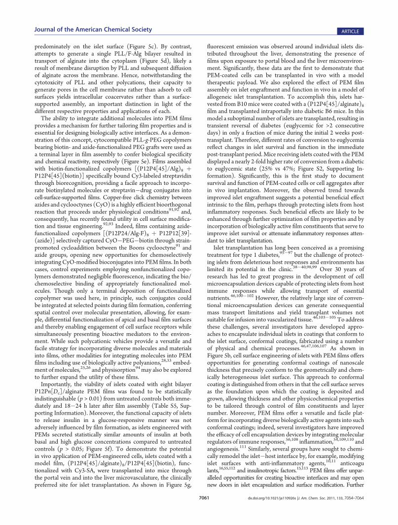
in Figure 5b, in which P12P24[25] was used as the polycation, aring of intense fluorescence emanating from F-Alg was observedsurrounding the islet periphery. By contrast, controls treated onlywith F-Alg in an LbL manner demonstrated essentially nofluorescent emission, indicating the necessity of the polycationin immobilizing alginate on the islet surface. Moreover, adramatic difference in fluorescent intensity was observed be-tween islets coated with eight and one bilayer(s). Collectively,these observations indicate that PEMs can be assembled onthe surface of islets via alternating deposition of P12Pn[Dc]copolymers and alginate. In accord with its role as a componentof a cell-surface-supported thin film, alginate was concentrated
Figure 5. Cell-surface-supported PEMs can be assembled on individual pancreatic islets through LbL deposition of P12Pn[Dc] copolymers and alginate.(a) Method to assemble PEMs on islets. (b) Representative confocal micrographs overlaid on bright-field images of islets coated using P12P24[25] andflourescein-labeled alginate (F-Alg) with eight bilayers (8�), a single bilayer (1�), or treated only with F-Alg (8� w/o cation). Comparable results wereobtained byusing P12P4[45], P12P12[30], andP12P40[22]. (c) F-Alg is localized on the extracellular surface of cells, confirming the cell-surface-supportednature of films. (d) By contrast, deposition of a single PLL/F-Alg bilayer resulted in intracellular internalization of alginate by peripheral cells. (e) Chemistryand reactivity of cell-surface-supported PEM films can be tailored through integration of biotin- and azide-functionalizedPLL-g-PEGcopolymers. (f) Insulinsecretion (mean( SE) by islets coatedwith a (P12P4[45]/alginate)8 film (gray bar) and untreated islets (black bar) in response to a step-change in glucose.(g) Confocal (left) overlaid on bright-field micrographs (right) of frozen sections of liver (L) after intraportal transplantation of islets (I) engineered withPEM films labeled with streptavidin�Cy3. Scale bars: b, e (top), g = 50 μm; c, d, e (bottom) = 10 μm.
7061 dx.doi.org/10.1021/ja110926s |J. Am. Chem. Soc. 2011, 133, 7054–7064
Journal of the American Chemical Society ARTICLE
predominately on the islet surface (Figure 5c). By contrast,attempts to generate a single PLL/F-Alg bilayer resulted intransport of alginate into the cytoplasm (Figure 5d), likely aresult of membrane disruption by PLL and subsequent diffusionof alginate across the membrane. Hence, notwithstanding thecytotoxicity of PLL and other polycations, their capacity togenerate pores in the cell membrane rather than adsorb to cellsurfaces yields intracellular coacervates rather than a surface-supported assembly, an important distinction in light of thedifferent respective properties and applications of each.The ability to integrate additional molecules into PEM films
provides a mechanism for further tailoring film properties and isessential for designing biologically active interfaces. As a demon-stration of this concept, cytocompatible PLL-g-PEG copolymersbearing biotin- and azide-functionalized PEG grafts were used asa terminal layer in film assembly to confer biological specificityand chemical reactivity, respectively (Figure 5e). Films assembledwith biotin-functionalized copolymers [(P12P4[45]/Alg)8 þP12P4[45](biotin)] specifically bound Cy3-labeled streptavidinthrough biorecognition, providing a facile approach to incorpo-rate biotinylated molecules or streptavin�drug conjugates intocell-surface-supported films. Copper-free click chemistry betweenazides and cyclooctynes (CyO) is a highly efficient bioorthogonalreaction that proceeds under physiological conditions91,92 and,consequently, has recently found utility in cell surface modifica-tion and tissue engineering.92,93 Indeed, films containing azide-functionalized copolymers [(P12P24/Alg-F)8 þ P12P12[39]-(azide)] selectively captured CyO�PEG�biotin through strain-promoted cycloaddition between the Boons cyclooctyne91 andazide groups, opening new opportunities for chemoselectivelyintegrating CyO-modified bioconjugates into PEM films. In bothcases, control experiments employing nonfunctionalized copo-lymers demonstrated negligible fluorescence, indicating the bio/chemoselective binding of appropriately functionalized mol-ecules. Though only a terminal deposition of functionalizedcopolymer was used here, in principle, such conjugates couldbe integrated at selected points during film formation, conferringspatial control over molecular presentation, allowing, for exam-ple, differential functionalization of apical and basal film surfacesand thereby enabling engagement of cell surface receptors whilesimultaneously presenting bioactive mediators to the environ-ment. While such polycationic vehicles provide a versatile andfacile strategy for incorporating diverse molecules and materialsinto films, other modalities for integrating molecules into PEMfilms including use of biologically active polyanions,28,33 embed-ment of molecules,25,26 and physisorption94may also be exploredto further expand the utility of these films.Importantly, the viability of islets coated with eight bilayer
P12Pn[Dc]/alginate PEM films was found to be statisticallyindistinguishable (p > 0.01) from untreated controls both imme-diately and 18�24 h later after film assembly (Table S5, Sup-porting Information). Moreover, the functional capacity of isletsto release insulin in a glucose-responsive manner was notadversely influenced by film formation, as islets engineered withPEMs secreted statistically similar amounts of insulin at bothbasal and high glucose concentrations compared to untreatedcontrols (p > 0.05; Figure 5f). To demonstrate the potentialin vivo application of PEM-engineered cells, islets coated with amodel film, (P12P4[45]/alginate)8/P12P4[45](biotin), func-tionalized with Cy3-SA, were transplanted into mice throughthe portal vein and into the liver microvasculature, the clinicallypreferred site for islet transplantation. As shown in Figure 5g,
fluorescent emission was observed around individual islets dis-tributed throughout the liver, demonstrating the presence offilms upon exposure to portal blood and the liver microenviron-ment. Significantly, these data are the first to demonstrate thatPEM-coated cells can be transplanted in vivo with a modeltherapeutic payload. We also explored the effect of PEM filmassembly on islet engraftment and function in vivo in a model ofallogeneic islet transplantation. To accomplish this, islets har-vested from B10 mice were coated with a (P12P4[45]/alginate)8film and transplanted intraportally into diabetic B6 mice. In thismodel a suboptimal number of islets are transplanted, resulting intransient reversal of diabetes (euglycemic for >2 consecutivedays) in only a fraction of mice during the initial 2 weeks post-transplant. Therefore, different rates of conversion to euglycemiareflect changes in islet survival and function in the immediatepost-transplant period.Mice receiving islets coated with the PEMdisplayed a nearly 2-fold higher rate of conversion from a diabeticto euglycemic state (25% vs 47%; Figure S2, Supporting In-formation). Significantly, this is the first study to documentsurvival and function of PEM-coated cells or cell aggregates afterin vivo implantation. Moreover, the observed trend towardsimproved islet engraftment suggests a potential beneficial effectintrinsic to the film, perhaps through protecting islets from hostinflammatory responses. Such beneficial effects are likely to beenhanced through further optimization of film properties and byincorporation of biologically active film constituents that serve toimprove islet survival or attenuate inflammatory responses atten-dant to islet transplantation.Islet transplantation has long been conceived as a promising
treatment for type 1 diabetes,95�97 but the challenge of protect-ing islets from deleterious host responses and environments haslimited its potential in the clinic.38�40,98,99 Over 30 years ofresearch has led to great progress in the development of cellmicroencapsulation devices capable of protecting islets from hostimmune responses while allowing transport of essentialnutrients.46,100�102 However, the relatively large size of conven-tional microencapsulation devices can generate consequentialmass transport limitations and yield transplant volumes notsuitable for infusion into vascularized tissue.46,103�105 To addressthese challenges, several investigators have developed appro-aches to encapsulate individual islets in coatings that conform tothe islet surface, conformal coatings, fabricated using a numberof physical and chemical processes.46,47,106,107 As shown inFigure 5b, cell surface engineering of islets with PEM films offersopportunities for generating conformal coatings of nanoscalethickness that precisely conform to the geometrically and chem-ically heterogeneous islet surface. This approach to conformalcoating is distinguished from others in that the cell surface servesas the foundation upon which the coating is deposited andgrown, allowing thickness and other physicochemical propertiesto be tailored through control of film constituents and layernumber. Moreover, PEM films offer a versatile and facile plat-form for incorporating diverse biologically active agents into suchconformal coatings; indeed, several investigators have improvedthe efficacy of cell encapsulation devices by integrating molecularregulators of immune responses,56,108 inflammation,54,109,110 andangiogenesis.111 Similarly, several groups have sought to chemi-cally remodel the islet�host interface by, for example, modifyingislet surfaces with anti-inflammatory agents,10,11 anticoagulants,16,55,112 and insulinotropic factors.15,113 PEM films offer unpar-alleled opportunities for creating bioactive interfaces and may opennew doors in islet encapsulation and surface modification. Further

7062 dx.doi.org/10.1021/ja110926s |J. Am. Chem. Soc. 2011, 133, 7054–7064
Journal of the American Chemical Society ARTICLE
exploration of the potential of this technology in islet transplantationand other areas is the subject of ongoing and future research.
’CONCLUSION
Cell-based therapies have recently found utility in the treat-ment of numerous pathologies, including heart and vasculardisease, stroke, spinal injury, musculoskeletal disorders, cancer,and diabetes, and cell surface engineering offers great potential toimprove clinical outcomes associated with this promising class oftherapeutic. Layer-by-layer assembly of PEM films has emergedas among the most versatile, modular, and useful surface en-gineering approaches, and, hence, is well-poised to greatlyexpand the molecular repertoire of available cell surface mod-ifications beyond what is currently possible with genetic andmetabolic approaches or covalent and noncovalent chemistries.However, this powerful technique has been largely inaccessibleto living cell surfaces owing to the toxicity associated with themajority of polycations when in direct contact with the cellmembrane. Indeed, in this and previous reports4,50 we havedemonstrated that polycations conventionally employed forPEM film assembly are highly toxic to pancreatic islets. Here,we have circumvented this molecular hurdle by exploiting PEG-dependent conformational changes in polycation structure tounveil a narrow window in PLL-g-PEG copolymer structurespace within which cytocompatible polycations can facilitate theassembly of a unique class of PEM films with tunable biologicaland physicochemical properties. These films can be assembleddirectly on the surface of fully viable and functional pancreaticislets via sequential deposition of cytocompatible PLL-g-PEGcopolymers and alginate, providing a powerful new platform forengineering cell surfaces layer-by-layer that offers superior ver-satility and modularity relative to conventional approaches.Furthermore, we have demonstrated the unprecedented use ofcells engineered with PEMs in vivo, opening exciting possibilitiesranging from nanoscale immunoisolation to localized and con-trolled release of therapeutic molecules from films. Althoughexemplified in the context of islet transplantation, potentialapplications are significantly broader in scope, and the extensionof this technology to other cell types is poised to offer newopportunities in drug and gene delivery, cell-based therapeutics,imaging, and tissue engineering, efforts which are ongoing in ourgroup. Collectively, these investigations have also provided aconceptual framework for the rational design of cell-surface-supported thin films and establish a new paradigm for translatingthe numerous and diverse biomedical applications of PEM filmsfrom abiotic substrates to living cellular interfaces.
’ASSOCIATED CONTENT
bS Supporting Information. Detailed Experimental Sec-tion, supplemental data, and complete ref 97. This material isavailable free of charge via the Internet at http://pubs.acs.org.
’AUTHOR INFORMATION
Corresponding [email protected]
’ACKNOWLEDGMENT
We are grateful to Prof. Clifford Henderson for assistance withthe ellipsometry. This work was supported by grants from the
National Institutes of Health (DK069275) and the JuvenileDiabetes Research Foundation. J.T.W. acknowledges theWhitakerFoundation for generous fellowship support.
’REFERENCES
(1) Saxon, E.; Bertozzi, C. R. Science 2000, 287, 2007–2010.(2) Stephan, M. T.; Moon, J. J.; Um, S. H.; Bershteyn, A.; Irvine, D. J.
Nat. Med. 2010, 16, 1035–41.(3) Zhao, W. A.; Teo, G. S. L.; Kumar, N.; Karp, J. M.Mater. Today
2010, 13, 14–21.(4) Wilson, J. T.; Krishnamurthy, V. R.; Cui, W.; Qu, Z.; Chaikof,
E. L. J. Am. Chem. Soc. 2009, 131, 18228–9.(5) Contreras, J. L.; Xie, D.; Mays, J.; Smyth, C. A.; Eckstein, C.;
Rahemtulla, F. G.; Young, C. J.; Anthony Thompson, J.; Bilbao, G.;Curiel, D. T.; Eckhoff, D. E. Surgery 2004, 136, 537–47.
(6) Medof, M. E.; Nagarajan, S.; Tykocinski, M. L. FASEB J. 1996,10, 574–86.
(7) Rabuka, D.; Forstner, M. B.; Groves, J. T.; Bertozzi, C. R. J. Am.Chem. Soc. 2008, 130, 5947–53.
(8) De Bank, P. A.; Kellam, B.; Kendall, D. A.; Shakesheff, K. M.Biotechnol. Bioeng. 2003, 81, 800–8.
(9) Mahal, L. K.; Bertozzi, C. R. Chem. Biol. 1997, 4, 415–22.(10) Stabler, C. L.; Sun, X. L.; Cui, W.; Wilson, J. T.; Haller, C. A.;
Chaikof, E. L. Bioconjugate Chem. 2007, 18, 1713–5.(11) Wilson, J. T.; Haller, C. A.; Qu, Z.; Cui, W.; Urlam, M. K.;
Chaikof, E. L. Acta Biomater. 2010, 6, 1895–903.(12) Koyfman, A. Y.; Braun, G. B.; Reich, N. O. J. Am. Chem. Soc.
2009, 131, 14237–9.(13) Sarkar, D.; Vemula, P. K.; Zhao, W. A.; Gupta, A.; Karnik, R.;
Karp, J. M. Biomaterials 2010, 31, 5266–5274.(14) Boonyarattanakalin, S.; Martin, S. E.; Sun, Q.; Peterson, B. R.
J. Am. Chem. Soc. 2006, 128, 11463–70.(15) Krishnamurthy, V. R.; Wilson, J. T.; Cui, W.; Song, X.;
Lasanajak, Y.; Cummings, R. D.; Chaikof, E. L. Langmuir 2010,25, 7675–8.
(16) Cabric, S.; Sanchez, J.; Lundgren, T.; Foss, A.; Felldin, M.;Kallen, R.; Salmela, K.; Tibell, A.; Tufveson, G.; Larsson, R.; Korsgren,O.; Nilsson, B. Diabetes 2007, 56, 2008–15.
(17) Gartner, Z. J.; Bertozzi, C. R. Proc. Natl. Acad. Sci. U. S. A. 2009,106, 4606–10.
(18) Kellam, B.; De Bank, P. A.; Shakesheff, K. M. Chem. Soc. Rev.2003, 32, 327–37.
(19) Decher, G. Science 1997, 277, 1232–1237.(20) Jiang, C. Y.; Markutsya, S.; Pikus, Y.; Tsukruk, V. V.Nat. Mater.
2004, 3, 721–728.(21) Krogman, K. C.; Lowery, J. L.; Zacharia, N. S.; Rutledge, G. C.;
Hammond, P. T. Nat. Mater. 2009, 8, 512–518.(22) Hiller, J.; Mendelsohn, J. D.; Rubner, M. F. Nat. Mater. 2002,
1, 59–63.(23) Podsiadlo, P.; Kaushik, A. K.; Arruda, E. M.; Waas, A. M.; Shim,
B. S.; Xu, J. D.; Nandivada, H.; Pumplin, B. G.; Lahann, J.; Ramamoorthy,A.; Kotov, N. A. Science 2007, 318, 80–83.
(24) Caruso, F.; Caruso, R. A.; Mohwald, H. Science 1998, 282,1111–1114.
(25) Mertz, D.; Vogt, C.; Hemmerle, J.; Mutterer, J.; Ball, V.; Voegel,J. C.; Schaaf, P.; Lavalle, P. Nat. Mater. 2009, 8, 731–735.
(26) Shutava, T. G.; Kommireddy, D. S.; Lvov, Y. M. J. Am. Chem.Soc. 2006, 128, 9926–9934.
(27) Liu, X. H.; Zhang, J. T.; Lynn, D. M. Adv. Mater. 2008,20, 4148–4153.
(28) Dimitrova, M.; Affolter, C.; Meyer, F.; Nguyen, I.; Richard,D. G.; Schuster, C.; Bartenschlager, R.; Voegel, J. C.; Ogier, J.; Baumert,T. F. Proc. Natl. Acad. Sci. U. S. A. 2008, 105, 16320–16325.
(29) Michel, M.; Vautier, D.; Voegel, J. C.; Schaaf, P.; Ball, V.Langmuir 2004, 20, 4835–4839.
(30) Yoo, P. J.; Nam, K. T.; Qi, J. F.; Lee, S. K.; Park, J.; Belcher,A. M.; Hammond, P. T. Nat. Mater. 2006, 5, 234–240.

7063 dx.doi.org/10.1021/ja110926s |J. Am. Chem. Soc. 2011, 133, 7054–7064
Journal of the American Chemical Society ARTICLE
(31) Picart, C.; Elkaim, R.; Richert, L.; Audoin, T.; Arntz, Y.;Cardoso, M. D.; Schaaf, P.; Voegel, J. C.; Frisch, B. Adv. Funct. Mater.2005, 15, 83–94.(32) Benkirane-Jessel, N.; Schwinte, P.; Falvey, P.; Darcy, R.; Haikel,
Y.; Schaaf, P.; Voegel, J. C.; Ogier, J. Adv. Funct. Mater. 2004,14, 174–182.(33) Smith, R. C.; Riollano, M.; Leung, A.; Hammond, P. T. Angew.
Chem.-Int. Ed. 2009, 48, 8974–8977.(34) Hunter, A. C. Adv. Drug Delivery Rev. 2006, 58, 1523–1531.(35) Chanana, M.; Gliozzi, A.; Diaspro, A.; Chodnevskaja, I.;
Huewel, S.; Moskalenko, V.; Ulrichs, K.; Galla, H. J.; Krol, S. Nano Lett.2005, 5, 2605–12.(36) Hong, S.; Leroueil, P. R.; Janus, E. K.; Peters, J. L.; Kober,
M. M.; Islam, M. T.; Orr, B. G.; Baker, J. R., Jr.; Banaszak Holl, M. M.Bioconjugate Chem. 2006, 17, 728–34.(37) Fischer, D.; Li, Y. X.; Ahlemeyer, B.; Krieglstein, J.; Kissel, T.
Biomaterials 2003, 24, 1121–1131.(38) Robertson, R. P. N. Engl. J. Med. 2004, 350, 694–705.(39) Ricordi, C.; Strom, T. B. Nat. Rev. Immunol. 2004, 4, 259–68.(40) Wilson, J. T.; Chaikof, E. L. J. Diabetes Sci. Technol. 2008,
2, 746–759.(41) Bennet, W.; Sundberg, B.; Groth, C. G.; Brendel, M. D.;
Brandhorst, D.; Brandhorst, H.; Bretzel, R. G.; Elgue, G.; Larsson, R.;Nilsson, B.; Korsgren, O. Diabetes 1999, 48, 1907–14.(42) Emamaullee, J. A.; Shapiro, A. M. Cell Transplant. 2007, 16, 1–8.(43) Lau, J.; Henriksnas, J.; Svensson, J.; Carlsson, P. O. Curr. Opin.
Organ Transplant. 2009, 14, 688–693.(44) Linn, T.; Schmitz, J.; Hauck-Schmalenberger, I.; Lai, Y.; Bretzel,
R. G.; Brandhorst, H.; Brandhorst, D. Clin. Exp. Immunol. 2006,144, 179–87.(45) Mattsson, G.; Jansson, L.; Carlsson, P. O. Diabetes 2002,
51, 1362–1366.(46) Wilson, J. T.; Chaikof, E. L. Adv. Drug Delivery Rev. 2008,
60, 124–45.(47) Sefton, M. V.; May, M. H.; Lahooti, S.; Babensee, J. E.
J. Controlled Release 2000, 65, 173–86.(48) Cruise, G. M.; Hegre, O. D.; Lamberti, F. V.; Hager, S. R.; Hill,
R.; Scharp, D. S.; Hubbell, J. A. Cell Transplant 1999, 8, 293–306.(49) Teramura, Y.; Kaneda, Y.; Iwata, H. Biomaterials 2007,
28, 4818–25.(50) Wilson, J. T.; Cui, W.; Chaikof, E. L. Nano Lett. 2008,
8, 1940–8.(51) Cabric, S.; Sanchez, J.; Johansson, U.; Larsson, R.; Nilsson, B.;
Korsgren, O.; Magnusson, P. U. Tissue Eng. Part A 2010, 16, 961–970.(52) Muller, S.; Koenig, G.; Charpiot, A.; Debry, C.; Voegel, J. C.;
Lavalle, P.; Vautier, D. Adv. Funct. Mater. 2008, 18, 1767–1775.(53) Chow, L. W.; Wang, L. J.; Kaufman, D. B.; Stupp, S. I.
Biomaterials 2010, 31, 6154–6161.(54) Cheung, C. Y.;McCartney, S. J.; Anseth, K. S.Adv. Funct. Mater.
2008, 18, 3119–3126.(55) Totani, T.; Teramura, Y.; Iwata, H. Biomaterials 2008, 29,
2878–83.(56) Hume, P. S.; Anseth, K. S. Biomaterials 2010, 31, 3166–3174.(57) Schoeler, B.; Kumaraswamy, G.; Caruso, F. Macromolecules
2002, 35, 889–897.(58) Glinel, K.; Moussa, A.; Jonas, A. M.; Laschewsky, A. Langmuir
2002, 18, 1408–1412.(59) Huang, N. P.; Michel, R.; Voros, J.; Textor, M.; Hofer, R.; Rossi,
A.; Elbert, D. L.; Hubbell, J. A.; Spencer, N. D. Langmuir 2001,17, 489–498.(60) Leroueil, P. R.; Hong, S.; Mecke, A.; Baker, J. R., Jr.; Orr, B. G.;
Banaszak Holl, M. M. Acc. Chem. Res. 2007, 40, 335–42.(61) Kujawa, P.; Moraille, P.; Sanchez, J.; Badia, A.; Winnik, F. M.
J. Am. Chem. Soc. 2005, 127, 9224–9234.(62) Podsiadlo, P.; Tang, Z. Y.; Shim, B. S.; Kotov, N. A. Nano Lett.
2007, 7, 1224–1231.(63) Schoeler, B.; Delorme, N.; Doench, I.; Sukhorukov, G. B.; Fery,
A.; Glinel, K. Biomacromolecules 2006, 7, 2065–2071.
(64) Elbert, D. L.; Herbert, C. B.; Hubbell, J. A. Langmuir 1999,15, 5355–5362.
(65) Choi, N. W.; Cabodi, M.; Held, B.; Gleghorn, J. P.; Bonassar,L. J.; Stroock, A. D. Nat. Mater. 2007, 6, 908–915.
(66) Picart, C.; Mutterer, J.; Richert, L.; Luo, Y.; Prestwich, G. D.;Schaaf, P.; Voegel, J. C.; Lavalle, P. Proc. Natl. Acad. Sci. U. S. A. 2002,99, 12531–12535.
(67) McAloney, R. A.; Sinyor, M.; Dudnik, V.; Goh, M. C. Langmuir2001, 17, 6655–6663.
(68) Stevens, M. M.; George, J. H. Science 2005, 310, 1135–1138.(69) Zheng, Y.; Bruening, M. L.; Baker, G. L. Macromolecules 2007,
40, 8212–8219.(70) Tang, Z. Y.; Kotov, N. A.; Magonov, S.; Ozturk, B. Nat. Mater.
2003, 2, 413–U8.(71) Nolte, A. J.; Rubner, M. F.; Cohen, R. E.Macromolecules 2005,
38, 5367–5370.(72) Nolte, A. J.; Treat, N. D.; Cohen, R. E.; Rubner, M. F.
Macromolecules 2008, 41, 5793–5798.(73) Pavoor, P. V.; Bellare, A.; Strom, A.; Yang, D. H.; Cohen, R. E.
Macromolecules 2004, 37, 4865–4871.(74) Schneider, A.; Francius, G.; Obeid, R.; Schwinte, P.; Hemmerle,
J.; Frisch, B.; Schaaf, P.; Voegel, J. C.; Senger, B.; Picart, C. Langmuir2006, 22, 1193–1200.
(75) Richert, L.; Engler, A. J.; Discher, D. E.; Picart, C. Biomacro-molecules 2004, 5, 1908–1916.
(76) Discher, D. E.; Janmey, P.; Wang, Y. L. Science 2005, 310,1139–1143.
(77) Levental, I.; Georges, P. C.; Janmey, P. A. Soft Matter 2007,3, 299–306.
(78) Ren, K. F.; Crouzier, T.; Roy, C.; Picart, C. Adv. Funct. Mater.2008, 18, 1378–1389.
(79) Lee, H.; Baker, J. R., Jr.; Larson, R. G. J. Phys. Chem. B 2006,110, 4014–9.
(80) Lee, H.; Larson, R. G. J. Phys. Chem. B 2006, 110, 18204–11.(81) Lee, H.; Larson, R. G. J. Phys. Chem. B 2008, 112, 12279–12285.(82) Lee, H.; Larson, R. G. J. Phys. Chem. B 2008, 112, 7778–84.(83) Lee, H.; Larson, R. G. Molecules 2009, 14, 423–38.(84) Lee, H.; Larson, R. G. J. Phys. Chem. B 2009, 113, 13202–7.(85) Illya, G.; Deserno, M. Biophys. J. 2008, 95, 4163–73.(86) Pal, S.; Milano, G.; Roccatano, D. J. Phys. Chem. B 2006,
110, 26170–9.(87) Ginzburg, V. V.; Balijepalli, S. Nano Lett. 2007, 7, 3716–22.(88) Feuz, L.; Leermakers, F. A. M.; Textor, M.; Borisov, O.
Langmuir 2008, 24, 7232–7244.(89) Bhat, R.; Timasheff, S. N. Protein Sci. 1992, 1, 1133–1143.(90) Millard, J. W.; Alvarez-Nunez, F. A.; Yalkowsky, S. H. Int.
J. Pharm. 2002, 245, 153–166.(91) Ning, X.; Guo, J.; Wolfert, M. A.; Boons, G. J. Angew. Chem.-Int.
Ed. 2008, 47, 2253–5.(92) Baskin, J. M.; Prescher, J. A.; Laughlin, S. T.; Agard, N. J.;
Chang, P. V.; Miller, I. A.; Lo, A.; Codelli, J. A.; Bertozzi, C. R. Proc. Natl.Acad. Sci. U. S. A. 2007, 104, 16793–7.
(93) DeForest, C. A.; Polizzotti, B. D.; Anseth, K. S. Nat. Mater.2009, 8, 659–664.
(94) Kharlampieva, E.; Tsukruk, T.; Slocik, J. M.; Ko, H.; Poulsen, N.;Naik, R. R.; Kroger, N.; Tsukruk, V. V. Adv. Mater. 2008, 20, 3274–3279.
(95) Williams, P. Br. Med. J. 1894, 2, 1303–1304.(96) Najarian, J. S.; Sutherland, D. E.; Matas, A. J.; Steffes, M. W.;
Simmons, R. L.; Goetz, F. C. Transplant. Proc. 1977, 9, 233–6.(97) Shapiro, A. M.; et al. N. Engl. J. Med. 2006, 355, 1318–30.(98) Shapiro, A. M. J.; Nanji, S. A.; Lakey, J. R. T. Immunol. Rev.
2003, 196, 219–236.(99) Korsgren, O.; Nilsson, B.; Berne, C.; Felldin, M.; Foss, A.;
Kallen, R.; Lundgren, T.; Salmela, K.; Tibell, A.; Tufveson, G. Trans-plantation 2005, 79, 1289–93.
(100) Lim, F.; Sun, A. M. Science 1980, 210, 908–10.(101) Lanza, R. P.; Hayes, J. L.; Chick, W. L. Nat. Biotechnol. 1996,
14, 1107–11.

7064 dx.doi.org/10.1021/ja110926s |J. Am. Chem. Soc. 2011, 133, 7054–7064
Journal of the American Chemical Society ARTICLE
(102) Chaikof, E. L. Annu. Rev. Biomed. Eng. 1999, 1, 103–27.(103) Calafiore, R.; Basta, G.; Luca, G.; Lemmi, A.; Montanucci,
M. P.; Calabrese, G.; Racanicchi, L.; Mancuso, F.; Brunetti, P. DiabetesCare 2006, 29, 137–8.(104) Leblond, F. A.; Simard, G.; Henley, N.; Rocheleau, B.; Huet,
P. M.; Halle, J. P. Cell Transplant. 1999, 8, 327–37.(105) Schneider, S.; von Mach, M. A.; Kraus, O.; Kann, P.; Feilen,
P. J. Artif. Organs 2003, 27, 1053–6.(106) Cruise, G. M.; Hegre, O. D.; Scharp, D. S.; Hubbell, J. A.
Biotechnol. Bioeng. 1998, 57, 655–65.(107) Teramura, Y.; Iwata, H. Adv. Drug Delivery Rev. 2010,
62, 827–840.(108) Cheung, C. Y.; Anseth, K. S. Bioconjugate Chem. 2006,
17, 1036–42.(109) Ricci, M.; Blasi, P.; Giovagnoli, S.; Rossi, C.; Macchiarulo, G.;
Luca, G.; Basta, G.; Calafiore, R. J. Controlled Release 2005, 107,395–407.(110) Chae, S. Y.; Lee, M.; Kim, S. W.; Bae, Y. H. Biomaterials 2004,
25, 843–50.(111) Lembert, N.; Wesche, J.; Petersen, P.; Doser, M.; Zschocke,
P.; Becker, H. D.; Ammon, H. P. Cell Transplant. 2005, 14, 97–108.(112) Teramura, Y.; Iwata, H. Bioconjugate Chem. 2008,
19, 1389–95.(113) Kizilel, S.; Scavone, A.; Liu, X. A.; Nothias, J. M.; Ostrega, D.;
Witkowski, P.; Millis, M. Tissue Eng. Part A 2010, 16, 2217–2228.