Embed Size (px)
Citation preview
REVIEW
Cell signalling in insulin secretion: the molecular targetsof ATP, cAMP and sulfonylurea
S. Seino
Received: 20 January 2012 /Accepted: 9 March 2012 /Published online: 4 May 2012# Springer-Verlag 2012
Abstract Clarification of the molecular mechanisms of in-sulin secretion is crucial for understanding the pathogenesisand pathophysiology of diabetes and for development ofnovel therapeutic strategies for the disease. Insulin secretionis regulated by various intracellular signals generated bynutrients and hormonal and neural inputs. In addition, avariety of glucose-lowering drugs including sulfonylureas,glinide-derivatives, and incretin-related drugs such as dipep-tidyl peptidase IV (DPP-4) inhibitors and glucagon-likepeptide 1 (GLP-1) receptor agonists are used for glycaemiccontrol by targeting beta cell signalling for improved insulinsecretion. There has been a remarkable increase in ourunderstanding of the basis of beta cell signalling over thepast two decades following the application of molecularbiology, gene technology, electrophysiology and bioimagingto beta cell research. This review discusses cell signalling ininsulin secretion, focusing on the molecular targets of ATP,cAMP and sulfonylurea, an essential metabolic signal inglucose-induced insulin secretion (GIIS), a critical signal in
the potentiation of GIIS, and the commonly used glucose-lowering drug, respectively.
Keywords ATP . cAMP .Epac . Incretin . Insulin secretion .
KATP channel . Review . Sulfonylurea
Abbreviations[Ca2+]i Intracellular calcium concentrationDAG DiacylglycerolDPP-4 Dipeptidyl peptidase IVECFP Enhanced cyan fluorescent proteinEpac Exchange protein activated by cAMPExoc3l Exocyst complex component 3-likeEYFP Enhanced yellow fluorescent proteinFRET Fluorescence resonance energy transferGEF Guanine nucleotide exchange factorGFP Green fluorescent proteinGIIS Glucose-induced insulin secretionGIP Glucose-dependent insulinotropic
polypeptideGLP-1 Glucagon-like peptide 1GR Glucose-responsiveIP3 Inositol 1,4,5-trisphosphateKATP channel ATP-sensitive K+ channelPHHI Persistent hyperinsulinaemic
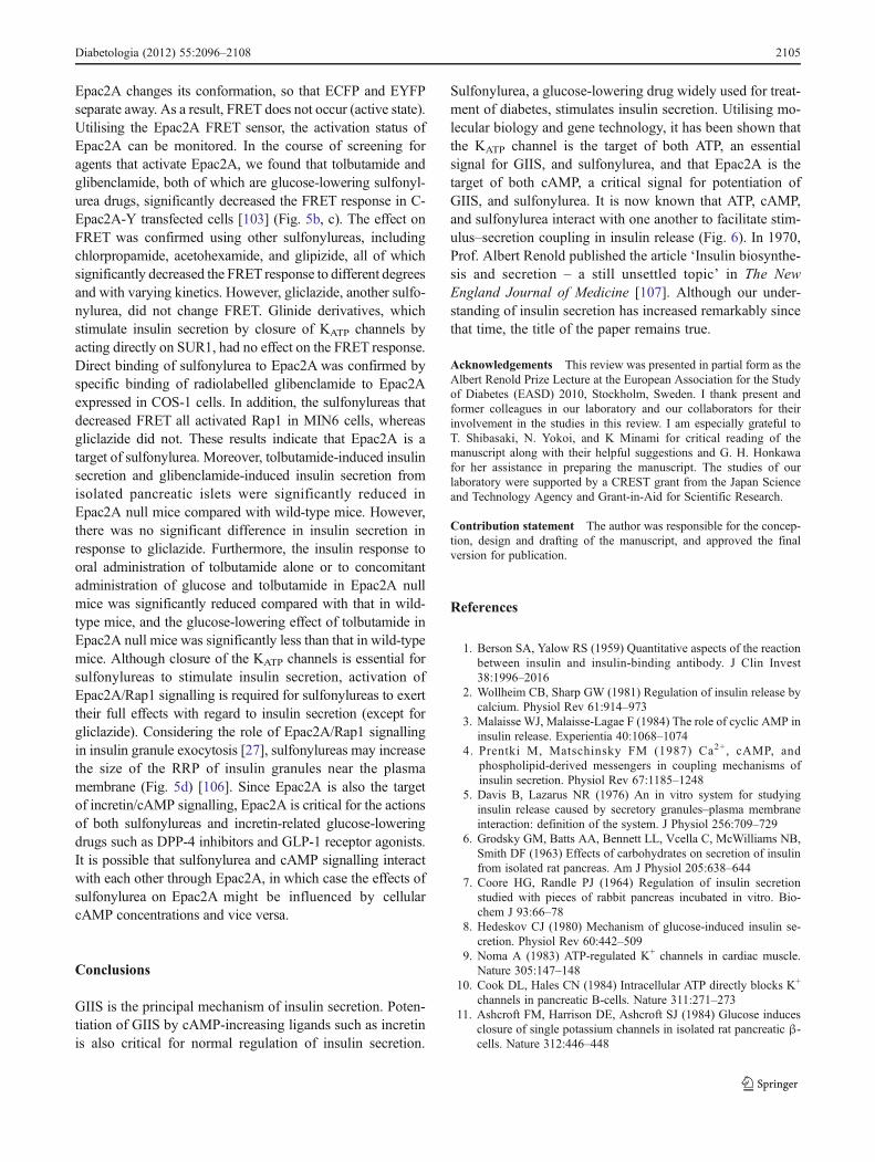
hypoglycaemia of infancyPKA Protein kinase ARP Reserve poolRRP Readily releasable poolSUR Sulfonylurea receptorTIRFM Total internal reflection fluorescence
microscopyTM TransmembraneTRP Transient receptor potentialVMH Ventromedial hypothalamus
This review was invited based on the Albert Renold Prize Lecture atthe EASD, September 2010.
S. Seino (*)Division of Diabetes and Endocrinology, Department of InternalMedicine, Kobe University Graduate School of Medicine,Kobe 650-0017, Japane-mail: [email protected]
S. SeinoDivision of Cellular and Molecular Medicine,Department of Physiology and Cell Biology,Kobe University Graduate School of Medicine,Kobe, Japan
S. SeinoCore Research for Evolutional Science and Technology (CREST),Japan Science and Technology Corp.,Kawaguchi, Saitama 332-0012, Japan
Diabetologia (2012) 55:2096–2108DOI 10.1007/s00125-012-2562-9

Introduction
The blood glucose level is tightly controlled by insulin secre-tion from pancreatic beta cells and insulin action in targettissues such as liver, muscle and adipose tissue. Pancreaticbeta cells secrete an appropriate amount of insulin in a processthat is precisely regulated temporally to maintain glucosehomeostasis. Insulin secretion is regulated by various factors,including nutrients and hormonal and neural inputs to the betacells, among which glucose is the most important physiolog-ical regulator. Beta cell dysfunction impairs normal regulationof insulin secretion and leads to diabetes or hypoglycaemia.The mechanisms of insulin secretion have been studied ex-tensively both in vivo and in vitro in the 50 years since theestablishment of the radioimmunoassay for insulin [1]. Ourunderstanding of the mechanisms of insulin secretion wasdeepened but remained incomplete. By the early 1980s, themajor intracellular signals in pancreatic beta cells for insulinsecretion had been identified by pharmacological, physiolog-ical and biochemical methods. These include Ca2+, ATP,cAMP and phospholipid-derived molecules such as diacyl-glycerol (DAG) and inositol 1,4,5-trisphosphate (IP3) [2–4].
Glucose-induced insulin secretion (GIIS) is the principalmechanism of insulin secretion (Fig. 1a). Early studies pro-posed two models of beta cell glucoreceptor signalling inGIIS: the regulatory-site model [5] and the substrate-sitemodel [6, 7], although most studies supported the lattermodel [8]. Following the discovery of the ATP-sensitiveK+ (KATP) channel in cardiomyocytes by electrophysiology[9], the KATP channel and the glucose-regulated K+ channelwere also found in pancreatic beta cells [10–12]. As theKATP channel couples glucose metabolism to electrical activ-ity of the beta cell [13], the discovery of the channel supportedthe notion that glucose metabolism is essential for GIIS.Accumulating evidence indicates that glucose induces insulinsecretion by two different pathways: the triggering pathway(KATP channel-dependent pathway) and the metabolic ampli-fying pathway (KATP channel-independent pathway), the for-mer of which is essential for GIIS [14]. According to thecurrent consensus on the triggering pathway of GIIS, glucosetransported into the beta cell through glucose transporters israpidly metabolised to yield an increase in the ATP concen-tration (ATP/ADP ratio), which causes closure of KATP chan-nels and depolarisation of the cell membrane. Membranedepolarisation opens the voltage-dependent Ca2+ channels,allowing Ca2+ influx. The resultant rise in intracellular Ca2+
concentration ([Ca2+]i) in the beta cell leads to fusion ofinsulin granules to the plasma membrane in a soluble N-ethyl-maleimide-sensitive factor attachment protein receptor(SNARE)-dependent process. In contrast to this well-established triggering pathway, the metabolic amplifyingpathway is more complex and its mechanism less well under-stood. It has been suggested that metabolic signals generated
by glucose, such as ATP, act on steps in the secretory processdistal to the [Ca2+]i rise [15–17]. However, this pathway doesnot influence insulin secretion if [Ca2+]i is not increased; themetabolic amplifying pathway does not function if the trig-gering pathway is not operational. In addition to metabolicsignals generated by glucose, intracellular signals such ascAMP, DAG and IP3, evoked by hormonal and neuronalinputs, are important for normal regulation of insulin secretion(neurohormonal amplifying pathway) [14]. Lipid metabolismis also important for regulating as well as modulating insulinsecretion [18]. The molecular bases for stimulus-secretioncoupling in GIIS and its potentiation were largely unknownuntil the early 1990s. By utilising molecular biology, genetechnology and bioimaging, many regulators and targets ofintracellular signals in insulin secretion have been identified,greatly enhancing our understanding of GIIS. This reviewdiscusses the targets of ATP, cAMP and the glucose-lowering drug sulfonylurea and their roles in GIIS, based onour recent studies.
Dynamics of insulin secretion
Insulin secretion is a highly dynamic process. Glucose inducesinsulin secretion in a biphasic pattern: there is an initial com-ponent (first phase) that develops rapidly but lasts only a fewminutes, and this is followed by a progressively increasing orsustained component (second phase) [14, 19, 20]. Loss of firstphase secretion and reduced second phase secretion are char-acteristic features of type 2 diabetes. It is known that there is adecrease in the first phase of GIIS in the early stage of type 2diabetes and in impaired glucose tolerance [21].
By analogy with the exocytosis of neurotransmitters inneurons [22], insulin granule exocytosis is thought to in-volve several steps, including recruitment, docking, primingand fusion [23]. It has been suggested that secretory vesiclesin pancreatic beta cells exist in functionally distinct poolsand that the sequential release of these pools underlies theseparable components in the dynamics of exocytosis [20].Pancreatic beta cells contain at least two pools of insulinsecretory granules that differ in release competence: a re-serve pool (RP) accounting for the vast majority of granules,and a readily releasable pool (RRP) accounting for theremaining <5%. A current hypothesis maintains that the firstphase of GIIS is caused by release of RRP granules and thatthe second phase of GIIS represents a subsequent supply ofnew granules mobilised from the RP [14, 20, 24].
Investigation of insulin granule dynamics has recently beenrefined by use of the total internal reflection fluorescencemicroscopy (TIRFM) system [25–28]. TIRF is a technologythat provides a means of selectively exciting fluorophores inan aqueous or cellular environment very near a solid surface(within 100 nm) without exciting fluorescence from regions
Diabetologia (2012) 55:2096–2108 2097
further from the surface [29]. This unique feature of TIRFManalysis has has led to its application in various different areasof biochemistry and cell biology. A previous TIRFM studyreported that insulin granule exocytosis occurs in two modes[26]. In one mode (mode 1), fusion events are caused bygranules that are predocked to the plasma membrane (referredto as ‘previously docked granules’ in [26] and ‘old face’ in
[27]). In the other mode, fusion events are caused by granulesthat are newly recruited to the plasma membrane (‘newcom-er’). Detailed analyses of insulin granule dynamics induced byvarious stimuli using primary cultured pancreatic beta cellsshow that ‘newcomer’ can be classified into two modes: onemode (mode 2), in which granules are newly recruited andimmediately fused to the plasma membrane without docking
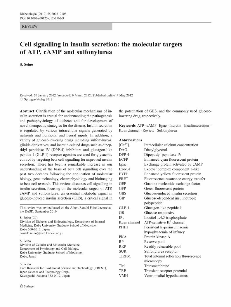
Insulin secretion
ATP
Glucose
VDCC
Glucosetransporter
Ca2+
cAMP
KATP channel
GsAC
Other metabolicsignals
Metabolism
IP3
DAG
Gq
PLCβ
Ca2+
Triggering pathwayMetabolic amplifying pathwayNeurohormonal amplifying pathway
SUR1Kir6.2 ΔΨ
Hormonal input(e.g. Incretin)
Neural input(e.g. ACh)
a
Predocked
Recruitment
Docked
Fusion
Fusion
Fusion
Recruitment
Old face
Restless newcomer
Resting newcomer
b
CellMembrane
RRPActinnetwork
RP
2nd phase1st phase
Existing model
RRPRP
New model
c
Fig. 1 a Glucose-induced insulin secretion and its potentiation. Glu-cose is transported thorough the glucose transporter into the pancreaticbeta cell. Metabolism of glucose increases ATP production (ATP/ADPratio), closing the KATP channels, depolarising beta cell membrane(ΔΨ), opening the voltage-dependent Ca2+ channels (VDCCs) andallowing Ca2+ influx, thereby triggering insulin secretion (formerlycalled the KATP channel-dependent pathway, presently called the trig-gering pathway). In addition to the triggering pathway, metabolicsignals generated by glucose metabolism amplify insulin secretion(formerly called the KATP channel-independent pathway, presentlycalled the metabolic amplifying pathway). Insulin secretion is alsoamplified by hormones and neurotransmitters that generate intracellu-lar signals such as cAMP, DAG, and IP3 (neurohormonal amplifyingpathways). PLCβ, phospholipase C-β; AC, adenylyl cyclase; ACh,acetylcholine. b Modes of insulin granule exocytosis. There are threemodes of insulin granule exocytosis based on the dynamics of thegranules. ‘Old face’: predocked granules that are fused to the plasma
membrane by stimulation. ‘Restless newcomer’: granules that arenewly recruited by stimulation and immediately fused to the plasmamembrane. ‘Resting newcomer’: granules that are newly recruited bystimulation, docked and fused to the plasma membrane by stimulation.Modified from [27] with permission. Copyright 2007 National Acad-emy of Sciences, U.S.A. c Models of glucose-induced insulin secre-tion. In the existing model of GIIS, the first phase of insulin secretionresults from an RRP comprising predocked insulin granules (old face);the second phase secretion results from an RP comprising granuleslocated farther away (resting newcomer), granules that are newlyrecruited upon stimulation, docked, and fused to the plasma membrane.In the new model, both phases are caused by restless newcomergranules that are recruited upon stimulation and immediately fused tothe plasma membrane without docking. Modified with permission ofthe American Society for Clinical Investigation, from [31]; permissionconveyed through Copyright Clearance Center, Inc.
2098 Diabetologia (2012) 55:2096–2108

(a docking state that can barely be detected by TIRFM)(‘restless newcomer’), and another mode (mode 3) in whichgranules are newly recruited, docked and then fused to theplasma membrane (‘resting newcomer’) [27] (Fig. 1b). Thethree modes of insulin granule exocytosis have been con-firmed by other studies [28, 30]. Unlike the original modelof GIIS, in which the first phase results from the RRP com-prising predocked granules and the second phase from RP, anew model in which both phases of GIIS are caused by‘restless newcomer’ has been proposed (Fig. 1c) [31].
In contrast, most K+-induced insulin granule exocytosisthat occurs immediately and transiently after stimulation rep-resents the release of predocked granules (‘old face’) [27, 32].The dynamics of insulin granule exocytosis vary according towhether stimulation is due to K+ or glucose. As K+ stimulationelicits only Ca2+ influx and glucose stimulation generatesvarious metabolic signals such as ATP in addition to Ca2+
influx in pancreatic beta cells, this difference in intracellularsignal may underlie the distinct modes of exocytosis.
Various proteins associated with insulin granule exocytosishave been identified [23, 33], among which Rab-interactingmolecule 2 (Rim2, Rim2α) was identified as a moleculeinteracting with exchange protein activated by cAMP (Epac)2A (cAMP-GEFII) [34]. In addition to Epac2A, Rim2α inter-acts with various exocytosis-related molecules, at least in vitro,including Rab3 [34], Munc13-1 [35], Rab8 [36], ELKS [37,38], Piccolo [39], and synaptotagmin 1 [40]. Although synap-totagmin 1 is produced in insulinoma cells, the synaptotagmingenes expressed in primary mouse beta cells are those encod-ing synaptotagmin 7 [41] and 9 [42]. Rim2α null (Rim2α−/−)mice exhibit a marked impairment in glucose tolerance [43].Analysis by TIRFM shows that both K+-induced insulin gran-ule exocytosis and glucose-induced insulin granule exocytosis,especially the first phase, are severely impaired in pancreaticbeta cells of Rim2α null mice [43]. Rim2α has been found todetermine the docking and priming states depending on inter-action with Rab3 or Munc13-1, respectively.
The exocyst is an octameric protein complex that ensuresspatial docking or tethering of exocytotic vesicles to fusionsites of the plasma membrane [44]. Eight subunits of theexocyst complex are expressed in both pancreatic islets andMIN6 cells. Exocyst complex component 3-like (Exoc3l),an isoform of Sec6, the core subunit of the exocyst complex,was identified by in silico screening [45]. Exoc3l formstertiary complexes consisting of Sec5, Sec8 and Sec10, allof which are binding partners of Sec6. Exoc3l is suggestedto be involved in the regulated exocytosis of insulin granulesthrough formation of the exocyst complex.
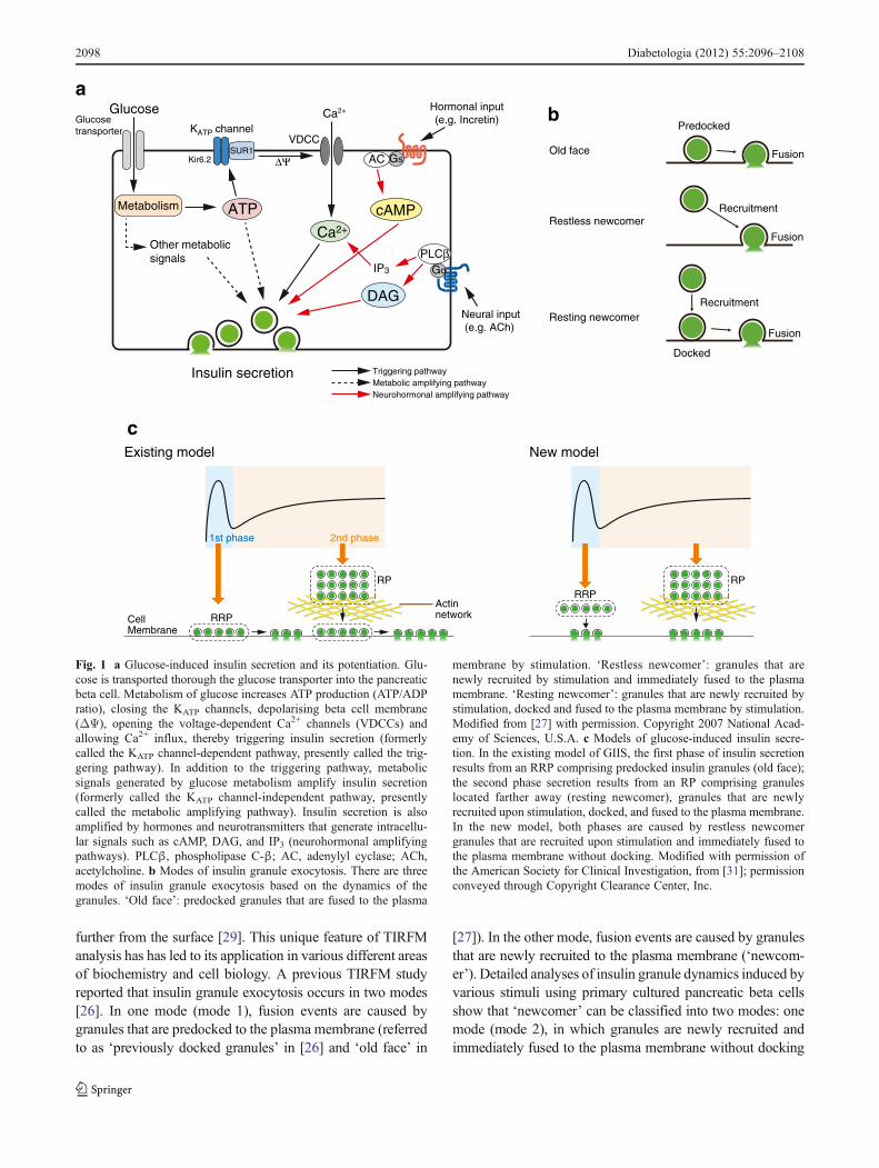
cAMP-increasing ligands potentiate both the first phaseand second phase of GIIS [4]. However, the potentiatingeffect of cAMP occurs only at glucose concentrations abovea certain threshold [46]. cAMP also affects various steps ofinsulin secretion. In normal pancreatic islets, the in vitro
concentration dependence of GIIS displays a sigmoidalcurve [47], in which a glucose concentration exceeding6 mmol/l is required to trigger insulin secretion. In addition,it has been reported that glucagon-like peptide 1 (GLP-1)renders glucose-insensitive beta cells glucose-competent,probably by modulating KATP channel activity [48]. Thesefindings suggest a mechanism by which cAMP might in-duce beta cell glucose responsiveness. Using a pancreaticperfusion system, we recently found that pretreatment withGLP-1 or glucose-dependent insulinotropic polypeptide(GIP) improved glucose responsiveness to some extent inKir6.2 null (Kir6.2−/−) mice, in which almost no insulinsecretion in response to glucose is detected [49], but theeffect of GLP-1 was stronger. The dynamics of GIIS isusually assessed by the insulin secretory response to a largeand prompt change in glucose concentration, e.g. from 2.8to 16.7 mmol/l in 1 min, using perfusion of the pancreas andperifusion of pancreatic islets. However, such a drasticchange in glucose concentration is unlikely to occur in thephysiological state. As regards the perfusion of the pan-creases of wild-type mice, when the glucose concentrationwas increased in a stepwise manner (1.4 mmol/l every5 min) from 2.8 to 12.5 mmol/l in the absence of cAMP-increasing agents (8-bromo-cAMP or GLP-1), no insulinsecretion was evoked (Fig. 2a) [50]. Interestingly, the pres-ence of these agents resulted in a dramatic induction of GIIS(Fig. 2a, b). Similar results also were found in Kir6.2 nullmice. This GIIS was almost completely abolished by treat-ment with niflumic acid, indicating that cAMP signallingalso evokes glucose responsiveness by activating niflumicacid-sensitive channels (Fig. 2c). Niflumic acid is often usedto block Cl− channels but it also acts on other channels,including transient receptor potential (TRP) channels. Sinceremoval of Na+ did not abolish the membrane depolarisationcaused by glucose and cAMP [50], Na+ influx through TRPchannels is unlikely to be a contributor to initial membranedepolarisation; Ca2+-activated Cl− channels are the morelikely candidate for the niflumic acid-sensitive channelsresponsible for depolarisation caused by glucose and cAMP.These findings indicate that cAMP signalling is importantnot only for potentiation of GIIS but also for induction ofglucose responsiveness in insulin secretion.
The role of cAMP signalling in insulin granule exocyto-sis has also been investigated by TIRFM. The cAMP ana-logue 8-bromo-cAMP alone did not cause either significantdocking or fusion events of insulin granules. However,8-bromo-cAMP clearly enhanced the frequency ofglucose-induced fusion events in both the first phase andthe second phase. 8-bromo-cAMP promoted fusion events byincreasing only ‘restless newcomer’. Comparison of the fu-sion sites induced by glucose stimulation and those inducedby 8-bromo-cAMP stimulation showed that new fusion sitesappeared upon 8-bromo-cAMP stimulation, suggesting that
Diabetologia (2012) 55:2096–2108 2099

cAMP signaling also participates in the spatial regulation ofinsulin granule exocytosis [27].
The KATP channel as a target of ATP and sulfonylurea
The KATP channel links cellular metabolic status to theelectrical activity of pancreatic beta cells [10–12] and is a
key molecule in the regulation of GIIS. The beta cell KATP
channel was also suggested to be the target for sulfonylurea[51], a widely used drug in the treatment of type 2 diabetes.However, whether the target (receptor) for sulfonylurea wasthe KATP channel itself or a molecule associated closely withthe KATP channel was not known. In 1995, a receptor(SUR1) for sulfonylurea was cloned by Aguilar-Bryan andcolleagues [52] (Fig. 3a). SUR1 was found to be a member
40 4530 3520 2510 150 5
Time (min)
0
1
2
Insu
lin (
pmol
/min
)G 2.8 4.2 5.6 6.9 8.3 9.7 11.1 12.5
a
40 4530 3520 2510 150 5
Time (min)
0
1
2
3
4
Insu
lin (
pmol
/min
)
10 nmol/l GLP-1
G 2.8 4.2 5.6 6.9 8.3 9.7 11.1 12.5
b
40 4530 3520 2510 150 5
Time (min)
0
1
2
Insu
lin (
pmol
/min
)
G 2.8 4.2 5.6 6.9 8.3 9.7 11.1 12.5
1 mmol/l 8-Bromo-cAMP
100 μmol/l Niflumic acidc
Fig. 2 a Insulin secretion in response to small and stepwise increases(increment of 1.4 mmol/l per 5 min) in glucose concentration in theabsence (white circles) and presence (white triangles) of 1 mmol/l8-bromo-cAMP. b Insulin secretion in response to small and stepwiseincreases in glucose concentration in the absence of 10 nmol/l GLP-1.c Effect of niflumic (100 μmol/l) acid on insulin secretion in responseto small and stepwise increases in glucose concentration in the pres-ence of 1 mmol/l 8-bromo-cAMP. Reproduced from [50] with permis-sion of Springer Science+Business Media
NBF-1 NBF-2
C
C
N
SUR1SUR1
SUR1
SUR1
Kir6.2Kir6.2
Kir6.2Kir6.2
0
10
20
30
40
50
0 20 40
Time (min)
60 80
Insu
lin (
pg i
slet
−1 m
in−1
)
Glucose (16.7 mmol/l)Tolbutamide (100 μmol/l)
b
c
TMD0 TMD1 TMD2
SUR1Kir6.2
N
a
Fig. 3 a Kir6.2 and SUR1. Kir6.2, which belongs to the inwardlyrectifying K+ channel family, is the pore-forming subunit. SUR1,which belongs to the ATP-binding cassette transporter family, is theregulatory subunit. b Subunit structure of the beta cell KATP channel. Abeta cell KATP channel is composed of Kir6.2 and SUR1 with 4 to 4stoichiometry. c Insulin secretion from perifused pancreatic islets ofKir6.2 null mice (black circles) and wild-type mice (white circles).NBF, nucleotide binding fold; TMD, transmembrane domain. Repro-duced from [62] with permission. Copyright 1998 National Academyof Sciences, USA
2100 Diabetologia (2012) 55:2096–2108
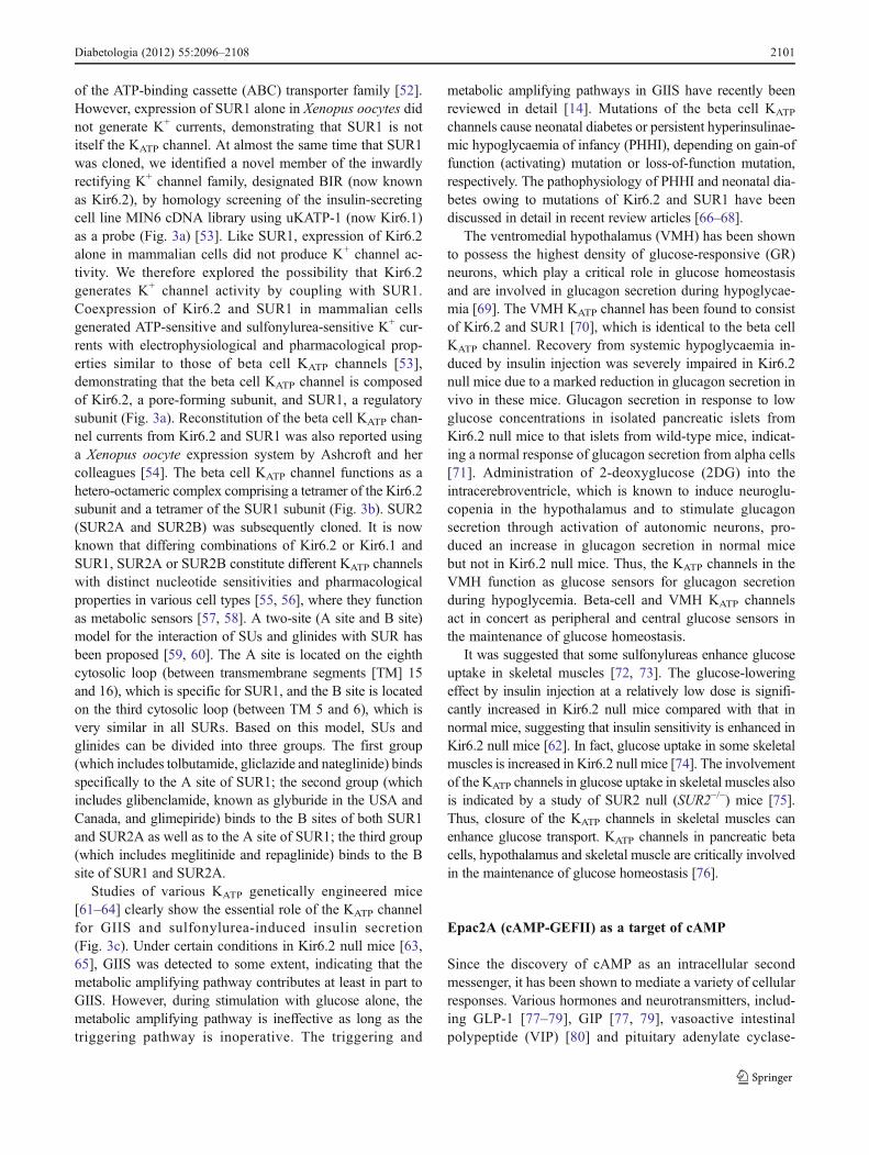
of the ATP-binding cassette (ABC) transporter family [52].However, expression of SUR1 alone in Xenopus oocytes didnot generate K+ currents, demonstrating that SUR1 is notitself the KATP channel. At almost the same time that SUR1was cloned, we identified a novel member of the inwardlyrectifying K+ channel family, designated BIR (now knownas Kir6.2), by homology screening of the insulin-secretingcell line MIN6 cDNA library using uKATP-1 (now Kir6.1)as a probe (Fig. 3a) [53]. Like SUR1, expression of Kir6.2alone in mammalian cells did not produce K+ channel ac-tivity. We therefore explored the possibility that Kir6.2generates K+ channel activity by coupling with SUR1.Coexpression of Kir6.2 and SUR1 in mammalian cellsgenerated ATP-sensitive and sulfonylurea-sensitive K+ cur-rents with electrophysiological and pharmacological prop-erties similar to those of beta cell KATP channels [53],demonstrating that the beta cell KATP channel is composedof Kir6.2, a pore-forming subunit, and SUR1, a regulatorysubunit (Fig. 3a). Reconstitution of the beta cell KATP chan-nel currents from Kir6.2 and SUR1 was also reported usinga Xenopus oocyte expression system by Ashcroft and hercolleagues [54]. The beta cell KATP channel functions as ahetero-octameric complex comprising a tetramer of the Kir6.2subunit and a tetramer of the SUR1 subunit (Fig. 3b). SUR2(SUR2A and SUR2B) was subsequently cloned. It is nowknown that differing combinations of Kir6.2 or Kir6.1 andSUR1, SUR2A or SUR2B constitute different KATP channelswith distinct nucleotide sensitivities and pharmacologicalproperties in various cell types [55, 56], where they functionas metabolic sensors [57, 58]. A two-site (A site and B site)model for the interaction of SUs and glinides with SUR hasbeen proposed [59, 60]. The A site is located on the eighthcytosolic loop (between transmembrane segments [TM] 15and 16), which is specific for SUR1, and the B site is locatedon the third cytosolic loop (between TM 5 and 6), which isvery similar in all SURs. Based on this model, SUs andglinides can be divided into three groups. The first group(which includes tolbutamide, gliclazide and nateglinide) bindsspecifically to the A site of SUR1; the second group (whichincludes glibenclamide, known as glyburide in the USA andCanada, and glimepiride) binds to the B sites of both SUR1and SUR2A as well as to the A site of SUR1; the third group(which includes meglitinide and repaglinide) binds to the Bsite of SUR1 and SUR2A.
Studies of various KATP genetically engineered mice[61–64] clearly show the essential role of the KATP channelfor GIIS and sulfonylurea-induced insulin secretion(Fig. 3c). Under certain conditions in Kir6.2 null mice [63,65], GIIS was detected to some extent, indicating that themetabolic amplifying pathway contributes at least in part toGIIS. However, during stimulation with glucose alone, themetabolic amplifying pathway is ineffective as long as thetriggering pathway is inoperative. The triggering and
metabolic amplifying pathways in GIIS have recently beenreviewed in detail [14]. Mutations of the beta cell KATP
channels cause neonatal diabetes or persistent hyperinsulinae-mic hypoglycaemia of infancy (PHHI), depending on gain-offunction (activating) mutation or loss-of-function mutation,respectively. The pathophysiology of PHHI and neonatal dia-betes owing to mutations of Kir6.2 and SUR1 have beendiscussed in detail in recent review articles [66–68].
The ventromedial hypothalamus (VMH) has been shownto possess the highest density of glucose-responsive (GR)neurons, which play a critical role in glucose homeostasisand are involved in glucagon secretion during hypoglycae-mia [69]. The VMH KATP channel has been found to consistof Kir6.2 and SUR1 [70], which is identical to the beta cellKATP channel. Recovery from systemic hypoglycaemia in-duced by insulin injection was severely impaired in Kir6.2null mice due to a marked reduction in glucagon secretion invivo in these mice. Glucagon secretion in response to lowglucose concentrations in isolated pancreatic islets fromKir6.2 null mice to that islets from wild-type mice, indicat-ing a normal response of glucagon secretion from alpha cells[71]. Administration of 2-deoxyglucose (2DG) into theintracerebroventricle, which is known to induce neuroglu-copenia in the hypothalamus and to stimulate glucagonsecretion through activation of autonomic neurons, pro-duced an increase in glucagon secretion in normal micebut not in Kir6.2 null mice. Thus, the KATP channels in theVMH function as glucose sensors for glucagon secretionduring hypoglycemia. Beta-cell and VMH KATP channelsact in concert as peripheral and central glucose sensors inthe maintenance of glucose homeostasis.
It was suggested that some sulfonylureas enhance glucoseuptake in skeletal muscles [72, 73]. The glucose-loweringeffect by insulin injection at a relatively low dose is signifi-cantly increased in Kir6.2 null mice compared with that innormal mice, suggesting that insulin sensitivity is enhanced inKir6.2 null mice [62]. In fact, glucose uptake in some skeletalmuscles is increased in Kir6.2 null mice [74]. The involvementof the KATP channels in glucose uptake in skeletal muscles alsois indicated by a study of SUR2 null (SUR2−/−) mice [75].Thus, closure of the KATP channels in skeletal muscles canenhance glucose transport. KATP channels in pancreatic betacells, hypothalamus and skeletal muscle are critically involvedin the maintenance of glucose homeostasis [76].
Epac2A (cAMP-GEFII) as a target of cAMP
Since the discovery of cAMP as an intracellular secondmessenger, it has been shown to mediate a variety of cellularresponses. Various hormones and neurotransmitters, includ-ing GLP-1 [77–79], GIP [77, 79], vasoactive intestinalpolypeptide (VIP) [80] and pituitary adenylate cyclase-
Diabetologia (2012) 55:2096–2108 2101
activating polypeptide (PACAP) [80], potentiate insulin se-cretion by promoting cAMP generation in pancreatic betacells. Eight adenylyl cyclase isoforms (types I–VIII) areexpressed in pancreatic islets and beta cell lines [81, 82].In fact, MDL12330A, an adenylyl cyclase inhibitor, com-pletely blocks both GLP-1- and GIP-induced cAMP produc-tion in islets and also markedly reduces both GLP-1- andGIP-potentiated insulin secretions [83].
Until recently, the action of cAMP in insulin secretionwas thought to primarily be mediated by protein kinase A(PKA), which phosphorylates various proteins associatedwith the secretory process [84]. Kir6.2, the pore-formingsubunit of KATP channels, and the α-subunit of the voltage-dependent Ca2+ channel can be phosphorylated by PKA onstimulation in beta cell lines [85, 86]. GLUT2 can also bephosphorylated by GLP-1 in purified beta bcells [87]. Al-though phosphorylation of these proteins influences theiractivities [85–87], a direct effect of phosphorylation oninsulin secretion has not been established. We recentlyfound that in MIN6 cells, Rip11, an effector of the smallG-protein Rab11, participates in the potentiation of exocy-tosis by cAMP plus glucose stimulation but not in that ofglucose stimulation alone [88]. In addition, Rip11 wasfound to be phosphorylated by PKA in MIN6 cells. Thesefindings indicate that Rip11, as a substrate of PKA, isinvolved in the regulation of insulin secretion potentiatedby cAMP in pancreatic beta cells.
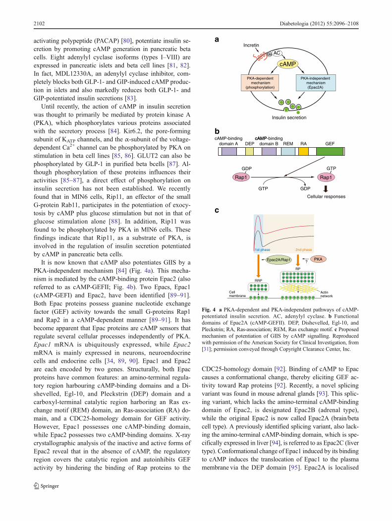
It is now known that cAMP also potentiates GIIS by aPKA-independent mechanism [84] (Fig. 4a). This mecha-nism is mediated by the cAMP-binding protein Epac2 (alsoreferred to as cAMP-GEFII; Fig. 4b). Two Epacs, Epac1(cAMP-GEFI) and Epac2, have been identified [89–91].Both Epac proteins possess guanine nucleotide exchangefactor (GEF) activity towards the small G-proteins Rap1and Rap2 in a cAMP-dependent manner [89–91]. It hasbecome apparent that Epac proteins are cAMP sensors thatregulate several cellular processes independently of PKA.Epac1 mRNA is ubiquitously expressed, while Epac2mRNA is mainly expressed in neurons, neuroendocrinecells and endocrine cells [34, 89, 90]. Epac1 and Epac2are each encoded by two genes. Structurally, both Epacproteins have common features: an amino-terminal regula-tory region harbouring cAMP-binding domains and a Di-shevelled, Egl-10, and Pleckstrin (DEP) domain and acarboxyl-terminal catalytic region harboring an Ras ex-change motif (REM) domain, an Ras-association (RA) do-main, and a CDC25-homology domain for GEF activity.However, Epac1 possesses one cAMP-binding domain,while Epac2 possesses two cAMP-binding domains. X-raycrystallographic analysis of the inactive and active forms ofEpac2 reveal that in the absence of cAMP, the regulatoryregion covers the catalytic region and autoinhibits GEFactivity by hindering the binding of Rap proteins to the
CDC25-homology domain [92]. Binding of cAMP to Epaccauses a conformational change, thereby eliciting GEF ac-tivity toward Rap proteins [92]. Recently, a novel splicingvariant was found in mouse adrenal glands [93]. This splic-ing variant, which lacks the amino-terminal cAMP-bindingdomain of Epac2, is designated Epac2B (adrenal type),while the original Epac2 is now called Epac2A (brain/betacell type). A previously identified splicing variant, also lack-ing the amino-terminal cAMP-binding domain, which is spe-cifically expressed in liver [94], is referred to as Epac2C (livertype). Conformational change of Epac1 induced by its bindingto cAMP induces the translocation of Epac1 to the plasmamembrane via the DEP domain [95]. Epac2A is localised
Insulin secretion
Incretina
b
c
PKA-dependent mechanism
(phosphorylation)
PKA-independent mechanism(Epac2A)
cAMP
Gs AC
DEPcAMP-binding
domain BcAMPcAMP-binding
domain A GEFREM RA
GDPGTP
GTP
Rap1
GDP
Cellular responses
Rap1
Actinnetwork
Cellmembrane
RRP
RP
Epac2A/Rap1 PKA?
2nd phase1st phase
Fig. 4 a PKA-dependent and PKA-independent pathways of cAMP-potentiated insulin secretion. AC, adenylyl cyclase. b Functionaldomains of Epac2A (cAMP-GEFII). DEP, Dishevelled, Egl-10, andPleckstrin; RA, Ras-association; REM, Ras exchange motif. c Proposedmechanism of potentiation of GIIS by cAMP signalling. Reproducedwith permission of the American Society for Clinical Investigation, from[31]; permission conveyed through Copyright Clearance Center, Inc.
2102 Diabetologia (2012) 55:2096–2108

FRET (+) FRET (-)
cAMP
Excitation(440 nm)
Inactive state(closed form)
Emission(527 nm)
EYFP
Excitation(440 nm)
GEFREMRA
B
DEPA
ECFP
GEF
RA
B A
DEP
CFP
YFPGEF
RA
B A
DEP
ECFP
EYFP
Active state(open form)
REM
Emission(480 nm)
KATP channel
cAMP
Incretin
Epac2A
Ca2+
SUR1
Sulfonylurea
Ca2+
VDCCGs receptor
PKA
Kir6.2
RRP
ΔΨ
Rap1
Insulin secretion
a
Time after stimulation (min)-2 0 2 4 6
0.92
0.96
1.00
1.04
R/R
0
-2 0 2 4 60.92
0.96
1.00
1.04R
/R0
Time after stimulation (min)
b c
d
Diabetologia (2012) 55:2096–2108 2103

to the plasma membrane through the interaction of the RAdomain with activated Ras proteins [96, 97]. The amino-terminal cAMP-binding domain A of Epac2A also mediatesits localisation to the plasma membrane [93]. Localisation ofEpac2A to the plasma membrane is independent of its bindingto cAMP.
Studies of Epac2A null (Epac2a−/−) mice and Rap1 knock-down in clonal mouse beta cells indicate that Epac2A/Rap1signalling is required for first phase potentiation of glucose-induced insulin granule exocytosis by cAMP [27]. It has beenproposed that activation of Epac2A/Rap1 signalling increasesthe size of the RRP and/or recruitment of insulin granulesfrom the RRP, while PKA signalling increases the size of theRP and/or recruitment of insulin granules from the RP(Fig. 4c) [31]. Rim2α has been found to be essential forEpac2A-mediated potentiation of GIIS by cAMP [43, 83].Epac2A has also been shown to be involved in mobilisationof Ca2+ from intracellular Ca2+ stores in pancreatic beta cells[98]. The effect of Epac2A on Ca2+ mobilisation has been
shown to be mediated by ryanodine receptors and IP3 recep-tors by studying ryanodine receptor null mice and phospholi-pase Cε null mice, respectively [98].
cAMP signals are known to be compartmentalised in dif-ferent regions of cardiac myocytes [99]. Such cAMP com-partmentalisation is thought to underlie the distinct biologicalresponses mediated by the different cAMP-increasing ligands.By analogy with the effects of cAMP in cardiac myocytes,cAMP compartmentalisation has also been proposed in pan-creatic beta cells [84].
Epac2A as a target of sulfonylurea
Sulfonylureas stimulate insulin secretion by closing KATP
channels through binding to SUR1, as mentioned above. Al-though sulfonylureas were also suggested to act intracellularlyto stimulate insulin granule exocytosis [100–102], the directtarget was not identified.
To screen for agents and ligands that activate Epac2A,Epac2A fluorescence resonance energy transfer (FRET) sen-sor, in which the full-length Epac2A was fused amino-terminally to enhanced cyan fluorescent protein (ECFP) andcarboxyl-terminally to enhanced yellow fluorescent protein(EYFP; termed C-Epac2A-Y) was established (Fig. 5a)[103]. FRET is the radiationless transfer of energy from aninitially excited donor to an acceptor [104]. It is dependent onthe proper spectral overlap of the donor and acceptor, theirdistance from each other and the relative orientation of thechromophore’s transition dipoles. In the case of green fluores-cent proteins (GFPs), including ECFP and EYFP, the distancebetween a donor and an acceptor must be within 5 nm forFRET to be detectable [105]. Epac2A is a closed form in theinactive state, so that ECFP and EYFP are located very closeto each other, which causes FRET. Upon binding of cAMP,
Fig. 5 a Epac2A FRET sensor. A FRET sensor was constructed inwhich the full-length Epac2Awas sandwiched between ECFP at the N-terminus and EYFP at the C-terminus. FRET occurs in the inactivestate (closed form). Upon cAMP binding, Epac2A changes its confor-mation and becomes the open form. As a result, FRET does not occur.DEP, Dishevelled, Egl-10, and Pleckstrin; RA, Ras-association; REM,Ras exchange motif. b, c Activation of Epac2A by sulfonylureas.Tolbutamide (b) and glibenclamide (c) decrease Epac2A FRET, indi-cating that they activate Epac2A. The EYFP/ECFP ratio (R) wasnormalised to R0 to describe FRET efficiency changes (FRETchange0R/R0), where R0 is the EYFP/ECFP ratio at time 0. Repro-duced from [103] with permission from AAAS. d Mechanisms ofsulfonylurea action in insulin secretion. Closure of KATP channels isrequired for sulfonylureas to stimulate insulin secretion; activation ofEpac2A/Rap1 signalling is required for sulfonylureas to exert their fulleffects on insulin secretion. ΔΨ, depolarising beta cell membrane,VDDC, voltage-dependent Ca2+ channel. Modified from [106] withpermission from the Asian Association for the Study of Diabetes andBlackwell Publishing Asia Pty Ltd
R
Opening of VDCC
Metabolism
Closure of KATP channel
Glucose Sulfonylurea Incretin
Ca2+ influx
cAMP
Epac2A PKA
Rap1
Fig. 6 Model of interactionof glucose, incretin andsulfonylurea. VDCC,voltage-dependentCa2+ channel
2104 Diabetologia (2012) 55:2096–2108
Epac2A changes its conformation, so that ECFP and EYFPseparate away. As a result, FRET does not occur (active state).Utilising the Epac2A FRET sensor, the activation status ofEpac2A can be monitored. In the course of screening foragents that activate Epac2A, we found that tolbutamide andglibenclamide, both of which are glucose-lowering sulfonyl-urea drugs, significantly decreased the FRET response in C-Epac2A-Y transfected cells [103] (Fig. 5b, c). The effect onFRET was confirmed using other sulfonylureas, includingchlorpropamide, acetohexamide, and glipizide, all of whichsignificantly decreased the FRET response to different degreesand with varying kinetics. However, gliclazide, another sulfo-nylurea, did not change FRET. Glinide derivatives, whichstimulate insulin secretion by closure of KATP channels byacting directly on SUR1, had no effect on the FRET response.Direct binding of sulfonylurea to Epac2A was confirmed byspecific binding of radiolabelled glibenclamide to Epac2Aexpressed in COS-1 cells. In addition, the sulfonylureas thatdecreased FRET all activated Rap1 in MIN6 cells, whereasgliclazide did not. These results indicate that Epac2A is atarget of sulfonylurea. Moreover, tolbutamide-induced insulinsecretion and glibenclamide-induced insulin secretion fromisolated pancreatic islets were significantly reduced inEpac2A null mice compared with wild-type mice. However,there was no significant difference in insulin secretion inresponse to gliclazide. Furthermore, the insulin response tooral administration of tolbutamide alone or to concomitantadministration of glucose and tolbutamide in Epac2A nullmice was significantly reduced compared with that in wild-type mice, and the glucose-lowering effect of tolbutamide inEpac2A null mice was significantly less than that in wild-typemice. Although closure of the KATP channels is essential forsulfonylureas to stimulate insulin secretion, activation ofEpac2A/Rap1 signalling is required for sulfonylureas to exerttheir full effects with regard to insulin secretion (except forgliclazide). Considering the role of Epac2A/Rap1 signallingin insulin granule exocytosis [27], sulfonylureas may increasethe size of the RRP of insulin granules near the plasmamembrane (Fig. 5d) [106]. Since Epac2A is also the targetof incretin/cAMP signalling, Epac2A is critical for the actionsof both sulfonylureas and incretin-related glucose-loweringdrugs such as DPP-4 inhibitors and GLP-1 receptor agonists.It is possible that sulfonylurea and cAMP signalling interactwith each other through Epac2A, in which case the effects ofsulfonylurea on Epac2A might be influenced by cellularcAMP concentrations and vice versa.
Conclusions
GIIS is the principal mechanism of insulin secretion. Poten-tiation of GIIS by cAMP-increasing ligands such as incretinis also critical for normal regulation of insulin secretion.
Sulfonylurea, a glucose-lowering drug widely used for treat-ment of diabetes, stimulates insulin secretion. Utilising mo-lecular biology and gene technology, it has been shown thatthe KATP channel is the target of both ATP, an essentialsignal for GIIS, and sulfonylurea, and that Epac2A is thetarget of both cAMP, a critical signal for potentiation ofGIIS, and sulfonylurea. It is now known that ATP, cAMP,and sulfonylurea interact with one another to facilitate stim-ulus–secretion coupling in insulin release (Fig. 6). In 1970,Prof. Albert Renold published the article ‘Insulin biosynthe-sis and secretion – a still unsettled topic’ in The NewEngland Journal of Medicine [107]. Although our under-standing of insulin secretion has increased remarkably sincethat time, the title of the paper remains true.
Acknowledgements This review was presented in partial form as theAlbert Renold Prize Lecture at the European Association for the Studyof Diabetes (EASD) 2010, Stockholm, Sweden. I thank present andformer colleagues in our laboratory and our collaborators for theirinvolvement in the studies in this review. I am especially grateful toT. Shibasaki, N. Yokoi, and K Minami for critical reading of themanuscript along with their helpful suggestions and G. H. Honkawafor her assistance in preparing the manuscript. The studies of ourlaboratory were supported by a CREST grant from the Japan Scienceand Technology Agency and Grant-in-Aid for Scientific Research.
Contribution statement The author was responsible for the concep-tion, design and drafting of the manuscript, and approved the finalversion for publication.
References
1. Berson SA, Yalow RS (1959) Quantitative aspects of the reactionbetween insulin and insulin-binding antibody. J Clin Invest38:1996–2016
2. Wollheim CB, Sharp GW (1981) Regulation of insulin release bycalcium. Physiol Rev 61:914–973
3. Malaisse WJ, Malaisse-Lagae F (1984) The role of cyclic AMP ininsulin release. Experientia 40:1068–1074
4. Prentki M, Matschinsky FM (1987) Ca2+, cAMP, andphospholipid-derived messengers in coupling mechanisms ofinsulin secretion. Physiol Rev 67:1185–1248
5. Davis B, Lazarus NR (1976) An in vitro system for studyinginsulin release caused by secretory granules–plasma membraneinteraction: definition of the system. J Physiol 256:709–729
6. Grodsky GM, Batts AA, Bennett LL, Vcella C, McWilliams NB,Smith DF (1963) Effects of carbohydrates on secretion of insulinfrom isolated rat pancreas. Am J Physiol 205:638–644
7. Coore HG, Randle PJ (1964) Regulation of insulin secretionstudied with pieces of rabbit pancreas incubated in vitro. Bio-chem J 93:66–78
8. Hedeskov CJ (1980) Mechanism of glucose-induced insulin se-cretion. Physiol Rev 60:442–509
9. Noma A (1983) ATP-regulated K+ channels in cardiac muscle.Nature 305:147–148
10. Cook DL, Hales CN (1984) Intracellular ATP directly blocks K+
channels in pancreatic B-cells. Nature 311:271–27311. Ashcroft FM, Harrison DE, Ashcroft SJ (1984) Glucose induces
closure of single potassium channels in isolated rat pancreatic β-cells. Nature 312:446–448
Diabetologia (2012) 55:2096–2108 2105
12. Rorsman P, Trube G (1985) Glucose dependent K+-channels inpancreatic β-cells are regulated by intracellular ATP. PflugersArch 405:305–309
13. Ashcroft FM, Rorsman P (1989) Electrophysiology of the pan-creatic β-cell. Prog Biophys Mol Biol 54:87–143
14. Henquin JC (2009) Regulation of insulin secretion: a matter ofphase control and amplitude modulation. Diabetologia 52:739–751
15. Detimary P, Gilon P, Nenquin M, Henquin JC (1994) Two sites ofglucose control of insulin release with distinct dependence on theenergy state in pancreatic B cells. Biochem J 297:455–461
16. Eliasson L, Renstrom E, Ding WG, Proks P, Rorsman P (1997)Rapid ATP-dependent priming of secretory granules precedes Ca2+-induced exocytosis in mouse pancreatic B cells. J Physiol503:399–412
17. Takahashi N, Kadowaki T, Yazaki Y, Ellis-Davies GC, MiyashitaY, Kasai H (1999) Post-priming actions of ATP on Ca2+-depen-dent exocytosis in pancreatic β cells. Proc Natl Acad Sci USA96:760–765
18. Nolan CJ, Madiraju MS, Delghingaro-Augusto V, Peyot ML,Prentki M (2006) Fatty acid signaling in the β-cell and insulinsecretion. Diabetes 55(Suppl 2):S16–S23
19. Curry DL, Bennett LL, Grodsky GM (1968) Dynamics of insulinsecretion by the perfused rat pancreas. Endocrinology 83:572–584
20. Rorsman P, Renstrom E (2003) Insulin granule dynamics inpancreatic β cells. Diabetologia 46:1029–1045
21. Porte D Jr (1991) Banting lecture 1990. β-cells in type II diabetesmellitus. Diabetes 40:166–180
22. Südhof TC, Rizo J (2011) Synaptic vesicle exocytosis. ColdSpring Harb Perspect Biol 3:a005637
23. Eliasson L, Abdulkader F, Braun M, Galvanovskis J, Hoppa MB,Rorsman P (2008) Novel aspects of the molecular mechanismscontrolling insulin secretion. J Physiol 586:3313–3324
24. Straub SG, Shanmugam G, Sharp GW (2004) Stimulation ofinsulin release by glucose is associated with an increase in thenumber of docked granules in the β-cells of rat pancreatic islets.Diabetes 53:3179–3183
25. Tsuboi T, Zhao C, Terakawa S, Rutter GA (2000) Simultaneousevanescent wave imaging of insulin vesicle membrane and cargoduring a single exocytotic event. Curr Biol 10:1307–1310
26. Ohara-Imaizumi M, Nakamichi Y, Tanaka T, Ishida H, Naga-matsu S (2002) Imaging exocytosis of single insulin secretorygranules with evanescent wave microscopy: distinct behavior ofgranule motion in biphasic insulin release. J Biol Chem277:3805–3808
27. Shibasaki T, Takahashi H, Miki T et al (2007) Essential role ofEpac2/Rap1 signaling in regulation of insulin granule dynamicsby cAMP. Proc Natl Acad Sci USA 104:19333–19338
28. Kasai K, Fujita T, Gomi H, Izumi T (2008) Docking is not aprerequisite but a temporal constraint for fusion of secretorygranules. Traffic 9:1191–1203
29. Axelrod D (1981) Cell–substrate contacts illuminated by totalinternal reflection fluorescence. J Cell Biol 89:141–145
30. Takahashi N, Hatakeyama H, Okado H, Noguchi J, Ohno M,Kasai H (2010) SNARE conformational changes that preparevesicles for exocytosis. Cell Metab 12:19–29
31. Seino S, Shibasaki T, Minami K (2011) Dynamics of insulinsecretion and the clinical implications for obesity and diabetes.J Clin Invest 121:2118–2125
32. Seino S, Takahashi H, Fujimoto W, Shibasaki T (2009) Roles ofcAMP signalling in insulin granule exocytosis. Diabetes ObesMetab 11(Suppl 4):180–188
33. Wang Z, Thurmond DC (2009) Mechanisms of biphasic insulin-granule exocytosis—roles of the cytoskeleton, small GTPasesand SNARE proteins. J Cell Sci 122:893–903
34. Ozaki N, Shibasaki T, Kashima Y et al (2000) cAMP-GEFII is adirect target of cAMP in regulated exocytosis. Nat Cell Biol2:805–811
35. Dulubova I, Lou X, Lu J et al (2005) A Munc13/RIM/Rab3tripartite complex: from priming to plasticity? EMBO J24:2839–2850
36. Fukuda M (2003) Distinct Rab binding specificity of Rim1,Rim2, rabphilin, and Noc2. Identification of a critical deter-minant of Rab3A/Rab27A recognition by Rim2. J Biol Chem278:15373–15380
37. Ohara-Imaizumi M, Ohtsuka T, Matsushima S et al (2005) ELKS,a protein structurally related to the active zone-associated proteinCAST, is expressed in pancreatic β cells and functions in insulinexocytosis: interaction of ELKS with exocytotic machinery ana-lyzed by total internal reflection fluorescence microscopy. MolBiol Cell 16:3289–3300
38. Inoue E, Deguchi-Tawarada M, Takao-Rikitsu E et al (2006)ELKS, a protein structurally related to the active zone proteinCAST, is involved in Ca2+-dependent exocytosis from PC12cells. Genes Cells 11:659–672
39. Fujimoto K, Shibasaki T, Yokoi N et al (2002) Piccolo, a Ca2+ sensorin pancreatic β-cells. Involvement of cAMP-GEFII⋅Rim2⋅Piccolocomplex in cAMP-dependent exocytosis. J Biol Chem 277:50497–50502
40. Shibasaki T, Sunaga Y, Fujimoto K, Kashima Y, Seino S (2004)Interaction of ATP sensor, cAMP sensor, Ca2+ sensor, andvoltage-dependent Ca2+ channel in insulin granule exocytosis. JBiol Chem 279:7956–7961
41. Gustavsson N, Lao Y, Maximov A et al (2008) Impaired insulinsecretion and glucose intolerance in synaptotagmin-7 null mutantmice. Proc Natl Acad Sci USA 105:3992–3997
42. Iezzi M, Kouri G, Fukuda M, Wollheim CB (2004) SynaptotagminVand IX isoforms control Ca2+-dependent insulin exocytosis. J CellSci 117:3119–3127
43. Yasuda T, Shibasaki T, Minami K et al (2010) Rim2α determinesdocking and priming states in insulin granule exocytosis. CellMetab 12:117–129
44. He B, Guo W (2009) The exocyst complex in polarized exocy-tosis. Curr Opin Cell Biol 21:537–542
45. Saito T, Shibasaki T, Seino S (2008) Involvement of Exoc3l, aprotein structurally related to the exocyst subunit Sec6, in insulinsecretion. Biomed Res 29:85–91
46. Weir GC, Mojsov S, Hendrick GK, Habener JF (1989) Glucagon-like peptide I (7–37) actions on endocrine pancreas. Diabetes38:338–342
47. Gembal M, Detimary P, Gilon P, Gao ZY, Henquin JC (1993)Mechanisms by which glucose can control insulin release inde-pendently from its action on adenosine triphosphate-sensitive K+
channels in mouse B cells. J Clin Invest 91:871–88048. Holz GG 4th, Kuhtreiber WM, Habener JF (1993) Pancreatic β-
cells are rendered glucose-competent by the insulinotropic hor-mone glucagon-like peptide-1(7–37). Nature 361:362–365
49. Miki T, Minami K, Shinozaki H et al (2005) Distinct effects ofglucose-dependent insulinotropic polypeptide and glucagon-likepeptide-1 on insulin secretion and gut motility. Diabetes54:1056–1063
50. Fujimoto W, Miki T, Ogura T et al (2009) Niflumic acid-sensitiveion channels play an important role in the induction of glucose-stimulated insulin secretion by cyclic AMP in mice. Diabetologia52:863–872
51. Sturgess NC, Ashford ML, Cook DL, Hales CN (1985) Thesulphonylurea receptor may be an ATP-sensitive potassium chan-nel. Lancet 2:474–475
52. Aguilar-Bryan L, Nichols CG, Wechsler SW et al (1995) Cloningof the β cell high-affinity sulfonylurea receptor: a regulator ofinsulin secretion. Science 268:423–426
2106 Diabetologia (2012) 55:2096–2108

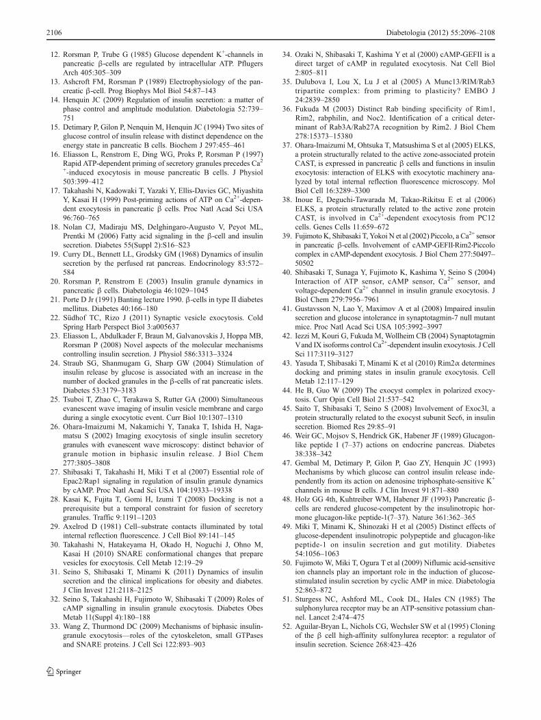
53. Inagaki N, Gonoi T, Clement JP 4th et al (1995) Reconstitution ofIKATP: an inward rectifier subunit plus the sulfonylurea receptor.Science 270:1166–1170
54. Sakura H, Ammala C, Smith PA, Gribble FM, Ashcroft FM(1995) Cloning and functional expression of the cDNA encodinga novel ATP-sensitive potassium channel subunit expressed inpancreatic β-cells, brain, heart and skeletal muscle. FEBS Letters377:338–344
55. Aguilar-Bryan L, Bryan J (1999) Molecular biology of adenosinetriphosphate-sensitive potassium channels. Endocrine reviews20:101–135
56. Seino S (1999) ATP-sensitive potassium channels: a model ofheteromultimeric potassium channel/receptor assemblies. AnnuRev Physiol 61:337–362
57. Seino S, Miki T (2003) Physiological and pathophysiologicalroles of ATP-sensitive K+ channels. Prog Biophys Mol Biol81:133–176
58. Gribble FM, Reimann F (2003) Sulphonylurea action revisited:the post-cloning era. Diabetologia 46:875–891
59. Winkler M, Stephan D, Bieger S, Kuhner P, Wolff F, Quast U(2007) Testing the bipartite model of the sulfonylurea receptorbinding site: binding of A-, B-, and A + B-site ligands. J Phar-macol Exp Ther 322:701–708
60. Proks P, Ashcroft FM (2009) Modeling KATP channel gating andits regulation. Prog Biophys Mol Biol 99:7–19
61. Miki T, Tashiro F, Iwanaga T et al (1997) Abnormalities ofpancreatic islets by targeted expression of a dominant-negativeKATP channel. Proc Natl Acad Sci USA 94:11969–11973
62. Miki T, Nagashima K, Tashiro F et al (1998) Defective insulinsecretion and enhanced insulin action in KATP channel-deficientmice. Proc Natl Acad Sci USA 95:10402–10406
63. Seghers V, Nakazaki M, DeMayo F, Aguilar-Bryan L, Bryan J(2000) Sur1 knockout mice. A model for KATP channel-independent regulation of insulin secretion. J Biol Chem275:9270–9277
64. Shiota C, Larsson O, Shelton KD et al (2002) Sulfonylureareceptor type 1 knock-out mice have intact feeding-stimulatedinsulin secretion despite marked impairment in their response toglucose. J Biol Chem 277:37176–37183
65. Ravier MA, Nenquin M, Miki T, Seino S, Henquin JC (2009)Glucose controls cytosolic Ca2+ and insulin secretion in mouseislets lacking adenosine triphosphate-sensitive K+ channels ow-ing to a knockout of the pore-forming subunit Kir6.2. Endocri-nology 150:33–45
66. Gloyn AL, Pearson ER, Antcliff JF et al (2004) Activatingmutations in the gene encoding the ATP-sensitive potassium-channel subunit Kir6.2 and permanent neonatal diabetes. N EnglJ Med 350:1838–1849
67. Remedi MS, Nichols CG (2009) Hyperinsulinism and diabetes:genetic dissection of β cell metabolism–excitation coupling inmice. Cell Metab 10:442–453
68. Ashcroft FM (2010) New uses for old drugs: neonatal diabetesand sulphonylureas. Cell Metab 11:179–181
69. BorgWP, During MJ, Sherwin RS, BorgMA, Brines ML, ShulmanGI (1994) Ventromedial hypothalamic lesions in rats suppresscounterregulatory responses to hypoglycemia. J Clin Invest93:1677–1682
70. Miki T, Liss B, Minami K et al (2001) ATP-sensitive K+ channelsin the hypothalamus are essential for the maintenance of glucosehomeostasis. Nat Neurosci 4:507–512
71. MacDonald PE, de Marinis YZ, Ramracheya R et al (2007) AKATP channel-dependent pathway within α cells regulates gluca-gon release from both rodent and human islets of Langerhans.PLoS Biol 5:e143
72. Wang PH, Moller D, Flier JS, Nayak RC, Smith RJ (1989)Coordinate regulation of glucose transporter function, number,
and gene expression by insulin and sulfonylureas in L6 ratskeletal muscle cells. J Clin Invest 84:62–67
73. Pulido N, Casla A, Suarez A, Casanova B, Arrieta FJ, Rovira A(1996) Sulphonylurea stimulates glucose uptake in rats throughan ATP-sensitive K+ channel dependent mechanism. Diabetolo-gia 39:22–27
74. Minami K, Morita M, Saraya A et al (2003) ATP-sensitive K+
channel-mediated glucose uptake is independent of IRS-1/phos-phatidylinositol 3-kinase signaling. Am J Physiol EndocrinolMetab 285:E1289–E1296
75. Chutkow WA, Samuel V, Hansen PA et al (2001) Disruption ofSur2-containingKATP channels enhances insulin-stimulated glucoseuptake in skeletal muscle. Proc Natl Acad Sci USA 98:11760–11764
76. Minami K, Miki T, Kadowaki T, Seino S (2004) Roles of ATP-sensitive K+ channels as metabolic sensors: studies of Kir6.x nullmice. Diabetes 53(Suppl 3):S176–S180
77. Drucker DJ (2006) The biology of incretin hormones. Cell Metab3:153–165
78. Holst JJ (2007) The physiology of glucagon-like peptide 1.Physiol Rev 87:1409–1439
79. Nauck MA (2009) Unraveling the science of incretin biology. AmJ Med 122:S3–S10
80. Winzell MS, Ahren B (2007) Role of VIP and PACAP in isletfunction. Peptides 28:1805–1813
81. Delmeire D, Flamez D, Hinke SA, Cali JJ, Pipeleers D, Schuit F(2003) Type VIII adenylyl cyclase in rat β cells: coincidencesignal detector/generator for glucose and GLP-1. Diabetologia46:1383–1393
82. Guenifi A, Portela-Gomes GM, Grimelius L, Efendic S, Abdel-Halim SM (2000) Adenylyl cyclase isoform expression in non-diabetic and diabetic Goto–Kakizaki (GK) rat pancreas. Evidencefor distinct overexpression of type-8 adenylyl cyclase in diabeticGK rat islets. Histochem Cell Biol 113:81–89
83. Kashima Y, Miki T, Shibasaki T et al (2001) Critical role ofcAMP–GEFII–Rim2 complex in incretin-potentiated insulin se-cretion. J Biol Chem 276:46046–46053
84. Seino S, Shibasaki T (2005) PKA-dependent and PKA-independent pathways for cAMP-regulated exocytosis. PhysiolRev 85:1303–1342
85. Leiser M, Fleischer N (1996) cAMP-dependent phosphorylationof the cardiac-type α1 subunit of the voltage-dependent Ca
2+ chan-nel in a murine pancreatic β-cell line. Diabetes 45:1412–1418
86. Safayhi H, Haase H, Kramer U et al (1997) L-type calciumchannels in insulin-secreting cells: biochemical characterizationand phosphorylation in RINm5F cells. Mol Endocrinol 11:619–629
87. Thorens B, Deriaz N, Bosco D et al (1996) Protein kinase A-dependent phosphorylation of GLUT2 in pancreatic β cells. JBiol Chem 271:8075–8081
88. Sugawara K, Shibasaki T, Mizoguchi A, Saito T, Seino S (2009)Rab11 and its effector Rip11 participate in regulation of insulingranule exocytosis. Genes Cells 14:445–456
89. de Rooij J, Zwartkruis FJ, Verheijen MH et al (1998) Epac is aRap1 guanine-nucleotide-exchange factor directly activated bycyclic AMP. Nature 396:474–477
90. Kawasaki H, Springett GM, Mochizuki N et al (1998) A familyof cAMP-binding proteins that directly activate Rap1. Science282:2275–2279
91. de Rooij J, Rehmann H, van Triest M, Cool RH, Wittinghofer A,Bos JL (2000) Mechanism of regulation of the Epac family ofcAMP-dependent RapGEFs. J Biol Chem 275:20829–20836
92. Gloerich M, Bos JL (2010) Epac: defining a new mechanism forcAMP action. Annu Rev Pharmacol Toxicol 50:355–375
93. Niimura M, Miki T, Shibasaki T, Fujimoto W, Iwanaga T, Seino S(2009) Critical role of the N-terminal cyclic AMP-binding
Diabetologia (2012) 55:2096–2108 2107

domain of Epac2 in its subcellular localization and function. JCell Physiol 219:652–658
94. Ueno H, Shibasaki T, Iwanaga T et al (2001) Characterization ofthe gene EPAC2: structure, chromosomal localization, tissue ex-pression, and identification of the liver-specific isoform.Genomics 78:91–98
95. Ponsioen B, Gloerich M, Ritsma L, Rehmann H, Bos JL, Jalink K(2009) Direct spatial control of Epac1 by cyclic AMP. Mol CellBiol 29:2521–2531
96. Li Y, Asuri S, Rebhun JF, Castro AF, Paranavitana NC, QuilliamLA (2006) The RAP1 guanine nucleotide exchange factor Epac2couples cyclic AMP and Ras signals at the plasma membrane. JBiol Chem 281:2506–2514
97. Liu C, Takahashi M, Li Y et al (2008) Ras is required for thecyclic AMP-dependent activation of Rap1 via Epac2. Mol CellBiol 28:7109–7125
98. Leech CA, Dzhura I, Chepurny OG et al (2011) Molecularphysiology of glucagon-like peptide-1 insulin secretagogue ac-tion in pancreatic β cells. Prog Biophys Mol Biol 107:236–247
99. Vandecasteele G, Rochais F, Abi-Gerges A, Fischmeister R(2006) Functional localization of cAMP signalling in cardiacmyocytes. Biochem Soc Trans 34:484–488
100. Ozanne SE, Guest PC, Hutton JC, Hales CN (1995) Intracellularlocalization and molecular heterogeneity of the sulphonylureareceptor in insulin-secreting cells. Diabetologia 38:277–282
101. Eliasson L, Renstrom E, Ammala C et al (1996) PKC-dependentstimulation of exocytosis by sulfonylureas in pancreatic β cells.Science 271:813–815
102. Eliasson L, Ma X, Renstrom E et al (2003) SUR1 regulates PKA-independent cAMP-induced granule priming in mouse pancreaticB cells. J Gen Physiol 121:181–197
103. Zhang CL, Katoh M, Shibasaki T et al (2009) The cAMP sensorEpac2 is a direct target of antidiabetic sulfonylurea drugs. Science325:607–610
104. Lakowicz JR (1999) Energy transfer. In: Lakowicz JR (ed) Prin-ciples of fluorescence spectroscopy. Kluwer Academic, NewYork, pp 368–391
105. Miyawaki A, Tsien RY (2000) Monitoring protein conformationsand interactions by fluorescence resonance energy transfer be-tween mutants of green fluorescent protein. Methods Enzymol327:472–500
106. Seino S, Zhang CL, Shibasaki T (2010) Sulfonylurea action re-revisited. J Diabetes Invest 1:37–39
107. Renold AE (1970) Insulin biosynthesis and secretion—a stillunsettled topic. N Engl J Med 282:173–182
2108 Diabetologia (2012) 55:2096–2108