Embed Size (px)
Citation preview
Cell shape and cell-wall organizationin Gram-negative bacteriaKerwyn Casey Huanga,1, Ranjan Mukhopadhyayb, Bingni Wena, Zemer Gitaia, and Ned S. Wingreena,2
aDepartment of Molecular Biology, Princeton University, Washington Road, Princeton, NJ 08544-1014; and bDepartment of Physics, Clark University,950 Main Street, Worcester, MA 01610
Edited by Michael E. Fisher, University of Maryland, College Park, MD, and approved October 14, 2008 (received for review June 4, 2008)
In bacterial cells, the peptidoglycan cell wall is the stress-bearingstructure that dictates cell shape. Although many molecular detailsof the composition and assembly of cell-wall components areknown, how the network of peptidoglycan subunits is organizedto give the cell shape during normal growth and how it is reor-ganized in response to damage or environmental forces have beenrelatively unexplored. In this work, we introduce a quantitativephysical model of the bacterial cell wall that predicts the mechan-ical response of cell shape to peptidoglycan damage and pertur-bation in the rod-shaped Gram-negative bacterium Escherichia coli.To test these predictions, we use time-lapse imaging experimentsto show that damage often manifests as a bulge on the sidewall,coupled to large-scale bending of the cylindrical cell wall aroundthe bulge. Our physical model also suggests a surprising robust-ness of cell shape to peptidoglycan defects, helping explain theobserved porosity of the cell wall and the ability of cells to growand maintain their shape even under conditions that limit peptidecrosslinking. Finally, we show that many common bacterial cellshapes can be realized within the same model via simple spatialpatterning of peptidoglycan defects, suggesting that minor pat-terning changes could underlie the great diversity of shapesobserved in the bacterial kingdom.
bacterial cell wall � biophysics � elasticity � peptidoglycan � morphology
Bacterial cells come in a wide variety of shapes and sizes (1),with the cell wall being the primary stress-bearing and
shape-maintaining element (2, 3). In recent years, cell shape hasbeen shown to play a critical role in regulating the importantbacterial functions of attachment, dispersal, motility, polar dif-ferentiation, predation, and cellular differentiation (for a review,see ref. 4). Importantly, to achieve cell growth, the cell wall mustcontinuously reorganize, with collateral risks to cell integrity.
In both Gram-negative and Gram-positive bacteria, the cellwall is constructed from the polymer peptidoglycan, a compositeof long strands of glycans crosslinked by stretchable peptides.The resulting elastic network protects the cell from lysis (5).Initially, glycans are polymerized as strands of up to 100 disac-charide subunits (6, 7). The steady-state length distribution ofglycan strands in Escherichia coli is extremely broad, with a meanof �20–30 disaccharide units depending on strain, conditions,and growth phase (7, 8), and some strand lengths rangingupwards of 80 units (2). Each disaccharide unit in a glycan strandis synthesized with a covalently linked peptide that can becrosslinked to a peptide emanating from another glycan strand.The orientation of peptide stems winds around each glycanstrand (9). A 3D structure of a relaxed dimeric peptidoglycansegment determined by NMR suggests that subsequent peptidesare spaced by �120° (10). Molecular dynamics simulationssuggest that the minimal energy conformation of an oligosac-charide peptidoglycan chain has a peptide rotation angle closerto 90°, with little restriction on rotations of up to 15° (9). Themolecular details of glycan strands are conserved among bac-teria, and although there is some variation in the biochemistryof crosslinking, the 3D NMR structure of peptidoglycan frag-ments is relatively insensitive to the type of peptide crosslinks
(10). In Gram-negative bacteria, the cell wall is only 1–3 layersthick (11), and in E. coli 80% or more of the peptidoglycan existsas a monolayer (12). Consistent with these earlier results, recentelectron cryotomography density profile measurements haverevealed that the thickness of the cell wall of both E. coli andanother Gram-negative bacteria Caulobacter crescentus is at most4 nm (13). Popular hypotheses for the organization of thebacterial cell wall (see ref. 14 for a review) have focused on ahorizontal orientation of the glycan strands, arranged in hoopsaround the circumference of the cylinder and crosslinked vialongitudinally oriented peptides.
Although the E. coli cell wall normally maintains a cylindricalshape during exponential growth (15), the cell shape can bealtered either genetically or environmentally. E. coli mutantslacking the high molecular-weight PBP2, a transpeptidase, swellup to resemble spheroplasts (16), while cells lacking the lowmolecular-weight PBPs 5 and 7 are often branched with 3 ormore poles (17, 18). During stationary phase, E. coli transformto a more spherical shape (19). Wild-type E. coli in micron-scaleagarose moldings grow in a variety of cell shapes determined bytheir confinement, and the new cell shape persists after thebacteria are released (20).
The nature of the crosslinked peptidoglycan network is suchthat bonds must be broken to permit new growth and division(21). The potential for deleterious consequences of peptidogly-can bond breakage has prompted the ‘‘make-before-break’’hypothesis in which new material is made and inserted into thecell wall before old material is removed (22). However, the needto always make before breaking assumes that the cell wall ishighly vulnerable to defects, which has not been established.Indeed, the peptidoglycan density can be reduced by �50% bylimiting the supply of the specific precursor mesodiaminopimelicacid without any detectable alteration in morphology or growth(23).
To test the robustness of the Gram-negative cell wall to defectsand damage and to probe cell-wall organization, we have devel-oped a physical model that extends the existing hypothesis ofpeptidoglycan as a 2D (single-layer) cell wall with horizontallyoriented glycan strands by explicitly incorporating the mechan-ical properties of the cell wall. Our physical model predicted thata local accumulation of peptide defects would reproduciblyresult in a cracked cell shape. To verify these predictions, we usedan E. coli strain sensitive to the antibiotic vancomycin, whichdisrupts the formation of peptide crosslinks. In the presence of
Author contributions: K.C.H., R.M., Z.G., and N.S.W. designed research; K.C.H. and B.W.performed research; K.C.H. and B.W. contributed new reagents/analytic tools; K.C.H., R.M.,B.W., Z.G., and N.S.W. analyzed data; and K.C.H., R.M., Z.G., and N.S.W. wrote the paper.
The authors declare no conflict of interest.
This article is a PNAS Direct Submission.
Freely available online through the PNAS open access option.
1Present address: Department of Bioengineering, Stanford University, Stanford, CA 94305.
2To whom correspondence should be addressed. E-mail: [email protected].
This article contains supporting information online at www.pnas.org/cgi/content/full/0805309105/DCSupplemental.
© 2008 by The National Academy of Sciences of the USA
www.pnas.org�cgi�doi�10.1073�pnas.0805309105 PNAS � December 9, 2008 � vol. 105 � no. 49 � 19281–19286
BIO
PHYS
ICS
vancomycin, these cells bulge and ‘‘crack’’ in the same manneras the model cell walls. We then used our verified physical modelto demonstrate the robustness of the cell wall to large amountsof defects and damage such as might arise during cell growth,including holes in the peptidoglycan large enough to account forthe observed porosity of the cell wall. Finally, we demonstratedthat crescent, helical, and lemon-shaped model cells, resemblingcommon bacterial shapes, can be generated via parsimoniouspatterning of peptidoglycan defects.
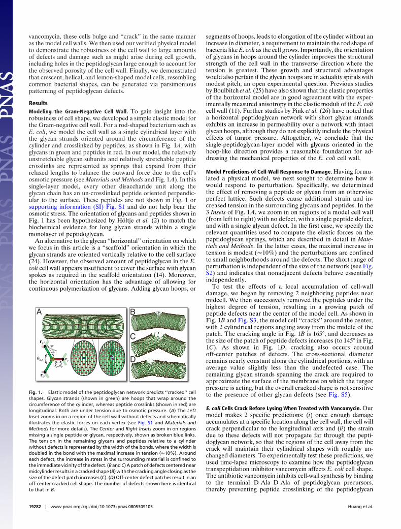
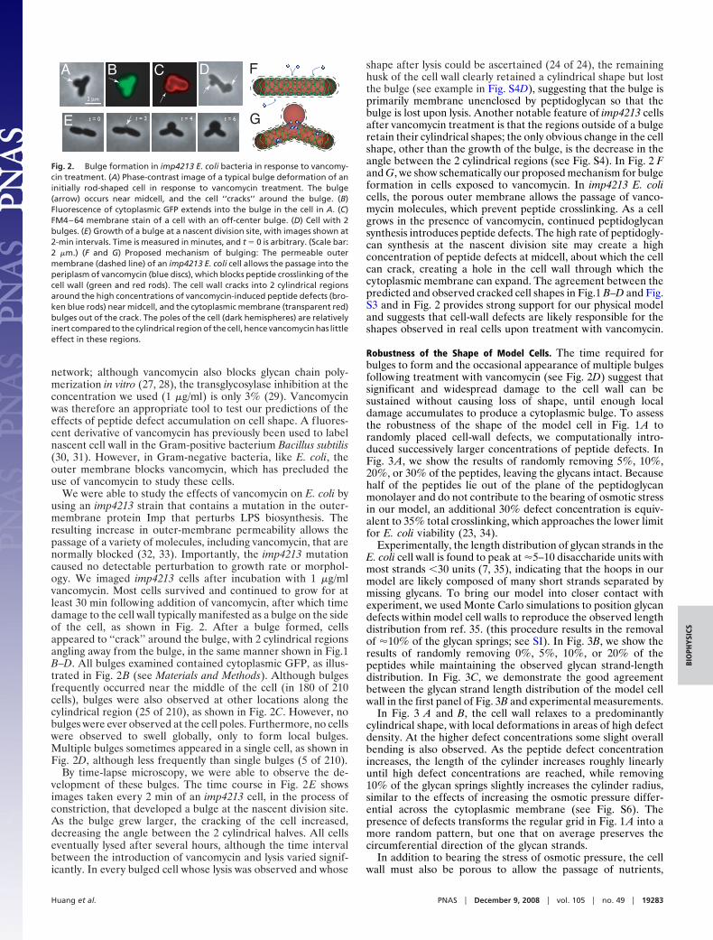
ResultsModeling the Gram-Negative Cell Wall. To gain insight into therobustness of cell shape, we developed a simple elastic model forthe Gram-negative cell wall. For a rod-shaped bacterium such asE. coli, we model the cell wall as a single cylindrical layer withthe glycan strands oriented around the circumference of thecylinder and crosslinked by peptides, as shown in Fig. 1A, withglycans in green and peptides in red. In our model, the relativelyunstretchable glycan subunits and relatively stretchable peptidecrosslinks are represented as springs that expand from theirrelaxed lengths to balance the outward force due to the cell’sosmotic pressure (see Materials and Methods and Fig. 1 A). In thissingle-layer model, every other disaccharide unit along theglycan chain has an un-crosslinked peptide oriented perpendic-ular to the surface. These peptides are not shown in Fig. 1 orsupporting information (SI) Fig. S1 and do not help bear theosmotic stress. The orientation of glycans and peptides shown inFig. 1 has been hypothesized by Holtje et al. (2) to match thebiochemical evidence for long glycan strands within a singlemonolayer of peptidoglycan.
An alternative to the glycan ‘‘horizontal’’ orientation on whichwe focus in this article is a ‘‘scaffold’’ orientation in which theglycan strands are oriented vertically relative to the cell surface(24). However, the observed amount of peptidoglycan in the E.coli cell wall appears insufficient to cover the surface with glycanspokes as required in the scaffold orientation (14). Moreover,the horizontal orientation has the advantage of allowing forcontinuous polymerization of glycans. Adding glycan hoops, or
segments of hoops, leads to elongation of the cylinder without anincrease in diameter, a requirement to maintain the rod shape ofbacteria like E. coli as the cell grows. Importantly, the orientationof glycans in hoops around the cylinder improves the structuralstrength of the cell wall in the transverse direction where thetension is greatest. These growth and structural advantageswould also pertain if the glycan hoops are in actuality spirals withmodest pitch, an open experimental question. Previous studiesby Boulbitch et al. (25) have also shown that the elastic propertiesof the horizontal model are in good agreement with the exper-imentally measured anisotropy in the elastic moduli of the E. colicell wall (11). Further studies by Pink et al. (26) have noted thata horizontal peptidoglycan network with short glycan strandsexhibits an increase in permeability over a network with intactglycan hoops, although they do not explicitly include the physicaleffects of turgor pressure. Altogether, we conclude that thesingle-peptidoglycan-layer model with glycans oriented in thehoop-like direction provides a reasonable foundation for ad-dressing the mechanical properties of the E. coli cell wall.
Model Predictions of Cell-Wall Response to Damage. Having formu-lated a physical model, we next sought to determine how itwould respond to perturbation. Specifically, we determinedthe effect of removing a peptide or glycan from an otherwiseperfect lattice. Such defects cause additional strain and in-creased tension in the surrounding glycans and peptides. In the3 Insets of Fig. 1 A, we zoom in on regions of a model cell wall(from left to right) with no defect, with a single peptide defect,and with a single glycan defect. In the first case, we specify therelevant quantities used to compute the elastic forces on thepeptidoglycan springs, which are described in detail in Mate-rials and Methods. In the latter cases, the maximal increase intension is modest (�10%) and the perturbations are confinedto small neighborhoods around the defects. The short range ofperturbation is independent of the size of the network (see Fig.S2) and indicates that nonadjacent defects behave essentiallyindependently.
To test the effects of a local accumulation of cell-walldamage, we began by removing 2 neighboring peptides nearmidcell. We then successively removed the peptides under thehighest degree of tension, resulting in a growing patch ofpeptide defects near the center of the model cell. As shown inFig. 1B and Fig. S3, the model cell ‘‘cracks’’ around the center,with 2 cylindrical regions angling away from the middle of thepatch. The cracking angle in Fig. 1B is 165°, and decreases asthe size of the patch of peptide defects increases (to 145° in Fig.1C). As shown in Fig. 1D, cracking also occurs aroundoff-center patches of defects. The cross-sectional diameterremains nearly constant along the cylindrical portions, with anaverage value slightly less than the undefected case. Theremaining glycan strands spanning the crack are required toapproximate the surface of the membrane on which the turgorpressure is acting, but the overall cracked shape is not sensitiveto the presence of other glycan defects (see Fig. S5).
E. coli Cells Crack Before Lysing When Treated with Vancomycin. Ourmodel makes 2 specific predictions: (i) once enough damageaccumulates at a specific location along the cell wall, the cell willcrack perpendicular to the longitudinal axis and (ii) the straindue to these defects will not propagate far through the pepti-doglycan network, so that the regions of the cell away from thecrack will maintain their cylindrical shapes with roughly un-changed diameters. To experimentally test these predictions, weused time-lapse microscopy to examine how the peptidoglycantranspeptidation inhibitor vancomycin affects E. coli cell shape.The antibiotic vancomycin inhibits cell-wall synthesis by bindingto the terminal D-Ala–D-Ala of peptidoglycan precursors,thereby preventing peptide crosslinking of the peptidoglycan
A
r
kg ,dg
kp,dp
rg1
θg1g2 rp
rg2
B
C
D
Fig. 1. Elastic model of the peptidoglycan network predicts ‘‘cracked’’ cellshapes. Glycan strands (shown in green) are hoops that wrap around thecircumference of the cylinder, whereas peptide crosslinks (shown in red) arelongitudinal. Both are under tension due to osmotic pressure. (A) The LeftInset zooms in on a region of the cell wall without defects and schematicallyillustrates the elastic forces on each vertex (see Fig. S1 and Materials andMethods for more details). The Center and Right Insets zoom in on regionsmissing a single peptide or glycan, respectively, shown as broken blue links.The tension in the remaining glycans and peptides relative to a cylinderwithout defects is represented by the width of the bonds, where the width isdoubled in the bond with the maximal increase in tension (�10%). Aroundeach defect, the increase in stress in the surrounding material is confined tothe immediate vicinity of the defect. (B and C) A patch of defects centered nearmidcylinder results in a cracked shape (B) with the cracking angle closing as thesize of the defect patch increases (C). (D) Off-center defect patches result in anoff-center cracked cell shape. The number of defects shown here is identicalto that in B.
19282 � www.pnas.org�cgi�doi�10.1073�pnas.0805309105 Huang et al.
network; although vancomycin also blocks glycan chain poly-merization in vitro (27, 28), the transglycosylase inhibition at theconcentration we used (1 �g/ml) is only 3% (29). Vancomycinwas therefore an appropriate tool to test our predictions of theeffects of peptide defect accumulation on cell shape. A fluores-cent derivative of vancomycin has previously been used to labelnascent cell wall in the Gram-positive bacterium Bacillus subtilis(30, 31). However, in Gram-negative bacteria, like E. coli, theouter membrane blocks vancomycin, which has precluded theuse of vancomycin to study these cells.
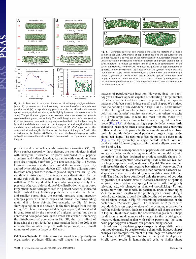
We were able to study the effects of vancomycin on E. coli byusing an imp4213 strain that contains a mutation in the outer-membrane protein Imp that perturbs LPS biosynthesis. Theresulting increase in outer-membrane permeability allows thepassage of a variety of molecules, including vancomycin, that arenormally blocked (32, 33). Importantly, the imp4213 mutationcaused no detectable perturbation to growth rate or morphol-ogy. We imaged imp4213 cells after incubation with 1 �g/mlvancomycin. Most cells survived and continued to grow for atleast 30 min following addition of vancomycin, after which timedamage to the cell wall typically manifested as a bulge on the sideof the cell, as shown in Fig. 2. After a bulge formed, cellsappeared to ‘‘crack’’ around the bulge, with 2 cylindrical regionsangling away from the bulge, in the same manner shown in Fig.1B–D. All bulges examined contained cytoplasmic GFP, as illus-trated in Fig. 2B (see Materials and Methods). Although bulgesfrequently occurred near the middle of the cell (in 180 of 210cells), bulges were also observed at other locations along thecylindrical region (25 of 210), as shown in Fig. 2C. However, nobulges were ever observed at the cell poles. Furthermore, no cellswere observed to swell globally, only to form local bulges.Multiple bulges sometimes appeared in a single cell, as shown inFig. 2D, although less frequently than single bulges (5 of 210).
By time-lapse microscopy, we were able to observe the de-velopment of these bulges. The time course in Fig. 2E showsimages taken every 2 min of an imp4213 cell, in the process ofconstriction, that developed a bulge at the nascent division site.As the bulge grew larger, the cracking of the cell increased,decreasing the angle between the 2 cylindrical halves. All cellseventually lysed after several hours, although the time intervalbetween the introduction of vancomycin and lysis varied signif-icantly. In every bulged cell whose lysis was observed and whose
shape after lysis could be ascertained (24 of 24), the remaininghusk of the cell wall clearly retained a cylindrical shape but lostthe bulge (see example in Fig. S4D), suggesting that the bulge isprimarily membrane unenclosed by peptidoglycan so that thebulge is lost upon lysis. Another notable feature of imp4213 cellsafter vancomycin treatment is that the regions outside of a bulgeretain their cylindrical shapes; the only obvious change in the cellshape, other than the growth of the bulge, is the decrease in theangle between the 2 cylindrical regions (see Fig. S4). In Fig. 2 Fand G, we show schematically our proposed mechanism for bulgeformation in cells exposed to vancomycin. In imp4213 E. colicells, the porous outer membrane allows the passage of vanco-mycin molecules, which prevent peptide crosslinking. As a cellgrows in the presence of vancomycin, continued peptidoglycansynthesis introduces peptide defects. The high rate of peptidogly-can synthesis at the nascent division site may create a highconcentration of peptide defects at midcell, about which the cellcan crack, creating a hole in the cell wall through which thecytoplasmic membrane can expand. The agreement between thepredicted and observed cracked cell shapes in Fig.1 B–D and Fig.S3 and in Fig. 2 provides strong support for our physical modeland suggests that cell-wall defects are likely responsible for theshapes observed in real cells upon treatment with vancomycin.
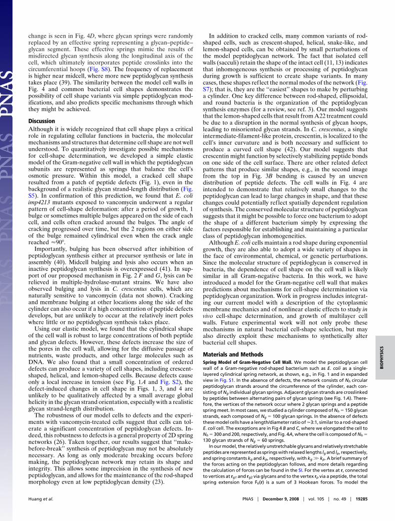
Robustness of the Shape of Model Cells. The time required forbulges to form and the occasional appearance of multiple bulgesfollowing treatment with vancomycin (see Fig. 2D) suggest thatsignificant and widespread damage to the cell wall can besustained without causing loss of shape, until enough localdamage accumulates to produce a cytoplasmic bulge. To assessthe robustness of the shape of the model cell in Fig. 1 A torandomly placed cell-wall defects, we computationally intro-duced successively larger concentrations of peptide defects. InFig. 3 A, we show the results of randomly removing 5%, 10%,20%, or 30% of the peptides, leaving the glycans intact. Becausehalf of the peptides lie out of the plane of the peptidoglycanmonolayer and do not contribute to the bearing of osmotic stressin our model, an additional 30% defect concentration is equiv-alent to 35% total crosslinking, which approaches the lower limitfor E. coli viability (23, 34).
Experimentally, the length distribution of glycan strands in theE. coli cell wall is found to peak at �5–10 disaccharide units withmost strands �30 units (7, 35), indicating that the hoops in ourmodel are likely composed of many short strands separated bymissing glycans. To bring our model into closer contact withexperiment, we used Monte Carlo simulations to position glycandefects within model cell walls to reproduce the observed lengthdistribution from ref. 35. (this procedure results in the removalof �10% of the glycan springs; see SI). In Fig. 3B, we show theresults of randomly removing 0%, 5%, 10%, or 20% of thepeptides while maintaining the observed glycan strand-lengthdistribution. In Fig. 3C, we demonstrate the good agreementbetween the glycan strand length distribution of the model cellwall in the first panel of Fig. 3B and experimental measurements.
In Fig. 3 A and B, the cell wall relaxes to a predominantlycylindrical shape, with local deformations in areas of high defectdensity. At the higher defect concentrations some slight overallbending is also observed. As the peptide defect concentrationincreases, the length of the cylinder increases roughly linearlyuntil high defect concentrations are reached, while removing10% of the glycan springs slightly increases the cylinder radius,similar to the effects of increasing the osmotic pressure differ-ential across the cytoplasmic membrane (see Fig. S6). Thepresence of defects transforms the regular grid in Fig. 1 A into amore random pattern, but one that on average preserves thecircumferential direction of the glycan strands.
In addition to bearing the stress of osmotic pressure, the cellwall must also be porous to allow the passage of nutrients,
v
v
v
v
v vE
A B C D
t = 0 t = 2 t = 4 t = 6
2 µmv
v vvF
G
Fig. 2. Bulge formation in imp4213 E. coli bacteria in response to vancomy-cin treatment. (A) Phase-contrast image of a typical bulge deformation of aninitially rod-shaped cell in response to vancomycin treatment. The bulge(arrow) occurs near midcell, and the cell ‘‘cracks’’ around the bulge. (B)Fluorescence of cytoplasmic GFP extends into the bulge in the cell in A. (C)FM4–64 membrane stain of a cell with an off-center bulge. (D) Cell with 2bulges. (E) Growth of a bulge at a nascent division site, with images shown at2-min intervals. Time is measured in minutes, and t � 0 is arbitrary. (Scale bar:2 �m.) (F and G) Proposed mechanism of bulging: The permeable outermembrane (dashed line) of an imp4213 E. coli cell allows the passage into theperiplasm of vancomycin (blue discs), which blocks peptide crosslinking of thecell wall (green and red rods). The cell wall cracks into 2 cylindrical regionsaround the high concentrations of vancomycin-induced peptide defects (bro-ken blue rods) near midcell, and the cytoplasmic membrane (transparent red)bulges out of the crack. The poles of the cell (dark hemispheres) are relativelyinert compared to the cylindrical region of the cell, hence vancomycin has littleeffect in these regions.
Huang et al. PNAS � December 9, 2008 � vol. 105 � no. 49 � 19283
BIO
PHYS
ICS
proteins, and even nucleic acids during transformation (36, 37).For a perfect network without defects, the peptidoglyan is tiledwith hexagonal ‘‘tesserae’’ or pores composed of 2 peptidecrosslinks and 4 disaccharide glycan units with a small, uniformpore size (roughly 5 nm2 for lp � 1 nm; see, e.g., Fig. 1 A Insets).However, previous studies have noted the increase in porositycaused by peptidoglycan defects (26), which link adjacent poresto create new pores with more edges and larger area. In Fig. 3D,we show a histogram of the tessera area distribution for themodel cell walls in the topmost and bottom images of Fig. 3Bwith 0 and 20% peptide defect concentrations, respectively. Thepresence of glycan defects alone (blue distribution) creates poreslarger than the uniform pore area in a perfect network (indicatedby the dashed line). Adding peptide defects creates both largerand smaller pores, since the osmotic pressure simultaneouslyenlarges pores with more edges and shrinks the surroundingmaterial if it lacks defects. For example, see Fig. 3D Inset,showing a region of the network in the third image from the topin Fig. 3B. There is an expanded decagonal pore, shown shadedin gray, formed by the removal of a glycan spring, but also acontracted hexagonal pore in the lower left corner. Comparingthe distributions of pore sizes in Fig. 3D, the main effect ofincreasing the peptide defect concentration from 0 to 20% is toincrease the number of pores with large areas, with smallnumbers of pores as large as 400 nm2.
Cell-Shape Variants. To date, consideration of how peptidoglycanorganization produces different cell shapes has focused on
patterns of peptidoglycan insertion. However, since the pepti-doglycan network appears capable of tolerating a large numberof defects, we decided to explore the possibility that specificpatterns of defects could induce specific cell shapes. We noticedthat the bending of the cylinders in Figs. 1 and 3 is reminiscentof the flexing of an elastic tube. For such a tube, certaindeformations (modes) require less energy than others to exciteto a given amplitude. Indeed, the most flexible mode of apeptidoglycan network similar to the one in Fig. 1 A is a bendmode (Fig. S7A). Although a single peptide defect causes littlechange to local shape, such a defect near midcell does contributeto this bend mode. In principle, the accumulation of bend frommultiple peptide defects could produce a large change in theglobal cell shape. The next most flexible mode is a twist mode(Fig. S7B). By symmetry, a single peptide defect does notproduce twist. However, a glycan defect at midcell produces bothbend and twist.
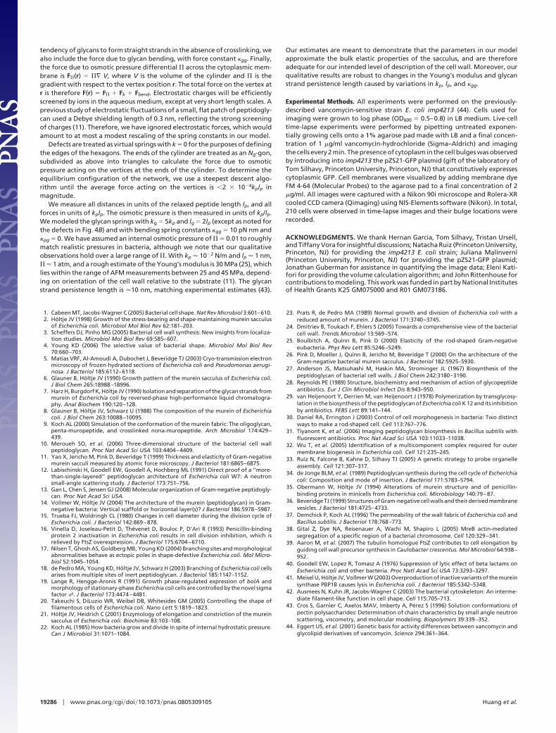
Guided by the correspondence of peptide defects with bendingand glycan defects with twisting, we modified our model cell withcollections of defects designed to produce specific shapes. In-troducing lines of peptide defects along 1 side of the cell resultedin a large amplitude bend, as shown in Fig. 4A. The resulting cellwall resembles the Gram-negative bacterium C. crescentus. Thisresult prompted us to ask whether other common bacterial cellshapes could also be produced by local modifications of the cellwall. Thus far, we have considered only the removal of peptidesor glycans, but a wider class of defects consisting of spatiallyvarying spring constants or spring lengths is both biologicallyrelevant, e.g., via changes in chemical crosslinking (3), andaccessible within our model. In particular, upon shortening by75% the relaxed lengths of the peptidoglycan springs along ahelical path winding around the cylinder, the cell relaxes into thehelical shape shown in Fig. 4B, resembling spirochaetes or thebacterium Helicobacter pylori. The removal of 2 patches ofpeptide defects on opposite sides of the cylinder, separated byhalf the cylinder length, leads to a snakelike cell shape as shownin Fig. 4C. In all these cases, the observed changes in cell shaperesult from a small number of changes to the peptidoglycannetwork, demonstrating that with subtle patterning, the samebasic peptidoglycan network can produce many shapes.
In addition to explaining species-specific shape differences,our model can also be used to explore chemically-induced shapeschanges. For example, treatment of Gram-negative bacteria withthe chemical A22 (38), an inhibitor of the cytoskeletal proteinMreB, often results in lemon-shaped cells. A similar shape
peptide defects only
10%
peptide and glycan defects
10%10%20%
5%
30%
0%10%
5%10%
20%10%
R = 1.0R0 L = 1.04L0
R = 1.0R0 L = 1.08L0
R = 1.0R0 L = 1.20L0
R = 1.0R0 L = 1.39L0
R = 1.03R0 L = 1.10L0
R = 1.04R0 L = 1.23L0
R = 1.03R0 L = 1.17L0
R = 1.04R0 L = 1.49L0
5 10 15 20 25 30 35 400
0% peptide defect concentration20% peptide defect concentration
Pore area (nm2)
Num
ber
of p
ores
5 10 15 20 25 30
0.01
0.02
0.03
0.04
0.05
0.06
00
Length distribution from Ref. [40]Modeled length distribution
Glycan strand length
Frac
tion
of g
lyca
ns
glycan strand-length distribution
0
500
1000
1500
2000
2500
3000
3500
pore-size distributionDC
A B
Fig. 3. Robustness of the shape of a model cell with peptidoglycan defects.(A and B) Upon removal of an increasing concentration of randomly chosenpeptide bonds (A) or peptide and glycan bonds (B), the cell wall maintains anapproximately cylindrical shape, with slightly increased dimensions as indi-cated. The peptide and glycan defect concentrations are shown as percent-ages in red and green, respectively. The radii, lengths, and defect concentra-tions are measured relative to a single-layer cell wall with radius R0 and lengthL0. In B, the defects are chosen so that the glycan strand-length distributionmatches the experimental distribution from ref. 35. (C) Comparison of thecomputed strand-length distribution of the topmost image in B with theexperimental distribution. (D) The glycan defects in B create large pores in thecell wall; shown are the distributions of pore areas in the topmost and bottomimages.
D
A
C
B
Fig. 4. Common bacterial cell shapes generated via defects in a modelcylindrical cell wall. (A) Removal of peptide bonds along the top surface of thecylinder results in a curved cell shape reminiscent of Caulobacter crescentus.(B) A reduction in the relaxed lengths of peptides and glycans along a helicalpath generates a helical cell shape similar to that of spirochaetes or thebacterium Helicobacter pylori. (C) Removal of 2 patches of peptide defects onopposite sides of the cylinder, separated by half the cylinder length, creates asnakelike cell shape similar to vancomycin-treated E. coli cells with multiplebulges. (D) Increased substitution of glycan–peptide–glycan segments in placeof glycans near the midplane of the cell creates a swelled cylinder, similar tothe lemon shape of cylindrical Gram-negative bacteria after treatment withthe MreB inhibitor A22.
19284 � www.pnas.org�cgi�doi�10.1073�pnas.0805309105 Huang et al.
change is seen in Fig. 4D, where glycan springs were randomlyreplaced by an effective spring representing a glycan–peptide–glycan segment. These effective springs mimic the results ofmisdirected glycan synthesis along the longitudinal axis of thecell, which ultimately incorporates peptide crosslinks into thecircumferential hoops (Fig. S8). The frequency of replacementis higher near midcell, where more new peptidoglycan synthesistakes place (39). The similarity between the model cell walls inFig. 4 and common bacterial cell shapes demonstrates thepossibility of cell shape variants via simple peptidoglycan mod-ifications, and also predicts specific mechanisms through whichthey might be achieved.
DiscussionAlthough it is widely recognized that cell shape plays a criticalrole in regulating cellular functions in bacteria, the molecularmechanisms and structures that determine cell shape are not wellunderstood. To quantitatively investigate possible mechanismsfor cell-shape determination, we developed a simple elasticmodel of the Gram-negative cell wall in which the peptidoglycansubunits are represented as springs that balance the cell’sosmotic pressure. Within this model, a cracked cell shaperesulted from a patch of peptide defects (Fig. 1), even in thebackground of a realistic glycan strand-length distribution (Fig.S5). In confirmation of this prediction, we found that E. coliimp4213 mutants exposed to vancomycin underwent a regularpattern of cell-shape deformation: after a period of growth, 1bulge or sometimes multiple bulges appeared on the side of eachcell, and cells often cracked around the bulges. The angle ofcracking progressed over time, but the 2 regions on either sideof the bulge remained cylindrical even when the crack anglereached �90°.
Importantly, bulging has been observed after inhibition ofpeptidoglycan synthesis either at precursor synthesis or late inassembly (40). Midcell bulging and lysis also occurs when aninactive peptidoglycan synthesis is overexpressed (41). In sup-port of our proposed mechanism in Fig. 2 F and G, lysis can berelieved in multiple-hydrolase-mutant strains. We have alsoobserved bulging and lysis in C. crescentus cells, which arenaturally sensitive to vancomycin (data not shown). Crackingand membrane bulging at other locations along the side of thecylinder can also occur if a high concentration of peptide defectsdevelops, but are unlikely to occur at the relatively inert poleswhere little or no peptidoglycan synthesis takes place.
Using our elastic model, we found that the cylindrical shapeof the cell wall is robust to large concentrations of both peptideand glycan defects. However, these defects increase the size ofthe pores in the cell wall, allowing for the diffusive passage ofnutrients, waste products, and other large molecules such asDNA. We also found that a small concentration of ordereddefects can produce a variety of cell shapes, including crescent-shaped, helical, and lemon-shaped cells. Because defects causeonly a local increase in tension (see Fig. 1 A and Fig. S2), thedefect-induced changes in cell shape in Figs. 1, 3, and 4 areunlikely to be qualitatively affected by a small average globalhelicity in the glycan strand orientation, especially with a realisticglycan strand-length distribution.
The robustness of our model cells to defects and the experi-ments with vancomycin-treated cells suggest that cells can tol-erate a significant concentration of peptidoglycan defects. In-deed, this robustness to defects is a general property of 2D springnetworks (26). Taken together, our results suggest that ‘‘make-before-break’’ synthesis of peptidoglycan may not be absolutelynecessary. As long as only moderate breaking occurs beforemaking, the peptidoglycan network may retain its shape andintegrity. This allows some imprecision in the synthesis of newpeptidoglycan, and allows for the maintenance of the rod-shapedmorphology even at low peptidoglycan density (23).
In addition to cracked cells, many common variants of rod-shaped cells, such as crescent-shaped, helical, snake-like, andlemon-shaped cells, can be obtained by small perturbations ofthe model peptidoglycan network. The fact that isolated cellwalls (sacculi) retain the shape of the intact cell (11, 13) indicatesthat inhomogeneous synthesis or processing of peptidoglycanduring growth is sufficient to create shape variants. In manycases, these shapes reflect the normal modes of the network (Fig.S7); that is, they are the ‘‘easiest’’ shapes to make by perturbinga cylinder. One key difference between rod-shaped, ellipsoidal,and round bacteria is the organization of the peptidoglycansynthesis enzymes (for a review, see ref. 3). Our model suggeststhat the lemon-shaped cells that result from A22 treatment couldbe due to a disruption in the normal synthesis of glycan hoops,leading to misoriented glycan strands. In C. crescentus, a singleintermediate-filament-like protein, crescentin, is localized to thecell’s inner curvature and is both necessary and sufficient toproduce a curved cell shape (42). Our model suggests thatcrescentin might function by selectively stabilizing peptide bondson one side of the cell surface. There are other related defectpatterns that produce similar shapes, e.g., in the second imagefrom the top in Fig. 3B bending is caused by an unevendistribution of peptide defects. The cell walls in Fig. 4 areintended to demonstrate that relatively small changes to thepeptidoglycan can lead to large changes in shape, and that thesechanges could potentially reflect spatially dependent regulationof synthesis. The conserved molecular structure of peptidoglycansuggests that it might be possible to force one bacterium to adoptthe shape of a different bacterium simply by expressing thefactors responsible for establishing and maintaining a particularclass of peptidoglycan inhomogeneities.
Although E. coli cells maintain a rod shape during exponentialgrowth, they are also able to adopt a wide variety of shapes inthe face of environmental, chemical, or genetic perturbations.Since the molecular structure of peptidoglycan is conserved inbacteria, the dependence of cell shape on the cell wall is likelysimilar in all Gram-negative bacteria. In this work, we haveintroduced a model for the Gram-negative cell wall that makespredictions about mechanisms for cell-shape determination viapeptidoglycan organization. Work in progress includes integrat-ing our current model with a description of the cytoplasmicmembrane mechanics and of nonlinear elastic effects to study invivo cell-shape determination, and growth of multilayer cellwalls. Future experimental work will not only probe thesemechanisms in natural bacterial cell-shape selection, but mayalso directly exploit these mechanisms to synthetically alterbacterial cell shapes.
Materials and MethodsSpring Model of Gram-Negative Cell Wall. We model the peptidoglycan cellwall of a Gram-negative rod-shaped bacterium such as E. coli as a single-layered cylindrical spring network, as shown, e.g., in Fig. 1 and in expandedview in Fig. S1. In the absence of defects, the network consists of Nh circularpeptidoglycan strands around the circumference of the cylinder, each con-sisting of Ng individual glycan springs. Adjacent glycan strands are crosslinkedby peptides between alternating pairs of glycan springs (see Fig. 1A). There-fore, the vertices of the network occur where 2 glycan springs and a peptidespring meet. In most cases, we studied a cylinder composed of Nh � 150 glycanstrands, each composed of Ng � 100 glycan springs. In the absence of defectsthese model cells have a length/diameter ratio of �3:1, similar to a rod-shapedE. coli cell. The exceptions are in Fig 4 B and C, where we elongated the cell toNh � 300 and 200, respectively, and Fig. 4A, where the cell is composed of Nh �130 glycan strands of Ng � 60 springs.
In our model, the relatively unstretchable glycans and relatively stretchablepeptides are represented as springs with relaxed lengths lg and lp, respectively,and spring constants kg and kp, respectively, with kg �� kp. A brief summary ofthe forces acting on the peptidoglycan follows, and more details regardingthe calculation of forces can be found in the SI. For the vertex at r, connectedto vertices at rg1 and rg2 via glycans and to the vertex rp via a peptide, the totalspring extension force Fk(r) is a sum of 3 Hookean forces. To model the
Huang et al. PNAS � December 9, 2008 � vol. 105 � no. 49 � 19285
BIO
PHYS
ICS
tendency of glycans to form straight strands in the absence of crosslinking, wealso include the force due to glycan bending, with force constant �gg. Finally,the force due to osmotic pressure differential � across the cytoplasmic mem-brane is F�(r) � �� V, where V is the volume of the cylinder and � is thegradient with respect to the vertex position r. The total force on the vertex atr is therefore F(r) � F� � Fk � Fbend. Electrostatic charges will be efficientlyscreened by ions in the aqueous medium, except at very short length scales. Aprevious study of electrostatic fluctuations of a small, flat patch of peptidogly-can used a Debye shielding length of 0.3 nm, reflecting the strong screeningof charges (11). Therefore, we have ignored electrostatic forces, which wouldamount to at most a modest rescaling of the spring constants in our model.
Defects are treated as virtual springs with k � 0 for the purposes of definingthe edges of the hexagons. The ends of the cylinder are treated as an Ng-gon,subdivided as above into triangles to calculate the force due to osmoticpressure acting on the vertices at the ends of the cylinder. To determine theequilibrium configuration of the network, we use a steepest descent algo-rithm until the average force acting on the vertices is �2 � 104kplp inmagnitude.
We measure all distances in units of the relaxed peptide length lp, and allforces in units of kplp. The osmotic pressure is then measured in units of kp/lp.We modeled the glycan springs with kg � 5kp and lg � 2lp (except as noted forthe defects in Fig. 4B) and with bending spring constants �gg � 10 pN nm and�pg � 0. We have assumed an internal osmotic pressure of � � 0.01 to roughlymatch realistic pressures in bacteria, although we note that our qualitativeobservations hold over a large range of �. With kp � 102 N/m and lp � 1 nm,� � 1 atm, and a rough estimate of the Young’s modulus is 30 MPa (25), whichlies within the range of AFM measurements between 25 and 45 MPa, depend-ing on orientation of the cell wall relative to the substrate (11). The glycanstrand persistence length is �10 nm, matching experimental estimates (43).
Our estimates are meant to demonstrate that the parameters in our modelapproximate the bulk elastic properties of the sacculus, and are thereforeadequate for our intended level of description of the cell wall. Moreover, ourqualitative results are robust to changes in the Young’s modulus and glycanstrand persistence length caused by variations in kp, lp, and �gg.
Experimental Methods. All experiments were performed on the previously-described vancomycin-sensitive strain E. coli imp4213 (44). Cells used forimaging were grown to log phase (OD600 � 0.5–0.8) in LB medium. Live-celltime-lapse experiments were performed by pipetting untreated exponen-tially growing cells onto a 1% agarose pad made with LB and a final concen-tration of 1 �g/ml vancomycin-hydrochloride (Sigma–Aldrich) and imagingthe cells every 2 min. The presence of cytoplasm in the cell bulges was observedby introducing into imp4213 the pZS21-GFP plasmid (gift of the laboratory ofTom Silhavy, Princeton University, Princeton, NJ) that constitutively expressescytoplasmic GFP. Cell membranes were visualized by adding membrane dyeFM 4-64 (Molecular Probes) to the agarose pad to a final concentration of 2�g/ml. All images were captured with a Nikon 90i microscope and Rolera-XRcooled CCD camera (Qimaging) using NIS-Elements software (Nikon). In total,210 cells were observed in time-lapse images and their bulge locations wererecorded.
ACKNOWLEDGMENTS. We thank Hernan Garcia, Tom Silhavy, Tristan Ursell,and Tiffany Vora for insightful discussions; Natacha Ruiz (Princeton University,Princeton, NJ) for providing the imp4213 E. coli strain; Juliana Malinverni(Princeton University, Princeton, NJ) for providing the pZS21-GFP plasmid;Jonathan Guberman for assistance in quantifying the image data; Eleni Kati-fori for providing the volume calculation algorithm; and John Rittenhouse forcontributions to modeling. This work was funded in part by National Institutesof Health Grants K25 GM075000 and R01 GM073186.
1. Cabeen MT, Jacobs-Wagner C (2005) Bacterial cell shape. Nat Rev Microbiol 3:601–610.2. Holtje JV (1998) Growth of the stress-bearing and shape-maintaining murein sacculus
of Escherichia coli. Microbiol Mol Biol Rev 62:181–203.3. Scheffers DJ, Pinho MG (2005) Bacterial cell wall synthesis: New insights from localiza-
tion studies. Microbiol Mol Biol Rev 69:585–607.4. Young KD (2006) The selective value of bacterial shape. Microbiol Mol Biol Rev
70:660–703.5. Matias VRF, Al-Amoudi A, Dubochet J, Beveridge TJ (2003) Cryo-transmission electron
microscopy of frozen-hydrated sections of Escherichia coli and Pseudomonas aerugi-nosa. J Bacteriol 185:6112–6118.
6. Glauner B, Holtje JV (1990) Growth pattern of the murein sacculus of Escherichia coli.J Biol Chem 265:18988–18996.
7. Harz H, Burgdorf K, Holtje JV (1990) Isolation and separation of the glycan strands frommurein of Escherichia coli by reversed-phase high-performance liquid chromatogra-phy. Anal Biochem 190:120–128.
8. Glauner B, Holtje JV, Schwarz U (1988) The composition of the murein of Escherichiacoli. J Biol Chem 263:10088–10095.
9. Koch AL (2000) Simulation of the conformation of the murein fabric: The oligoglycan,penta-muropeptide, and crosslinked nona-muropeptide. Arch Microbiol 174:429–439.
10. Meroueh SO, et al. (2006) Three-dimensional structure of the bacterial cell wallpeptidoglycan. Proc Nat Acad Sci USA 103:4404–4409.
11. Yao X, Jericho M, Pink D, Beveridge T (1999) Thickness and elasticity of Gram-negativemurein sacculi measured by atomic force microscopy. J Bacteriol 181:6865–6875.
12. Labischinski H, Goodell EW, Goodell A, Hochberg ML (1991) Direct proof of a ‘‘more-than-single-layered’’ peptidoglycan architecture of Escherichia coli W7: A neutronsmall-angle scattering study. J Bacteriol 173:751–756.
13. Gan L, Chen S, Jensen GJ (2008) Molecular organization of Gram-negative peptidogly-can. Proc Nat Acad Sci USA.
14. Vollmer W, Holtje JV (2004) The architecture of the murein (peptidoglycan) in Gram-negative bacteria: Vertical scaffold or horizontal layer(s)? J Bacteriol 186:5978–5987.
15. Trueba FJ, Woldringh CL (1980) Changes in cell diameter during the division cycle ofEscherichia coli. J Bacteriol 142:869–878.
16. Vinella D, Joseleau-Petit D, Thevenet D, Bouloc P, D’Ari R (1993) Penicillin-bindingprotein 2 inactivation in Escherichia coli results in cell division inhibition, which isrelieved by FtsZ overexpression. J Bacteriol 175:6704–6710.
17. Nilsen T, Ghosh AS, Goldberg MB, Young KD (2004) Branching sites and morphologicalabnormalities behave as ectopic poles in shape-defective Escherichia coli. Mol Micro-biol 52:1045–1054.
18. de Pedro MA, Young KD, Holtje JV, Schwarz H (2003) Branching of Escherichia coli cellsarises from multiple sites of inert peptidoglycan. J Bacteriol 185:1147–1152.
19. Lange R, Hengge-Aronis R (1991) Growth phase-regulated expression of bolA andmorphology of stationary-phase Escherichia coli cells are controlled by the novel sigmafactor �s. J Bacteriol 173:4474–4481.
20. Takeuchi S, DiLuzio WR, Weibel DB, Whitesides GM (2005) Controlling the shape offilamentous cells of Escherichia coli. Nano Lett 5:1819–1823.
21. Holtje JV, Heidrich C (2001) Enzymology of elongation and constriction of the mureinsacculus of Escherichia coli. Biochimie 83:103–108.
22. Koch AL (1985) How bacteria grow and divide in spite of internal hydrostatic pressure.Can J Microbiol 31:1071–1084.
23. Prats R, de Pedro MA (1989) Normal growth and division of Escherichia coli with areduced amount of murein. J Bacteriol 171:3740–3745.
24. Dmitriev B, Toukach F, Ehlers S (2005) Towards a comprehensive view of the bacterialcell wall. Trends Microbiol 13:569–574.
25. Boulbitch A, Quinn B, Pink D (2000) Elasticity of the rod-shaped Gram-negativeeubacteria. Phys Rev Lett 85:5246–5249.
26. Pink D, Moeller J, Quinn B, Jericho M, Beveridge T (2000) On the architecture of theGram-negative bacterial murein sacculus. J Bacteriol 182:5925–5930.
27. Anderson JS, Matsuhashi M, Haskin MA, Strominger JL (1967) Biosynthesis of thepeptidoglycan of bacterial cell walls. J Biol Chem 242:3180–3190.
28. Reynolds PE (1989) Structure, biochemistry and mechanism of action of glycopeptideantibiotics. Eur J Clin Microbiol Infect Dis 8:943–950.
29. van Heijenoort Y, Derrien M, van Heijenoort J (1978) Polymerization by transglycosy-lation in the biosynthesis of the peptidoglycan of Escherichia coli K 12 and its inhibitionby antibiotics. FEBS Lett 89:141–144.
30. Daniel RA, Errington J (2003) Control of cell morphogenesis in bacteria: Two distinctways to make a rod-shaped cell. Cell 113:767–776.
31. Tiyanont K, et al. (2006) Imaging peptidoglycan biosynthesis in Bacillus subtilis withfluorescent antibiotics. Proc Nat Acad Sci USA 103:11033–11038.
32. Wu T, et al. (2005) Identification of a multicomponent complex required for outermembrane biogenesis in Escherichia coli. Cell 121:235–245.
33. Ruiz N, Falcone B, Kahne D, Silhavy TJ (2005) A genetic strategy to probe organelleassembly. Cell 121:307–317.
34. de Jonge BLM, et al. (1989) Peptidoglycan synthesis during the cell cycle of Escherichiacoli: Composition and mode of insertion. J Bacteriol 171:5783–5794.
35. Obermann W, Holtje JV (1994) Alterations of murein structure and of penicillin-binding proteins in minicells from Escherichia coli. Microbiology 140:79–87.
36. Beveridge TJ (1999) Structures of Gram-negative cell walls and their derived membranevesicles. J Bacteriol 181:4725–4733.
37. Demchick P, Koch AL (1996) The permeability of the wall fabric of Escherichia coli andBacillus subtilis. J Bacteriol 178:768–773.
38. Gitai Z, Dye NA, Reisenauer A, Wachi M, Shapiro L (2005) MreB actin-mediatedsegregation of a specific region of a bacterial chromosome. Cell 120:329–341.
39. Aaron M, et al. (2007) The tubulin homologue FtsZ contributes to cell elongation byguiding cell wall precursor synthesis in Caulobacter crescentus. Mol Microbiol 64:938–952.
40. Goodell EW, Lopez R, Tomasz A (1976) Suppression of lytic effect of beta lactams onEscherichia coli and other bacteria. Proc Natl Acad Sci USA 73:3293–3297.
41. Meisel U, Holtje JV, Vollmer W (2003) Overproduction of inactive variants of the mureinsynthase PBP1B causes lysis in Escherichia coli. J Bacteriol 185:5342–5348.
42. Ausmees N, Kuhn JR, Jacobs-Wagner C (2003) The bacterial cytoskeleton: An interme-diate filament-like function in cell shape. Cell 115:705–713.
43. Cros S, Garnier C, Axelos MAV, Imberty A, Perez S (1996) Solution conformations ofpectin polysaccharides: Determination of chain characteristics by small angle neutronscattering, viscometry, and molecular modeling. Biopolymers 39:339–352.
44. Eggert US, et al. (2001) Genetic basis for activity differences between vancomycin andglycolipid derivatives of vancomycin. Science 294:361–364.
19286 � www.pnas.org�cgi�doi�10.1073�pnas.0805309105 Huang et al.