Embed Size (px)
Citation preview

BIO 199: Basic Anatomy and Physiology Lab Lab 3: Handout
Page 1 of 8
Cell Membrane and Transport Review Sheet
Transport of nutrients, ions, and excretory substances from one side to the other is a major function of
the cell membrane. A number of different means have been developed to fulfill this function.
Generally, the permeation of small molecules across the membrane is quite different from engulfing
molecules too large to penetrate membrane.
Transport of small molecules
Depending on whether a cell pays for the transport (energetically) or not, we talk about passive (free)
and active transport.
Passive transport
no sweat
Simple diffusion
no sweat AND no helpers !
The simplest form of transport is passive diffusion. It is a real freebie; it does not even need helpers.
Water diffusion: osmosis
Lipid membranes are semi-permeable; some substances pass through freely (water) some don’t (ions).
Membrane transport
Small molecules
Large molecules
water
nonpolar
simple diffusion facilitated diffusion
active transport
cootransporters
exocytosis pinocytosisendocytosis
pumps
passive transport
ions
permeases
channels

BIO 199: Basic Anatomy and Physiology Lab Lab 3: Handout
Page 2 of 8
Consider two water solutions, one rich in ions and the other not, which are separated by a semi-
permeable membrane. Water can move across the membrane in both directions, but because ions
attract water and impede its random diffusion, water is retarded on the ion-rich side, therefore the rate
from the ion-rich side is less than the rate of ions permeating the membrane from the other side.
The net movement of water toward the ion-rich solution builds up hydrostatic pressure, called osmotic
pressure, which at some point will counteract the attraction of ions. The two sides will then be at
equilibrium.
Whenever two solutions are separated by a semi-permeable membrane, net movement of water will be
toward the solution more concentrated in substances that do not permeate the membrane.

BIO 199: Basic Anatomy and Physiology Lab Lab 3: Handout
Page 3 of 8
We say, the more concentrated solution is hypertonic with respect to solution less rich in the
impermeant substance. The water will always try to rush in to make the more concentrated solution less
hypertonic.
The less concentrated solution is referred to as hypotonic, water will attempt to leave this compartment
and thereby decrease concentration of impermeant solute.
When two compartments are equally concentrated they are isotonic with respect to each other, and
there is no net diffusion of water.
Diffusion through semipermeable
membrane
BIO 199: Basic Anatomy and Physiology Lab Lab 3: Handout
Page 4 of 8
Passive diffusion of water regulated size of lymphocytes (white blood cells): when a lymphocyte is
placed in a hypertonic solution the cell shrinks internal pH decreases export of H+ and HCO3-
and import of Na+ and Cl- are triggered intracellular NaCl increases water flows in the cell
swells up, countering the original shrinkage.
Uncharged molecules
Facilitated diffusion
helpers, cars and channels
Some molecules diffuse freely but with the help of another molecule.
BIO 199: Basic Anatomy and Physiology Lab Lab 3: Handout
Page 5 of 8
The kinetics of facilitated (with a helper) transport are different from those of simple diffusion. In the
latter, the rate of diffusion is proportional to the concentration of the diffusing molecules; the more of
them, the more of them will diffuse across the membrane per unit time.
In facilitated diffusion, however, the rate is limited by the availability of the helper molecules. Once all
the helpers are saturated, the increasing concentration of diffusing molecules will only increase a
waiting line for the helper and will not increase rate of transport.
Such a saturation kinetics is characteristic of any event (transport, chemical reaction) that requires the
help of other molecules.
Active transport
no pain - no gain
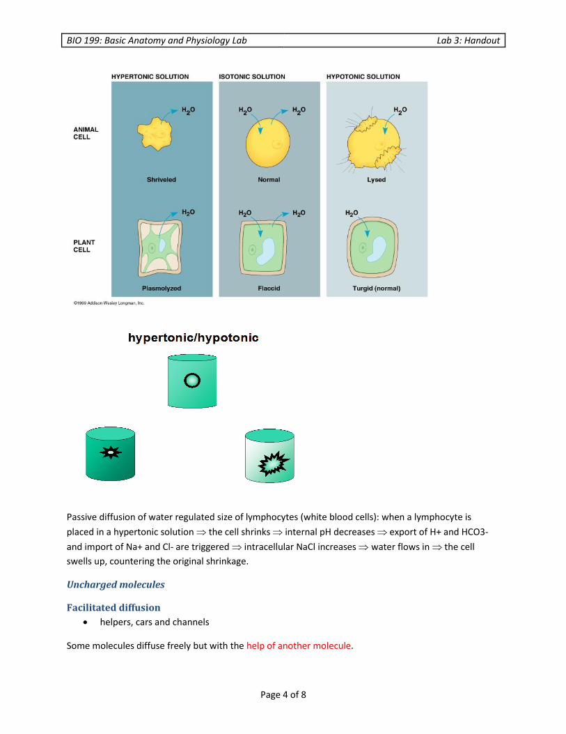
Often the transport has to happen in the direction opposite to the concentration gradient. In order to
accomplish this, membranes have evolved elaborate schemes to pump the substance from the area of
smaller concentration to a compartment with higher concentration. All these schemes cost the cell
energy and thus are called active transport.
Examples are ATPase and co-transporters.
Na/K ATPase (pump)
This pump is an ATPase, which means that the enzyme derives its energy from the hydrolysis of ATP.
The function of Na/K ATPase is to set up the electrochemical gradient of the membrane.
It does so by pumping Na out of the cell and pumping K into the cell.
The net effect is to create a chemical potential consisting of two concentration gradients (for Na and for
K), as well as electrical potential because three positive charges are pumped out while two positive
charges are pumped in. A negative potential inside the cell is thus created.

BIO 199: Basic Anatomy and Physiology Lab Lab 3: Handout
Page 6 of 8
Mechanism: inside of the cell, Na binding triggers phosphorylation by ATP eversion to outside of the
cell Na release K binding triggers dephosphorylation inversion to inside of the cell K release.
Transport of large molecules
Membranes transport molecules too big to permeate the membrane by engulfing the substance and
forming internal vesicles.
Uptake of substances by such a mechanism is called endocytosis; the secretion is called exocytosis.
Exocytosis
In exocytosis, the transport vesicle fuses with the plasma membrane, making the inside of the vesicle
continuous with the outside of the cell.

BIO 199: Basic Anatomy and Physiology Lab Lab 3: Handout
Page 7 of 8
Exocytosis is used in secretion of protein hormones (insulin), serum proteins, extracellular matrix
(collagen).
Endocytosis
Endocytosis occurs mainly in animal cells, as plants have rigid cell walls.
Mechanism:
Receptor-mediated endocytosis
Uptake of cholesterol proceeds as follows: LDL (cholesterol-containing particles) are recognized by the
receptors on the surface of the cell receptors aggregate clathrin crosslinks the ends of receptors
on the interior side of membrane, forcing the curvature membrane pinches off to form a clathrin-
coated vesicle clathrin falls off to form an uncoated vesicle: an endosome fusion with CURL vesicle
low pH releases the receptor which recycles receptor to the membrane.
Phagocytosis
Removal of foreign materials or dead cells by immune cells is a form of endocytosis.
exocytosis

BIO 199: Basic Anatomy and Physiology Lab Lab 3: Handout
Page 8 of 8
For example, phagocytes are macrophages that line blood channels of liver (spleen) and eat up aging
rbc's; monocytes penetrate inflamed tissue and remove the invading bacteria.
a complement reaction labels
antibody for recognition by the antibody receptors in the membrane of macrophages formation of
pseudopodia particles are engulfed by binding of antibodies by receptors.
Pinocytosis
Pinocytosis is a nonspecific uptake of extracellular solution. Whatever is in the solution is taken up by
the cell.
© Heidi Peterson and Indian Hills Community College
pinocytosis