Embed Size (px)
Citation preview
JOURNAL OF VIROLOGY, JUlY 1988, p. 2490-2497 Vol. 62, No. 70022-538X/88/072490-08$02.00/0Copyright © 1988, American Society for Microbiology
Cell-Mediated Immunity Induced in Mice after Vaccination with aProtease Activation Mutant, TR-2, of Sendai Virus
MASATO TASHIRO,'t* YOICHI FUJII,2t KIYOTO NAKAMURA,1 AND MORIO HOMMA3Department of Bacteriology1 and Department of Pathology,2 Yamagata University School of Medicine, Zao-Iida,
Yamagata 990-23, and Department of Microbiology, Kobe University School of Medicine, Chuo-ku, Kobe,Hyogo 650,3 Japan
Received 28 December 1987/Accepted 29 March 1988
Our previous study has shown that, although a trypsin-resistant mutant of Sendai virus, TR-2, replicatesonly in a single cycle in mouse lung with a negligible lesion, the animal acquires a strong immunity against lethalinfection with wild-type Sendai virus, suggesting that TR-2 could be used as a new type of live vaccine (M.Tashiro and M. Homma, J. Virol. 53:228-234, 1985). In the present study, we investigated the immunologicalresponse elicited in TR-2-infected mice, particularly with respect to cell-mediated immunity. Analyses ofcytotoxic activities of spleen cells with 51Cr release assays revealed that Sendai virus-specific T lymphocytes(CTL), in addition to natural killer activity and antiviral antibodies, were induced in DBA/2 and C3H/He miceinfected intranasally with TR-2. Proteolytic activation of the fusion glycoprotein F was required for theprimary induction of CTL, though not necessarily for stimulation of natural killer and antibody responses.Memory of the CTL induced by TR-2 was long-lasting and was recalled in vivo immediately after challengewith wild-type Sendai virus. In contrast to TR-2, immunization with inactive split vaccine failed to induce theCTL response, but it elicited a high titer of serum antibody and a low level of natural killer activity.
Sendai virus, a parainfluenza virus type 1, is prevalentworldwide as a contaminant in laboratory colonies of ro-dents (11, 31, 47). Under conventional conditions, mostanimals are affected by enzootic infections of the virus,which usually proceed subclinically in the upper respiratorytract. On the other hand, devastating acute epizootic lunginfections often take place in mouse colonies, resulting ininterruption of the maintenance and breeding of experimen-tal animals. Control of Sendai virus infection is therefore agreat practical problem. Although several attempts at vacci-nation with killed vaccines have been made (15, 21, 30, 47,60), to date they are far from general use.
Sendai virus possesses two kinds of envelope glycopro-tein, HANA and F (44). HANA mediates virus attachmentto sialic acid-containing cellular receptors (49). The fusionglycoprotein, F, plays an essential role in virus entry; itmediates a membrane fusion of viral envelope with cellularmembrane, and the genome RNA is introduced into hostcells by this process (29, 49). F is synthesized in infectedcells as a functionally inactive precursor and is proteolyti-cally activated by trypsin or trypsinlike proteases into twodisulfide-linked fragments, F1 and F2 (27, 29, 49-51). Theproteolytic activation of F is therefore required for the virusto be infectious. Most tissue culture cell lines lack theactivating protease; hence, the infection is limited to asingle-cycle replication in these cells (27, 50, 54). On theother hand, if the activating protease exists in the hosts, e.g.,chorioallantoic and amniotic cavities of embryonatedchicken eggs (8, 45), certain primary culture cells (54), ormouse lungs (58), multiple cycles of replication are sup-ported.
Sendai virus has exclusive pneumotropism in mice (11, 31,
* Corresponding author.t Present address: Department of Virology, Jichi Medical School,
Tochigi-ken, 329-04 Japan.t Present address: Institute of Virology, National Environment
Research Council, Oxford OX1 3SR, United Kingdom.
57) despite wide distribution of the cellular receptors for thevirus in various organs (34). Based on the cleavage-activa-tion phenomenon, we have indicated that pneumotropismand pathogenicity of Sendai virus are primarily determinedby the presence of an activating protease(s) in mouse lungs(28, 57, 58). The wild-type Sendai virus replicates in multiplefashion in the bronchial epithelial cells, which contain atrypsinlike protease(s) for activation of F, and causes a fatallung lesion (58). We isolated a Sendai virus mutant, TR-2, inwhich F was resistant to both trypsin and the activator inmouse lungs but could be activated in vitro by chymotrypsin(33, 57). After activation with chymotrypsin, it can infectmouse lungs by a natural route of infection. However, thereplication is limited to a single cycle and the lung lesion isonly scanty, revealing that a chymotrypsinlike proteasewhich activates TR-2 is absent in the lung (57). Neverthe-less, the mice become immune and are strongly protectedfrom challenge with wild-type Sendai virus (59). Consider-able titers of hemagglutination-inhibiting (HI) antibody in thesera and neutralizing antibody of the immunoglobulin A(IgA) class in the bronchoalveolar lavages are found (59).These results indicated that TR-2 could be used as a noveltype of live vaccine against Sendai virus infection (28, 59).We are therefore investigating the mechanism of immuneprotection induced in mice after single-step replication ofTR-2 in the lung. In the present study, we examined cell-mediated immunity of mouse spleen cells after intranasalinfection with TR-2.
MATERIALS AND METHODS
Wild-type Sendai virus. The Fushimi strain of Sendai viruswas grown in 10-day-old chicken eggs and used as thewild-type virus. A mouse-adapted virus was obtained byserial passages of lung homogenates of mice infected intra-nasally with the wild-type virus. The adapted virus was 20times as virulent for mice as the original wild-type virus andwas used for the challenge experiments (59).A trypsin-resistant mutant, TR-2. The procedures for
2490
on Novem
ber 19, 2018 by guesthttp://jvi.asm
.org/D
ownloaded from
CELLULAR IMMUNITY TO SENDAI VIRUS 2491
isolation and characterization of a trypsin-resistant mutantof Sendai virus, TR-2, were described previously (33, 57).Active TR-2 was obtained by treating the LLC-MK2 cell-grown virus with 25 p.g of chymotrypsin (Sigma ChemicalCo., St. Louis, Mo.) per ml (pH 7.2) for 10 min at 37°C. Theenzyme action was then stopped by soybean trypsin inhibi-tor (Sigma) at 50 ,ug/ml (57).
Solubilization and UV irradiation of Sendai virus. SplitSendai virus was prepared by the method of Tsukui et al.(60). Egg-grown wild-type virus (2 x 10⁶ hemagglutinatingunits [HAU]; 20 mg of protein) was solubilized in 0.125%Tween 80 in 100 mM Tris hydrochloride (pH 7.2), to whichwas then added an equal volume of diethyl ether (WAKOPure Chemical Co., Osaka, Japan). After shaking the mix-ture vigorously, the water phase was collected and irradiatedwith a UV lamp at 3.5 x 10⁻⁵ J/mm2 per s for 2 min.Formalin and glucose were then added to final concentra-tions of 0.2 and 5%, respectively, and the mixture waslyophilized. The preparation was suspended in saline beforeimmunization. It expressed hemagglutinating activity (5 x10⁵ HAU/mg of protein) but lost infectivity to chicken eggs.Animals. Specific-pathogen-free, 4- to 5-week-old male
mice of strains C3H/HeNCrj (H-2k) and DBA/2NCrj (H-2")were purchased from Charles River Japan, Inc., and keptunder bioclean conditions at 23°C and 55% humiditythroughout the experiment.
Infection of animals. Mice were infected intranasally with25 ,ul of TR-2 at 128 HAU/ml (3.8 x 10⁶ PFU per mouse)under mild anesthesia with ether. Ten weeks later, the micewere challenged intranasally with 8.5 x 10⁶ PFU of themouse-adapted wild-type virus, which corresponded to 2050% lethal doses (59). For immunization with split virus, 100,ug of the vaccine was given intraperitoneally. At intervals,mice were sacrificed and spleens were removed for thecytotoxicity test. In some experiments, 100 ,ul of a 10-fold-diluted IgG fraction of rabbit antiserum against purifiedneutral glycolipid ganglio-N-tetra-osylceramide (ASGM1)derived from bovine brain tissue, a kind gift from M. Naiki ofthe Hokkaido University Faculty of Veterinary Medicine,was intravenously injected 24 h prior to the cytotoxicityassay. Serum HI antibody was measured by the standardmicrotitration method (57).
Preparation of spleen cells. Single cell suspensions ofspleen cells were obtained by grinding the spleens in a glasstissue homogenizer. Erythrocytes were removed by lysiswith ACT buffer (140 mM NH4CI, 17 mM Tris hydrochlo-ride, pH 7.2) at room temperature for 2 min. The cellsuspension was washed three times and suspended in Iscovemodified Dulbecco medium (GIBCO Laboratories, ChagrinFalls, Ohio) supplemented with 10% heat-inactivated fetalcalf serum (FCS; Flow Laboratories, Inc., McLean, Va.).The number of viable cells was determined by trypan bluedye exclusion.
In vitro secondary stimulation of anti-Sendai virus cytotoxicT lymphocytes (CTL). For secondary in vitro stimulation ofeffector lymphocytes, 5 x 10⁶ responder spleen cells frommice infected with TR-2 were placed in each well of 24-welltissue culture plates. Suspensions containing 105 syngeneiccells (L929 cells for C3H/He mice; P815 cells for DBA/2mice) infected with wild-type Sendai virus for 12 h wereadded to each well as stimulator cells after X-ray irradiationat 2,000 R. Alternatively, UV-inactivated egg-grown wild-type viral particles (8 HAU per well) were added to eachwell. After incubation in Iscove modified Dulbecco mediumcontaining 5 x 10⁻⁵ M 2-mercaptoethanol at 37°C for 5 days
in a CO2 incubator, the viable lymphocytes were countedand used for the cytotoxicity test.
Antibody treatment of spleen cells. A spleen cell suspen-sion at 107 cells per ml was incubated on ice for 45 min withanti-Thy-1.2 mouse monoclonal antibody of the IgM class(Serotec, Tokyo, Japan) at a dilution of 1:250, anti-Lyt-2.1mouse monoclonal antibody of the IgG3 class (CederlaneLaboratory, Hornby, Ontario, Canada) at a dilution of 1:500,or rabbit immunoglobulins against purified ASGM1 at adilution of 1:10. After being washed twice, the cells wereincubated with a 1:15-diluted Low-Tox rabbit complement(Cederlane Laboratory) for 45 min at 37°C. The cells werewashed twice and suspended in Iscove modified Dulbeccomedium, and their viability was determined.
Preparation of target cells. The following three cell lineswere used as target cells. The L929 cell line (H-2k) is afibroblast line derived from the liver of a C3H mouse andsubcultured in minimum essential medium plus 10% bovineserum. YAC-1 cells (H-2"), natural killer (NK)-sensitivecells derived from Molony murine leukemia virus-inducedlymphoma in an A/Sn mouse, were maintained as a suspen-sion culture in RPMI 1640 medium plus 10% FCS. P815 cells(H-2d), NK-resistant cells, were derived from a methylcholanthrene-induced mastocytoma of a DBA/2 mouse andmaintained either in the same conditions as YAC-1 cells or inthe peritoneal cavity of DBA/2 mice. For testing Sendaivirus-specific CTL, L929 cells infected with wild-type Sen-dai virus at a multiplicity of infection of 5 PFU per cell for 12h at 37°C were dispersed by 0.05% EDTA with or without0.05% trypsin in phosphate-buffered saline (pH 7.2) andsuspended in RPMI 1640 medium plus 10% FCS. P815 cells(5 x 10⁶/ml) were coated with UV-inactivated Sendai virus(128 HAU/ml) in the same medium at 37°C for 2 h andlabeled with 51Cr. One milliliter of cell suspension in RPMI1640 medium plus 10% FCS containing 100 ,uCi of sodiumchromate (Na251CrO4; The Radiochemical Centre, Amer-sham, Bucks, U.K.) was incubated for 2 h at 37°C in plasticflasks with occasional gentle shaking. The labeled cells werewashed three times and suspended in RPMI 1640 mediumplus 10% FCS to yield 2 x 10⁵ cells per ml.
Cytotoxicity test. Samples (0.1 ml) of spleen cell suspen-sions (serial twofold dilutions from 1 x 10⁷ to 2.5 x 10⁶ cellsper ml) were seeded in triplicate into the wells of 96-wellhalf-area tissue culture plates (Costar, Cambridge, Mass.).To each well was added 50 ,ul of target cell suspension. Theplates were incubated in a CO2 incubator for 4 h at 37°C, and0.1 ml of the supernatant was removed from each well forcounting radioactivities in a gamma counter (ARC-351;Aloka, Tokyo, Japan). For measurement of spontaneous andmaximum releases of 51Cr, 0.1 ml of Iscove modified Dul-becco medium and 1 N HCI were added, respectively. Thepercentage of 51Cr release was calculated according to thefollowing formula: percent specific release = [(test release -spontaneous release)/(maximum release - spontaneous re-lease)] x 100. Spontaneous releases of 51Cr against maxi-mum release were as follows: uninfected L929 cells, 12%;Sendai virus-infected L929 cells, 23%; P815 cells, 14%;YAC-1 cells, 11%; Sendai virus-coated P815 cells, 21%. Alldata presented are the means of triplicate determinations.
RESULTS
Infection of C3HI/He and DBA/2 mice with TR-2. WhenTR-2, which had been activated in vitro by chymotrypsin,was inoculated intranasally into C3H/He and DBA/2 mice,progeny virus in the lung was produced as an inactive form;
VOL. 62, 1988
on Novem
ber 19, 2018 by guesthttp://jvi.asm
.org/D
ownloaded from
2492 TASHIRO ET AL.
~30 I-
>. i: +-_____o
10
_ ~~~~~C3H/He
0 10 15
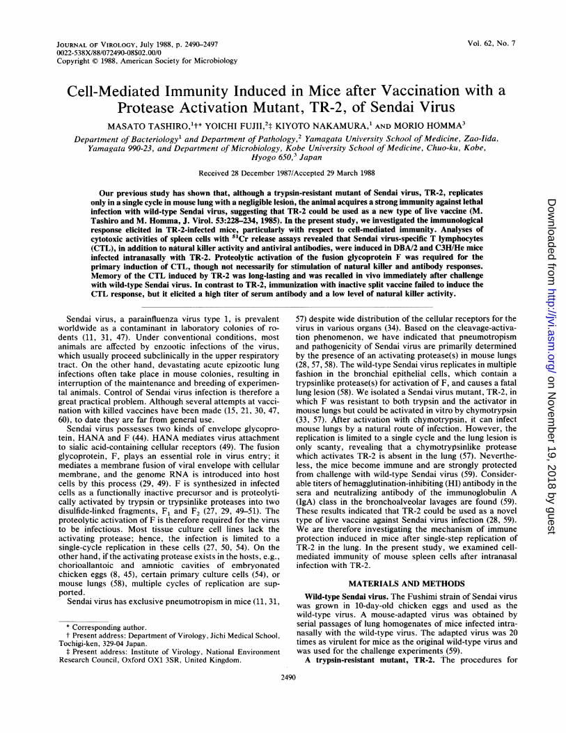
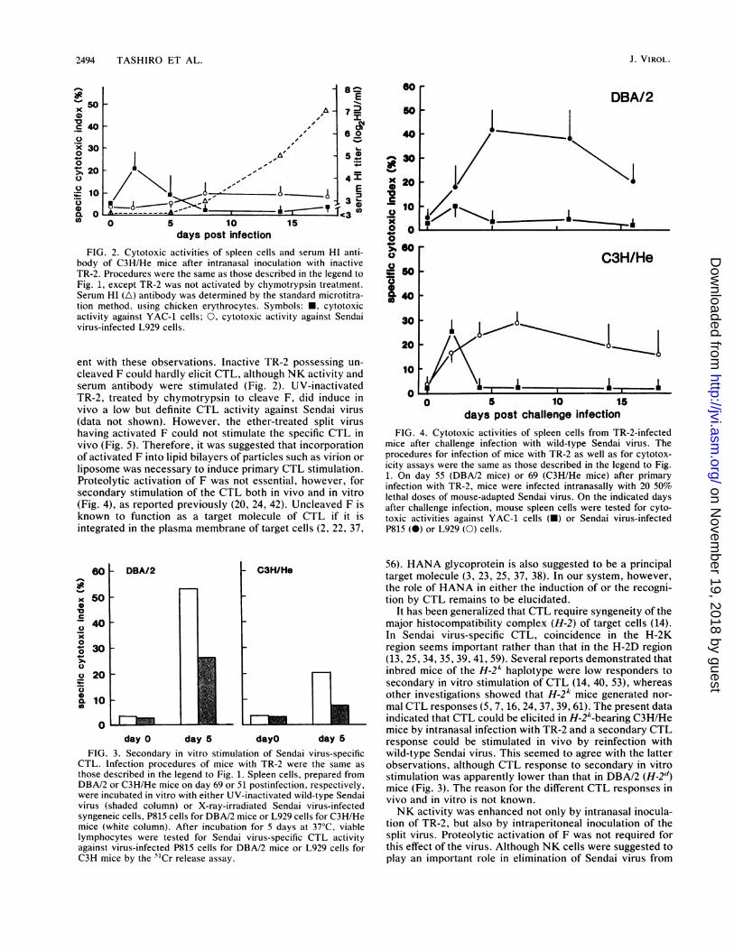
days post infectionFIG. 1. Cytotoxic activities of spleen cells in mice after intra-
nasal infection with chymotrypsin-activated TR-2. Male mice, 5weeks old, of DBA/2 (H-2d) and C3H/He (H-2A) strains were
infected intranasally with 25 ,ul of TR-2 (128 HAU/ml) grown inLLC-MK2 cells and activated in vitro by chymotrypsin. Spleen cellsuspensions were prepared on the days indicated for testing NKactivity against YAC-1 cells (-) and CTL activity against Sendai-virus-infected P815 (H-2d) (0) or L929 (H-2k) (0) cells by 51Crrelease assays. NK activity of control mice against YAC-1 cells (O)is included. Each plot indicates the mean of two to five mice, withbar indicating standard deviation.
hence, the infection terminated after single-cycle replication.As a result, lung lesions were barely induced and the miceremained healthy. Nevertheless, mice became immune fromchallenge with wild-type Sendai virus (data not shown), theresults being comparable to those found previously in ICRmice (57, 59). Further experiments were carried out with 4-to 5-week-old C3H/He and DBA/2 mice.NK activity of spleen cells after infection with TR-2. After
intranasal infection of mice with TR-2, cytotoxic activityagainst YAC-1 cells appeared the next day and predomi-nated until day 5 (Fig. 1). This activity was reduced drasti-cally when the spleen cells were treated with anti-ASGM1antibody and complement (Table 1). Furthermore, when themice were injected intravenously with anti-ASGM1 IgGfraction 24 h prior to assay, cytotoxic activity was almostcompletely eliminated (Table 2). This treatment did not haveany significant effect on virus titer in the lung and lungpathology (data not shown). These results showed thatintranasal infection with TR-2 induced a transient NK activ-ity in mice. It should also be noted in Fig. 1 that spontaneousNK activity of uninfected mice increased gradually fromweek 7 of life, whereas NK activity of TR-2-infected miceremained low after the transient appearance. The increase in
NK activity in control animals could be explained by age
dependency of NK activity in mice (32).Sendai virus-specific CTL induced after infection with
TR-2. After intranasal infection with activated TR-2, cyto-toxic activity of spleen cells against Sendai virus antigen-
bearing target cells was detected between 5 and 14 dayspostinfection (Fig. 1). The activity was elicited only whenthe H-2 phenotype was compatible between mouse strainand target cells and was abolished by treating the spleen cellswith anti-Thy-1.2 or anti-Lyt-2.1 antibody plus complement(Table 1). These results indicated that cytotoxic activity wasdue to CTL against Sendai virus. The CTL activity waspartially suppressed by in vitro treatment with anti-ASGM1plus complement (Table 1). This was consistent with anobservation that part of the CTL population shares a com-mon antigen marker of ASGM1 with NK cells (55). Sendaivirus-infected L929 cells were equally sensitive to CTLirrespective of the activation state of F on their cell mem-branes, since CTL activity did not change even after treat-ment of the target cells with trypsin (data not shown).
In contrast to activated TR-2, when the same amount ofinactive TR-2 was inoculated intranasally-into mice, only amarginal CTL activity was elicited, although a transient NKactivity and serum HI antibody response appeared (Fig. 2).The NK activity might be mediated by interferons whichwere stimulated by the noninfectious virus. These datashowed that induction of CTL specific for wild-type Sendaivirus was definite in mice after a single-step replication ofTR-2 in the lungs and that inoculation of the activated viruswas required for primary CTL induction in vivo.
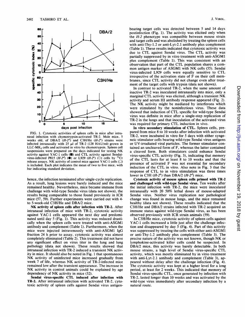
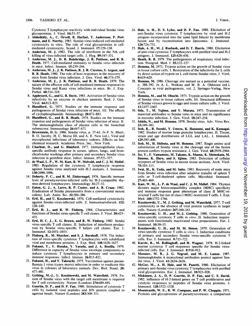
In vitro secondary stimulation of CTL. Spleen cells, pre-pared from mice 8 to 10 weeks after infection with activatedTR-2, were incubated in vitro for 5 days with either synge-neic stimulator cell-s bearing wild-type Sendai virus antigensor UV-irradiated viral particles. The former stimulator con-tained an uncleaved form of F, whereas the latter containeda cleaved form. Both stimulators could enhance Sendaivirus-specific CTL activity (Fig. 3), indicating that memoryof the CTL lasts for at least 8 to 10 weeks and that thepresence of activated F was not essential for secondaryinduction of the CTL in vitro. Also note in Fig. 3 that theresponse of CTL to in vitro stimulation was three timeslower in C3H (-t2k) than DBA/2 (H-2d) mice.
Cytotoxic activity of mouse spleen cells primed with TR-2and challenged with wild-type Sendai virus. Ten weeks afterthe initial infection with TR-2, the mice were inoculatedintranasally with 20 50% lethal doses of mouse-adaptedSendai virus. Neither virus replication nor pathologicalchange was found in mouse lungs, and the mice remainedhealthy (data not shown). These results indicated that theC3H/He and DBA/2 strains infected with TR-2 acquired animmune status against wild-type Sendai virus, as has beenobserved previously with ICR strain animals (59).
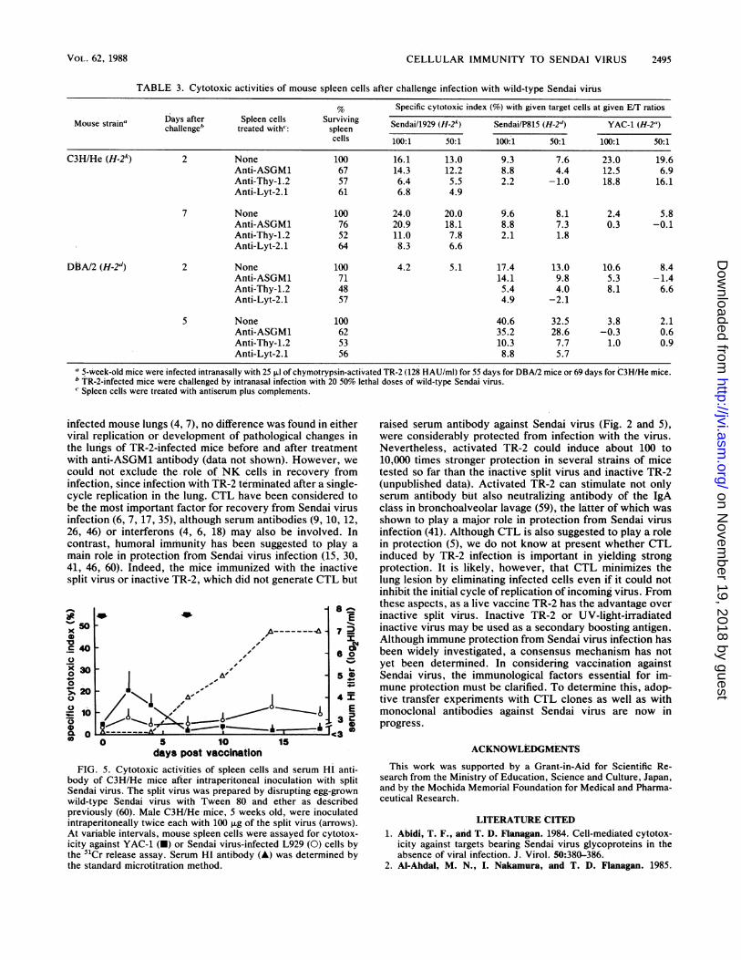
In C3H/He mice, cytotoxic activity of spleen cells againstYAC-1 cells increased a little just after the challenge infec-tion and disappeared by day 5 (Fig. 4). Part of this activitywas suppressed by treating the cells with either anti-ASGM1or anti-Thy-1.2 antibody plus complement (Table 3). Theprecise nature of the activity was not known, though NK orlymphokine-activated killer cells could be suspected. InDBA/2 mice, this activity was barely detectable. In bothmouse strains, a high level of Sendai virus-specific CTLactivity, which was mostly eliminated by in vitro treatmentwith anti-Lyt-2.1 antibody and complement (Table 3), ap-peared without delay after the challenge infection (Fig. 4).The cytotoxic activity was kept at a higher level for a longperiod, at least for 2 weeks. This indicated that memory ofSendai virus-specific CTL, once generated by infection withTR-2, lasted longer than 10 weeks and was activated by thewild-type virus immediately after secondary infection by anatural route.
J. VIROL.
on Novem
ber 19, 2018 by guesthttp://jvi.asm
.org/D
ownloaded from
CELLULAR IMMUNITY TO SENDAI VIRUS 2493
TABLE 1. Cytotoxic activities of mouse spleen cells after intranasal infection of activated TR-2
Specific cytotoxic index (%) with given target cells at given E/T ratios
Days Spleen cells Surviving L929 (H-24) P815 (H-2") YAC-1 (H-2")ousesramp i, treated with': spleen - Virus + Virus" - Virus + Virus" -Virus
cells
100:1 50:1 100:1 50:1 100:1 50:1 100:1 50:1 100:1 50:1
C3H/He (H-2k) 4 None 100 4.8 3.6 7.3 5.3 6.2 3.4 15.6 10.0 36.8 24.2Anti-ASGM1 68 2.3 2.5 4.3 4.1 8.8 6.5Anti-Thy-1.2 53 6.9 3.3 9.2 6.4 31.1 20.8
11 None 100 8.4 3.3 26.0 22.1 10.4 6.1 8.7 4.4 6.8 3.6Anti-ASGM1 64 22.1 16.4 6.6 4.0 2.3 2.0Anti-Thy-1.2 49 11.6 8.6 2.3 2.2 5.1 4.6Anti-Lyt-2.1 56 10.9 8.1
DBA/2 (H-2d) 4 None 100 4.8 2.9 6.3 5.3 5.1 4.0 14.0 11.3 33.5 28.1Anti-ASGM1 70 2.1 3.0 4.4 2.0 4.6 2.1Anti-Thy-1.2 59 3.8 3.0 8.9 7.1 29.0 25.4
12 None 100 6.9 5.5 8.1 6.6 11.4 4.6 42.2 35.1 4.8 2.0Anti-ASGM1 72 36.9 29.0 -0.3 0.8Anti-Thy-1.2 46 11.1 8.5 2.8 -0.4Anti-Lyt-2.1 51 12.9 9.6
"5-week-old male mice.b Intranasal inoculation with 25 ,ul of chymotrypsin-activated TR-2 (128 HAU/ml). p.i., Postinfection.' Spleen cells were treated with antiserum plus complements.d L929 cells were infected with wild-type Sendai virus for 12 h." UV-irradiated egg-grown wild-type Sendai virus was incubated with the cells for 2 h at 37°C.
Cytotoxic activity of mouse spleen cells after immunization intranasal infection with TR-2, which replicated only in awith split Sendai virus. Intraperitoneal inoculation of split single cycle in bronchial epithelia and caused little patholog-Sendai virus was reported to induce serum HI antibody ical change in the lung (Fig. 1; Table 1). These mice wereresponse and protection of mice from challenge with the strongly protected from lethal challenge with wild-type Sen-virulent virus (60). It was therefore of interest to compare dai virus (59), and memory of the CTL in mice lasted for >10cellular immunity-inducing capacity between TR-2 and the weeks and could be recalled immediately after challengesplit virus. When C3H/He mice were inoculated intraperito- (Fig. 4). Since intranasal infection with wild-type Sendaineally with 100 ,ug of the split virus, serum HI antibody to virus causes severe pneumonia in mice which results inSendai virus was detected at high titers (Fig. 5). NK activity animal death as early as 10 days, it is difficult to investigatewas also detected for a few days (Fig. 5), although the the Sendai virus-specific CTL induced after a natural routeactivity was significantly lower than that in the case of TR-2 of infection. Therefore, most previous studies of CTL have(Fig. 1). Specific CTL activity remained at a marginal level been made by intraperitoneal inoculation of UV-inactivatedthroughout the experimental period even when a booster virus or disrupted virus components. In contrast, TR-2 isimmunization was done. A secondary in vitro stimulation by nonpathogenic in mice, although it replicates in mouse lungsSendai virus-infected L929 cells failed to elicit a significant after intranasal inoculation (57). In this respect, TR-2 couldincrease in CTL activity (data not shown). When DBA/2 be a valuable tool for investigation of cellular immunitymice were used as hosts, the same results were obtained induced by respiratory infection with Sendai virus.(data not shown). Previous reports on Sendai virus-specific CTL have indi-
DISCUSSIONcated that proteolytic activation of F glycoprotein is requiredfor the primary induction of CTL (19, 20, 39, 42, 52), but
The present study showed that Sendai virus-specific CTL replication of the inoculum virus is not necessary (19, 20,as well as NK activity were elicited in mouse spleens after 52). The present results with TR-2 were principally consist-
TABLE 2. Effect of intravenous injection of anti-ASGM1 antibody on cytotoxic activities of spleen cells of C3H/He miceinfected intranasally with TR-2
Specific cytotoxic index (%) with given target cells at given E/T ratiosDays Mice treated Sendai/L929 (H-24) Sendai/P815 (H-2d) YAC-1 (H-2a)pA." with": _____________________
100:1 50:1 100:1 50:1 100:1 50:1
4 None 7.1 4.8 12.0 7.6 40.2 29.4Anti-ASGM1 5.5 3.3 5.9 2.3 8.8 4.0
14 None 21.3 16.8 3.7 4.6 9.0 7.7Anti-ASGM1 20.6 13.3 5.6 6.4 0.3 -0.2
"5-week-old male C3H/He mice were inoculated intranasally with 25 ,ul of chymotrypsin-activated TR-2 (128 HAU/ml). p.i., Postinfection.b Mice were injected intravenously with 0.1 ml of anti-ASGM1 antibody 24 h prior to cytotoxic assays.
VOL. 62, 1988
on Novem
ber 19, 2018 by guesthttp://jvi.asm
.org/D
ownloaded from
2494 TASHIRO ET AL.
50
40
30
20
10
0
E7 M
60o
A~~~~
~~---~4EI
A- A-~~~~~~~~~~~~~~~~ r----E-T~~~~~~~~~~~~~.........
0 5 10 15days post infection
FIG. 2. Cytotoxic activities of spleen cells and serum HI anti-body of C3H/He mice after intranasal inoculation with inactiveTR-2. Procedures were the same as those described in the legend toFig. 1, except TR-2 was not activated by chymotrypsin treatment.Serum HI (A) antibody was determined by the standard microtitra-tion method, using chicken erythrocytes. Symbols: *, cytotoxicactivity against YAC-1 cells; 0, cytotoxic activity against Sendaivirus-infected L929 cells.
60 rDBA/2
soF
40F
30
-
x 20
0
60
0
30
20
ent with these observations. Inactive TR-2 possessing un-cleaved F could hardly elicit CTL, although NK activity andserum antibody were stimulated (Fig. 2). UV-inactivatedTR-2, treated by chymotrypsin to cleave F, did induce invivo a low but definite CTL activity against Sendai virus(data not shown). However, the ether-treated split virushaving activated F could not stimulate the specific CTL invivo (Fig. 5). Therefore, it was suggested that incorporationof activated F into lipid bilayers of particles such as virion orliposome was necessary to induce primary CTL stimulation.Proteolytic activation of F was not essential, however, forsecondary stimulation of the CTL both in vivo and in vitro(Fig. 4), as reported previously (20, 24, 42). Uncleaved F isknown to function as a target molecule of CTL if it isintegrated in the plasma membrane of target cells (2, 22, 37,
10
0
F1 \1
0A.\, A_I
C3H/He
0 5 10 15days post challenge infection
FIG. 4. Cytotoxic activities of spleen cells from TR-2-infectedmice after challenge infection with wild-type Sendai virus. Theprocedures for infection of mice with TR-2 as well as for cytotox-icity assays were the same as those described in the legend to Fig.1. On day 55 (DBA/2 mice) or 69 (C3H/He mice) after primaryinfection with TR-2, mice were infected intranasally with 20 50%lethal doses of mouse-adapted Sendai virus. On the indicated daysafter challenge infection, mouse spleen cells were tested for cyto-toxic activities against YAC-1 cells (-) or Sendai virus-infectedP815 (0) or L929 (0) cells.
60
x 500
40_
0
° 30>0
o 20
D0a 10
O I I I I I
day O day 5 dayO day 5
FIG. 3. Secondary in vitro stimulation of Sendai virus-specificCTL. Infection procedures of mice with TR-2 were the same as
those described in the legend to Fig. 1. Spleen cells, prepared fromDBA/2 or C3H/He mice on day 69 or 51 postinfection, respectively,were incubated in vitro with either UV-inactivated wild-type Sendaivirus (shaded column) or X-ray-irradiated Sendai virus-infectedsyngeneic cells, P815 cells for DBA/2 mice or L929 cells for C3H/Hemice (white column). After incubation for 5 days at 37°C, viablelymphocytes were tested for Sendai virus-specific CTL activityagainst virus-infected P815 cells for DBA/2 mice or L929 cells forC3H mice by the 5'Cr release assay.
56). HANA glycoprotein is also suggested to be a principaltarget molecule (3, 23, 25, 37, 38). In our system, however,the role of HANA in either the induction of or the recogni-tion by CTL remains to be elucidated.
It has been generalized that CTL require syngeneity of themajor histocompatibility complex (H-2) of target cells (14).In Sendai virus-specific CTL, coincidence in the H-2Kregion seems important rather than that in the H-2D region(13, 25, 34, 35, 39, 41, 59). Several reports demonstrated thatinbred mice of the H-2k haplotype were low responders tosecondary in vitro stimulation of CTL (14, 40, 53), whereasother investigations showed that H-2k mice generated nor-mal CTL responses (5, 7, 16, 24, 37, 39, 61). The present dataindicated that CTL could be elicited in H-2k-bearing C3H/Hemice by intranasal infection with TR-2 and a secondary CTLresponse could be stimulated in vivo by reinfection withwild-type Sendai virus. This seemed to agree with the latterobservations, although CTL response to secondary in vitrostimulation was apparently lower than that in DBA/2 (H-2d)mice (Fig. 3). The reason for the different CTL responses invivo and in vitro is not known.NK activity was enhanced not only by intranasal inocula-
tion of TR-2, but also by intraperitoneal inoculation of thesplit virus. Proteolytic activation of F was not required forthis effect of the virus. Although NK cells were suggested toplay an important role in elimination of Sendai virus from
x10c.0
._
x4-0
>_00
(0)
J. VIROL.
on Novem
ber 19, 2018 by guesthttp://jvi.asm
.org/D
ownloaded from
CELLULAR IMMUNITY TO SENDAI VIRUS 2495
TABLE 3. Cytotoxic activities of mouse spleen cells after challenge infection with wild-type Sendai virius
% Specific cytotoxic index (%) with given target cells at given E/T ratiosDays after Spleen cells Surviving Sendai/1929 (H-2A) Sendai/P815 (H-2d) YAC-i (H-2")Mouse strain' challenge' treated with": spleen Sna199H2_Sed_P5 _dYAC_1_H_2__
cells 100:1 50:1 100:1 50:1 100:1 50:1
C3H/He (H-2k) 2 None 100 16.1 13.0 9.3 7.6 23.0 19.6Anti-ASGM1 67 14.3 12.2 8.8 4.4 12.5 6.9Anti-Thy-1.2 57 6.4 5.5 2.2 - 1.0 18.8 16.1Anti-Lyt-2.1 61 6.8 4.9
7 None 100 24.0 20.0 9.6 8.1 2.4 5.8Anti-ASGM1 76 20.9 18.1 8.8 7.3 0.3 -0.1Anti-Thy-1.2 52 11.0 7.8 2.1 1.8Anti-Lyt-2.1 64 8.3 6.6
DBA/2 (H-2d) 2 None 100 4.2 5.1 17.4 13.0 10.6 8.4Anti-ASGM1 71 14.1 9.8 5.3 -1.4Anti-Thy-1.2 48 5.4 4.0 8.1 6.6Anti-Lyt-2.1 57 4.9 -2.1
S None 100 40.6 32.5 3.8 2.1Anti-ASGM1 62 35.2 28.6 -0.3 0.6Anti-Thy-1.2 53 10.3 7.7 1.0 0.9Anti-Lyt-2.1 56 8.8 5.7
a 5-week-old mice were infected intranasally with 25 p.1 of chymotrypsin-activated TR-2 (128 HAU/ml) for 55 days for DBA/2 mice or 69 days for C3H/He mice.b TR-2-infected mice were challenged by intranasal infection with 20 50% lethal doses of wild-type Sendai virus.Spleen cells were treated with antiserum plus complements.
infected mouse lungs (4, 7), no difference was found in eitherviral replication or development of pathological changes inthe lungs of TR-2-infected mice before and after treatmentwith anti-ASGM1 antibody (data not shown). However, wecould not exclude the role of NK cells in recovery frominfection, since infection with TR-2 terminated after a single-cycle replication in the lung. CTL have been considered tobe the most important factor for recovery from Sendai virusinfection (6, 7, 17, 35), although serum antibodies (9, 10, 12,26, 46) or interferons (4, 6, 18) may also be involved. Incontrast, humoral immunity has been suggested to play amain role in protection from Sendai virus infection (15, 30,41, 46, 60). Indeed, the mice immunized with the inactivesplit virus or inactive TR-2, which did not generate CTL but
a0
10
.r-
0x
0
0-
c 4
0
0
x 3oo,;L1 iv
to
A
8
7
a
Al-I0 .j5N,-,j.1.1A, I - ~4
10--.1.30 5 10
days post vaccination
c
E"-%
0
0
EDa
15
FIG. 5. Cytotoxic activities of spleen cells and serum HI anti-body of C3H/He mice after intraperitoneal inoculation with splitSendai virus. The split virus was prepared by disrupting egg-grownwild-type Sendai virus with Tween 80 and ether as describedpreviously (60). Male C3H/He mice, S weeks old, were inoculatedintraperitoneally twice each with 100 ,ug of the split virus (arrows).At variable intervals, mouse spleen cells were assayed for cytotox-icity against YAC-1 (-) or Sendai virus-infected L929 (0) cells bythe 51Cr release assay. Serum HI antibody (A) was determined bythe standard microtitration method.
raised serum antibody against Sendai virus (Fig. 2 and 5),were considerably protected from infection with the virus.Nevertheless, activated TR-2 could induce about 100 to10,000 times stronger protection in several strains of micetested so far than the inactive split virus and inactive TR-2(unpublished data). Activated TR-2 can stimulate not onlyserum antibody but also neutralizing antibody of the IgAclass in bronchoalveolar lavage (59), the latter of which wasshown to play a major role in protection from Sendai virusinfection (41). Although CTL is also suggested to play a rolein protection (5), we do not know at present whether CTLinduced by TR-2 infection is important in yielding strongprotection. It is likely, however, that CTL minimizes thelung lesion by eliminating infected cells even if it could notinhibit the initial cycle of replication of incoming vitus. Fromthese aspects, as a live vaccine TR-2 has the advantage overinactive split virus. Inactive TR-2 or UV-light-irradiatedinactive virus may be used as a secondary boosting antigen.Although immune protection from Sendai virus infection hasbeen widely investigated, a consensus mechanism has notyet been determined. In considering vaccination againstSendai virus, the immunological factors essential for im-mune protection must be clarified. To determine this, adop-tive transfer experiments with CTL clones as well as withmonoclonal antibodies against Sendai virus are now inprogress.
ACKNOWLEDGMENTS
This work was supported by a Grant-in-Aid for Scientific Re-search from the Ministry of Education, Science and Culture, Japan,and by the Mochida Memorial Foundation for Medical and Pharma-ceutical Research.
LITERATURE CITED1. Abidi, T. F., and T. D. Flanagan. 1984. Cell-mediated cytotox-
icity against targets bearing Sendai virus glycoproteins in theabsence of viral infection. J. Virol. 50:380-386.
2. Al-Ahdal, M. N., I. Nakamura, and T. D. Flanagan. 1985.
VOL. 62, 1988
v
on Novem
ber 19, 2018 by guesthttp://jvi.asm
.org/D
ownloaded from
2496 TASHIRO ET AL.
Cytotoxic T-lymphocyte reactivity with individual Sendai virusglycoproteins. J. Virol. 54:53-57.
3. Alsheikhly, A., C. Orvell, B. Harfast, T. Andersson, P. Perl-mann, and E. Norrby. 1983. Sendai-virus-induced cell-mediatedcytotoxicity in vitro. The role of viral glycoproteins in cell-mediated cytotoxicity. Scand. J. Immunol. 17:129-138.
4. Anderson, M. J. 1982. The role of interferon in the NK cellkilling of virus-infected target cells. J. Hyg. 89:347-351.
5. Anderson, M. J., D. R. Bainbridge, J. R. Pattison, and R. B.Heath. 1977. Cell-mediated immunity to Sendai virus infectionin mice. Infect. Immun. 15:239-244.
6. Anderson, M. J., J. R. Pattison, R. J. R. Cureton, S. Argent, andR. B. Heath. 1980. The role of host responses in the recovery ofmice from Sendai virus infection. J. Gen. Virol. 46:373-379.
7. Anderson, M. J., J. R. Pattison, and R. B. Heath. 1979. Thenature of the effector cells of cell-mediated immune responses toSendai virus and kunz virus infections in mice. Br. J. Exp.Pathol. 60:314-319.
8. Appleyard, G., and G. B. Davis. 1983. Activation of Sendai virusinfectivity by an enzyme in chicken amniotic fluid. J. Gen.Virol. 64:813-823.
9. Blandford, G. 1975. Studies on the immune response andpathogenesis of Sendai virus infection of mice. III. The effectsof cyclophosphamide. I'mmunology 28:871-883.
10. Blandford, G., and R. B. Heath. 1974. Studies on the immuneresponse and pathogenesis of Sendai virus infection of mice. II.The immunoglobulin class of plasma cells in the bronchialsubmucosa. Immunology 26:667-671.
11. Brownstein, D. G. 1986. Sendai virus, p. 37-61. In P. N. Bhatt,R. 0. Jacoby, H. C. Morse III, and A. E. New (ed.), Viral andmycoplasmal infections of laboratory rodents. Effects on bio-chemical research. Academic Press, Inc., New York.
12. Charlton, D., and G. Blanford. 1977. Immunoglobulin class-specific antibody response in serum, spleen, lungs, and bron-choalveolar washings after primary and secondary Sendai virusinfection in germfree mice. Infect. Immun. 17:521-527.
13. de Waal, L. P., W. M. Kast, R. W. Melvold, and C. J. M. Melief.1983. Regulation of the cytotoxic T lymphocyte responseagainst Sendai virus analyzed with H-2 mutants. J. Immunol.130:1090-1096.
14. Doherty, P. C., and R. M. Zinkernagel. 1976. Specific immunelysis of paramyxovirus-infected cells by H-2-compatible thy-mus-derived lymphocytes. Immunology 31:27-32.
15. Eaton, G. J., A. Lerro, R. P. Custer, and A. R. Crane. 1982.Eradication of Sendai pneumonitis from a conventional mousecolony. Lab. Anim. Sci. 32:384-386.
16. Ertl, H., and U. Koszinowski. 1976. Cell-mediated cytotoxicityagainst Sendai-virus-infected cells. Z. Immunitaetsforsch. 152:128-140.
17. Ertl, H. J., and R. W. Finberg. 1984. Characteristics andfunctions of Sendai virus-specific T-cell clones. J. Virol. 50:425-431.
18. Ertl, H. C. J., E. G. Brown, and R. W. Finberg. 1982. Sendaivirus-specific T cell clones. II. Induction of interferon produc-tion by Sendai virus-specific T helper cell clones. Eur. J.Immunol. 12:1051-1053.
19. Finberg, R., M. Mescher, and S. J. Burakoff. 1978. The induc-tion of virus-specific cytotoxic T lymphocytes with solubilizedviral and membrane proteins. J. Exp. Med. 148:1620-1627.
20. Fukami, Y., Y. Hosaka, Y. Yasuda, and J. A. Bonilla. 1979.Difference in capacity of Sendai virus envelope components toinduce cytotoxic T lymphocytes in primary and secondaryimmune responses. Infect. Immun. 26:815-821.
21. Fukumi, H., and Y. Takeuchi. 1975. Vaccination against parain-fluenza 1 virus (types muris) infection in order to eradicate thisvirus in colonies of laboratory animals. Dev. Biol. Stand. 28:477-481.
22. Gething, M.-J., U. Koszinowski, and M. Waterfield. 1978. Fu-sion of Sendai virus with the target cell membrane is requiredfor T cell cytotoxicity. Nature (London) 274:689-691.
23. Guertin, D. P., and D. P. Fan. 1980. Stimulation of cytotoxic Tcells by isolated viral peptides and HN protein coupled toagarose beads. Nature (London) 283:308-311.
24. Hale, A. H., ID. S. Lyles, and D. P. Fan. 1980. Elicitation ofanti-Sendai virus cytotoxic T lymphocytes by viral and H-2antigens incorporated into the same lipid bilayer by membranefusion and by reconstitution into liposomes. J. Immunol.124:724-731.
25. Hale, A. H., M. J. Ruebush, and D. T. Harris. 1980. Elicitationof anti-viral cytotoxic T lymphocytes with purified viral and H-2antigens. J. Immunol. 125:428-430.
26. Heath, R. B. 1979. The pathogenesis of respiratory viral infec-tion. Postgrad. Med. J. 55:122-127.
27. Homma, M. 1971. Trypsin action on the growth of Sendai virusin tissue culture cells. I. Restoration of the infectivity for L cellsby direct action of trypsin on L cell-borne Sendai virus. J. Virol.8:619-629.
28. Homma, M. 1986. Cleavage site mutant as a potential vaccine,p. 388-392. In A. L. Notkins and M. B. A. Oldstone (ed.),Concepts in viral pathogenesis, vol. 2. Springer-Verlag, NewYork.
29. Homma, M., and M. Ohuchi. 1973. Trypsin action on the growthof Sendai virus in tissue culture cells. III. Structural differenceof Sendai viruses grown in eggs and tissue culture cells. J. Virol.12:1457-1465.
30. lida, T., M. Tajima, and Y. Murata. 1973. Transmission ofmaternal antibodies to Sendai virus in mice and its significancein enzootic infection. J. Gen. Viroi. 18:247-254.
31. Ishida, N., and M. Homma. 1978. Sendai virus. Adv. Virus Res.23:349-383.
32. Itoh, K., R. Suzuki, Y. Umezu, K. Hanaumi, and K. Kumagai.1982. Studies of murine large granular lymphocytes. II. Tissue,strain and age distributions of LGL and LAL. J. Immunol.129:395-400.
33. Itoh, M., H. Shibuta, and M. Homma. 1987. Single amino acidsubstitution of Sendai virus at the cleavage site of the fusionprotein confers trypsin resistance. J. Gen. Virol. 68:2939-2944.
34. Ito, Y., F. Yamamoto, M. Takano, K. Maeno, K. Shimokata, M.Iinuma, K. Hara, and S. lijima. 1983. Detection of cellularreceptors of Sendai virus in mouse tissue sections. Arch. Virol.75:103-113.
35. Iwai, H., K. Ueda, and M. Saito. 1983. Recovery of nude micefrom Sendai virus infection after adoptive transfer of spleen Tcells or T-cell-depleted spleen cells. Microbiol. Immunol.27:465-469.
36. Kast, W. M., L. P. de Waal, and C. J. M. Melief. 1984. Thymusdictates major histocompatibility complex (MHC) specificityand immune response gene phenotype of class II MHC-re-stricted T cells but not of class I MHC-restricted T cells. J. Exp.Med. 160:1752-1766.
37. Koszinowski, U., M. J. Gething, and M. Waterfield. 1977. T-cellcytotoxicity in the absence of viral protein synthesis in targetcells. Nature (London) 267:160-163.
38. Koszinowski, U. H., and M.-J. Gething. 1980. Generation ofvirus-specific cytotoxic T cells in vitro. II. Induction require-ments with functionally inactivated virus preparations. Eur. J.Immunol. 10:30-35.
39. Koszinowski, U. H., and M. M. Simon. 1979. Generation ofvirus-specific cytotoxic T cells in vitro. I. Induction conditionsof primary and secondary Sendai virus-specific cytotoxic Tcells. Eur. J. Immunol. 9:715-722.
40. Kurrle, R., M. Rollinghoff, and H. Wagner. 1978. H-2-linkedmurine cytotoxic T cell responses specific for Sendai virus-infected cells. Eur. J. Immunol. 8:910-912.
41. Mazanec, M. B., J. G. Negrud, and M. E. Lamm. 1987.Immunoglobulin A monoclonal antibodies protect against Sen-dai virus. J. Virol. 61:2624-2626.
42. McGee, M., A. H. Hale, and M. Panetti. 1980. Elicitation ofprimary anti-Sendai virus cytotoxic T lymphocytes with purifiedviral glycoproteins. Eur. J. Immunol. 10:923-928.
43. Miskimen, J. A., D. P. Guertin, D. P. Fan, and C. S. David.1982. Influence of 1--2-linked genes on T cell proliferative andcytolytic responses to peptides of Sendai virus proteins. J.Immunol. 128:1522-1528.
44. Mountcastle, W. E., R. W. Compans, and P. W. Choppin. 1971.Proteins and glycoproteins of paramyxoviruses: a comparison
J. VIROL.
on Novem
ber 19, 2018 by guesthttp://jvi.asm
.org/D
ownloaded from
CELLULAR IMMUNITY TO SENDAI VIRUS 2497
of simian 5, Newcastle disease virus, and Sendai virus. J. Virol.7:47-52.
45. Muramatsu, M., and M. Homma. 1980. Trypsin action on thegrowth of Sendai virus in tissue culture cells. V. An activatingenzyme for Sendai virus in the chorioallantoic fluid of theembryonated chicken eggs. Microbiol. Immunol. 24:113-122.
46. Orvell, C., and M. Grandien. 1982. The effect of monoclonalantibodies on biologic activities of structural proteins of Sendaivirus. J. Immunol. 129:2779-2787.
47. Parker, J. C. 1980. The possibility and limitations of viruscontrol in laboratory animals, p. 162-172. In A. Spiegel, S.Erichsen, and H. A. Solleveld (ed.), Animal quality and modelsin biochemical research. Gustav Fischer Verlag, Stuttgart,Federal Republic of Germany.
48. Portner, A. 1981. The HN glycoprotein of Sendai virus. Analy-sis of site(s) involved in hemagglutinating and neuraminidaseactivities. Virology 115:375-384.
49. Scheid, A., and P. W. Choppin. 1974. Identification of biologicalactivities of paramyxovirus glycoproteins. Activation of cellfusion, hemolysis, and infectivity by proteolytic cleavage of aninactive precursor protein of Sendai virus. Virology 57:475-490.
50. Scheid, A., and P. W. Choppin. 1976. Protease activation mutantof Sendai virus. Activation of biological properties by specificproteases. Virology 69:265-277.
51. Scheid, A., and P. W. Choppin. 1977. Two disulfide-linkedpolypeptide chains constitute the active F protein of paramyxo-viruses. Virology 80:54-66.
52. Schrader, J. W., and G. M. Edelman. 1977. Joint recognition bycytotoxic T cells of inactivated Sendai virus and products of themajor histocompatibility complex. J. Exp. Med. 145:523-539.
53. Shapiro, M. E., S. J. Burakoff, B. Benacerraf, and R. W.
Finberg. 1981. Ir gene control of the cytotoxic T lymphocyteresponse to Sendai virus: H-2k mice are low responders toSendai. Immunology 127:2571-2574.
54. Silver, S. M., A. Scheid, and P. W. Choppin. 1978. Loss on serialpassage of rhesus monkey kidney cells of proteolytic activityrequired for Sendai virus activation. Infect. Immun. 20:235-241.
55. Stitz, L., J. Baenzger, H. Pircher, H. Hengartner, and R. M.Zinkernagel. 1986. Effect of rabbit anti-asialo GM1 treatment invivo or with anti-asialo GM1 plus complement in vitro oncytotoxic T cell activities. J. Immunol. 136:4674-4680.
56. Sugamura, K., K. Shimizu, D. A. Zerling, and F. H. Bach. 1977.Role of Sendai virus fusion glycoprotein in target cell suscepti-bility of cytotoxic T cells. Nature (London) 270:251-253.
57. Tashiro, M., and M. Homma. 1983. Pneumotropism of Sendaivirus in relation to protease-mediated activation in mouse lungs.Infect. Immun. 39:879-888.
58. Tashiro, M., and M. Homma. 1983. Evidence of proteolyticactivation of Sendai virus in mouse lung. Arch. Virol. 77:127-137.
59. Tashiro, M., and M. Homma. 1985. Protection of mice fromwild-type Sendai virus infection by a trypsin-resistant mutant,TR-2. J. Virol. 53:228-234.
60. Tsukui, M., H. Ito, M. Tada, M. Nakata, H. Miyajima, and K.Fujiwara. 1982. Protective effect of inactivated virus vaccine onSendai virus infection in rats. Lab. Anim. Sci. 32:143-146.
61. Zinkernagel, R. M., A. Althage, S. Cooper, G. Kreeb, P. A.Klein, B. Sefton, L. Flaherty, J. Stimpfling, D. Shreffler, and J.Klein. 1978. Ir-genes in H-2 regulate generation of antiviralcytotoxic T cells. Mapping to K or D and dominance ofunresponsiveness. J. Exp. Med. 148:592-606.
VOL. 62, 1988
on Novem
ber 19, 2018 by guesthttp://jvi.asm
.org/D
ownloaded from