Embed Size (px)
Citation preview
Cell Growth and Size Control: controlling the cell cycle
Peter TakizawaDepartment of Cell Biology
•Start and commitment to cell division
•Regulation of entry into cell cycle: mitogens and anti-mitogens
•DNA damage and arresting the cell cycle
•Cell senescence
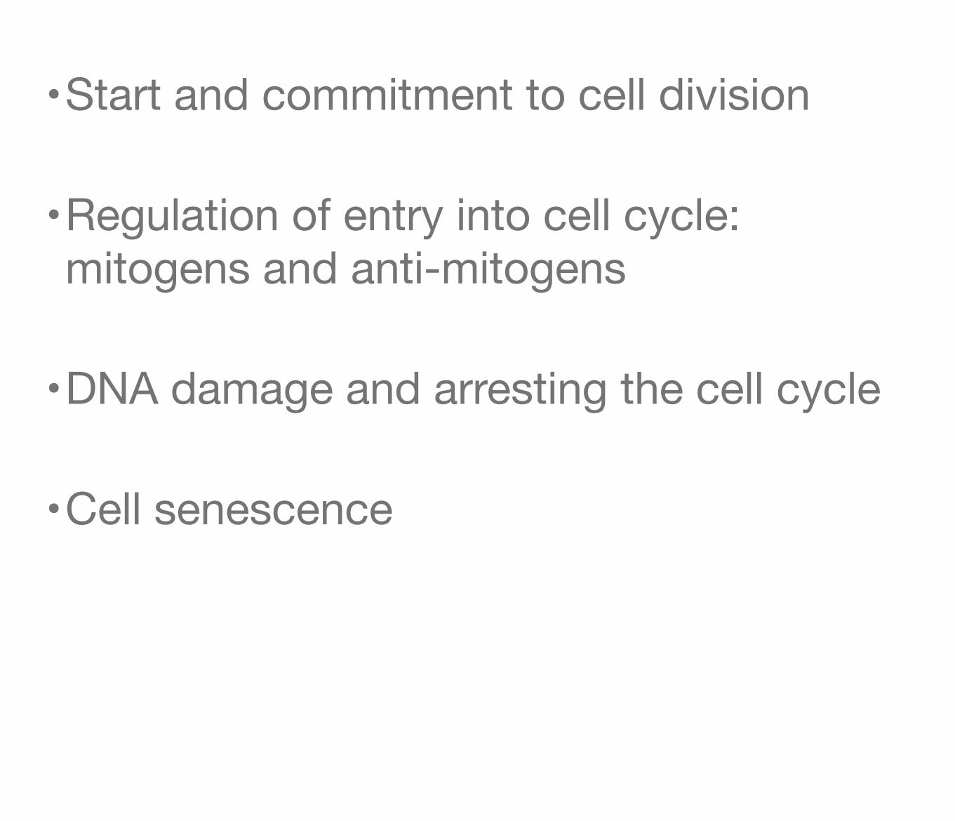
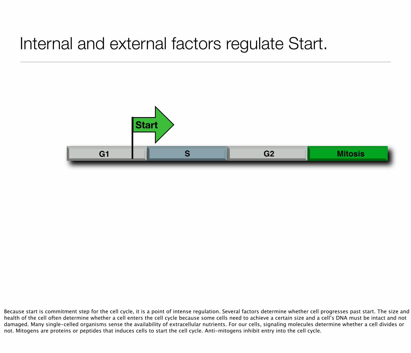
Start marks the initiation of DNA replication and an irreversible commitment to cell division.
G1 S G2 Mitosis
Start
The decision to enter the cell cycle is made at Start in G1. After Start, cell committed to cell division because the cell will commence chromosome replication and must finish the cell cycle.
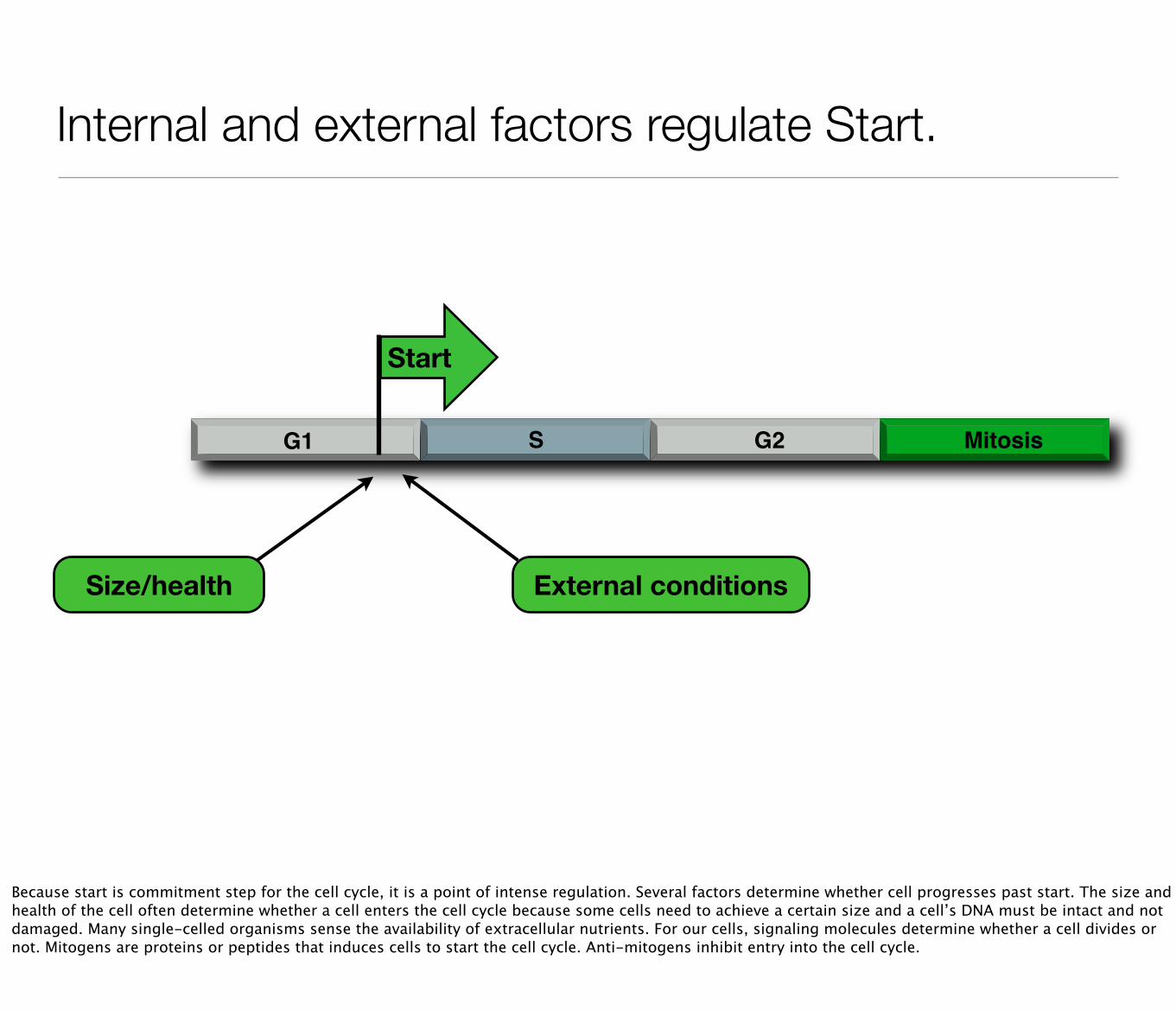
Internal and external factors regulate Start.
Size/health External conditions
G1 S G2 Mitosis
Start
Because start is commitment step for the cell cycle, it is a point of intense regulation. Several factors determine whether cell progresses past start. The size and health of the cell often determine whether a cell enters the cell cycle because some cells need to achieve a certain size and a cell’s DNA must be intact and not damaged. Many single-celled organisms sense the availability of extracellular nutrients. For our cells, signaling molecules determine whether a cell divides or not. Mitogens are proteins or peptides that induces cells to start the cell cycle. Anti-mitogens inhibit entry into the cell cycle.
Internal and external factors regulate Start.
G1 S G2 Mitosis
Start
Because start is commitment step for the cell cycle, it is a point of intense regulation. Several factors determine whether cell progresses past start. The size and health of the cell often determine whether a cell enters the cell cycle because some cells need to achieve a certain size and a cell’s DNA must be intact and not damaged. Many single-celled organisms sense the availability of extracellular nutrients. For our cells, signaling molecules determine whether a cell divides or not. Mitogens are proteins or peptides that induces cells to start the cell cycle. Anti-mitogens inhibit entry into the cell cycle.
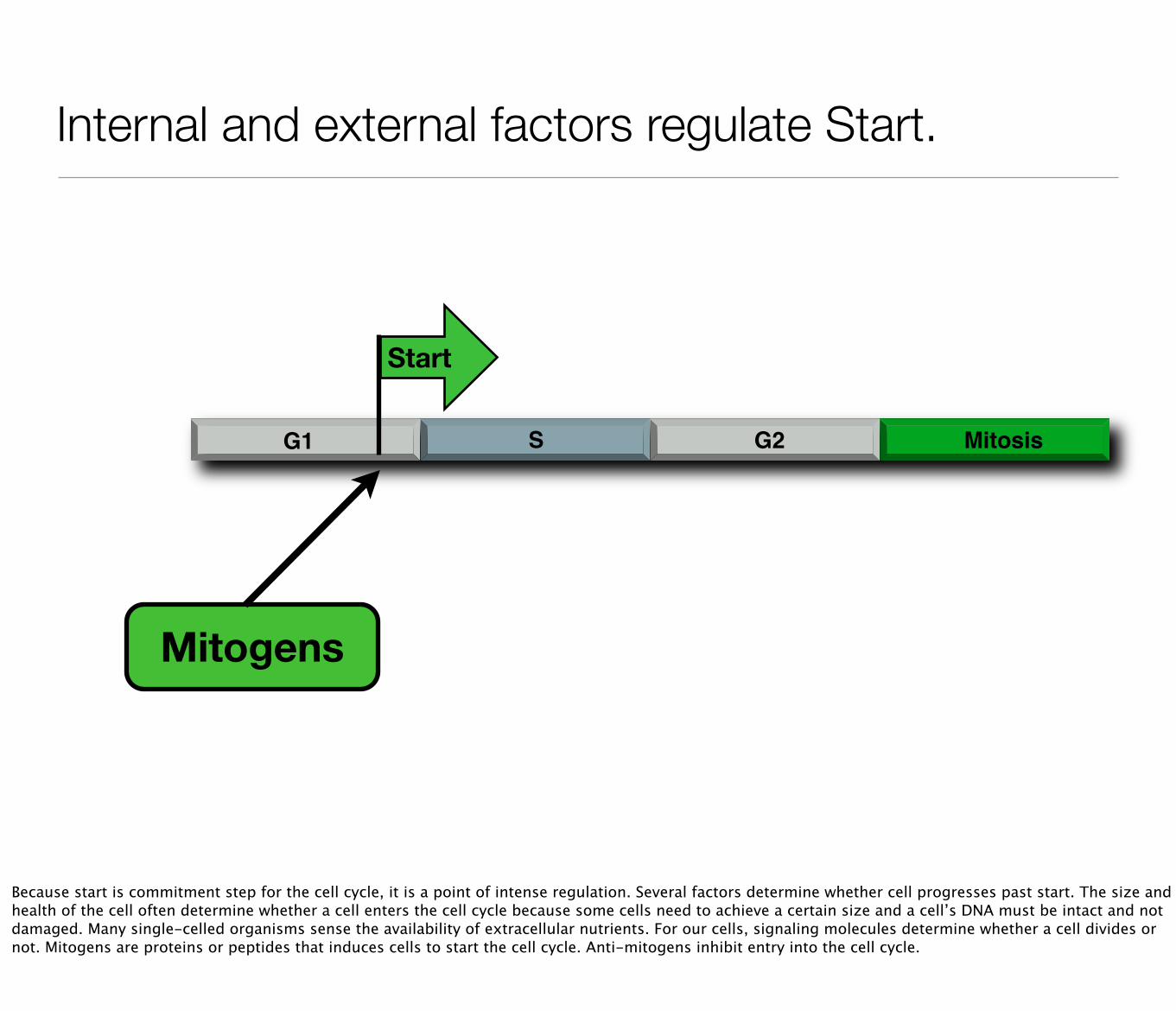
Internal and external factors regulate Start.
G1 S G2 Mitosis
Start
Mitogens
Because start is commitment step for the cell cycle, it is a point of intense regulation. Several factors determine whether cell progresses past start. The size and health of the cell often determine whether a cell enters the cell cycle because some cells need to achieve a certain size and a cell’s DNA must be intact and not damaged. Many single-celled organisms sense the availability of extracellular nutrients. For our cells, signaling molecules determine whether a cell divides or not. Mitogens are proteins or peptides that induces cells to start the cell cycle. Anti-mitogens inhibit entry into the cell cycle.
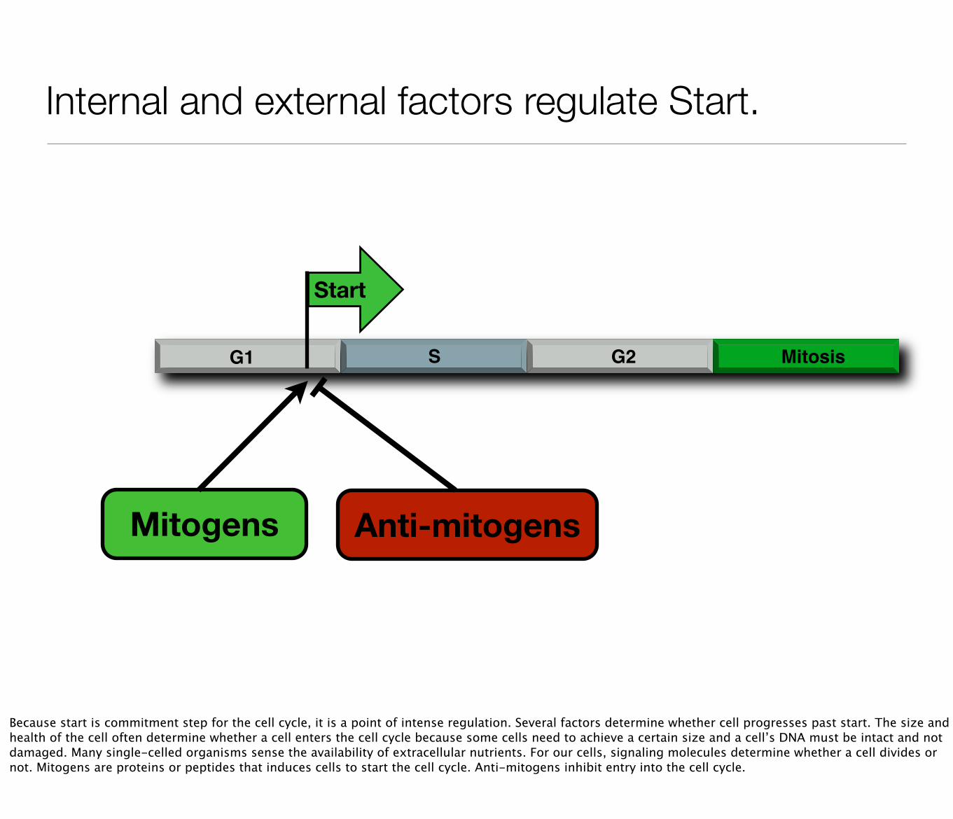
Internal and external factors regulate Start.
G1 S G2 Mitosis
Start
Mitogens Anti-mitogens
Because start is commitment step for the cell cycle, it is a point of intense regulation. Several factors determine whether cell progresses past start. The size and health of the cell often determine whether a cell enters the cell cycle because some cells need to achieve a certain size and a cell’s DNA must be intact and not damaged. Many single-celled organisms sense the availability of extracellular nutrients. For our cells, signaling molecules determine whether a cell divides or not. Mitogens are proteins or peptides that induces cells to start the cell cycle. Anti-mitogens inhibit entry into the cell cycle.
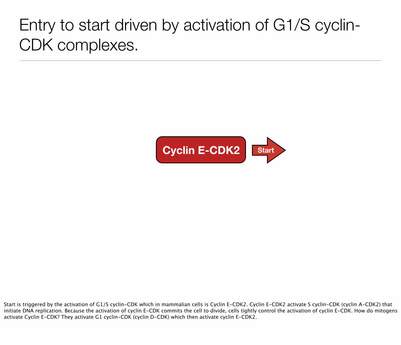
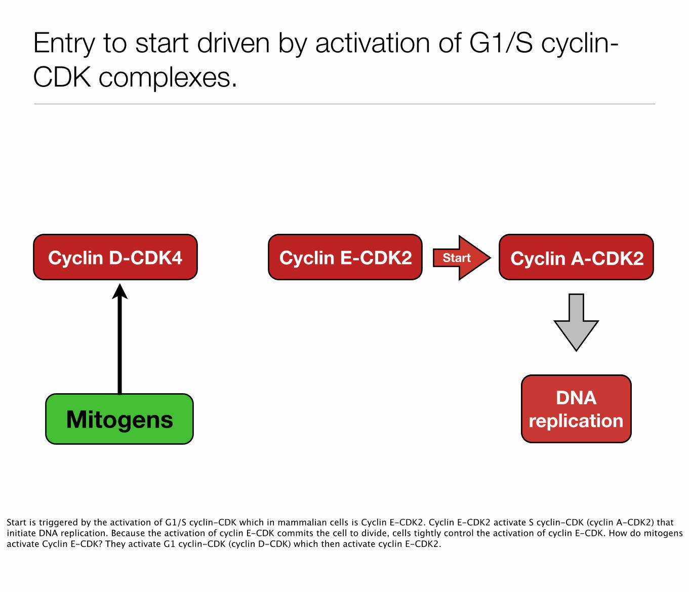
Entry to start driven by activation of G1/S cyclin-CDK complexes.
Cyclin E-CDK2 Start
Start is triggered by the activation of G1/S cyclin-CDK which in mammalian cells is Cyclin E-CDK2. Cyclin E-CDK2 activate S cyclin-CDK (cyclin A-CDK2) that initiate DNA replication. Because the activation of cyclin E-CDK commits the cell to divide, cells tightly control the activation of cyclin E-CDK. How do mitogens activate Cyclin E-CDK? They activate G1 cyclin-CDK (cyclin D-CDK) which then activate cyclin E-CDK2.
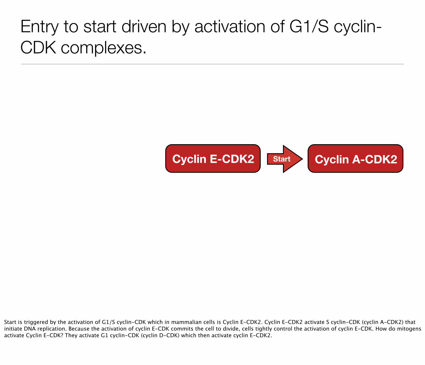
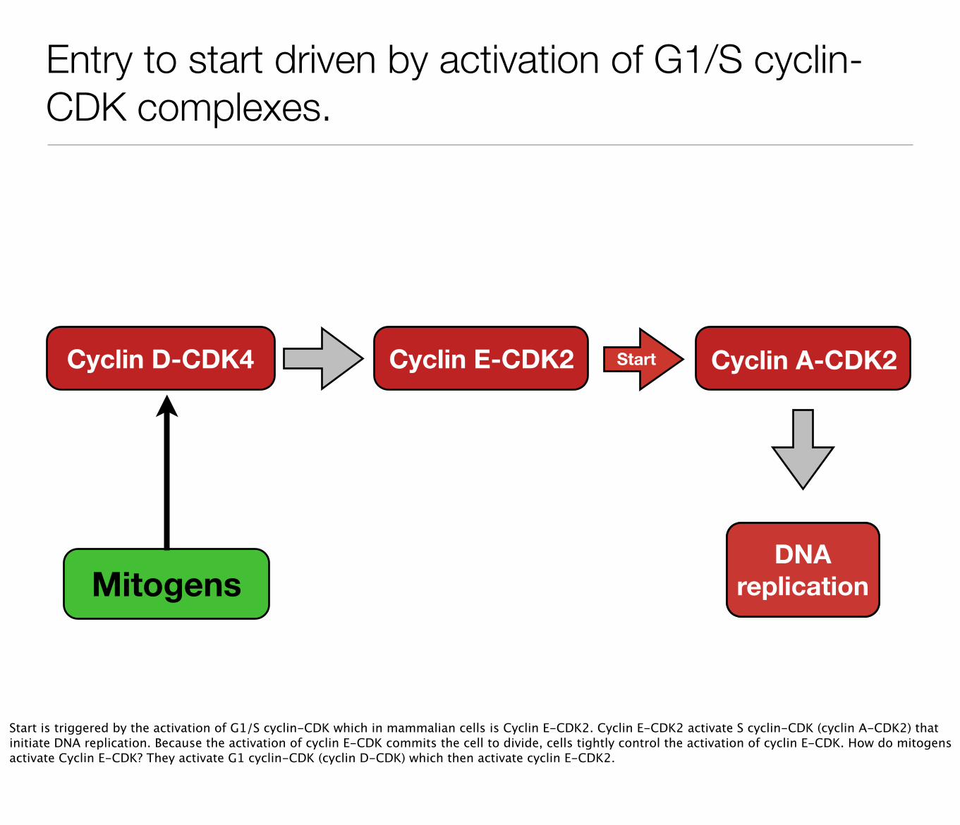
Entry to start driven by activation of G1/S cyclin-CDK complexes.
Cyclin E-CDK2 Cyclin A-CDK2Start
Start is triggered by the activation of G1/S cyclin-CDK which in mammalian cells is Cyclin E-CDK2. Cyclin E-CDK2 activate S cyclin-CDK (cyclin A-CDK2) that initiate DNA replication. Because the activation of cyclin E-CDK commits the cell to divide, cells tightly control the activation of cyclin E-CDK. How do mitogens activate Cyclin E-CDK? They activate G1 cyclin-CDK (cyclin D-CDK) which then activate cyclin E-CDK2.
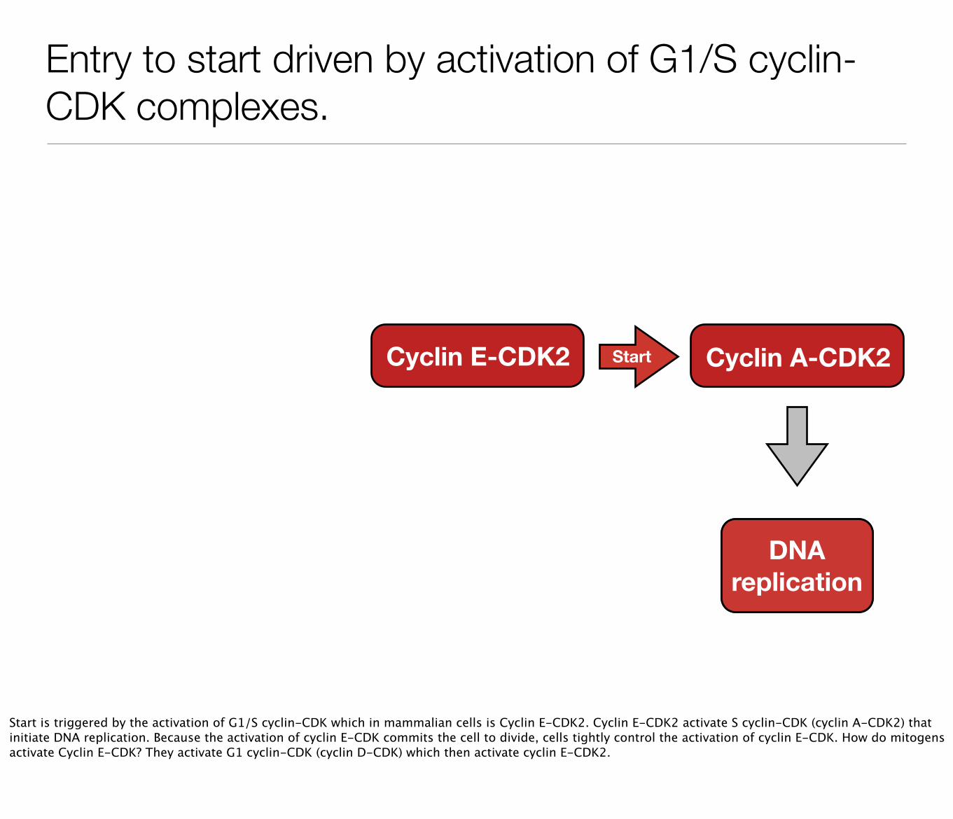
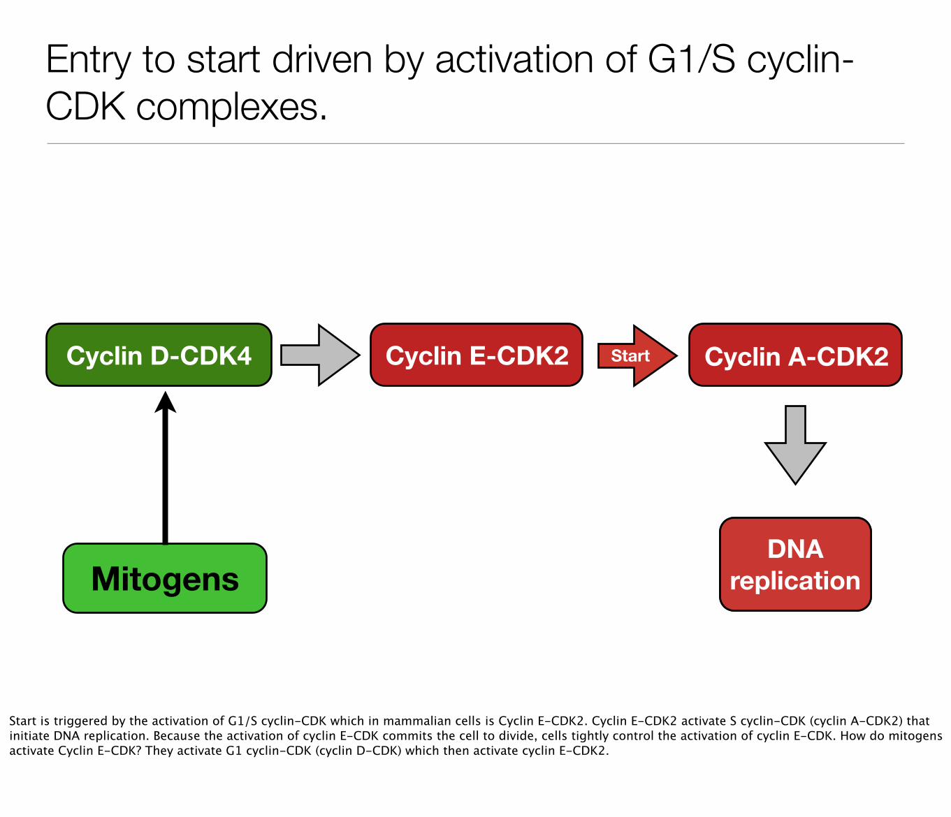
Entry to start driven by activation of G1/S cyclin-CDK complexes.
Cyclin E-CDK2
DNA replication
Cyclin A-CDK2Start
Start is triggered by the activation of G1/S cyclin-CDK which in mammalian cells is Cyclin E-CDK2. Cyclin E-CDK2 activate S cyclin-CDK (cyclin A-CDK2) that initiate DNA replication. Because the activation of cyclin E-CDK commits the cell to divide, cells tightly control the activation of cyclin E-CDK. How do mitogens activate Cyclin E-CDK? They activate G1 cyclin-CDK (cyclin D-CDK) which then activate cyclin E-CDK2.
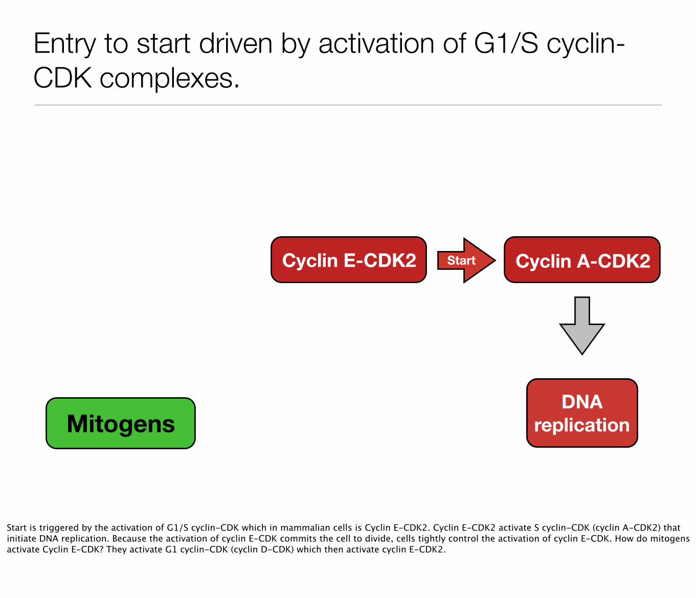
Entry to start driven by activation of G1/S cyclin-CDK complexes.
Cyclin E-CDK2
MitogensDNA
replication
Cyclin A-CDK2Start
Start is triggered by the activation of G1/S cyclin-CDK which in mammalian cells is Cyclin E-CDK2. Cyclin E-CDK2 activate S cyclin-CDK (cyclin A-CDK2) that initiate DNA replication. Because the activation of cyclin E-CDK commits the cell to divide, cells tightly control the activation of cyclin E-CDK. How do mitogens activate Cyclin E-CDK? They activate G1 cyclin-CDK (cyclin D-CDK) which then activate cyclin E-CDK2.
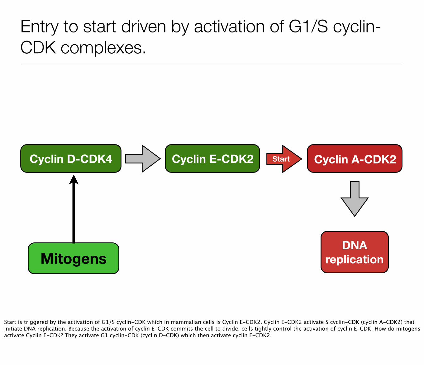
Entry to start driven by activation of G1/S cyclin-CDK complexes.
Cyclin D-CDK4 Cyclin E-CDK2
MitogensDNA
replication
Cyclin A-CDK2Start
Start is triggered by the activation of G1/S cyclin-CDK which in mammalian cells is Cyclin E-CDK2. Cyclin E-CDK2 activate S cyclin-CDK (cyclin A-CDK2) that initiate DNA replication. Because the activation of cyclin E-CDK commits the cell to divide, cells tightly control the activation of cyclin E-CDK. How do mitogens activate Cyclin E-CDK? They activate G1 cyclin-CDK (cyclin D-CDK) which then activate cyclin E-CDK2.
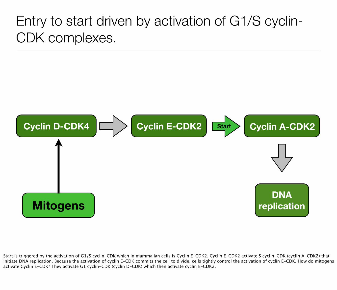
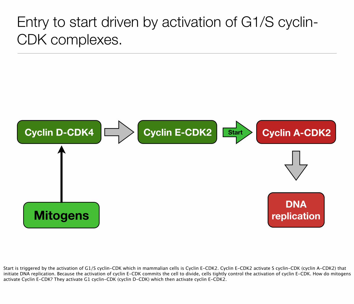
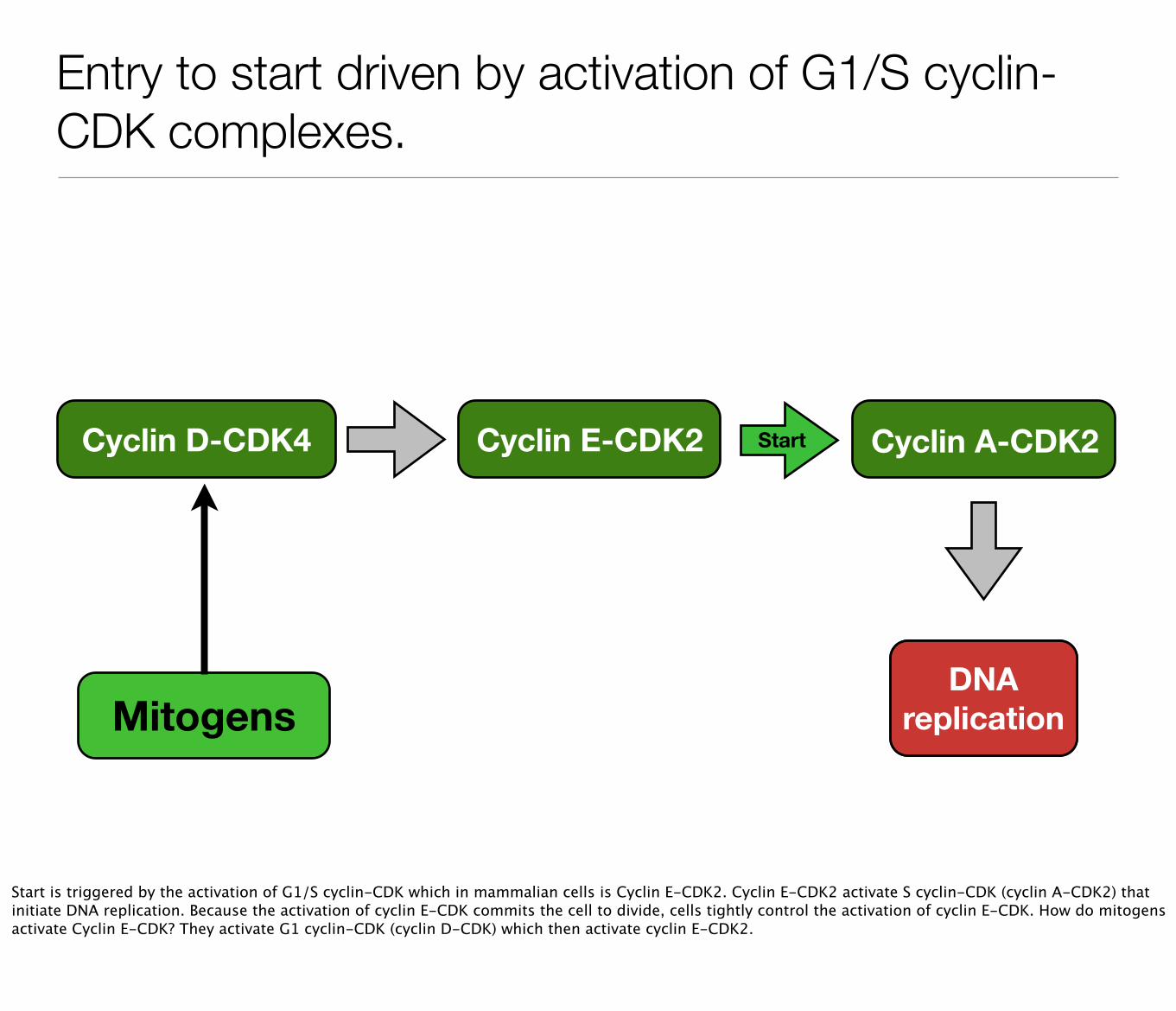
How CyclinD-CDK activate expression of G1/S cyclins
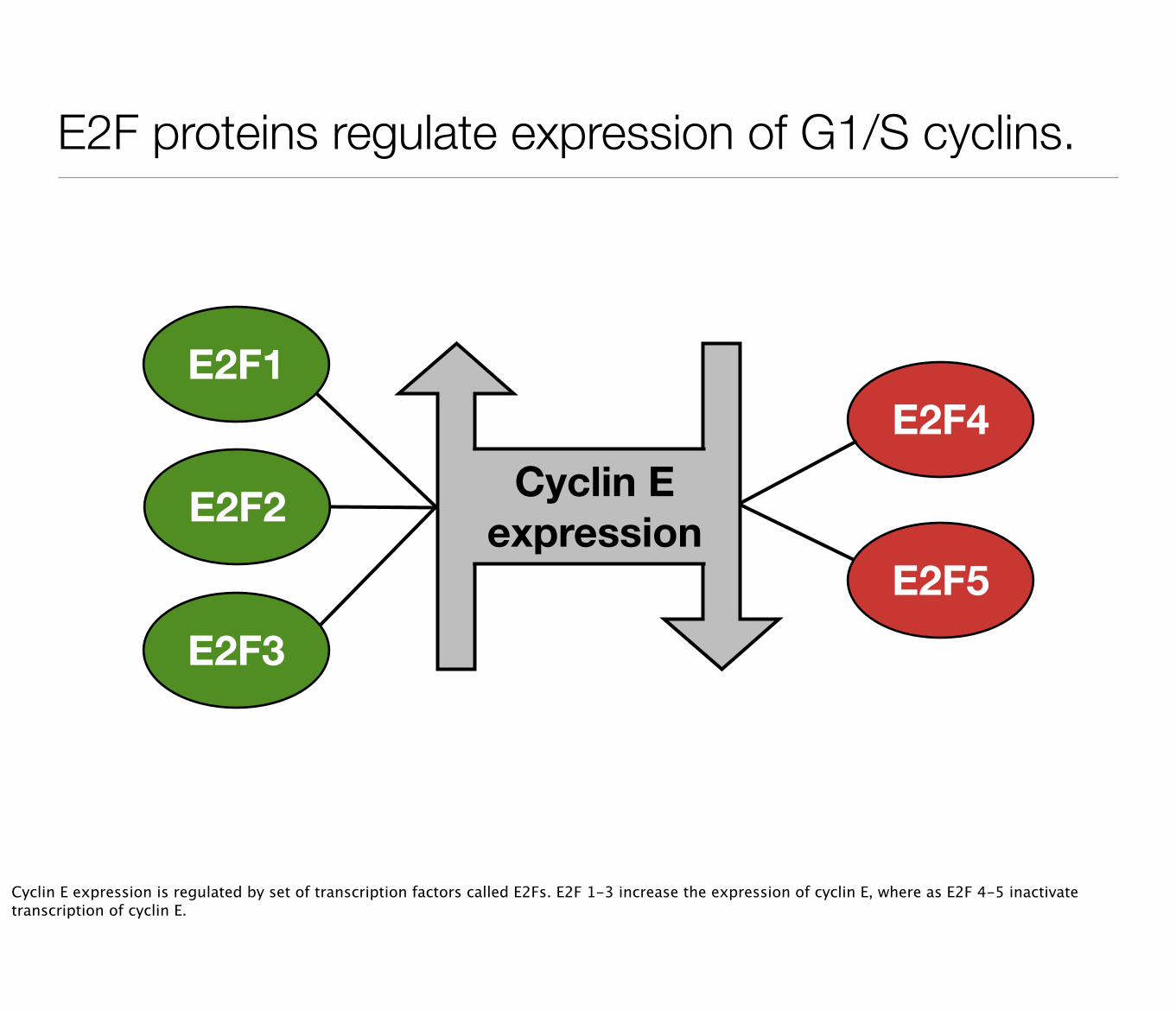
E2F proteins regulate expression of G1/S cyclins.
E2F1
E2F2
E2F3
E2F4
E2F5
Cyclin E expression
Cyclin E expression is regulated by set of transcription factors called E2Fs. E2F 1-3 increase the expression of cyclin E, where as E2F 4-5 inactivate transcription of cyclin E.
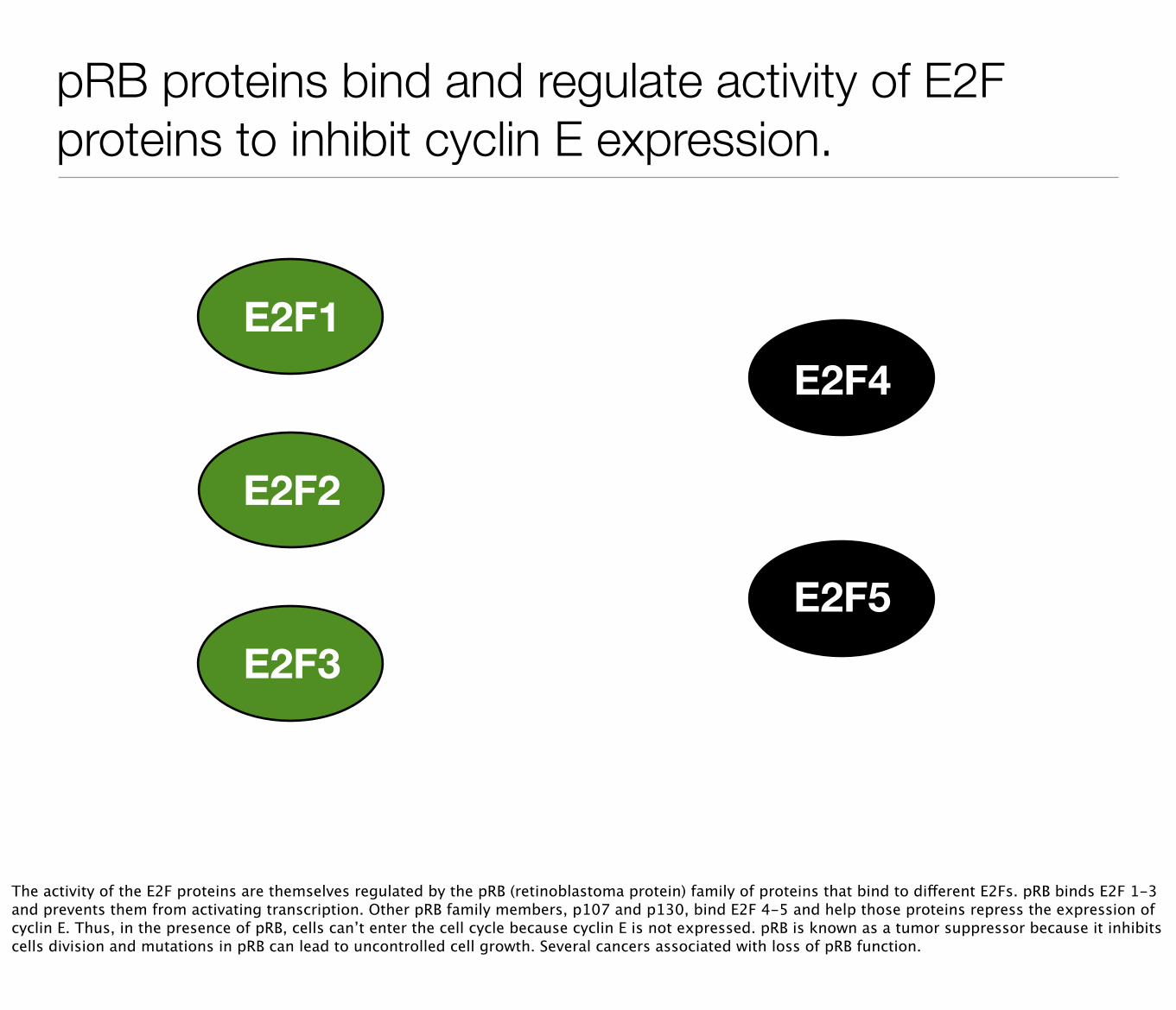
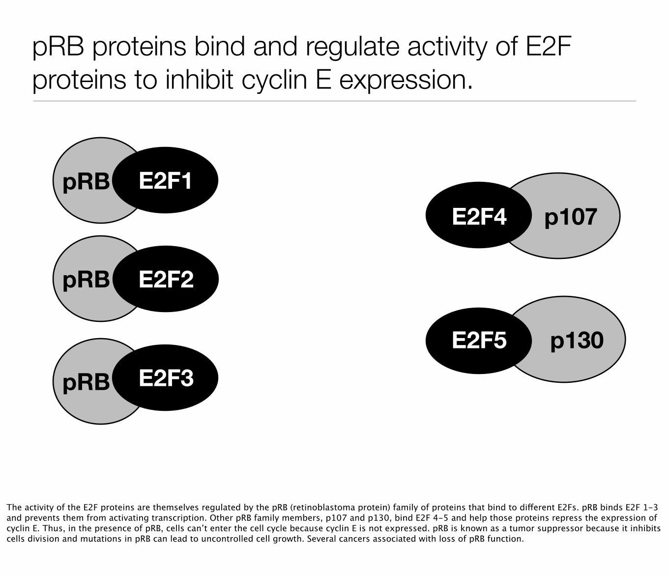
pRB proteins bind and regulate activity of E2F proteins to inhibit cyclin E expression.
E2F1
E2F3
E2F2
E2F4
E2F5
The activity of the E2F proteins are themselves regulated by the pRB (retinoblastoma protein) family of proteins that bind to different E2Fs. pRB binds E2F 1-3 and prevents them from activating transcription. Other pRB family members, p107 and p130, bind E2F 4-5 and help those proteins repress the expression of cyclin E. Thus, in the presence of pRB, cells can’t enter the cell cycle because cyclin E is not expressed. pRB is known as a tumor suppressor because it inhibits cells division and mutations in pRB can lead to uncontrolled cell growth. Several cancers associated with loss of pRB function.
pRB
pRB
pRB
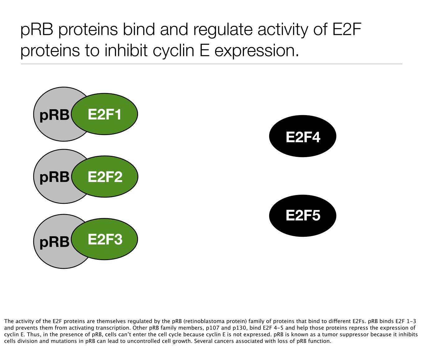
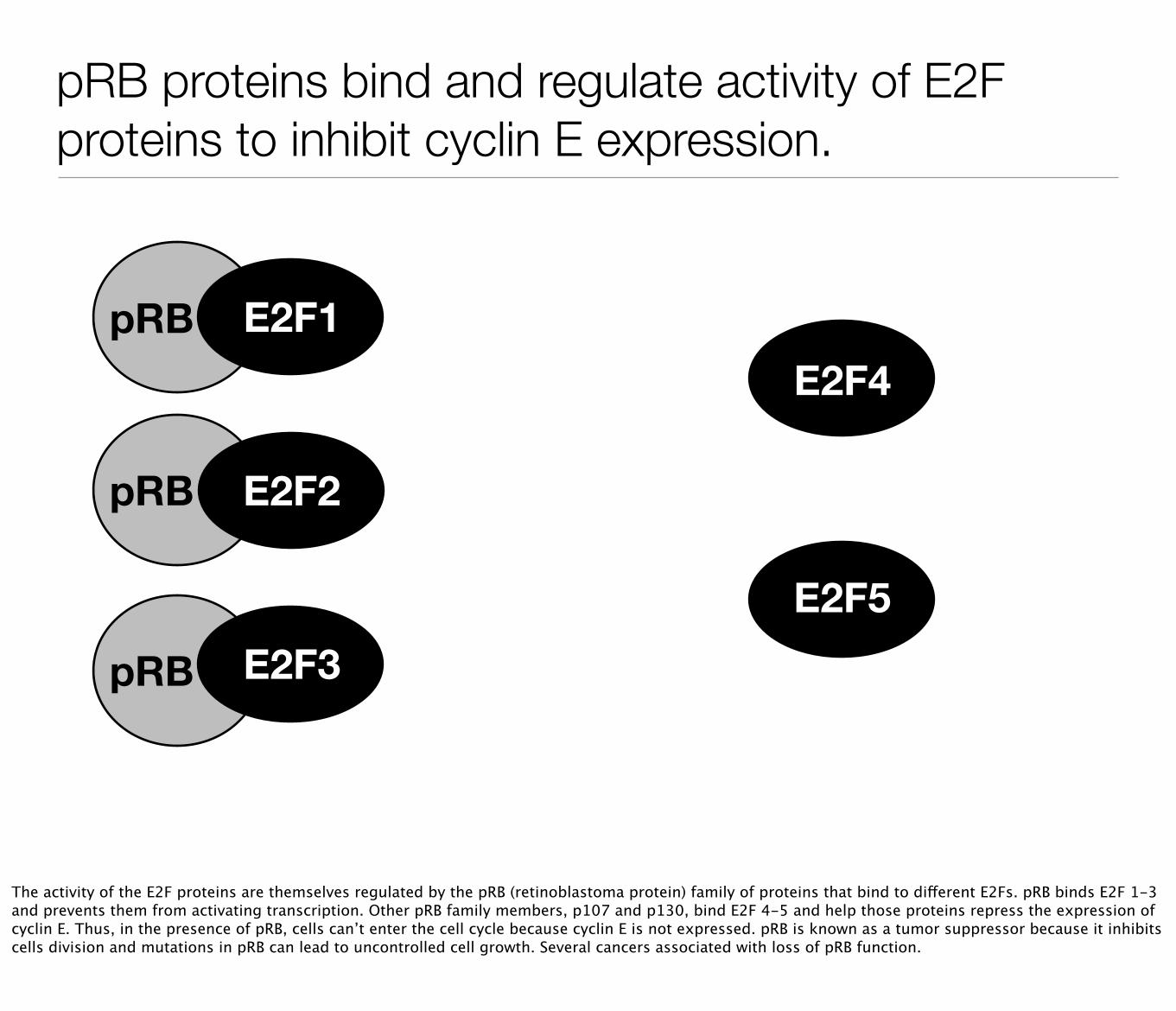
pRB proteins bind and regulate activity of E2F proteins to inhibit cyclin E expression.
E2F1
E2F3
E2F2
E2F4
E2F5
The activity of the E2F proteins are themselves regulated by the pRB (retinoblastoma protein) family of proteins that bind to different E2Fs. pRB binds E2F 1-3 and prevents them from activating transcription. Other pRB family members, p107 and p130, bind E2F 4-5 and help those proteins repress the expression of cyclin E. Thus, in the presence of pRB, cells can’t enter the cell cycle because cyclin E is not expressed. pRB is known as a tumor suppressor because it inhibits cells division and mutations in pRB can lead to uncontrolled cell growth. Several cancers associated with loss of pRB function.
p130
p107
pRB
pRB
pRB
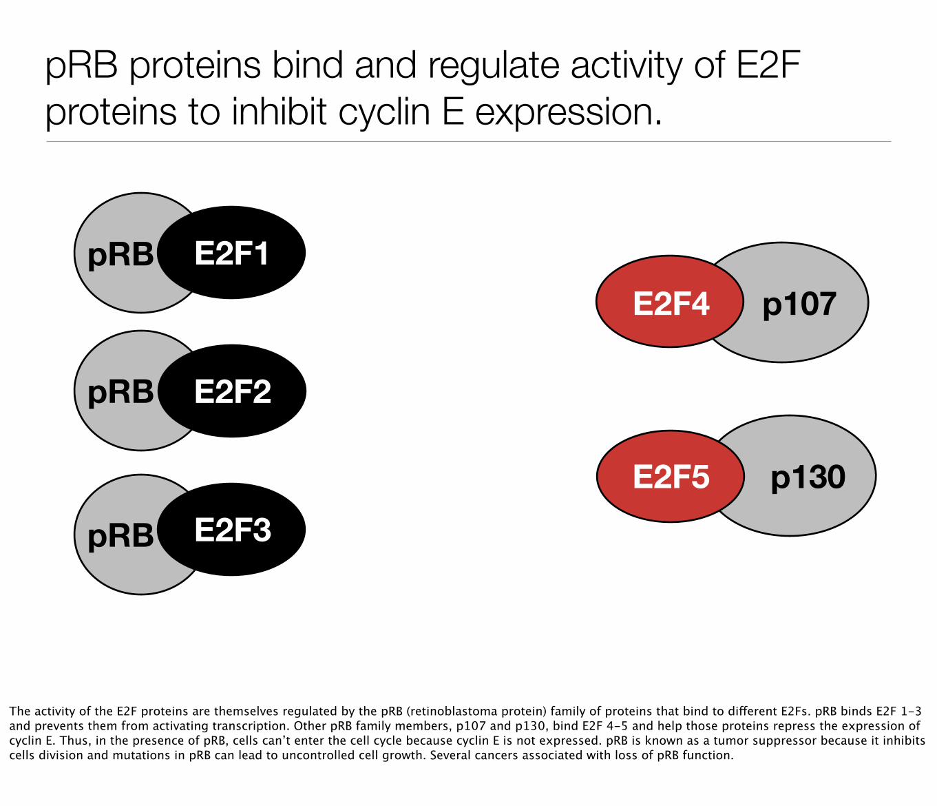
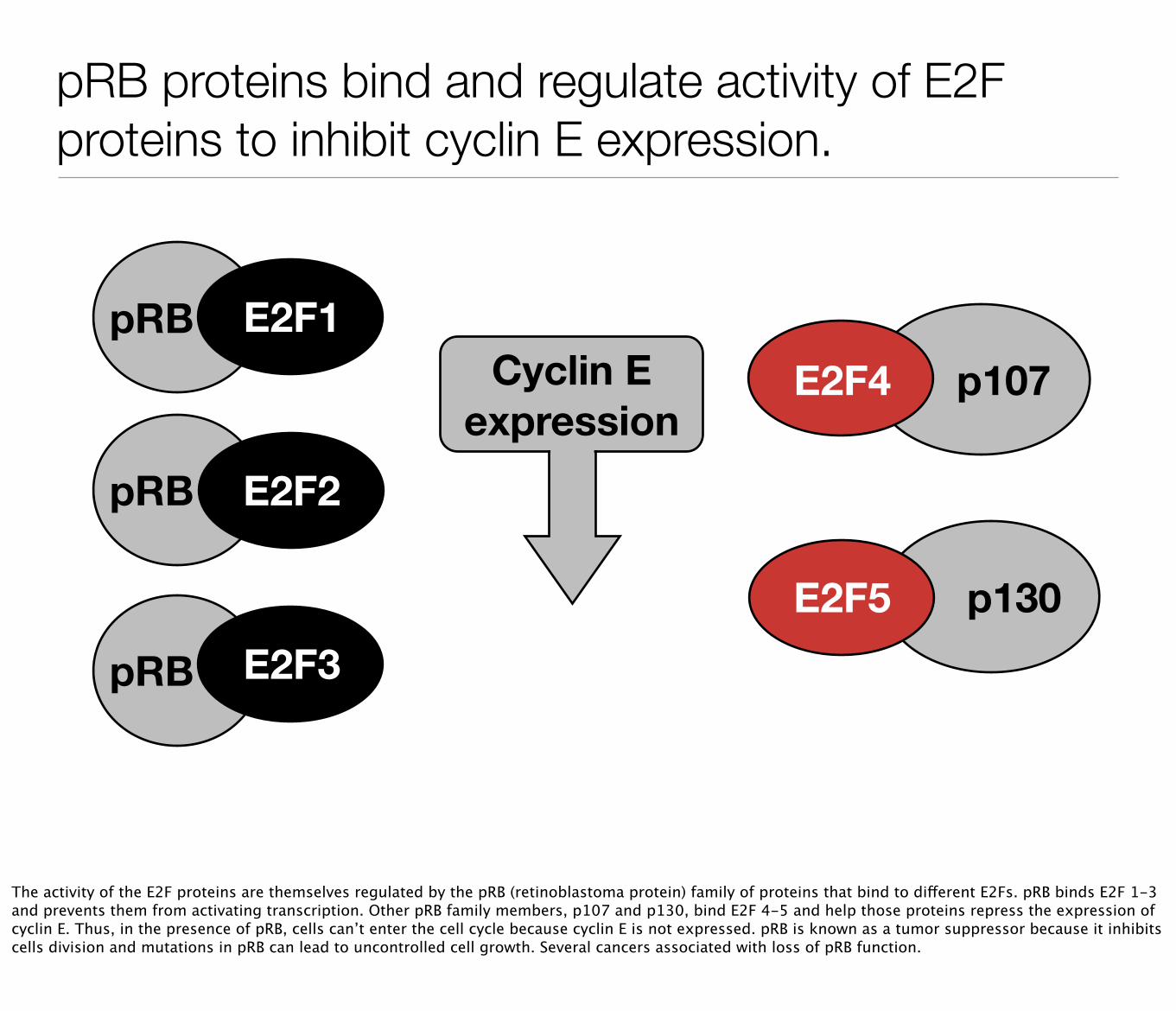
pRB proteins bind and regulate activity of E2F proteins to inhibit cyclin E expression.
E2F1
E2F3
E2F2
E2F4
E2F5
Cyclin E expression
The activity of the E2F proteins are themselves regulated by the pRB (retinoblastoma protein) family of proteins that bind to different E2Fs. pRB binds E2F 1-3 and prevents them from activating transcription. Other pRB family members, p107 and p130, bind E2F 4-5 and help those proteins repress the expression of cyclin E. Thus, in the presence of pRB, cells can’t enter the cell cycle because cyclin E is not expressed. pRB is known as a tumor suppressor because it inhibits cells division and mutations in pRB can lead to uncontrolled cell growth. Several cancers associated with loss of pRB function.
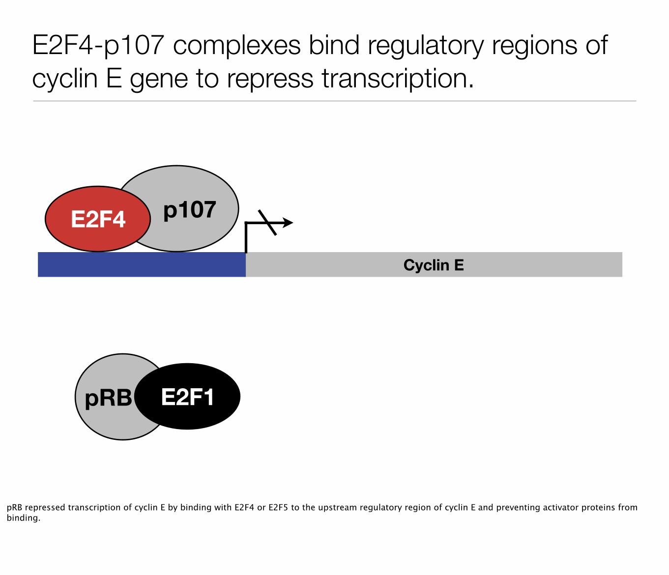
E2F4-p107 complexes bind regulatory regions of cyclin E gene to repress transcription.
Cyclin E
p107E2F4
pRB E2F1
pRB repressed transcription of cyclin E by binding with E2F4 or E2F5 to the upstream regulatory region of cyclin E and preventing activator proteins from binding.
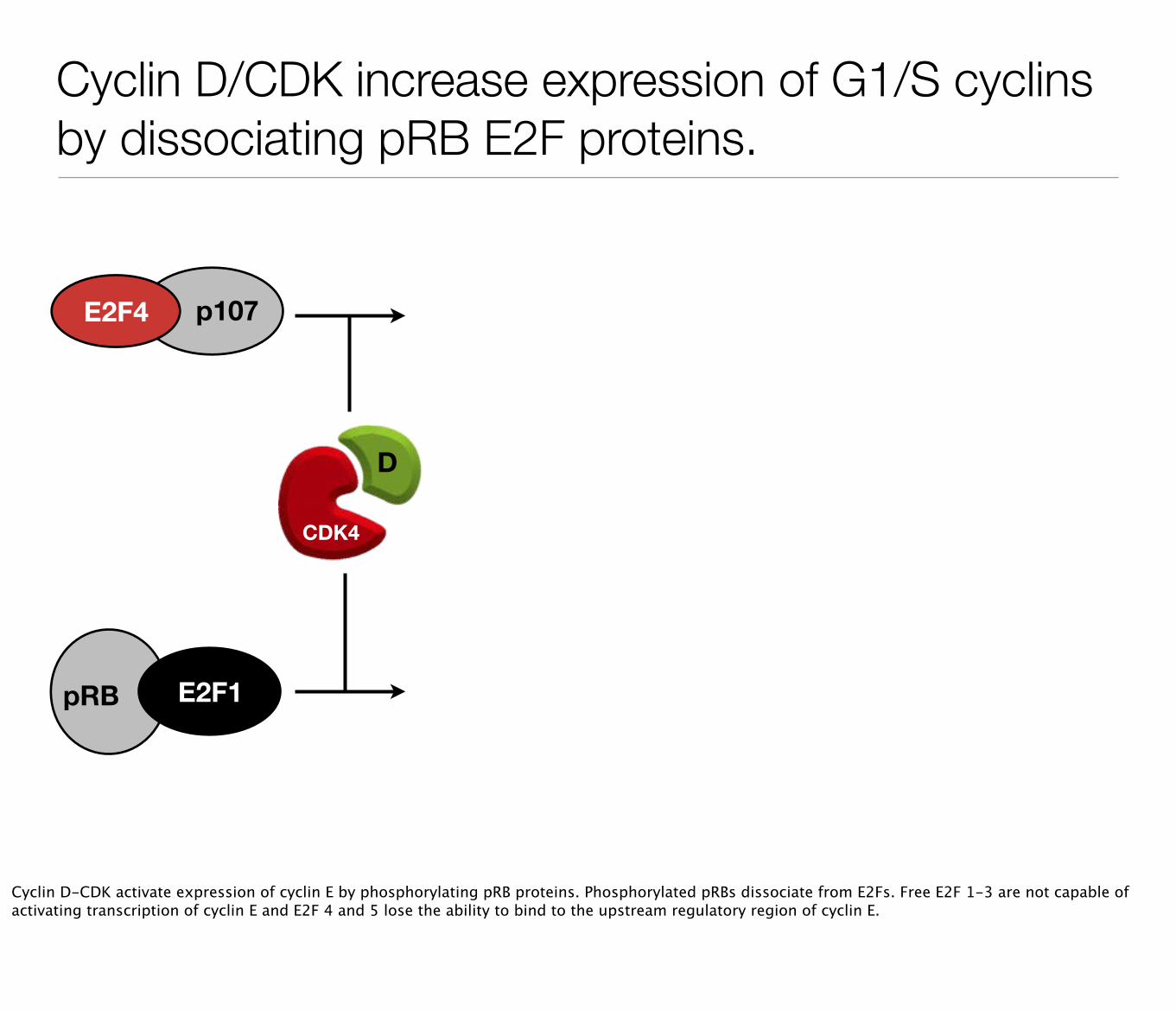
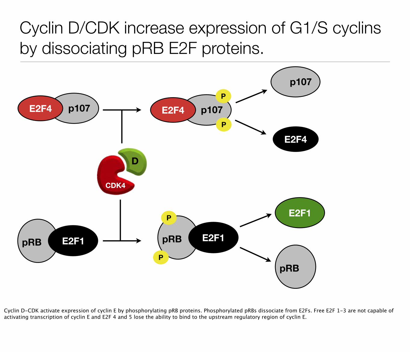
Cyclin D/CDK increase expression of G1/S cyclins by dissociating pRB E2F proteins.
pRB E2F1
p107E2F4
CDK4
D
Cyclin D-CDK activate expression of cyclin E by phosphorylating pRB proteins. Phosphorylated pRBs dissociate from E2Fs. Free E2F 1-3 are not capable of activating transcription of cyclin E and E2F 4 and 5 lose the ability to bind to the upstream regulatory region of cyclin E.
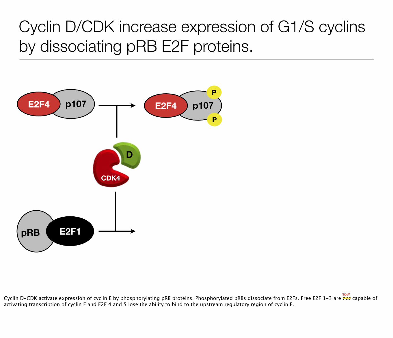
Cyclin D/CDK increase expression of G1/S cyclins by dissociating pRB E2F proteins.
pRB E2F1
p107E2F4 p107E2F4P
P
CDK4
D
Cyclin D-CDK activate expression of cyclin E by phosphorylating pRB proteins. Phosphorylated pRBs dissociate from E2Fs. Free E2F 1-3 are not capable of activating transcription of cyclin E and E2F 4 and 5 lose the ability to bind to the upstream regulatory region of cyclin E.
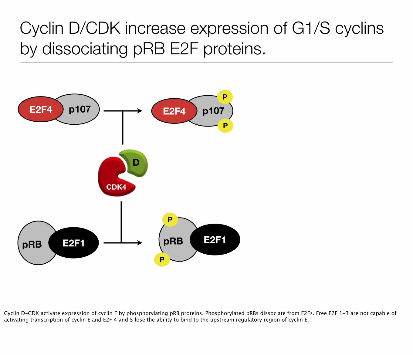
Cyclin D/CDK increase expression of G1/S cyclins by dissociating pRB E2F proteins.
pRB E2F1
p107E2F4 p107E2F4P
P
pRB E2F1
P
P
CDK4
D
Cyclin D-CDK activate expression of cyclin E by phosphorylating pRB proteins. Phosphorylated pRBs dissociate from E2Fs. Free E2F 1-3 are not capable of activating transcription of cyclin E and E2F 4 and 5 lose the ability to bind to the upstream regulatory region of cyclin E.
Cyclin D/CDK increase expression of G1/S cyclins by dissociating pRB E2F proteins.
pRB E2F1
p107E2F4 p107E2F4P
P
pRB E2F1
P
P
CDK4
D
p107
E2F4
Cyclin D-CDK activate expression of cyclin E by phosphorylating pRB proteins. Phosphorylated pRBs dissociate from E2Fs. Free E2F 1-3 are not capable of activating transcription of cyclin E and E2F 4 and 5 lose the ability to bind to the upstream regulatory region of cyclin E.
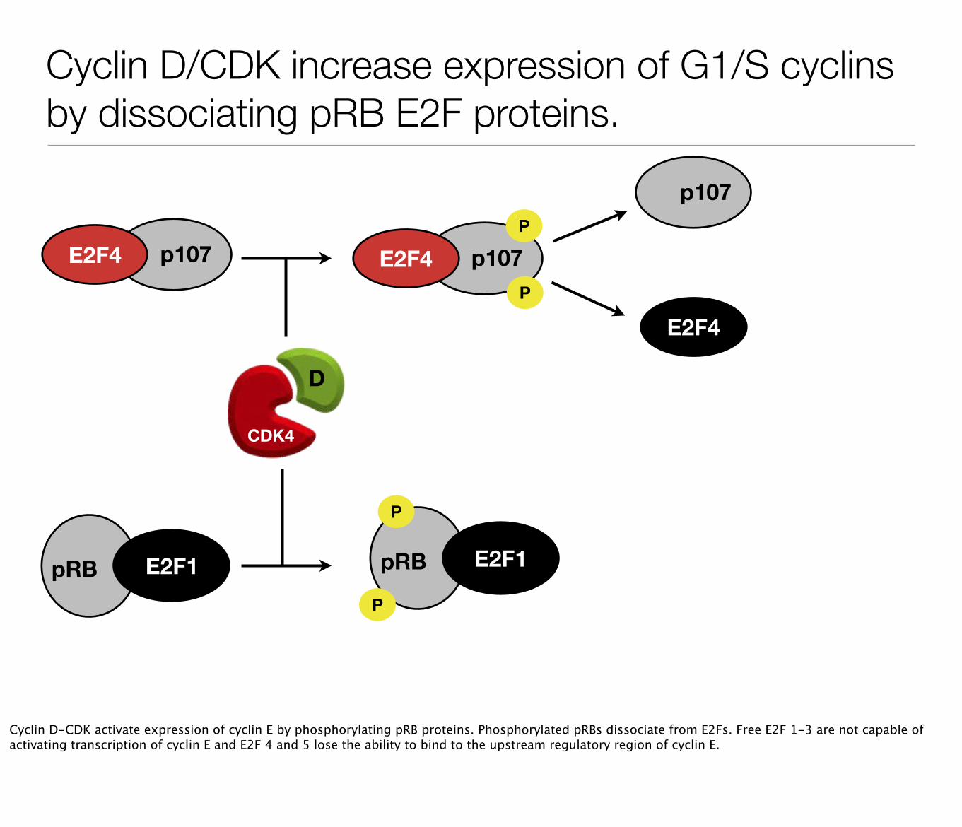
Cyclin D/CDK increase expression of G1/S cyclins by dissociating pRB E2F proteins.
pRB E2F1
p107E2F4 p107E2F4P
P
pRB E2F1
P
P
CDK4
D
p107
E2F4
E2F1
pRB
Cyclin D-CDK activate expression of cyclin E by phosphorylating pRB proteins. Phosphorylated pRBs dissociate from E2Fs. Free E2F 1-3 are not capable of activating transcription of cyclin E and E2F 4 and 5 lose the ability to bind to the upstream regulatory region of cyclin E.
p107
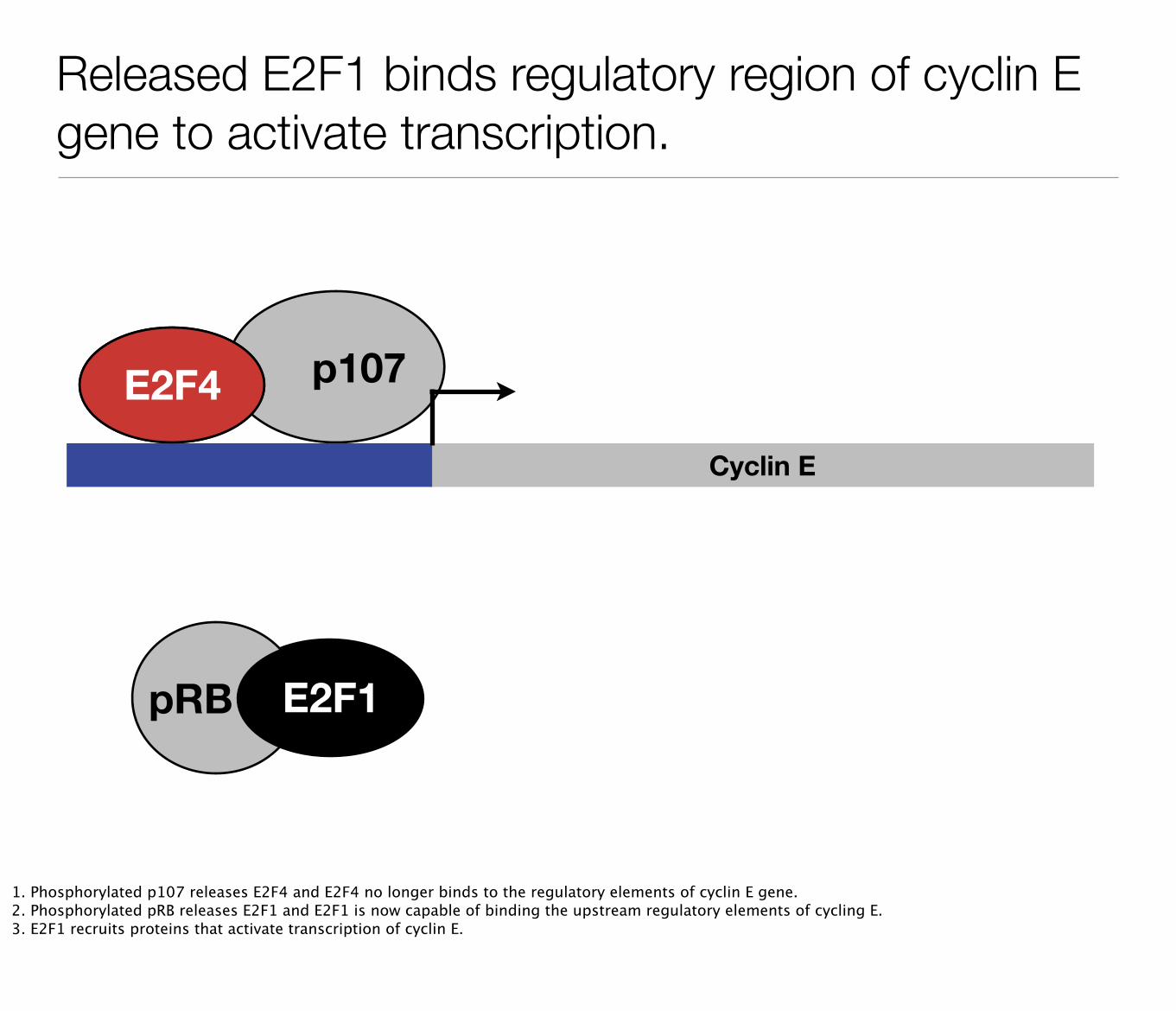
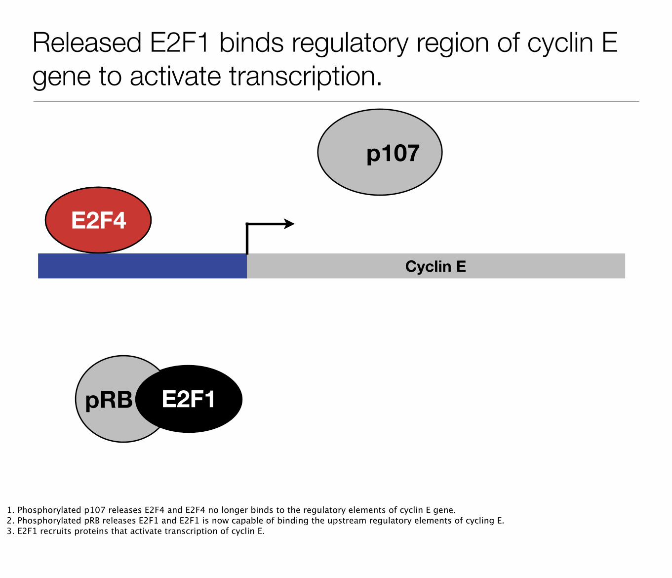
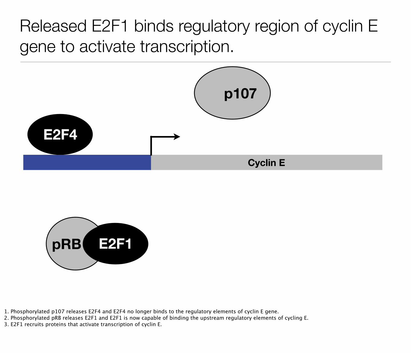
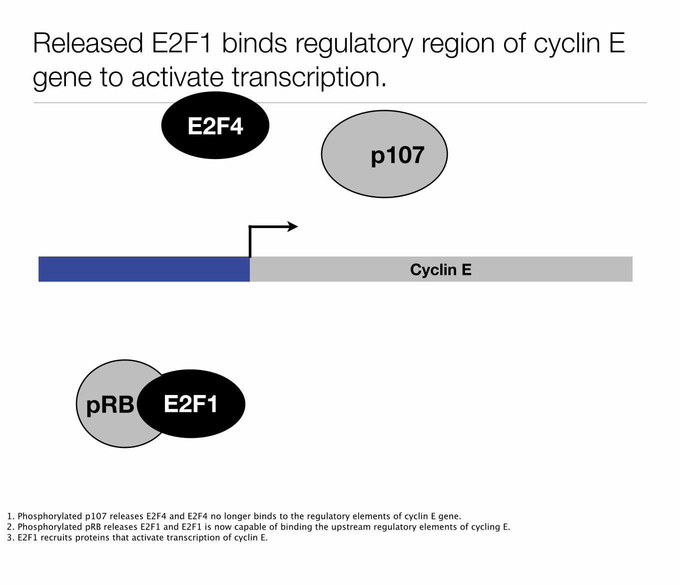
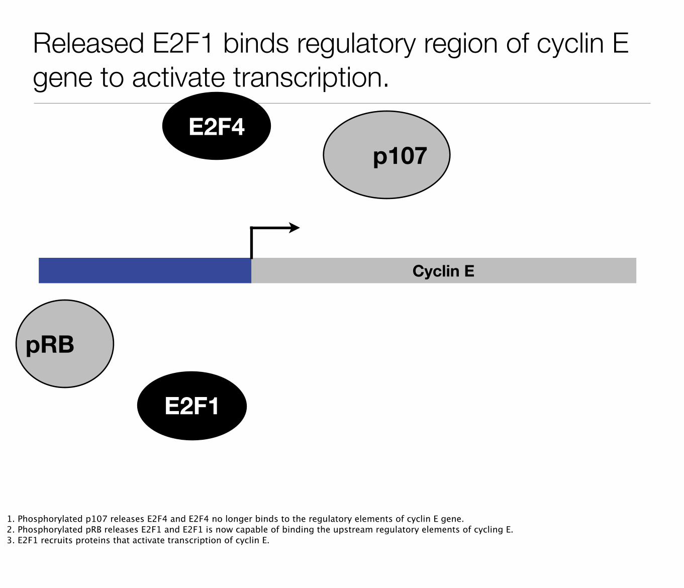
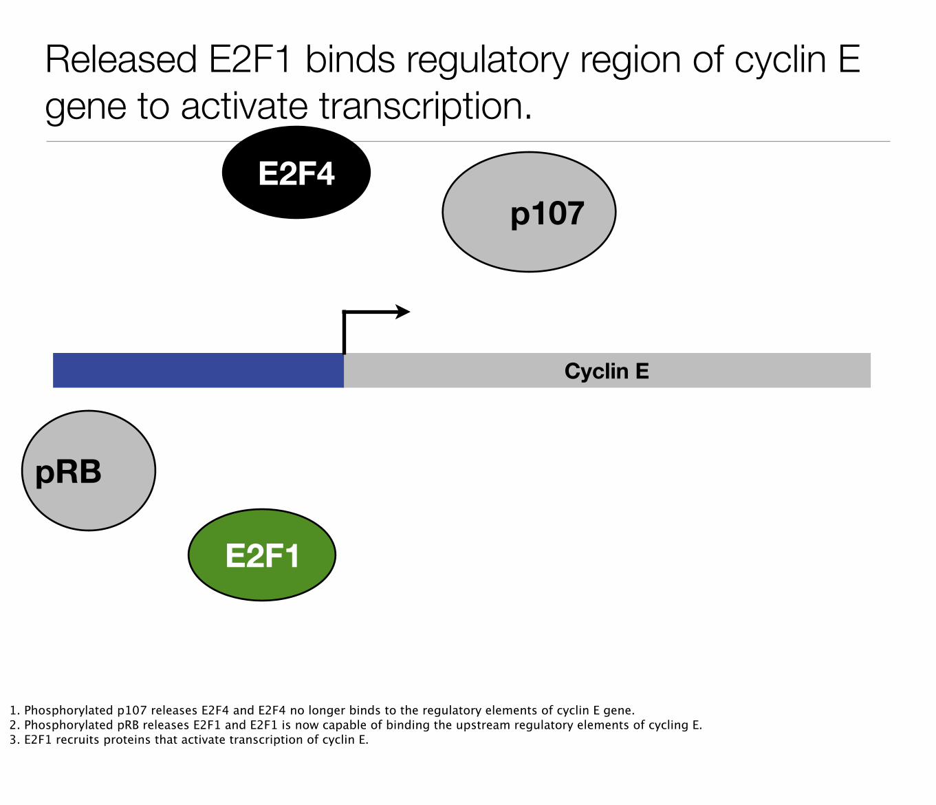
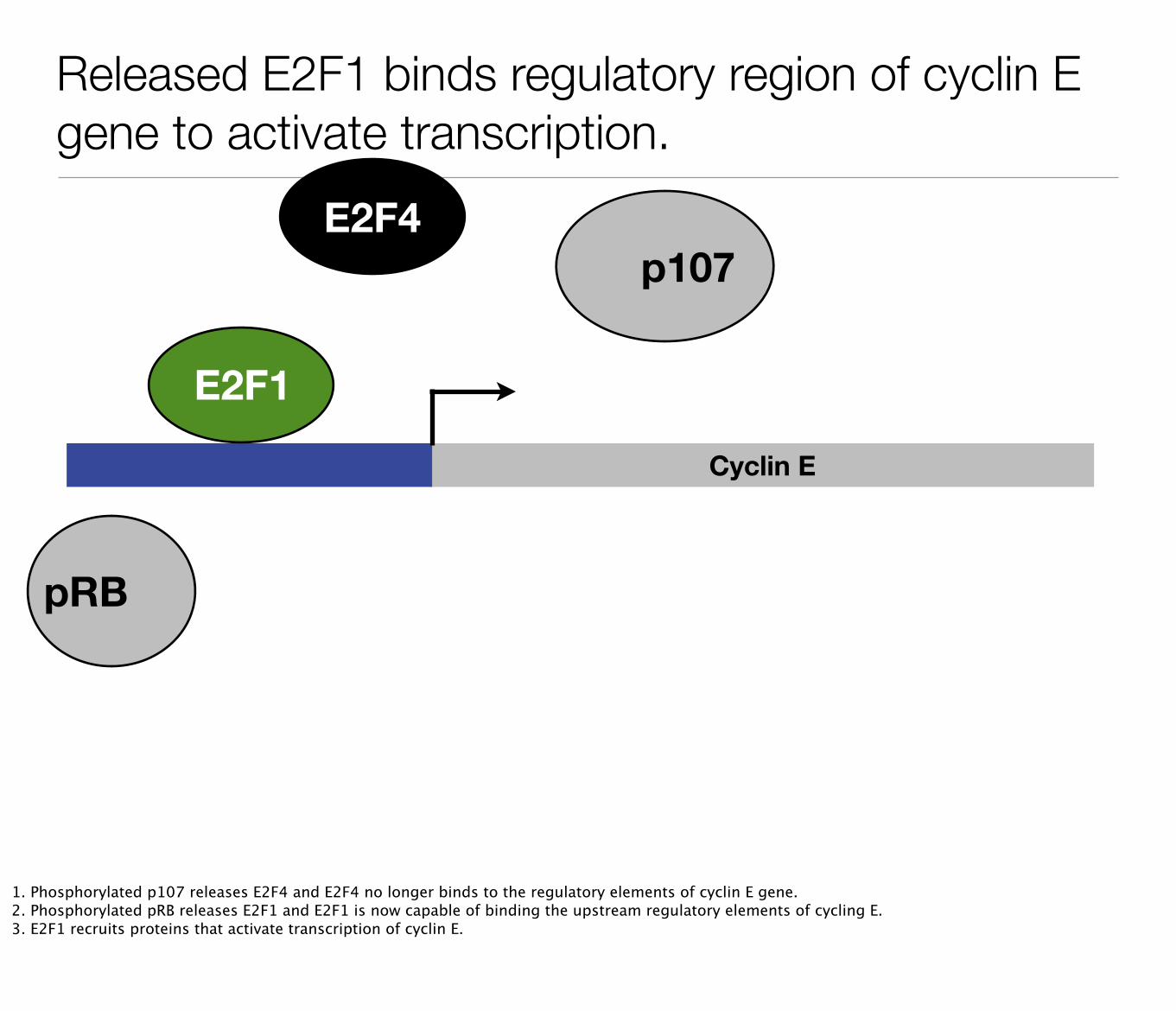
Released E2F1 binds regulatory region of cyclin E gene to activate transcription.
Cyclin E
E2F1
E2F4
pRB E2F1
1. Phosphorylated p107 releases E2F4 and E2F4 no longer binds to the regulatory elements of cyclin E gene.2. Phosphorylated pRB releases E2F1 and E2F1 is now capable of binding the upstream regulatory elements of cycling E.3. E2F1 recruits proteins that activate transcription of cyclin E.
p107
Released E2F1 binds regulatory region of cyclin E gene to activate transcription.
Cyclin E
E2F1
E2F4
pRB
E2F1
1. Phosphorylated p107 releases E2F4 and E2F4 no longer binds to the regulatory elements of cyclin E gene.2. Phosphorylated pRB releases E2F1 and E2F1 is now capable of binding the upstream regulatory elements of cycling E.3. E2F1 recruits proteins that activate transcription of cyclin E.
p107
Released E2F1 binds regulatory region of cyclin E gene to activate transcription.
Cyclin E
E2F1
E2F4
pRB
1. Phosphorylated p107 releases E2F4 and E2F4 no longer binds to the regulatory elements of cyclin E gene.2. Phosphorylated pRB releases E2F1 and E2F1 is now capable of binding the upstream regulatory elements of cycling E.3. E2F1 recruits proteins that activate transcription of cyclin E.
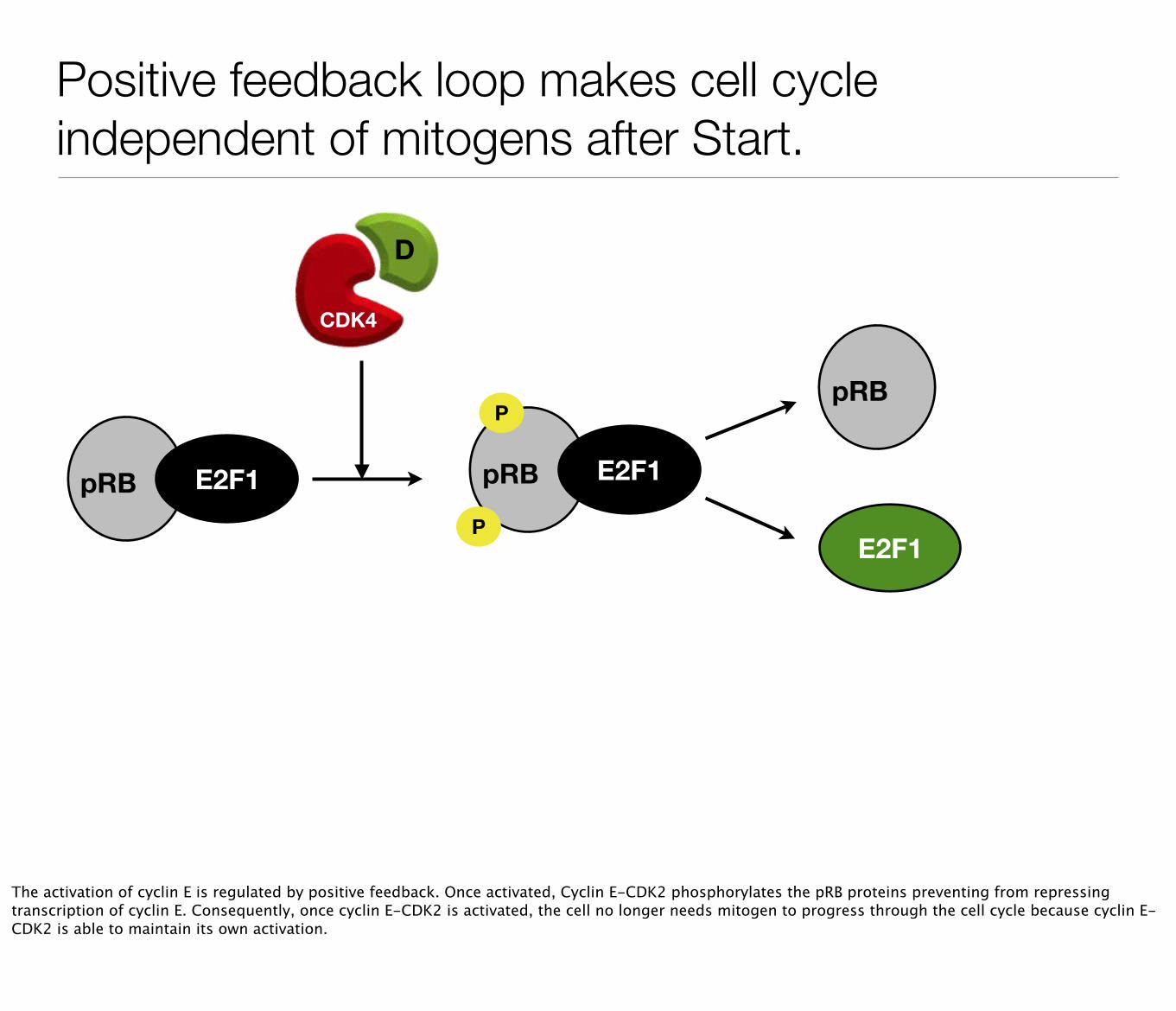
Positive feedback loop makes cell cycle independent of mitogens after Start.
pRB E2F1 pRB E2F1
P
P
CDK4
D
E2F1
pRB
The activation of cyclin E is regulated by positive feedback. Once activated, Cyclin E-CDK2 phosphorylates the pRB proteins preventing from repressing transcription of cyclin E. Consequently, once cyclin E-CDK2 is activated, the cell no longer needs mitogen to progress through the cell cycle because cyclin E-CDK2 is able to maintain its own activation.
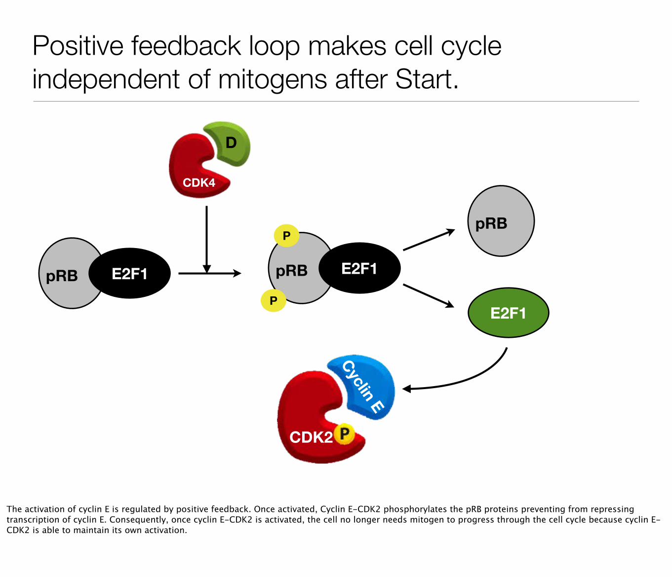
Positive feedback loop makes cell cycle independent of mitogens after Start.
pRB E2F1 pRB E2F1
P
P
CDK4
D
E2F1
pRB
Cyclin E
CDK2
The activation of cyclin E is regulated by positive feedback. Once activated, Cyclin E-CDK2 phosphorylates the pRB proteins preventing from repressing transcription of cyclin E. Consequently, once cyclin E-CDK2 is activated, the cell no longer needs mitogen to progress through the cell cycle because cyclin E-CDK2 is able to maintain its own activation.
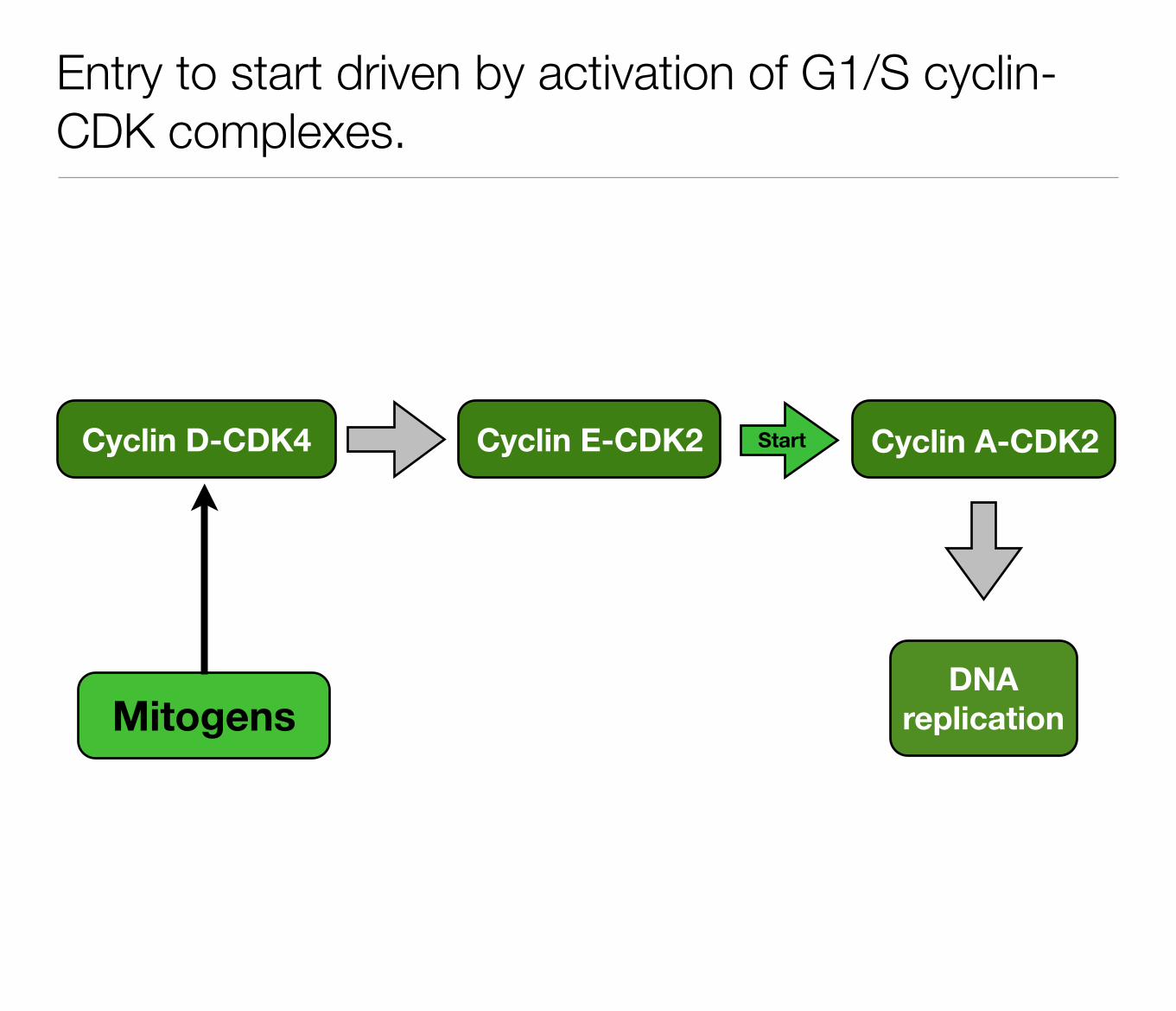
Entry to start driven by activation of G1/S cyclin-CDK complexes.
Cyclin D-CDK4 Cyclin E-CDK2
MitogensDNA
replication
Cyclin A-CDK2Start
How mitogens and anti-mitogens regulate activity of Cyclin D-CDK.
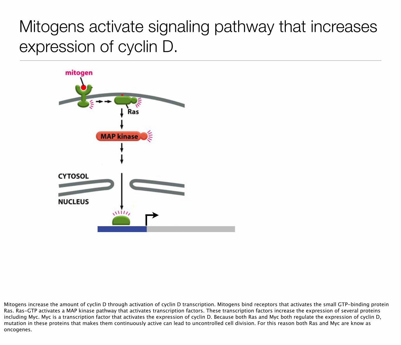
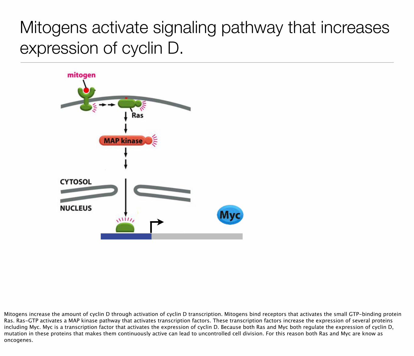
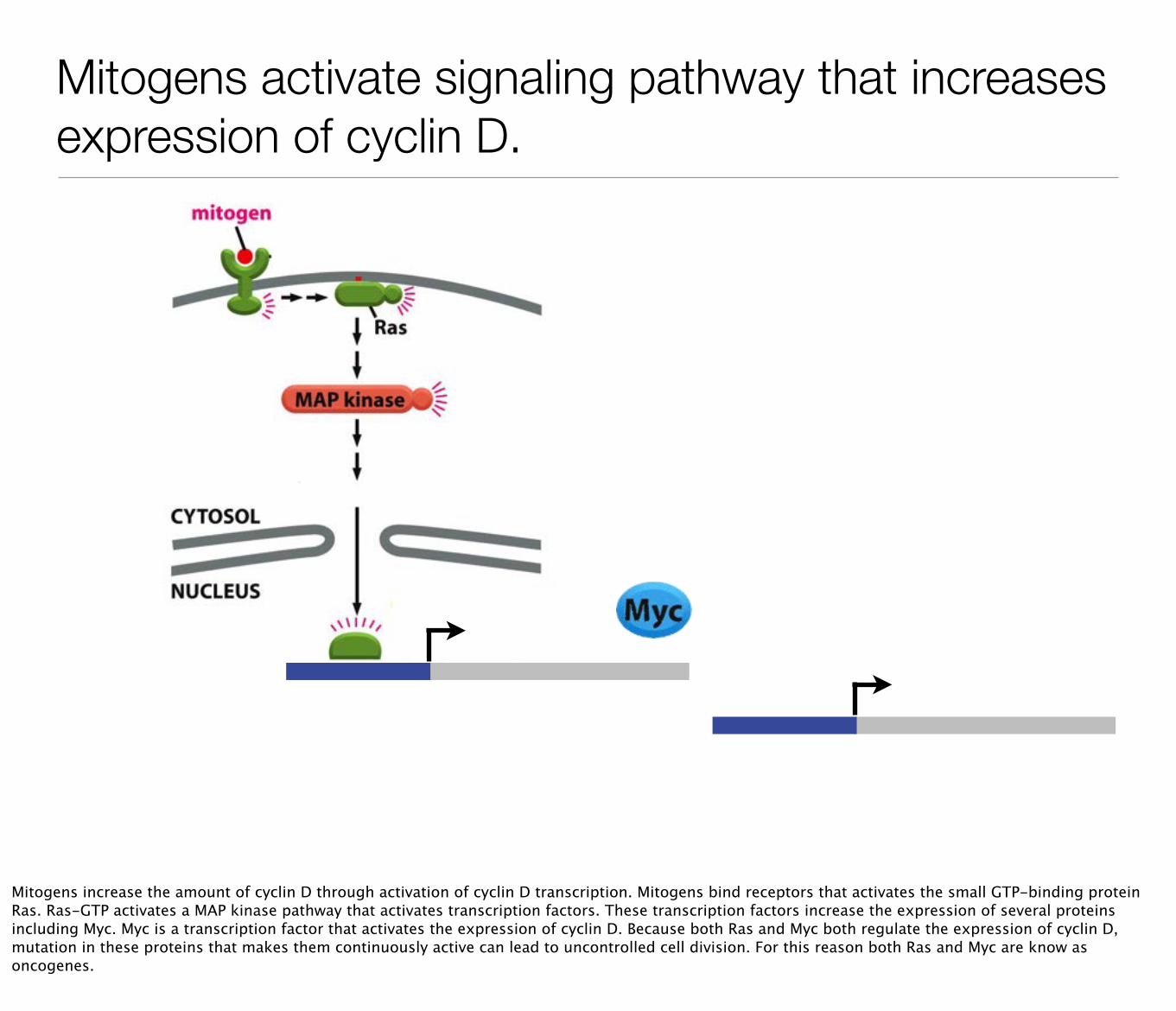
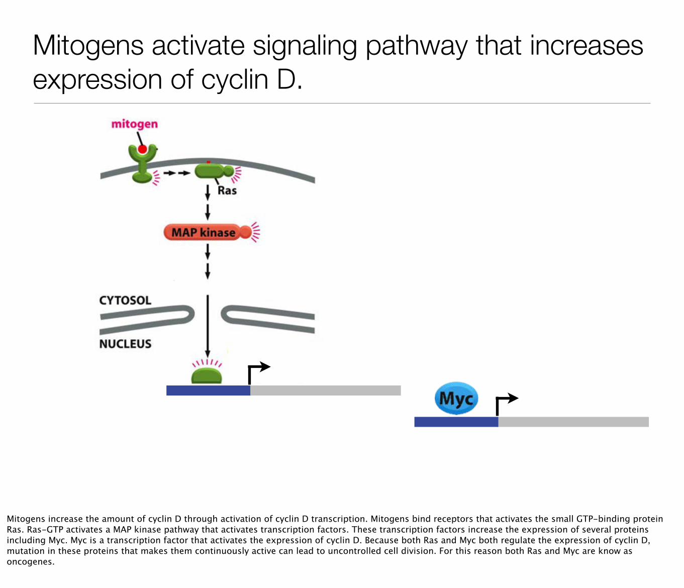
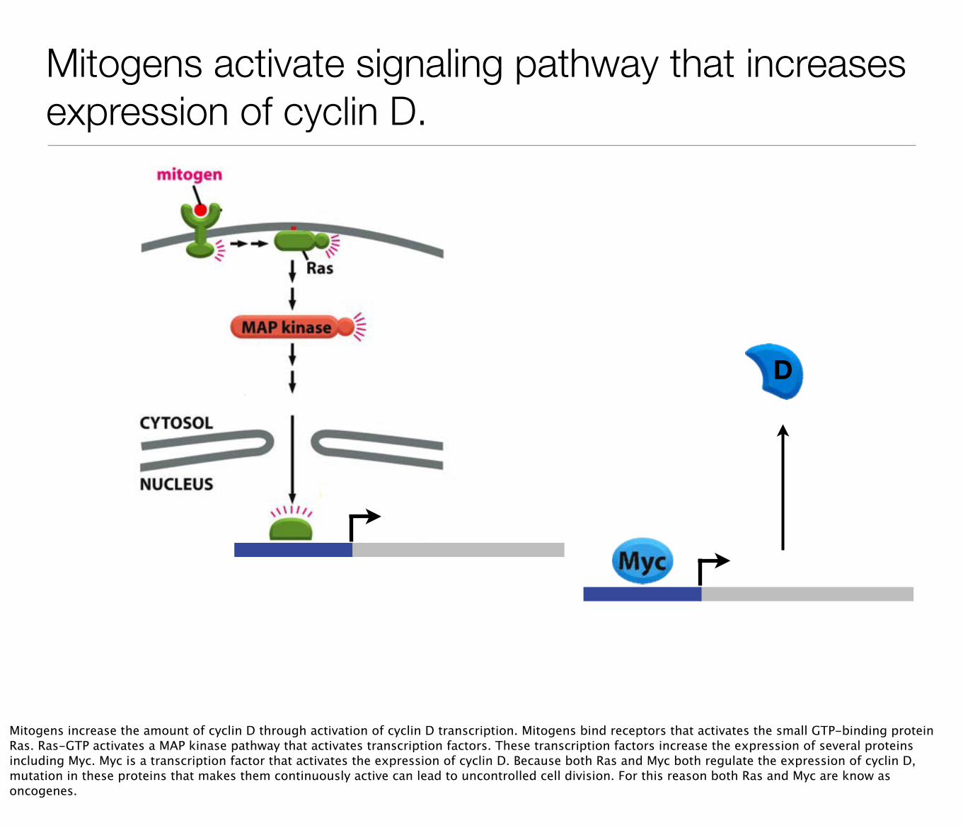
Mitogens activate signaling pathway that increases expression of cyclin D.
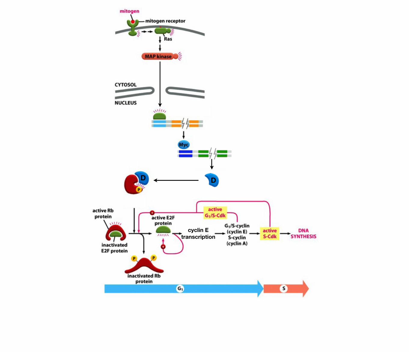
Mitogens increase the amount of cyclin D through activation of cyclin D transcription. Mitogens bind receptors that activates the small GTP-binding protein Ras. Ras-GTP activates a MAP kinase pathway that activates transcription factors. These transcription factors increase the expression of several proteins including Myc. Myc is a transcription factor that activates the expression of cyclin D. Because both Ras and Myc both regulate the expression of cyclin D, mutation in these proteins that makes them continuously active can lead to uncontrolled cell division. For this reason both Ras and Myc are know as oncogenes.
Mitogens activate signaling pathway that increases expression of cyclin D.
D
Mitogens increase the amount of cyclin D through activation of cyclin D transcription. Mitogens bind receptors that activates the small GTP-binding protein Ras. Ras-GTP activates a MAP kinase pathway that activates transcription factors. These transcription factors increase the expression of several proteins including Myc. Myc is a transcription factor that activates the expression of cyclin D. Because both Ras and Myc both regulate the expression of cyclin D, mutation in these proteins that makes them continuously active can lead to uncontrolled cell division. For this reason both Ras and Myc are know as oncogenes.
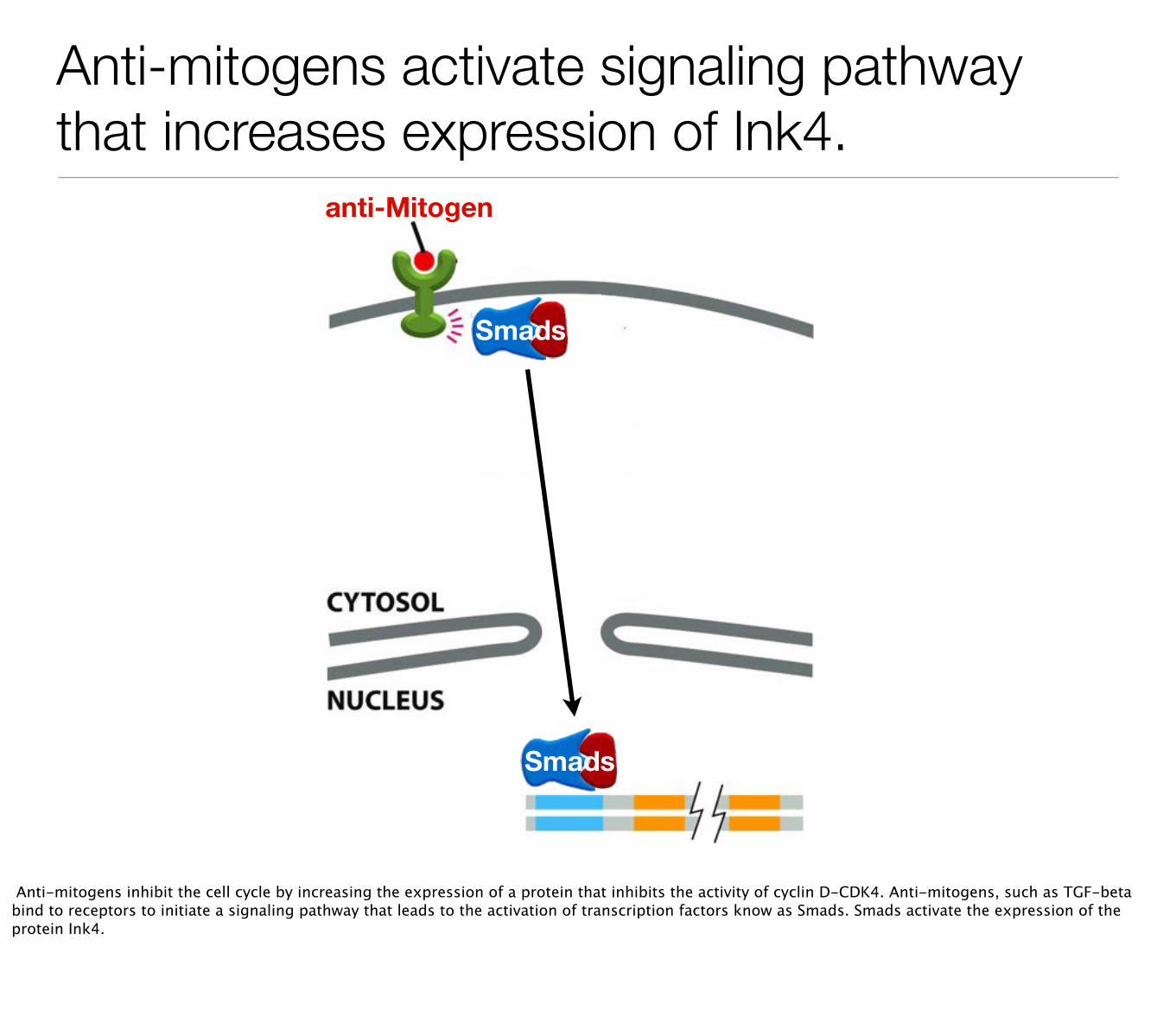
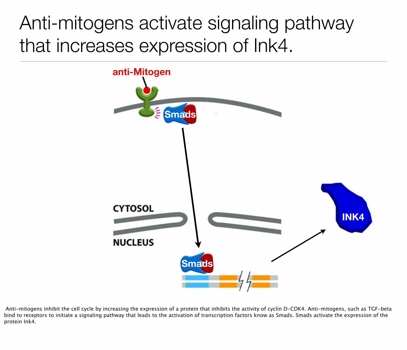
Anti-mitogens activate signaling pathway that increases expression of Ink4.
Smads
anti-Mitogen
Smads
Anti-mitogens inhibit the cell cycle by increasing the expression of a protein that inhibits the activity of cyclin D-CDK4. Anti-mitogens, such as TGF-beta bind to receptors to initiate a signaling pathway that leads to the activation of transcription factors know as Smads. Smads activate the expression of the protein Ink4.
Anti-mitogens activate signaling pathway that increases expression of Ink4.
Smads
anti-Mitogen
Smads
INK4
Anti-mitogens inhibit the cell cycle by increasing the expression of a protein that inhibits the activity of cyclin D-CDK4. Anti-mitogens, such as TGF-beta bind to receptors to initiate a signaling pathway that leads to the activation of transcription factors know as Smads. Smads activate the expression of the protein Ink4.
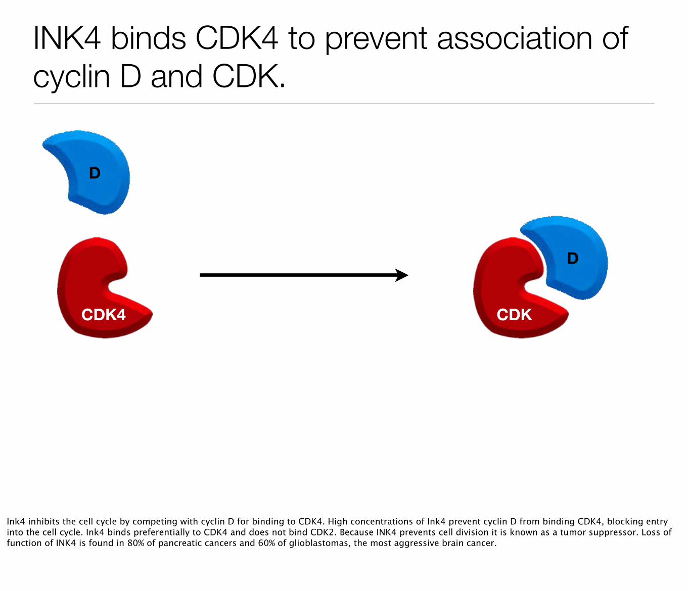
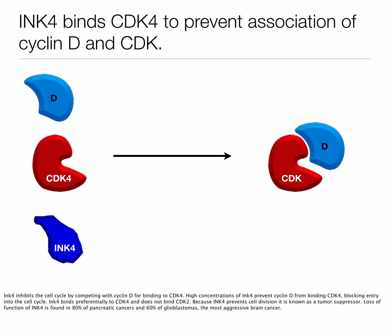
INK4 binds CDK4 to prevent association of cyclin D and CDK.
CDK4
D
CDK
D
Ink4 inhibits the cell cycle by competing with cyclin D for binding to CDK4. High concentrations of Ink4 prevent cyclin D from binding CDK4, blocking entry into the cell cycle. Ink4 binds preferentially to CDK4 and does not bind CDK2. Because INK4 prevents cell division it is known as a tumor suppressor. Loss of function of INK4 is found in 80% of pancreatic cancers and 60% of glioblastomas, the most aggressive brain cancer.
INK4 binds CDK4 to prevent association of cyclin D and CDK.
CDK4
D
CDK
D
INK4
Ink4 inhibits the cell cycle by competing with cyclin D for binding to CDK4. High concentrations of Ink4 prevent cyclin D from binding CDK4, blocking entry into the cell cycle. Ink4 binds preferentially to CDK4 and does not bind CDK2. Because INK4 prevents cell division it is known as a tumor suppressor. Loss of function of INK4 is found in 80% of pancreatic cancers and 60% of glioblastomas, the most aggressive brain cancer.
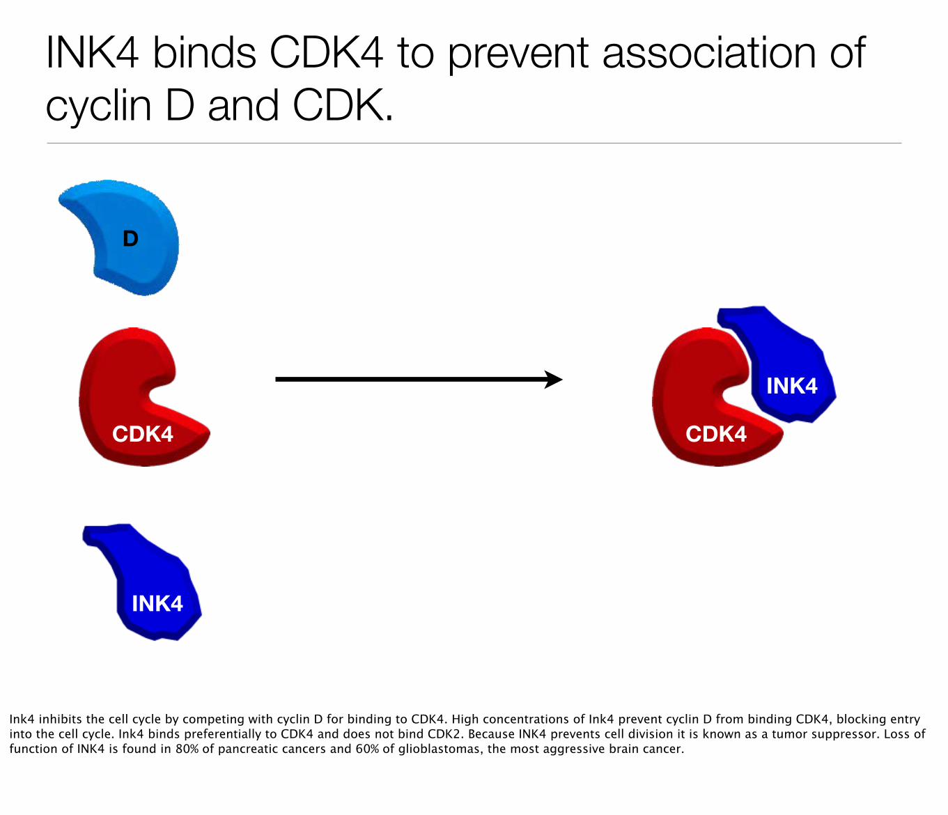
INK4 binds CDK4 to prevent association of cyclin D and CDK.
CDK4
D
INK4
INK4
CDK4
Ink4 inhibits the cell cycle by competing with cyclin D for binding to CDK4. High concentrations of Ink4 prevent cyclin D from binding CDK4, blocking entry into the cell cycle. Ink4 binds preferentially to CDK4 and does not bind CDK2. Because INK4 prevents cell division it is known as a tumor suppressor. Loss of function of INK4 is found in 80% of pancreatic cancers and 60% of glioblastomas, the most aggressive brain cancer.
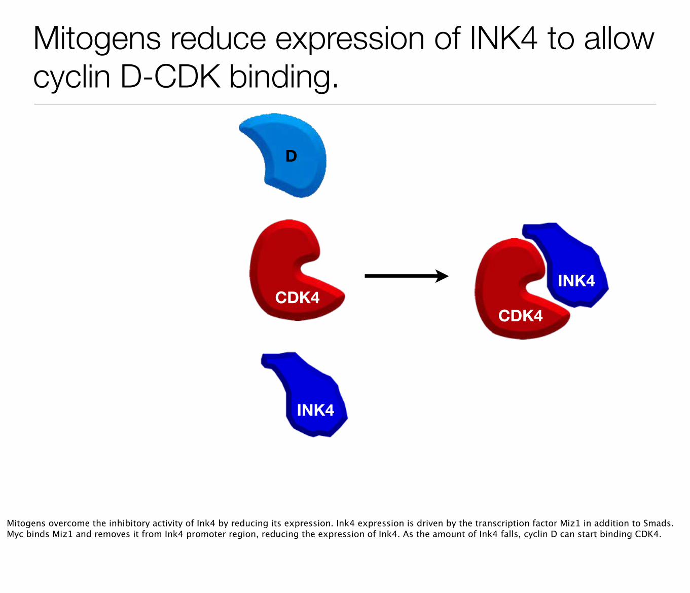
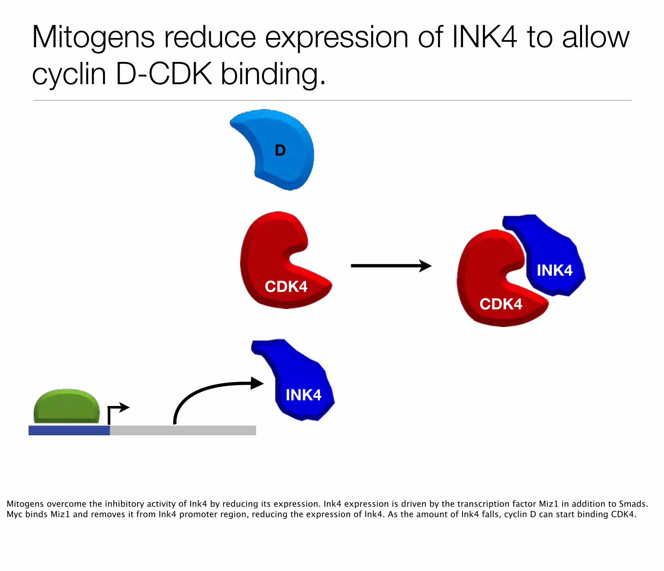
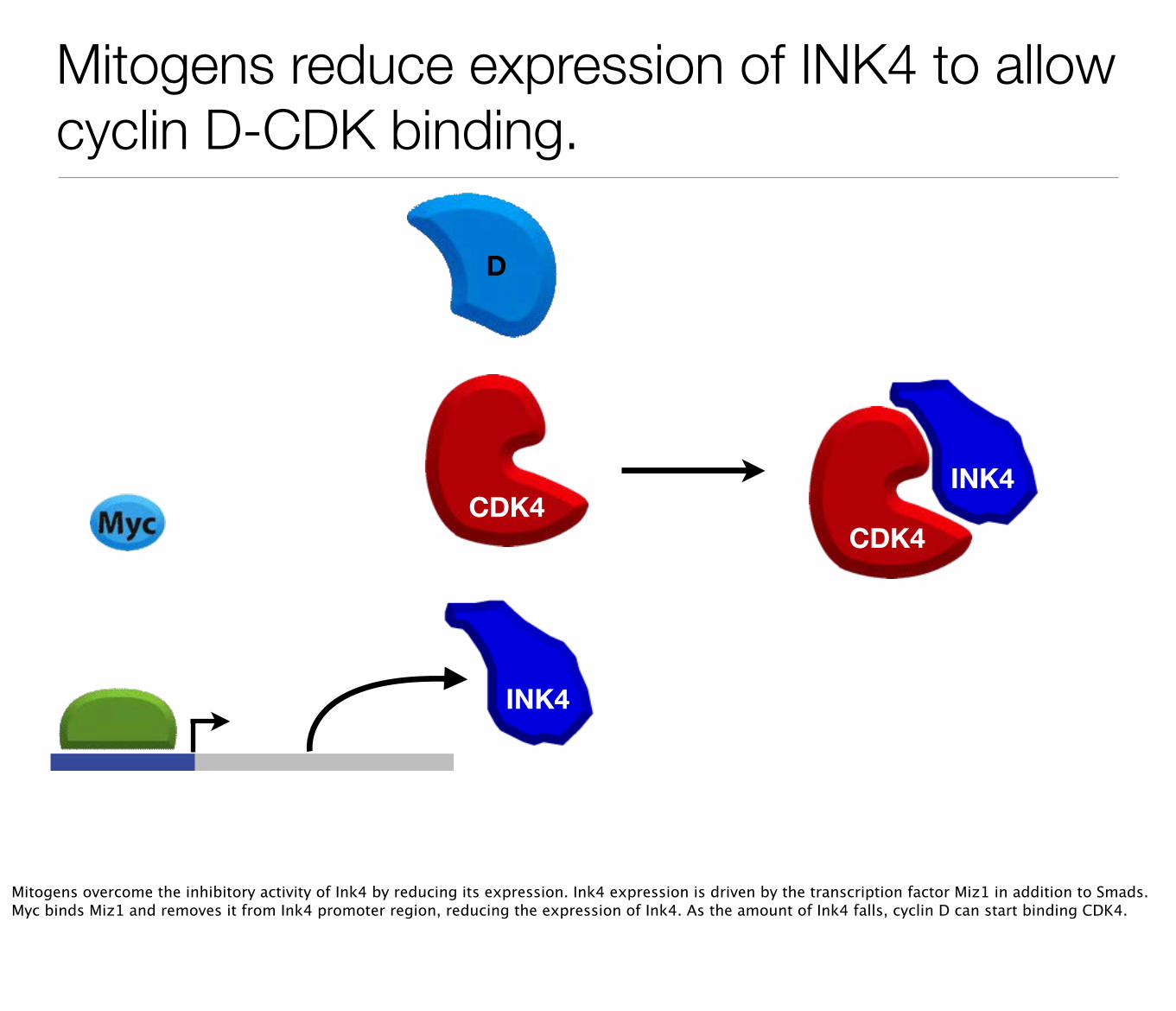
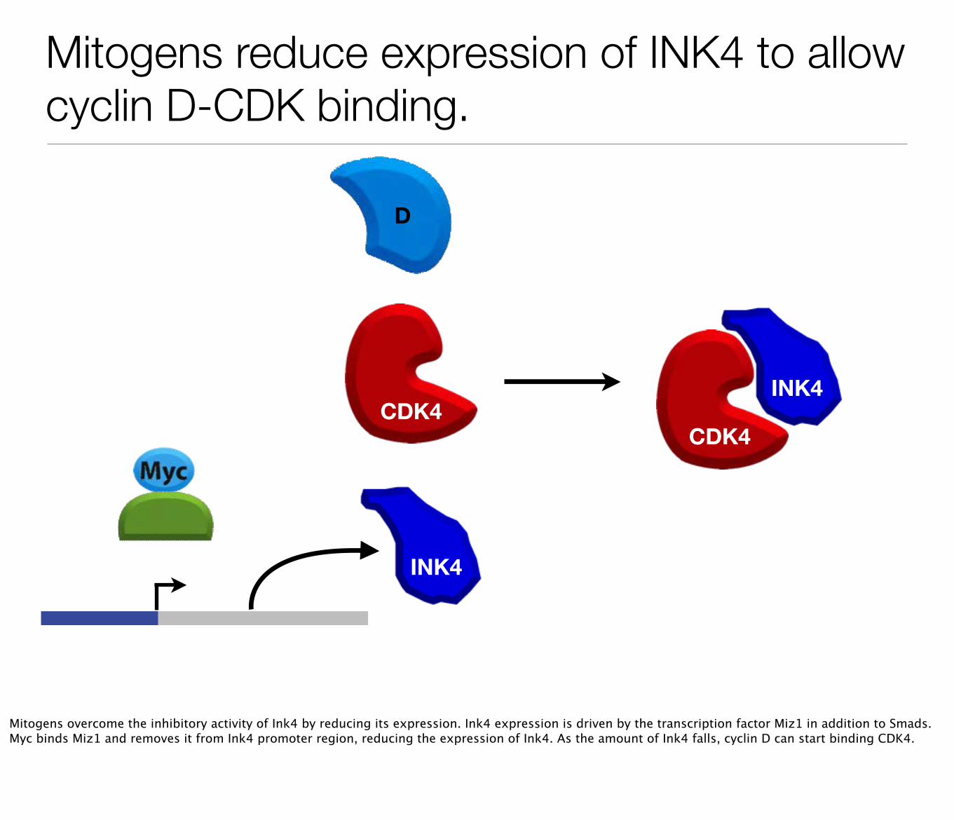
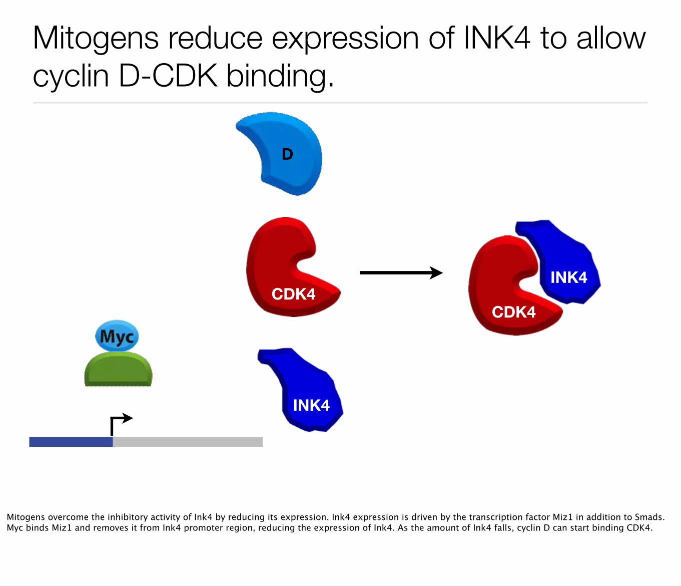
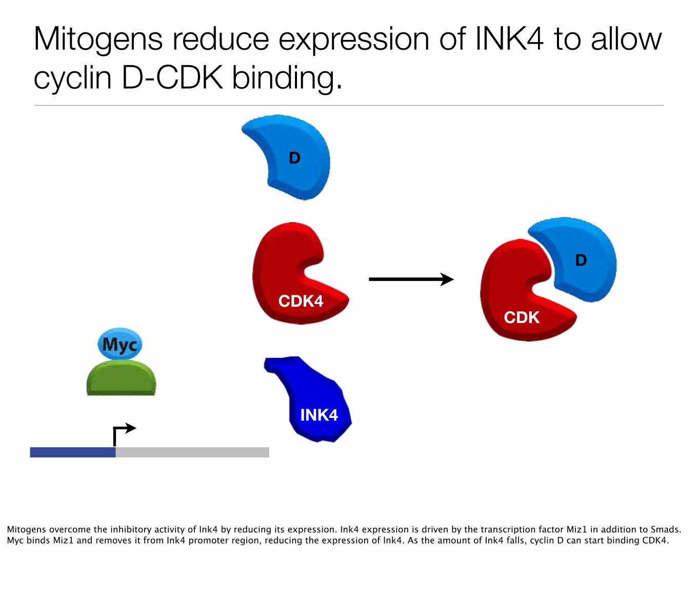
Mitogens reduce expression of INK4 to allow cyclin D-CDK binding.
CDK4
D
INK4
INK4
CDK4
Mitogens overcome the inhibitory activity of Ink4 by reducing its expression. Ink4 expression is driven by the transcription factor Miz1 in addition to Smads. Myc binds Miz1 and removes it from Ink4 promoter region, reducing the expression of Ink4. As the amount of Ink4 falls, cyclin D can start binding CDK4.
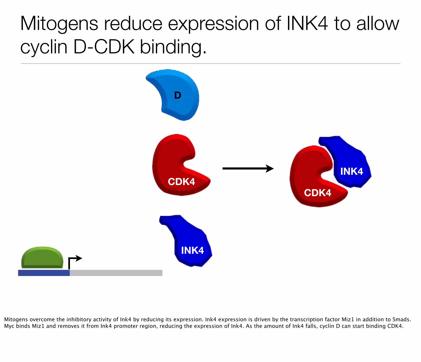
Mitogens reduce expression of INK4 to allow cyclin D-CDK binding.
CDK4
D
CDK
D
INK4
Mitogens overcome the inhibitory activity of Ink4 by reducing its expression. Ink4 expression is driven by the transcription factor Miz1 in addition to Smads. Myc binds Miz1 and removes it from Ink4 promoter region, reducing the expression of Ink4. As the amount of Ink4 falls, cyclin D can start binding CDK4.
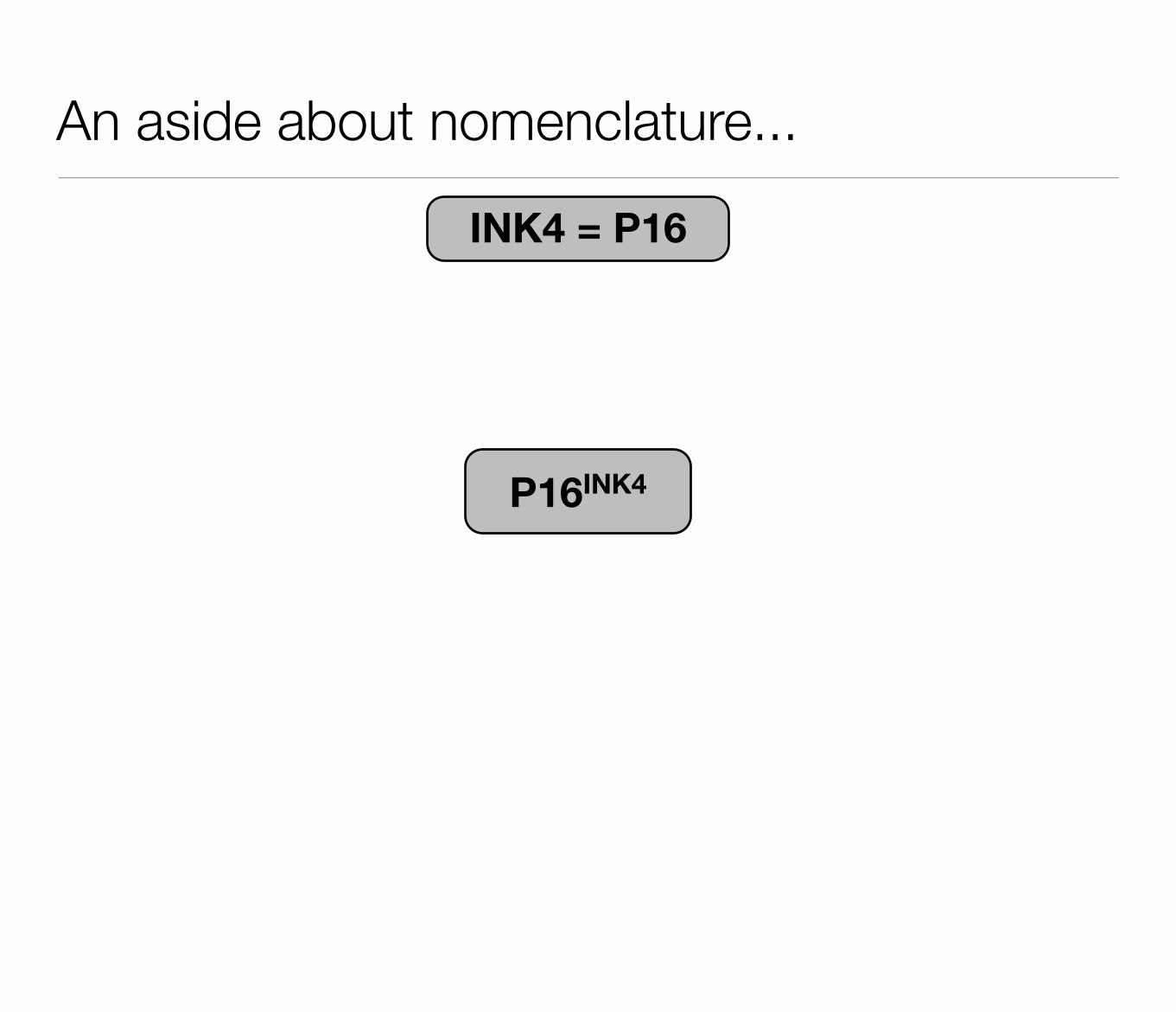
An aside about nomenclature...
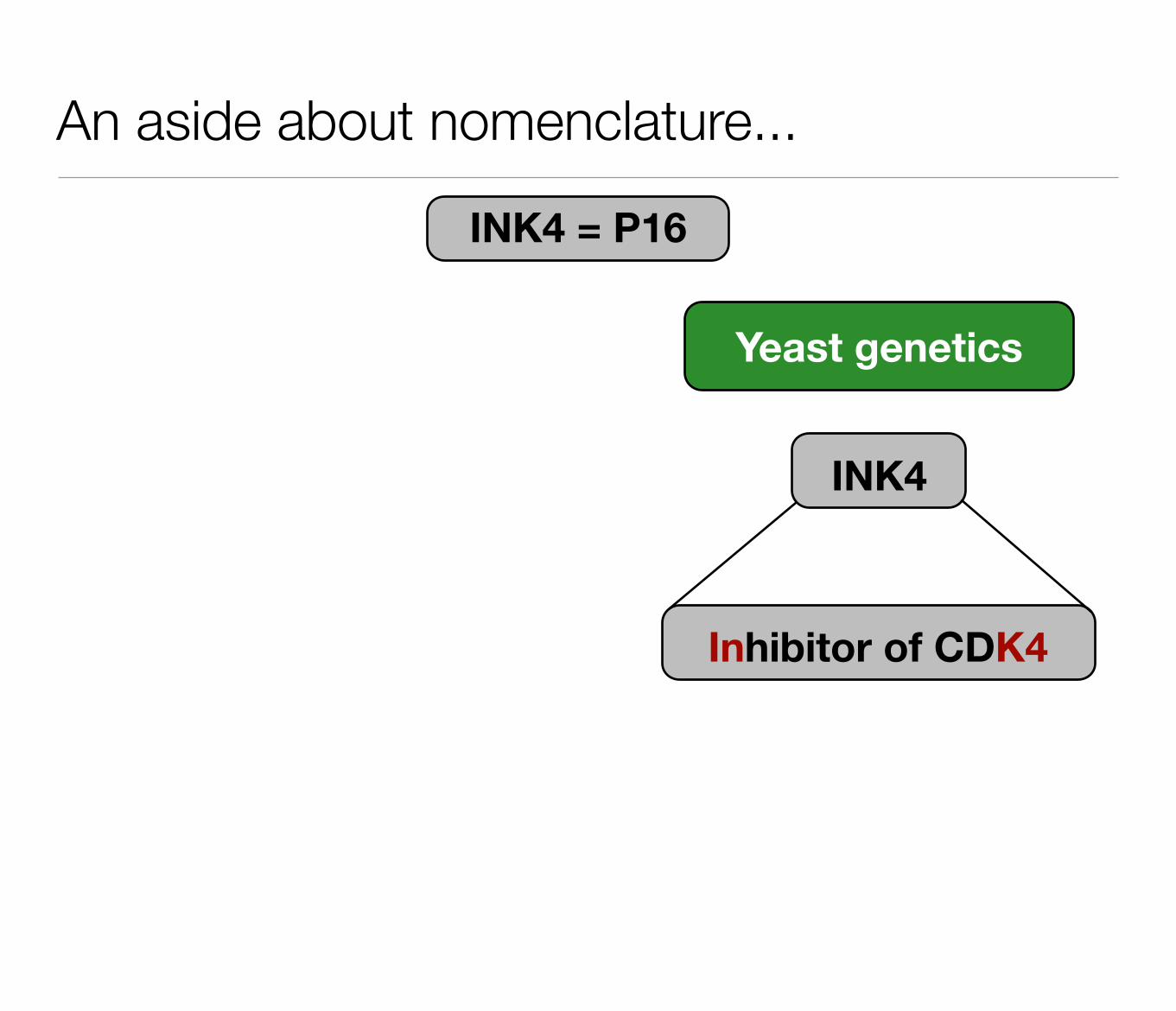
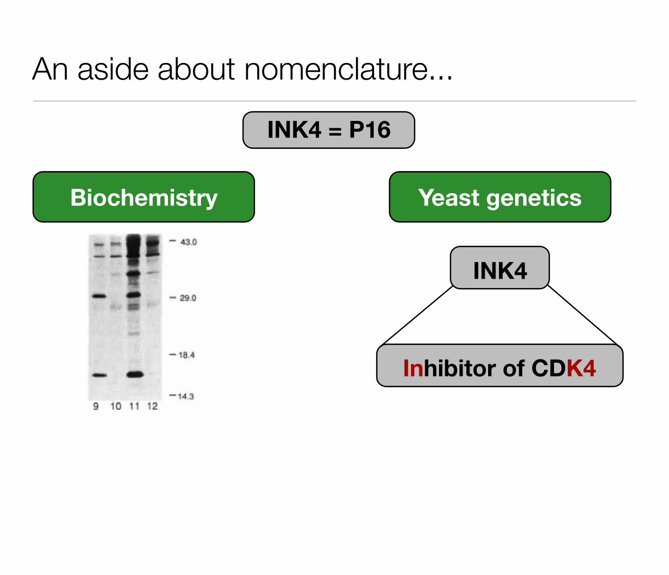
INK4 = P16
An aside about nomenclature...
INK4 = P16
INK4
Inhibitor of CDK4
Yeast genetics
An aside about nomenclature...
INK4 = P16
INK4
Inhibitor of CDK4
Yeast genetics
© 1993 Nature Publishing Group
Biochemistry
An aside about nomenclature...
INK4 = P16
P16INK4
DD
cyclin E transcription
How DNA damage arrests the cell cycle
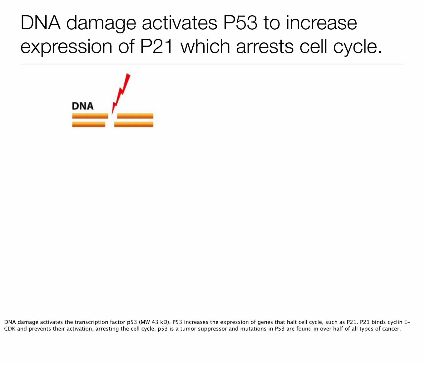
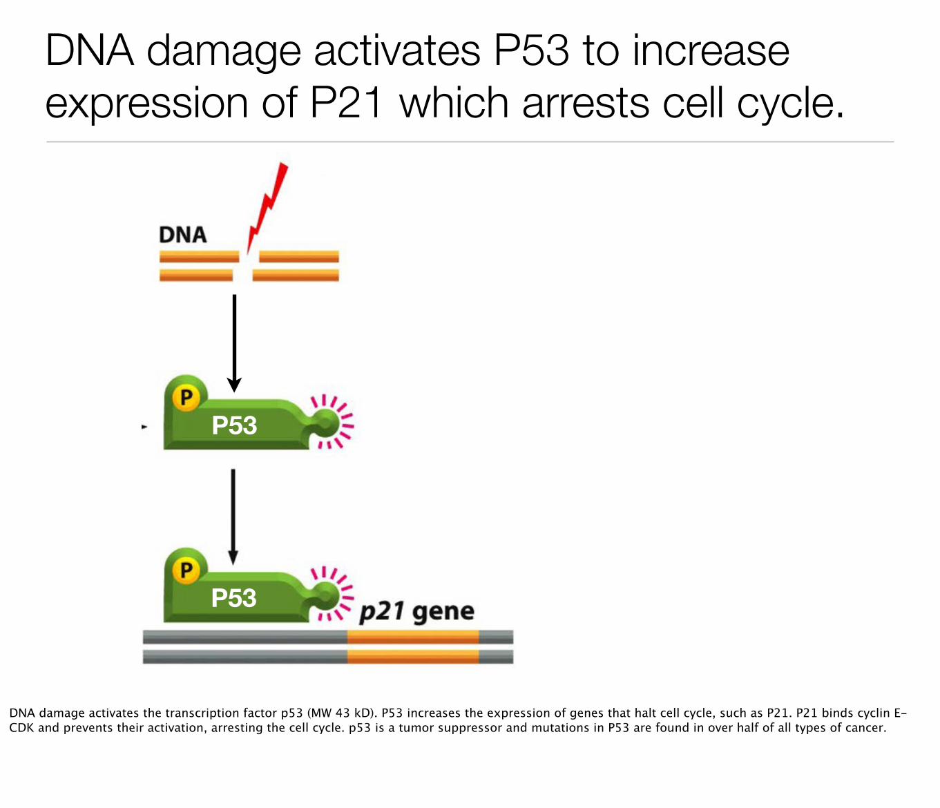
DNA damage activates P53 to increase expression of P21 which arrests cell cycle.
P53
P53
DNA damage activates the transcription factor p53 (MW 43 kD). P53 increases the expression of genes that halt cell cycle, such as P21. P21 binds cyclin E-CDK and prevents their activation, arresting the cell cycle. p53 is a tumor suppressor and mutations in P53 are found in over half of all types of cancer.
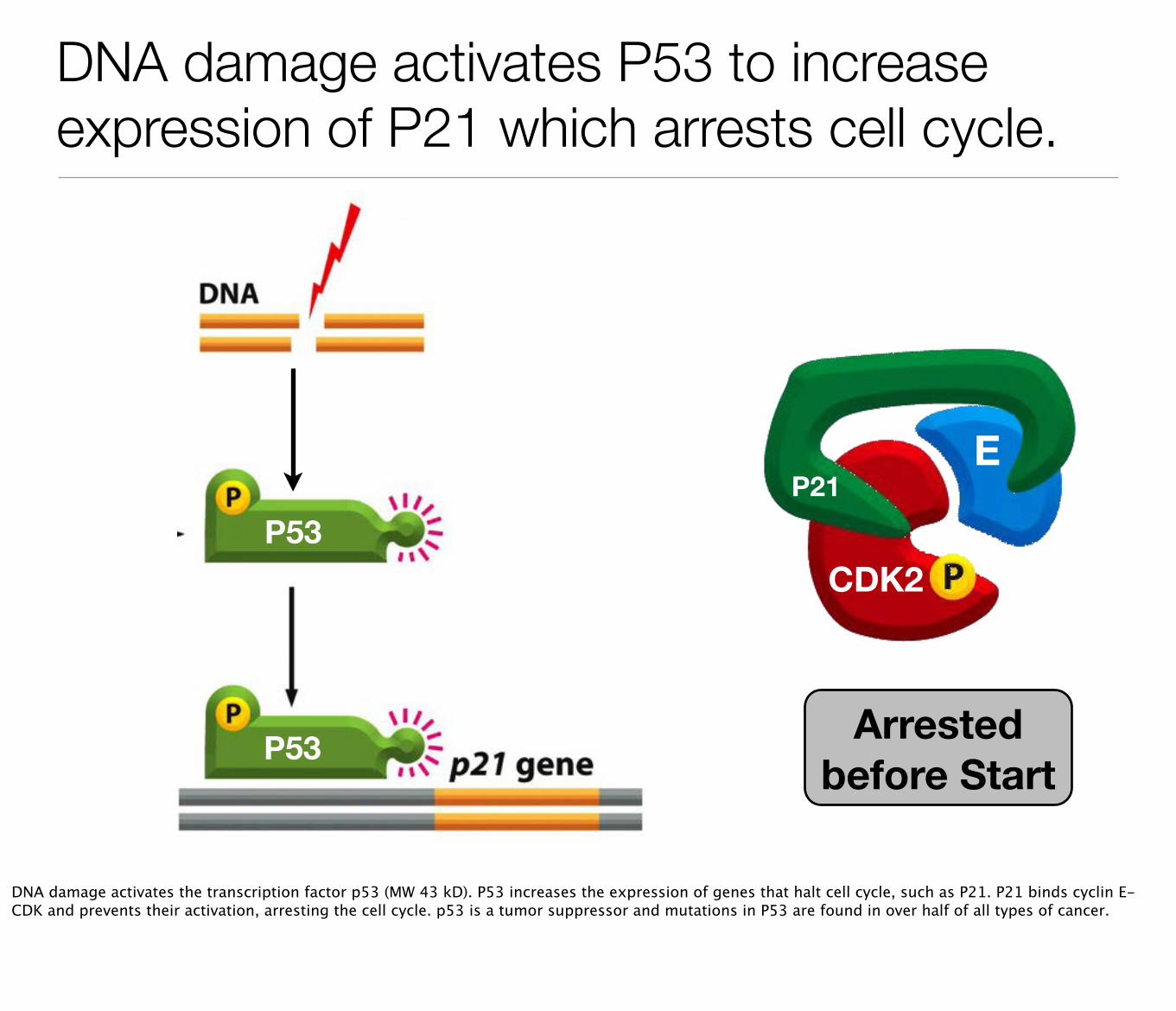
DNA damage activates P53 to increase expression of P21 which arrests cell cycle.
P53
P53
E
CDK2
P21
Arrested before Start
DNA damage activates the transcription factor p53 (MW 43 kD). P53 increases the expression of genes that halt cell cycle, such as P21. P21 binds cyclin E-CDK and prevents their activation, arresting the cell cycle. p53 is a tumor suppressor and mutations in P53 are found in over half of all types of cancer.
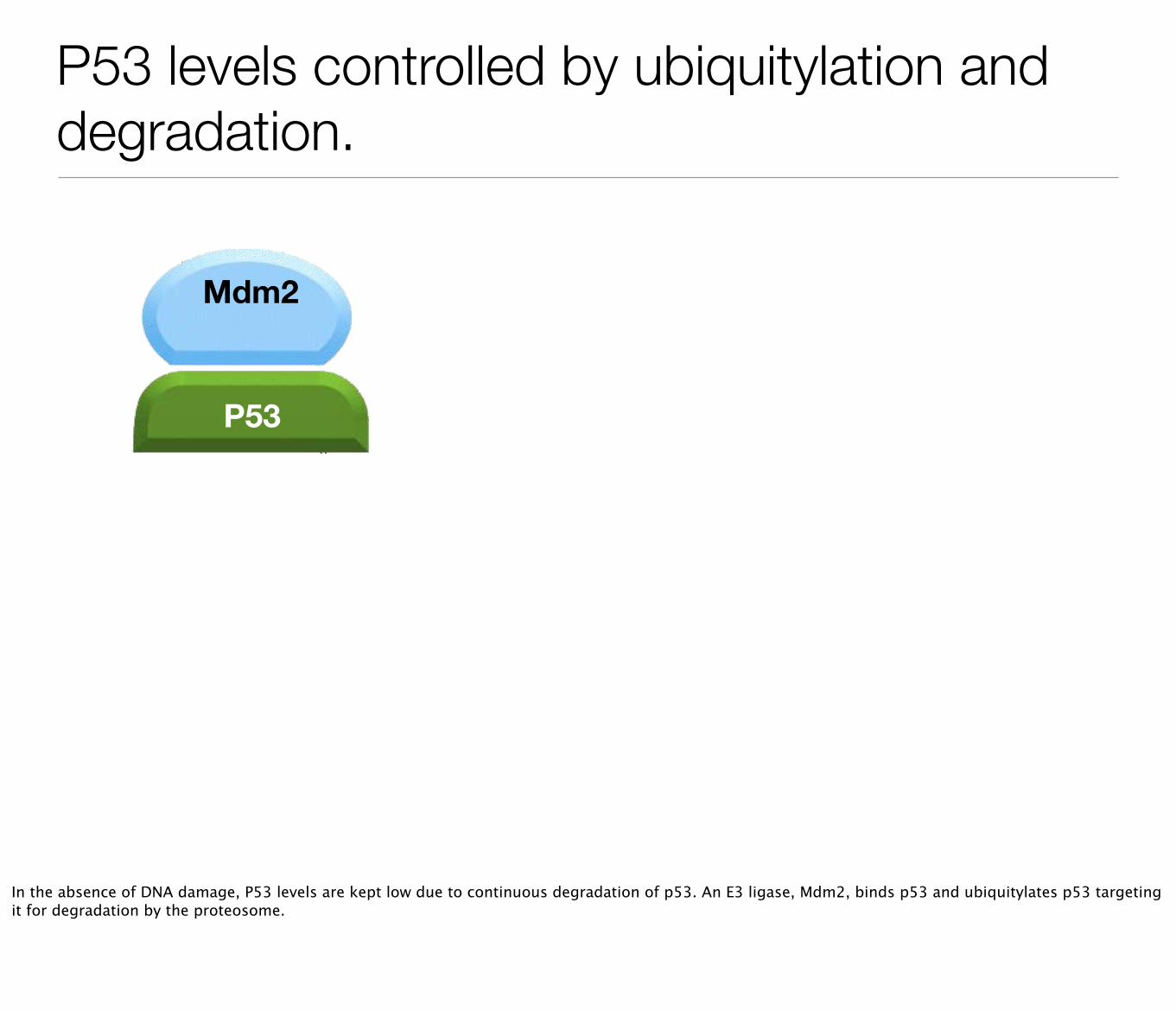
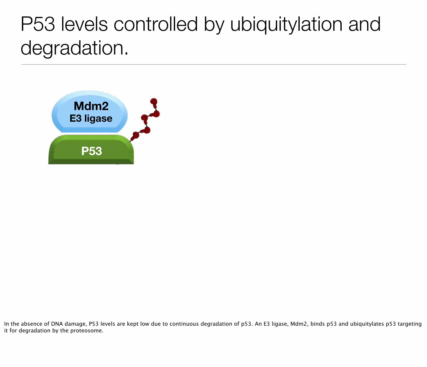
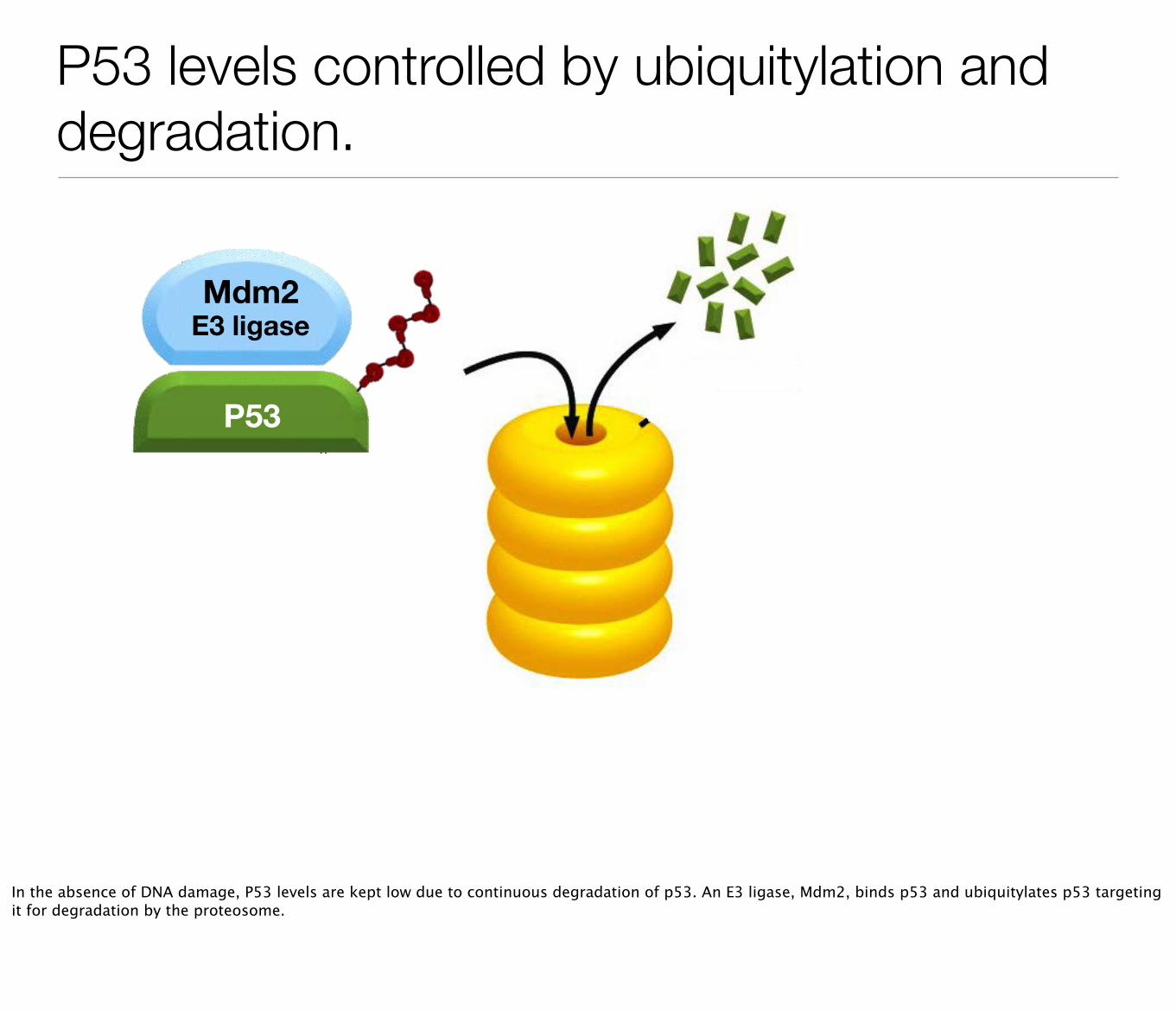
P53 levels controlled by ubiquitylation and degradation.
P53
Mdm2
In the absence of DNA damage, P53 levels are kept low due to continuous degradation of p53. An E3 ligase, Mdm2, binds p53 and ubiquitylates p53 targeting it for degradation by the proteosome.
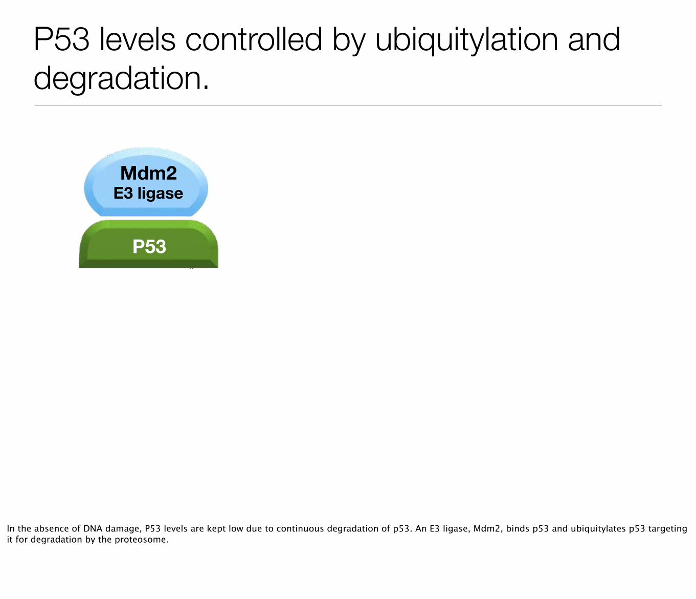
P53 levels controlled by ubiquitylation and degradation.
P53
Mdm2E3 ligase
In the absence of DNA damage, P53 levels are kept low due to continuous degradation of p53. An E3 ligase, Mdm2, binds p53 and ubiquitylates p53 targeting it for degradation by the proteosome.
P53 levels controlled by ubiquitylation and degradation.
P53
Mdm2E3 ligase
In the absence of DNA damage, P53 levels are kept low due to continuous degradation of p53. An E3 ligase, Mdm2, binds p53 and ubiquitylates p53 targeting it for degradation by the proteosome.
P53 levels controlled by ubiquitylation and degradation.
P53
Mdm2E3 ligase
In the absence of DNA damage, P53 levels are kept low due to continuous degradation of p53. An E3 ligase, Mdm2, binds p53 and ubiquitylates p53 targeting it for degradation by the proteosome.
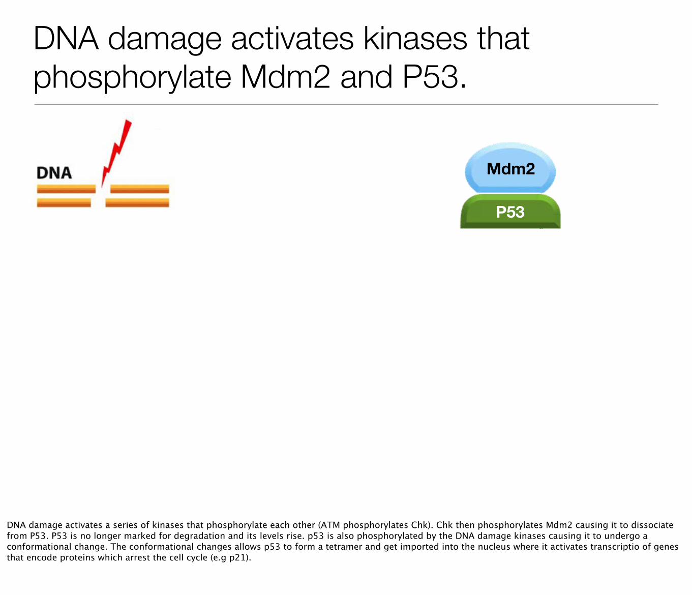
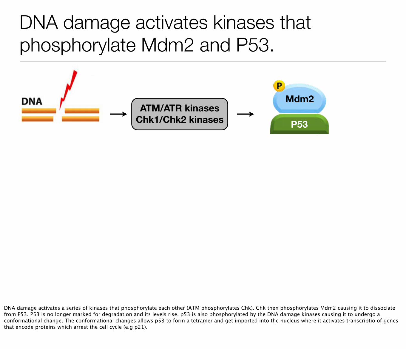
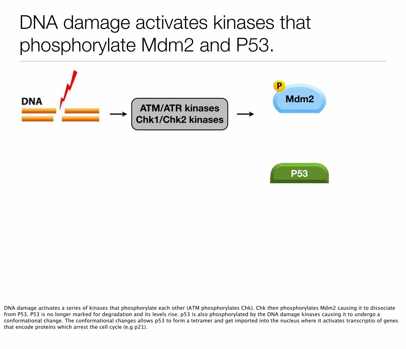
DNA damage activates kinases that phosphorylate Mdm2 and P53.
P53
Mdm2
DNA damage activates a series of kinases that phosphorylate each other (ATM phosphorylates Chk). Chk then phosphorylates Mdm2 causing it to dissociate from P53. P53 is no longer marked for degradation and its levels rise. p53 is also phosphorylated by the DNA damage kinases causing it to undergo a conformational change. The conformational changes allows p53 to form a tetramer and get imported into the nucleus where it activates transcriptio of genes that encode proteins which arrest the cell cycle (e.g p21).

DNA damage activates kinases that phosphorylate Mdm2 and P53.
ATM/ATR kinasesChk1/Chk2 kinases P53
Mdm2
DNA damage activates a series of kinases that phosphorylate each other (ATM phosphorylates Chk). Chk then phosphorylates Mdm2 causing it to dissociate from P53. P53 is no longer marked for degradation and its levels rise. p53 is also phosphorylated by the DNA damage kinases causing it to undergo a conformational change. The conformational changes allows p53 to form a tetramer and get imported into the nucleus where it activates transcriptio of genes that encode proteins which arrest the cell cycle (e.g p21).
DNA damage activates kinases that phosphorylate Mdm2 and P53.
ATM/ATR kinasesChk1/Chk2 kinases
P53
Mdm2
DNA damage activates a series of kinases that phosphorylate each other (ATM phosphorylates Chk). Chk then phosphorylates Mdm2 causing it to dissociate from P53. P53 is no longer marked for degradation and its levels rise. p53 is also phosphorylated by the DNA damage kinases causing it to undergo a conformational change. The conformational changes allows p53 to form a tetramer and get imported into the nucleus where it activates transcriptio of genes that encode proteins which arrest the cell cycle (e.g p21).

DNA damage activates kinases that phosphorylate Mdm2 and P53.
ATM/ATR kinasesChk1/Chk2 kinases
P53
Mdm2
DNA damage activates a series of kinases that phosphorylate each other (ATM phosphorylates Chk). Chk then phosphorylates Mdm2 causing it to dissociate from P53. P53 is no longer marked for degradation and its levels rise. p53 is also phosphorylated by the DNA damage kinases causing it to undergo a conformational change. The conformational changes allows p53 to form a tetramer and get imported into the nucleus where it activates transcriptio of genes that encode proteins which arrest the cell cycle (e.g p21).
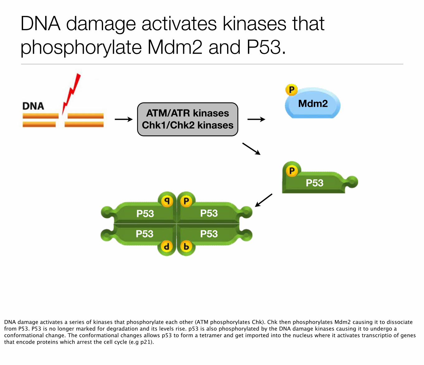
DNA damage activates kinases that phosphorylate Mdm2 and P53.
ATM/ATR kinasesChk1/Chk2 kinases
P53
Mdm2
P53
P53
P53
P53
DNA damage activates a series of kinases that phosphorylate each other (ATM phosphorylates Chk). Chk then phosphorylates Mdm2 causing it to dissociate from P53. P53 is no longer marked for degradation and its levels rise. p53 is also phosphorylated by the DNA damage kinases causing it to undergo a conformational change. The conformational changes allows p53 to form a tetramer and get imported into the nucleus where it activates transcriptio of genes that encode proteins which arrest the cell cycle (e.g p21).
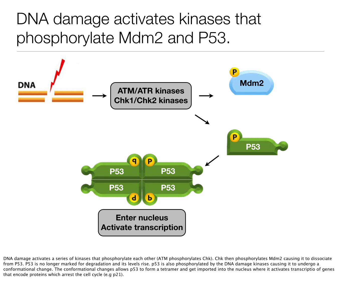
DNA damage activates kinases that phosphorylate Mdm2 and P53.
ATM/ATR kinasesChk1/Chk2 kinases
P53
Mdm2
P53
P53
P53
P53
Enter nucleusActivate transcription
DNA damage activates a series of kinases that phosphorylate each other (ATM phosphorylates Chk). Chk then phosphorylates Mdm2 causing it to dissociate from P53. P53 is no longer marked for degradation and its levels rise. p53 is also phosphorylated by the DNA damage kinases causing it to undergo a conformational change. The conformational changes allows p53 to form a tetramer and get imported into the nucleus where it activates transcriptio of genes that encode proteins which arrest the cell cycle (e.g p21).
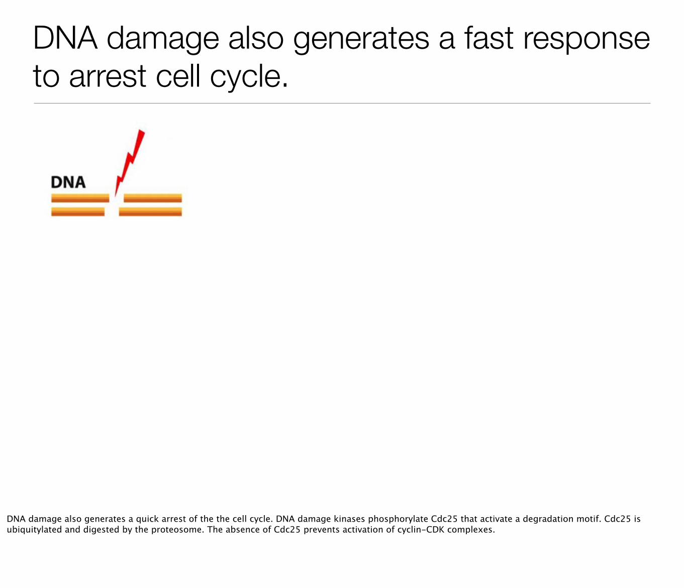
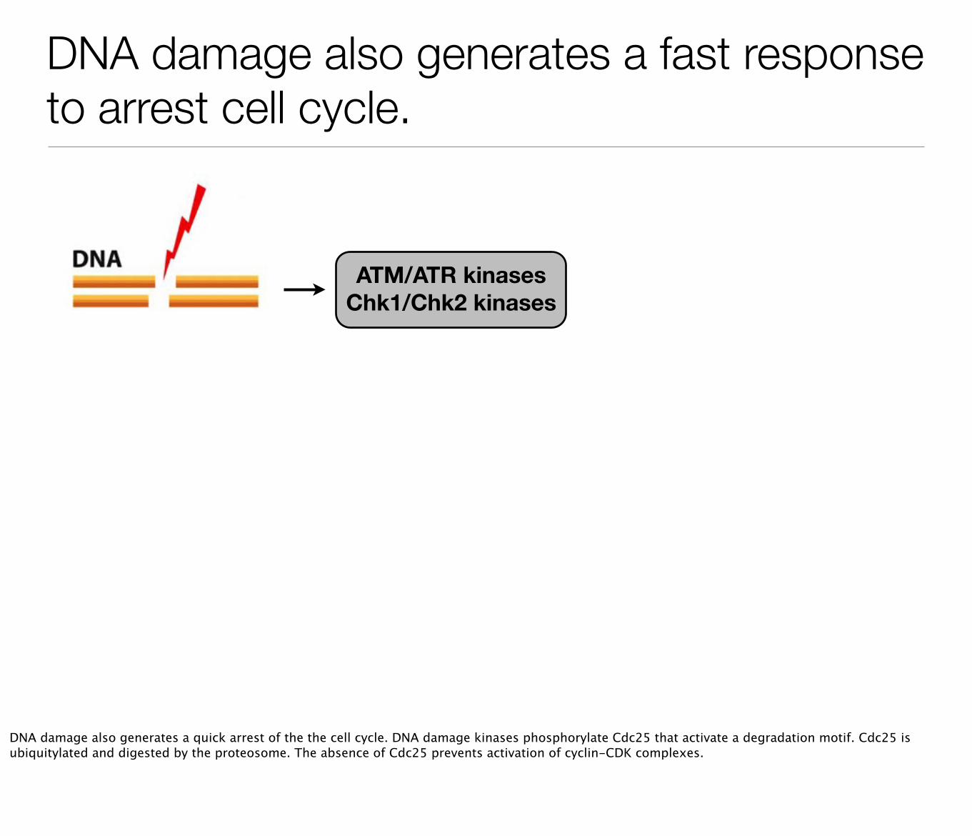
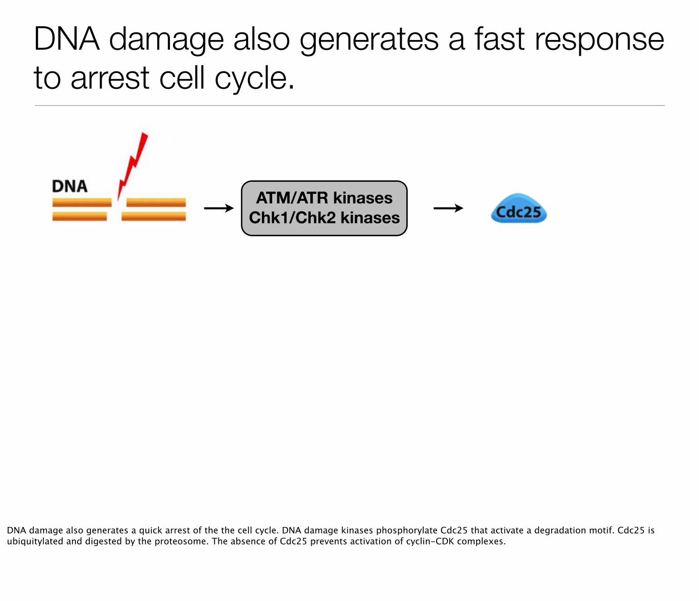
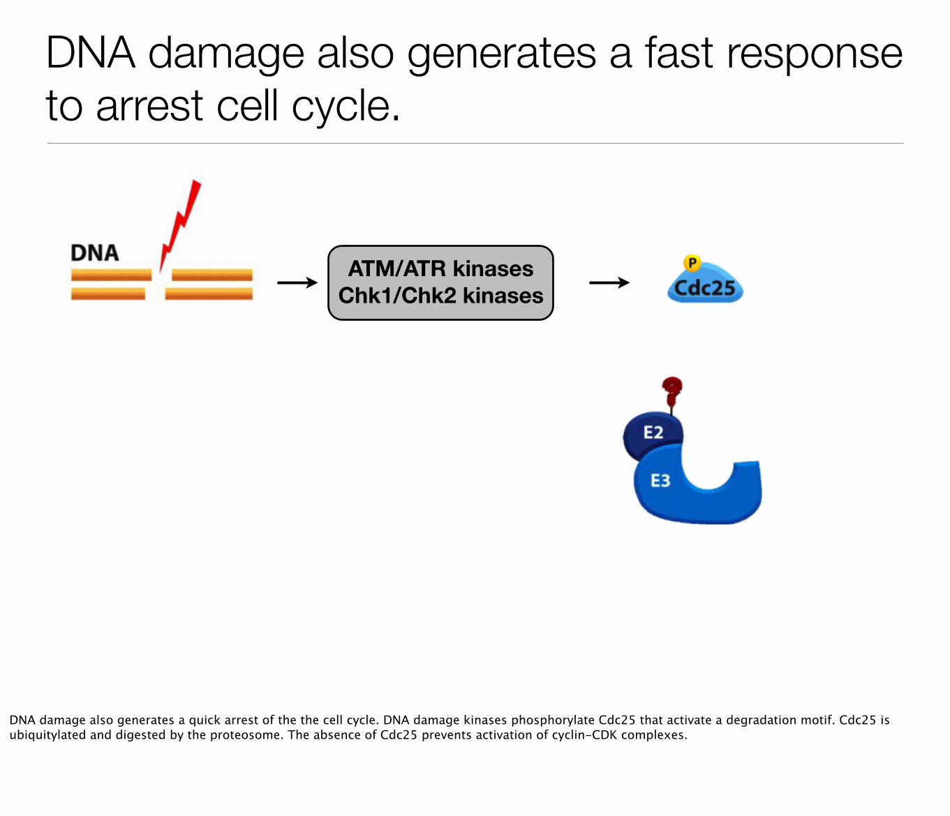
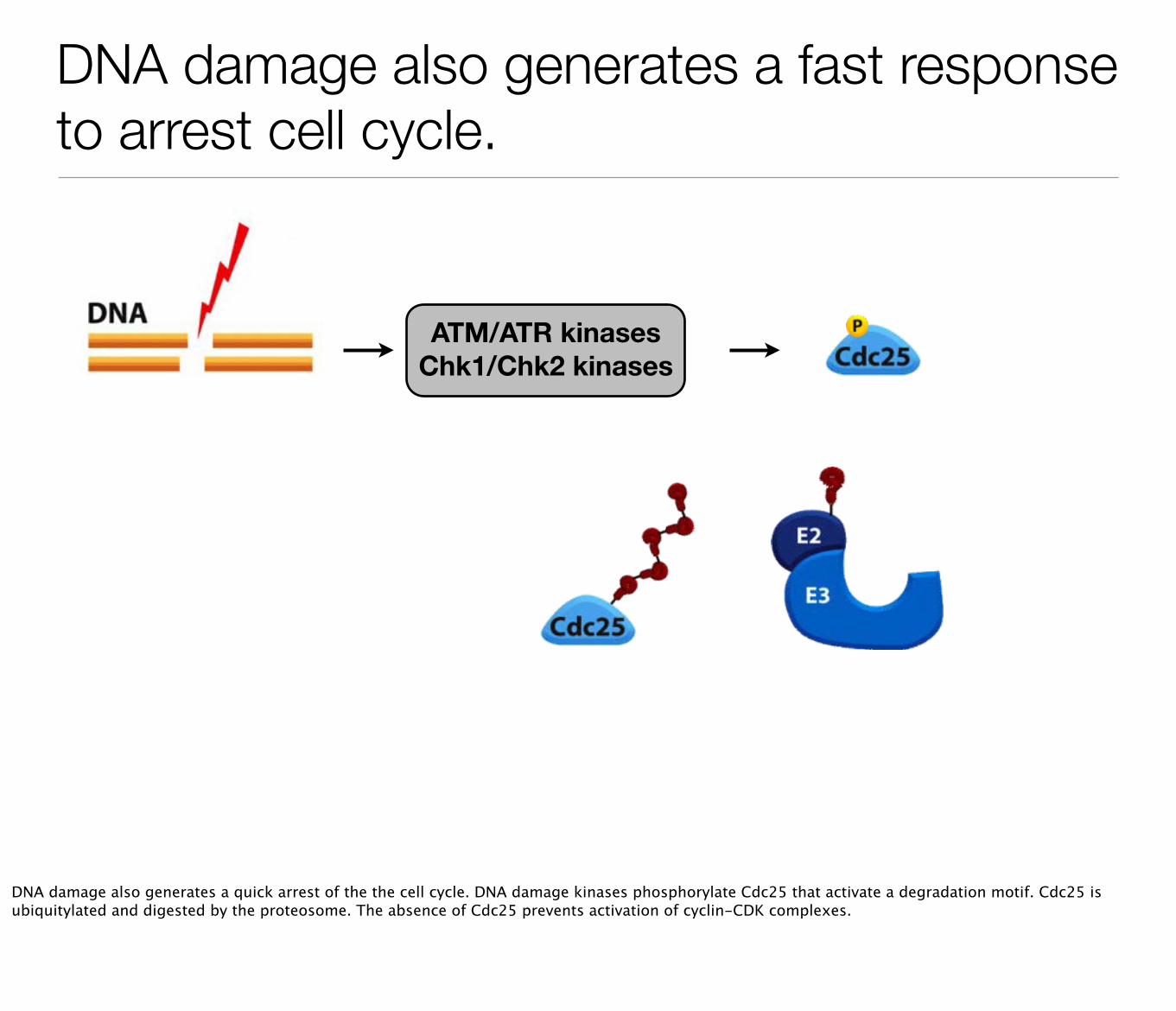
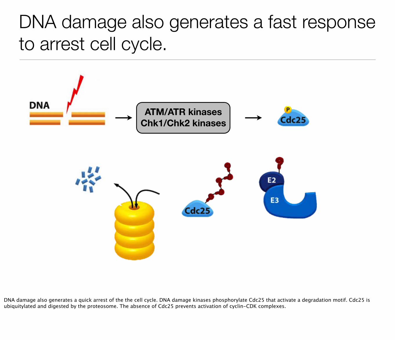
DNA damage also generates a fast response to arrest cell cycle.
DNA damage also generates a quick arrest of the the cell cycle. DNA damage kinases phosphorylate Cdc25 that activate a degradation motif. Cdc25 is ubiquitylated and digested by the proteosome. The absence of Cdc25 prevents activation of cyclin-CDK complexes.
DNA damage also generates a fast response to arrest cell cycle.
ATM/ATR kinasesChk1/Chk2 kinases
DNA damage also generates a quick arrest of the the cell cycle. DNA damage kinases phosphorylate Cdc25 that activate a degradation motif. Cdc25 is ubiquitylated and digested by the proteosome. The absence of Cdc25 prevents activation of cyclin-CDK complexes.
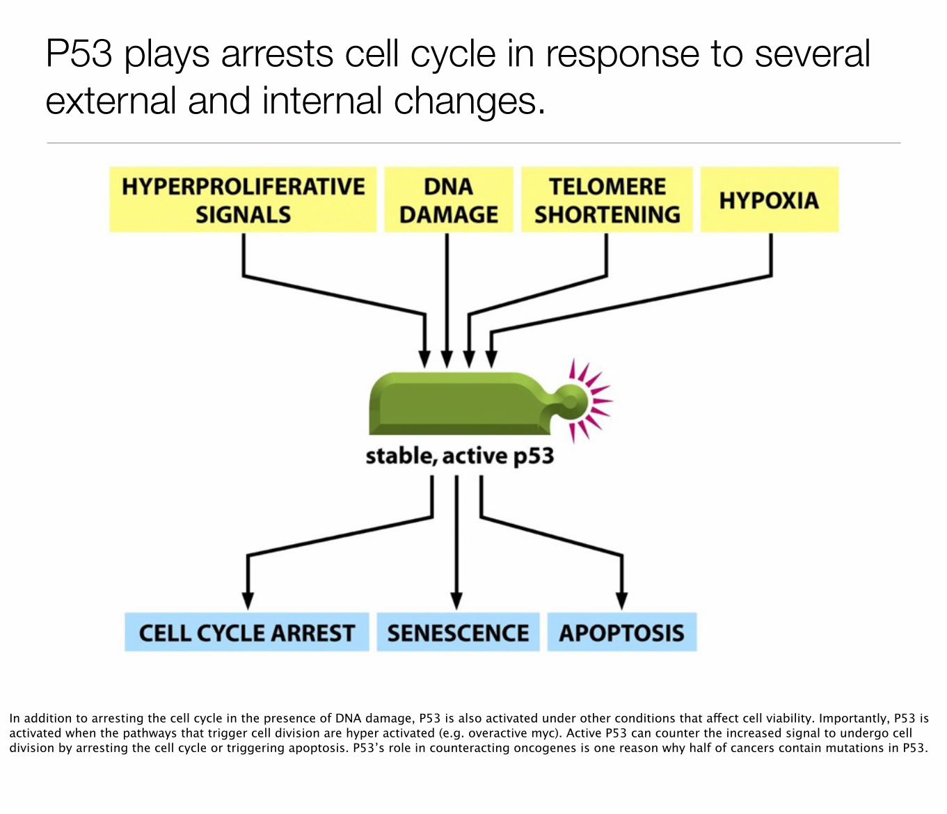
P53 plays arrests cell cycle in response to several external and internal changes.
In addition to arresting the cell cycle in the presence of DNA damage, P53 is also activated under other conditions that affect cell viability. Importantly, P53 is activated when the pathways that trigger cell division are hyper activated (e.g. overactive myc). Active P53 can counter the increased signal to undergo cell division by arresting the cell cycle or triggering apoptosis. P53’s role in counteracting oncogenes is one reason why half of cancers contain mutations in P53.
Cell senescence
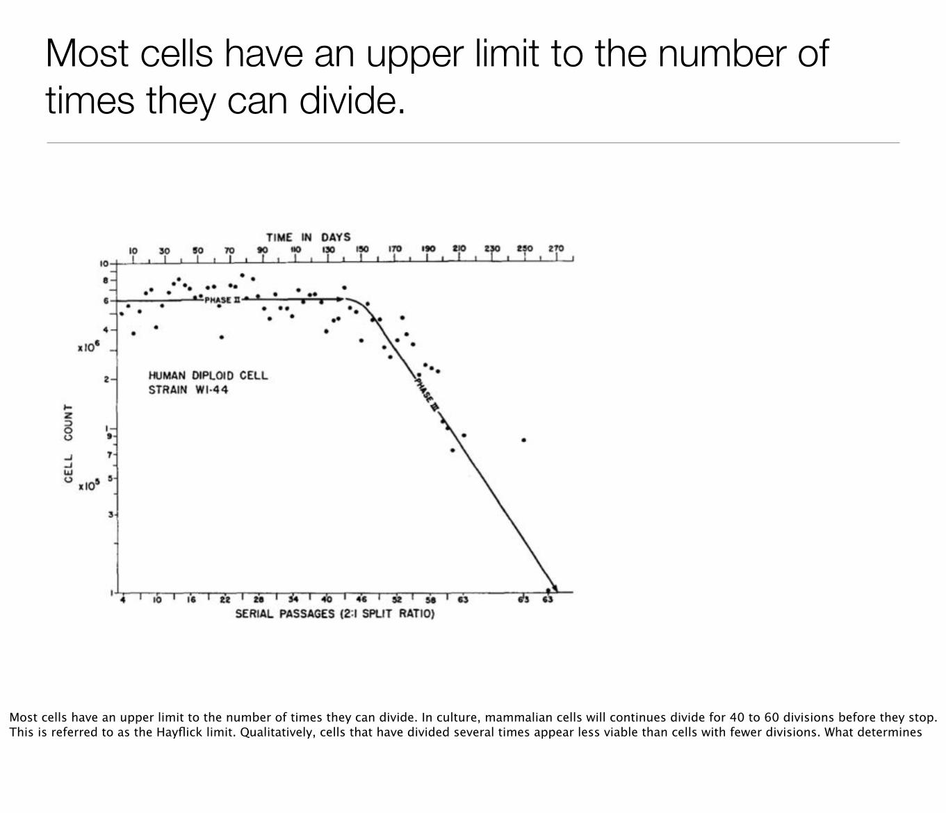
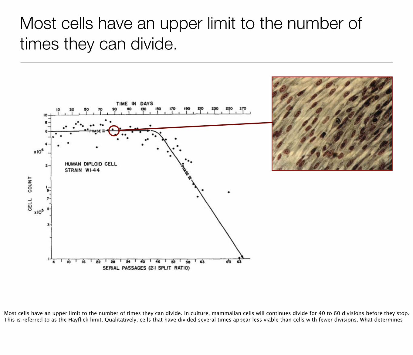
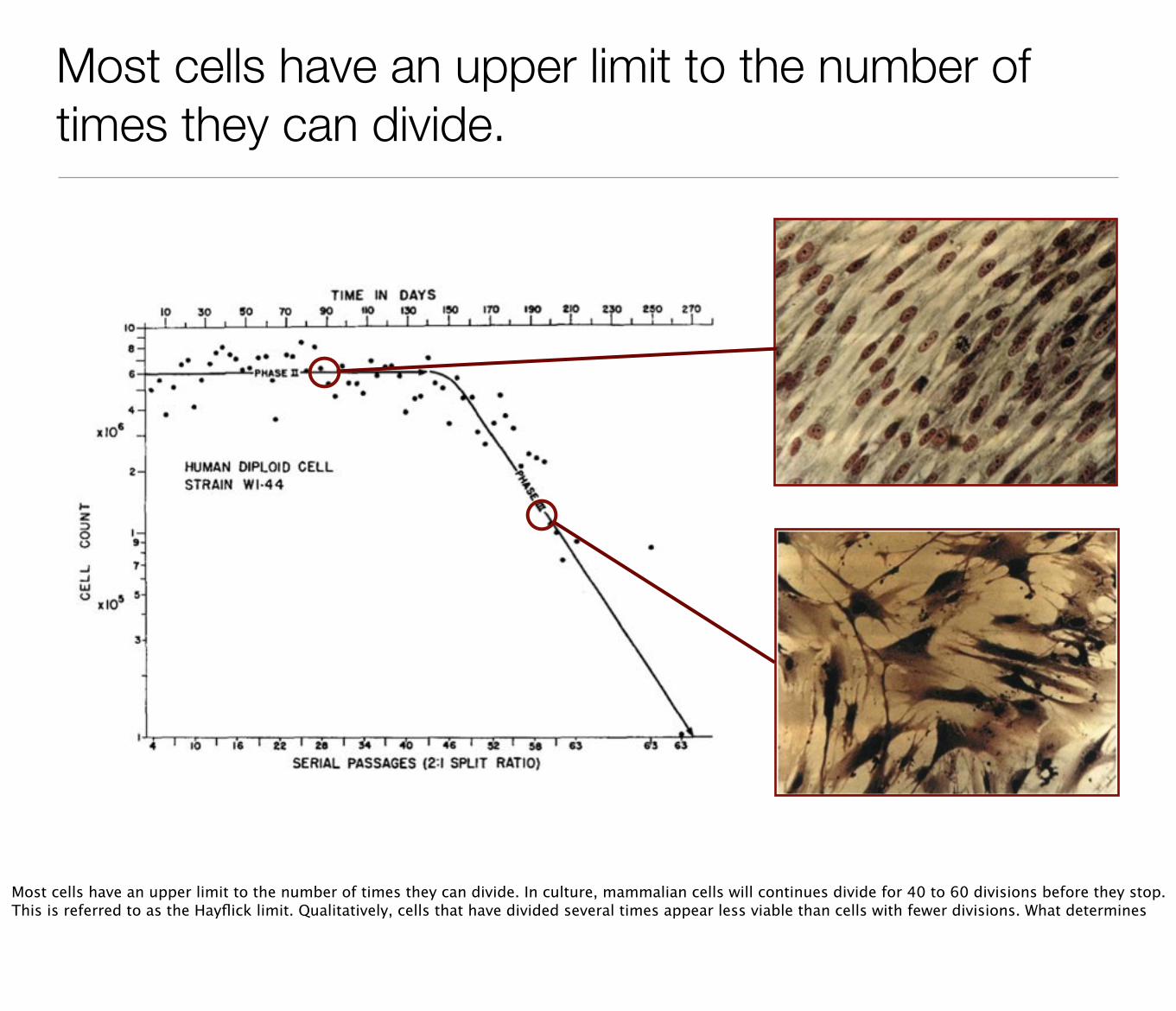
Most cells have an upper limit to the number of times they can divide.
Most cells have an upper limit to the number of times they can divide. In culture, mammalian cells will continues divide for 40 to 60 divisions before they stop. This is referred to as the Hayflick limit. Qualitatively, cells that have divided several times appear less viable than cells with fewer divisions. What determines
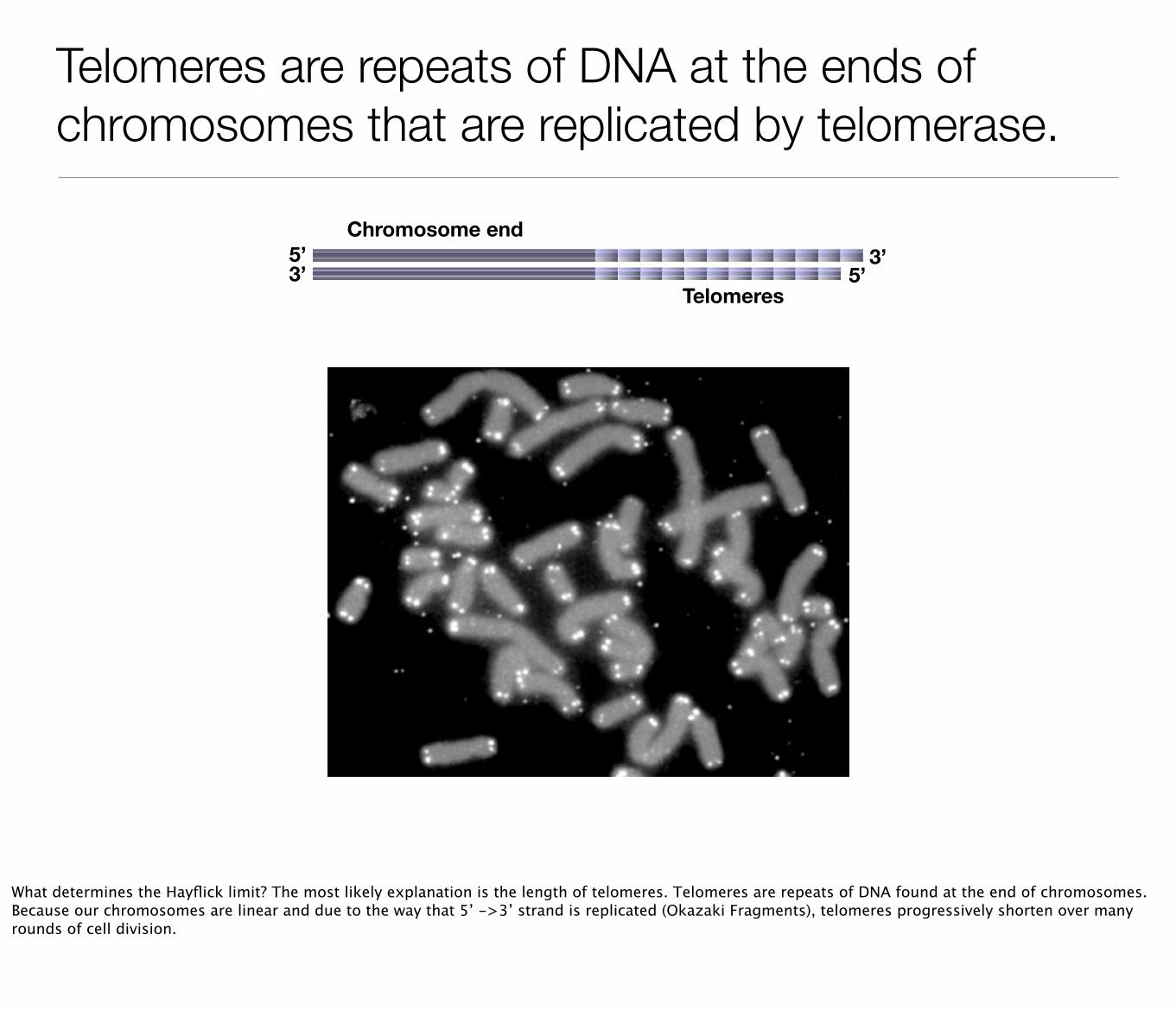
Telomeres are repeats of DNA at the ends of chromosomes that are replicated by telomerase.
Telomeres
5’5’
3’3’
Chromosome end
What determines the Hayflick limit? The most likely explanation is the length of telomeres. Telomeres are repeats of DNA found at the end of chromosomes. Because our chromosomes are linear and due to the way that 5’ ->3’ strand is replicated (Okazaki Fragments), telomeres progressively shorten over many rounds of cell division.
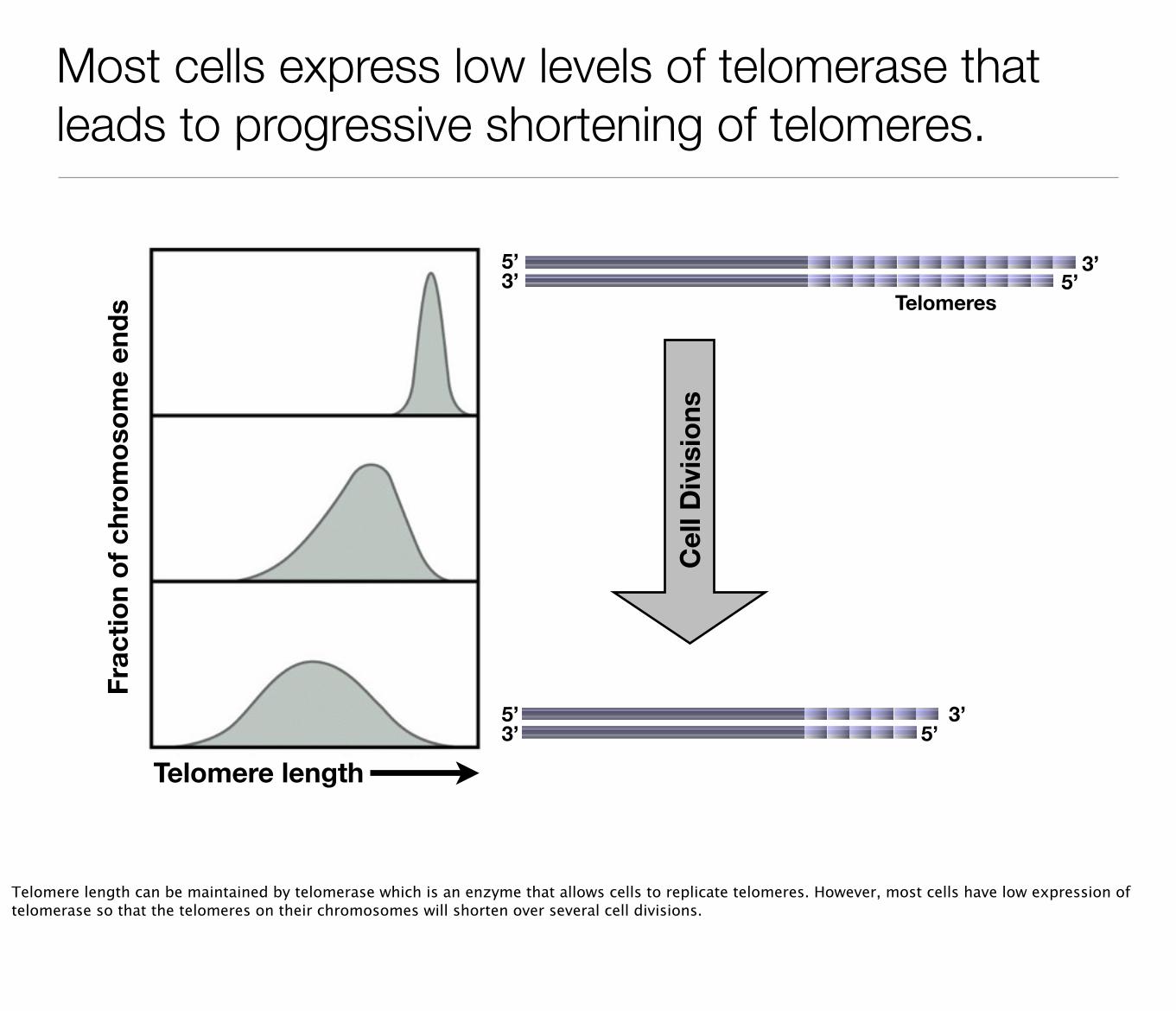
Most cells express low levels of telomerase that leads to progressive shortening of telomeres.
Telomere length
Frac
tion
of c
hrom
osom
e en
ds
Cel
l Div
isio
ns
Telomeres
5’5’
3’3’
5’3’5’
3’
Telomere length can be maintained by telomerase which is an enzyme that allows cells to replicate telomeres. However, most cells have low expression of telomerase so that the telomeres on their chromosomes will shorten over several cell divisions.
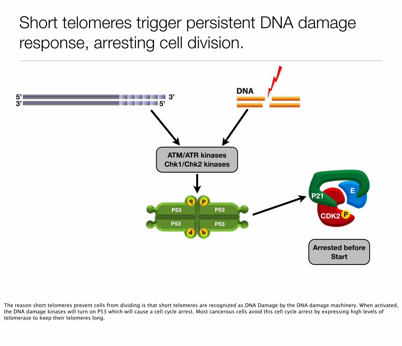
Short telomeres trigger persistent DNA damage response, arresting cell division.
5’3’5’
3’
ATM/ATR kinasesChk1/Chk2 kinases
P53
P53
P53
P53
E
CDK2
P21
Arrested before Start
The reason short telomeres prevent cells from dividing is that short telomeres are recognized as DNA Damage by the DNA damage machinery. When activated, the DNA damage kinases will turn on P53 which will cause a cell cycle arrest. Most cancerous cells avoid this cell cycle arrest by expressing high levels of telomerase to keep their telomeres long.
Coordination of cell growth and cell division
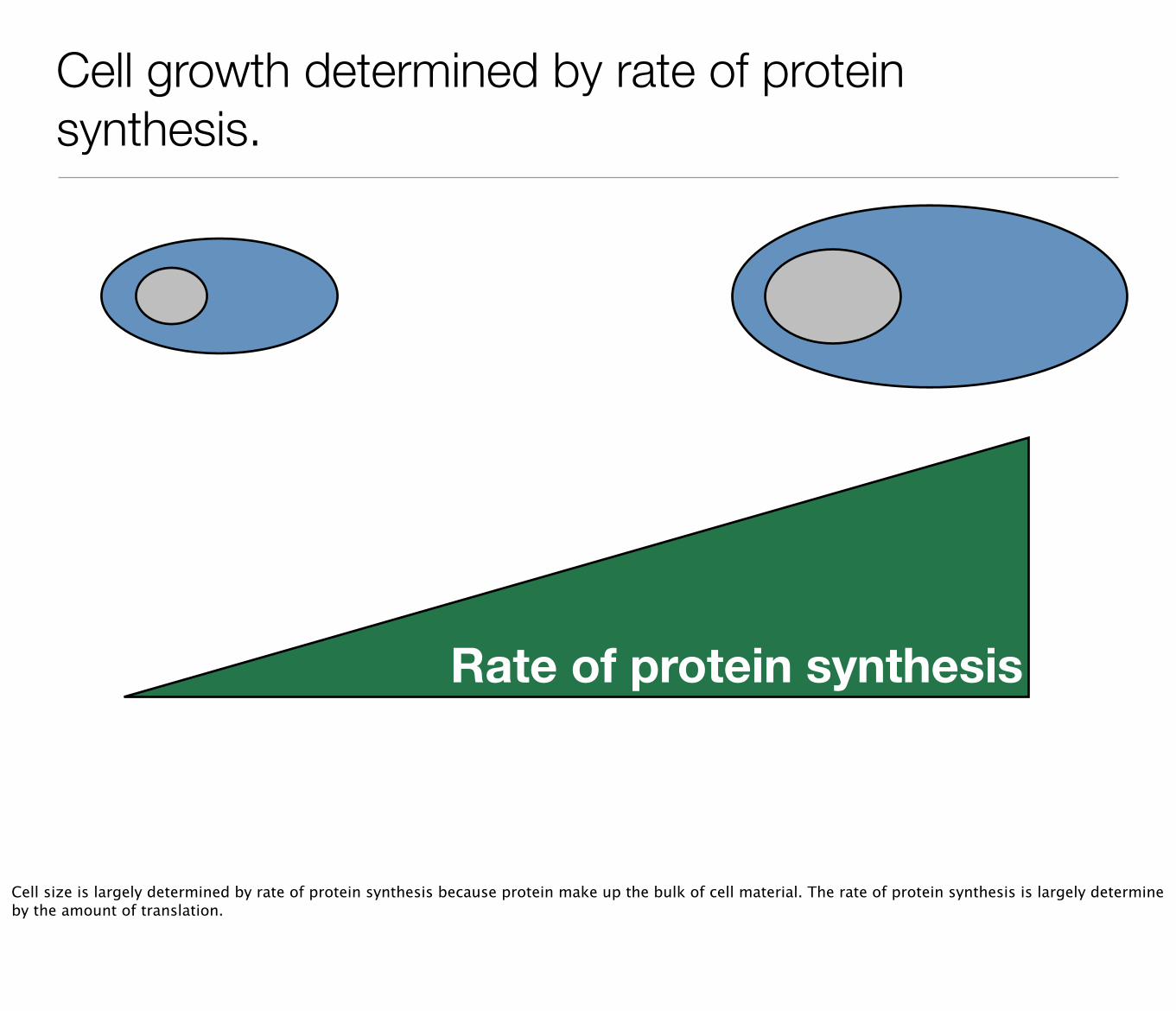
Cell growth determined by rate of protein synthesis.
Rate of protein synthesis
Cell size is largely determined by rate of protein synthesis because protein make up the bulk of cell material. The rate of protein synthesis is largely determine by the amount of translation.
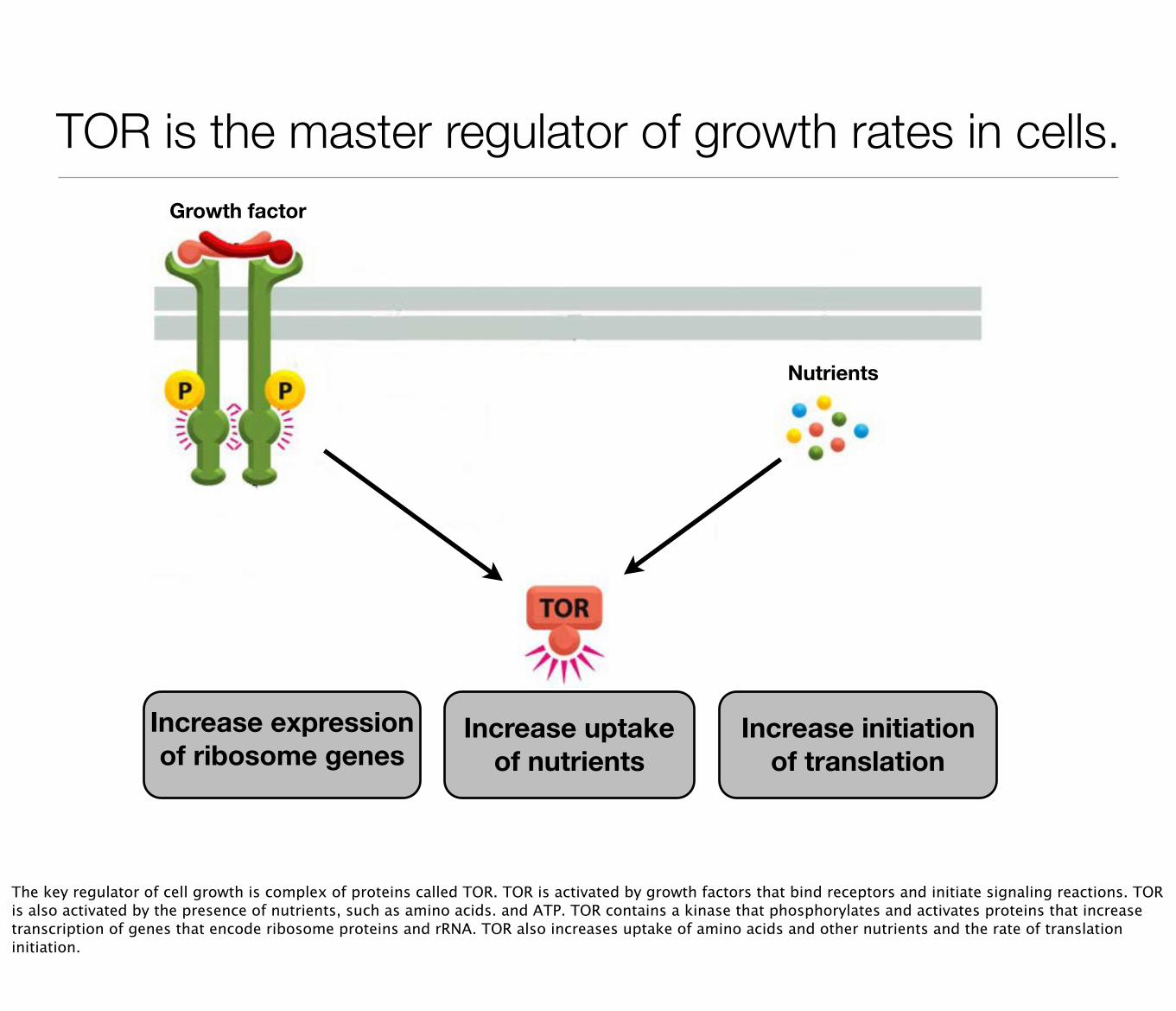
TOR is the master regulator of growth rates in cells.Growth factor
Nutrients
Increase expression of ribosome genes
Increase initiation of translation
Increase uptake of nutrients
The key regulator of cell growth is complex of proteins called TOR. TOR is activated by growth factors that bind receptors and initiate signaling reactions. TOR is also activated by the presence of nutrients, such as amino acids. and ATP. TOR contains a kinase that phosphorylates and activates proteins that increase transcription of genes that encode ribosome proteins and rRNA. TOR also increases uptake of amino acids and other nutrients and the rate of translation initiation.
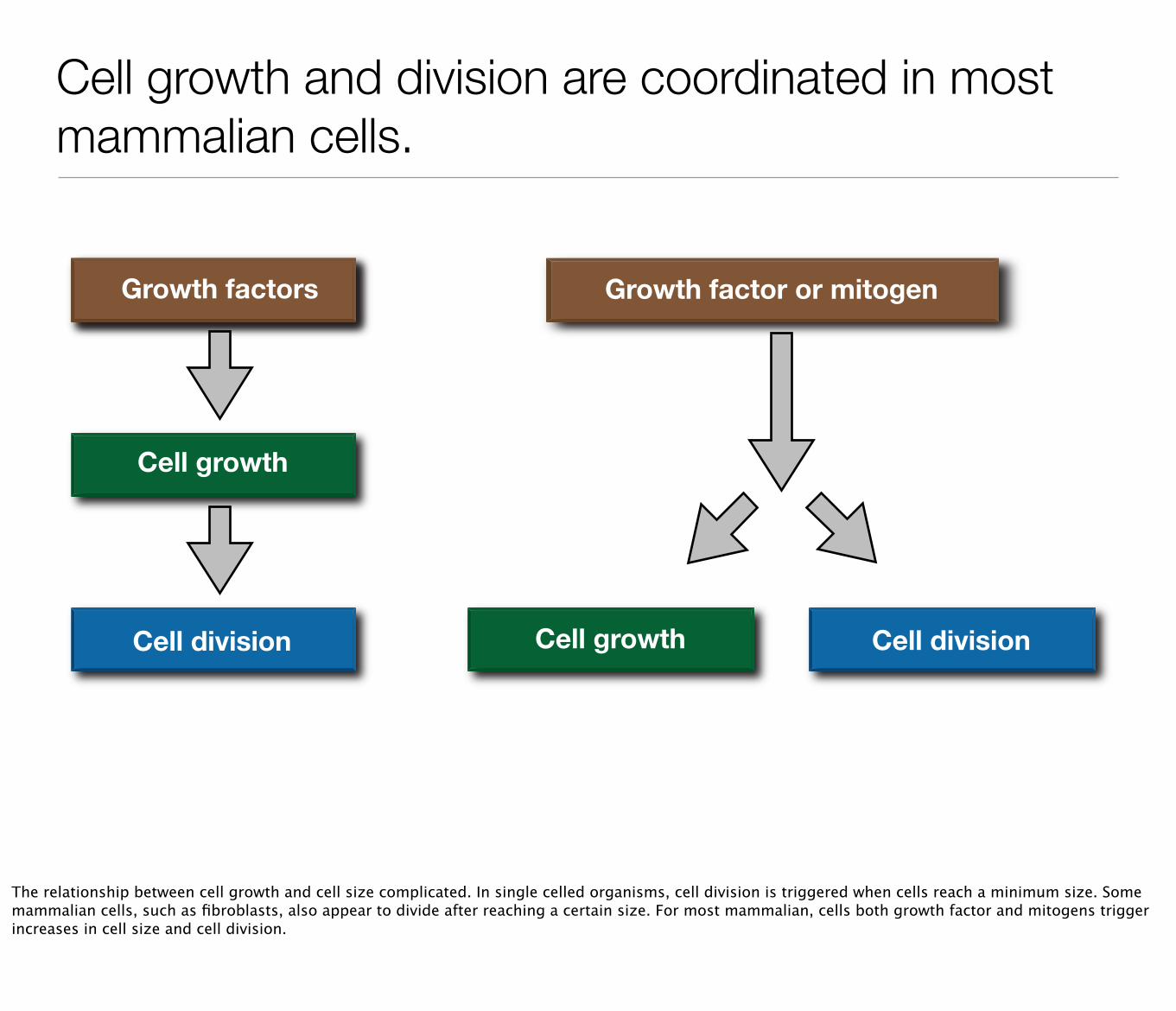
Cell growth and division are coordinated in most mammalian cells.
Nutrie
Cell growth
Growth factors
Cell division
Growth factor or mitogen
Cell growth Cell division
The relationship between cell growth and cell size complicated. In single celled organisms, cell division is triggered when cells reach a minimum size. Some mammalian cells, such as fibroblasts, also appear to divide after reaching a certain size. For most mammalian, cells both growth factor and mitogens trigger increases in cell size and cell division.
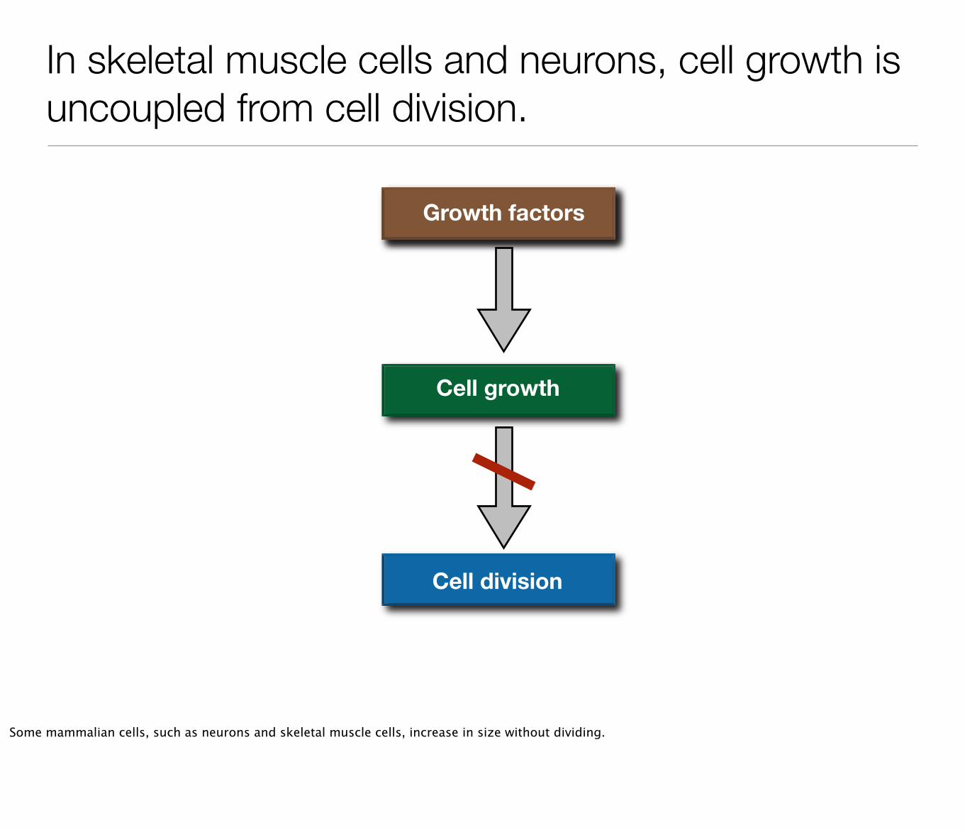
In skeletal muscle cells and neurons, cell growth is uncoupled from cell division.
Nutrie
Cell growth
Growth factors
Cell division
Some mammalian cells, such as neurons and skeletal muscle cells, increase in size without dividing.
Take home points...
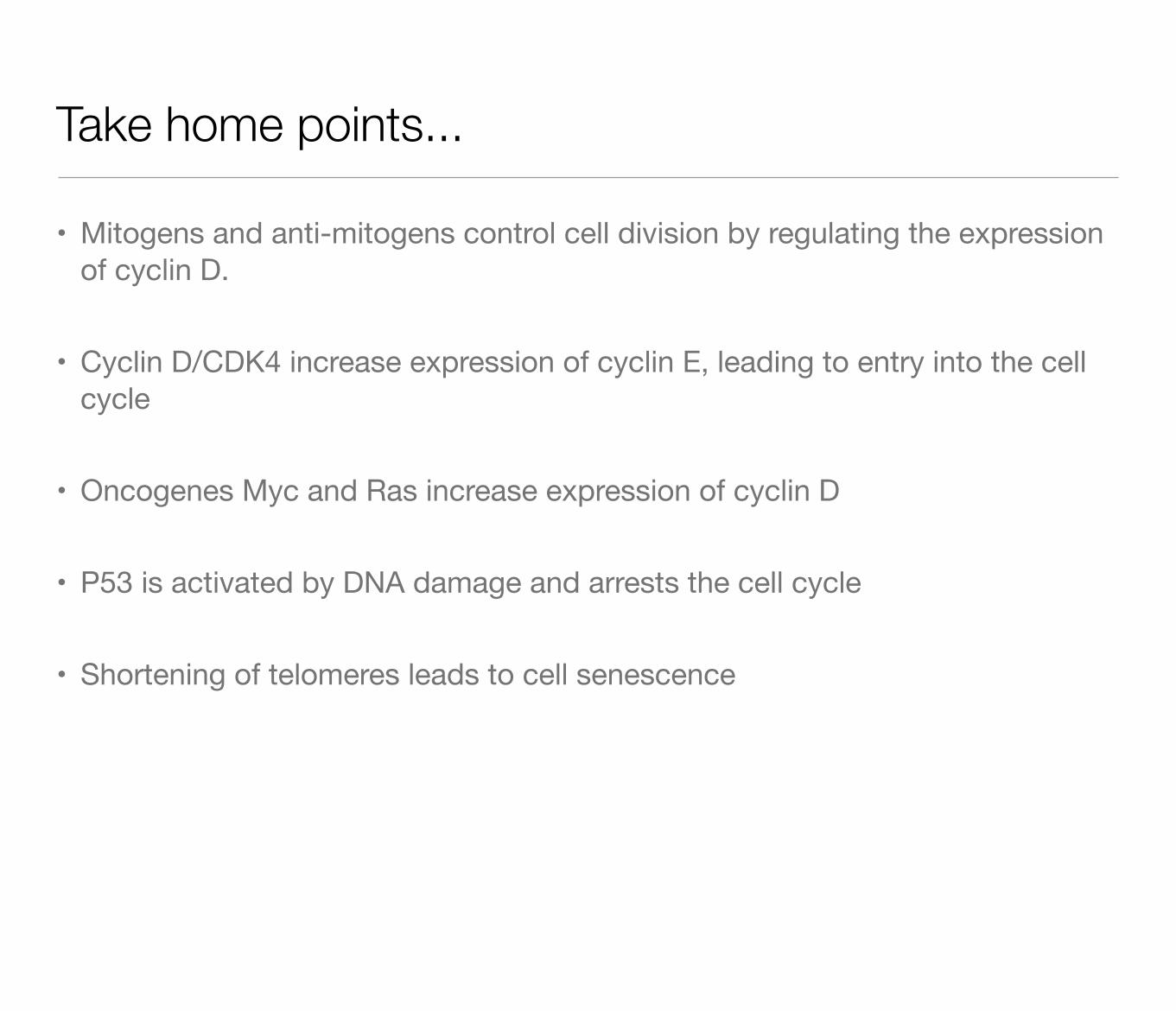
• Mitogens and anti-mitogens control cell division by regulating the expression of cyclin D.
• Cyclin D/CDK4 increase expression of cyclin E, leading to entry into the cell cycle
• Oncogenes Myc and Ras increase expression of cyclin D
• P53 is activated by DNA damage and arrests the cell cycle
• Shortening of telomeres leads to cell senescence