Embed Size (px)
Citation preview
1
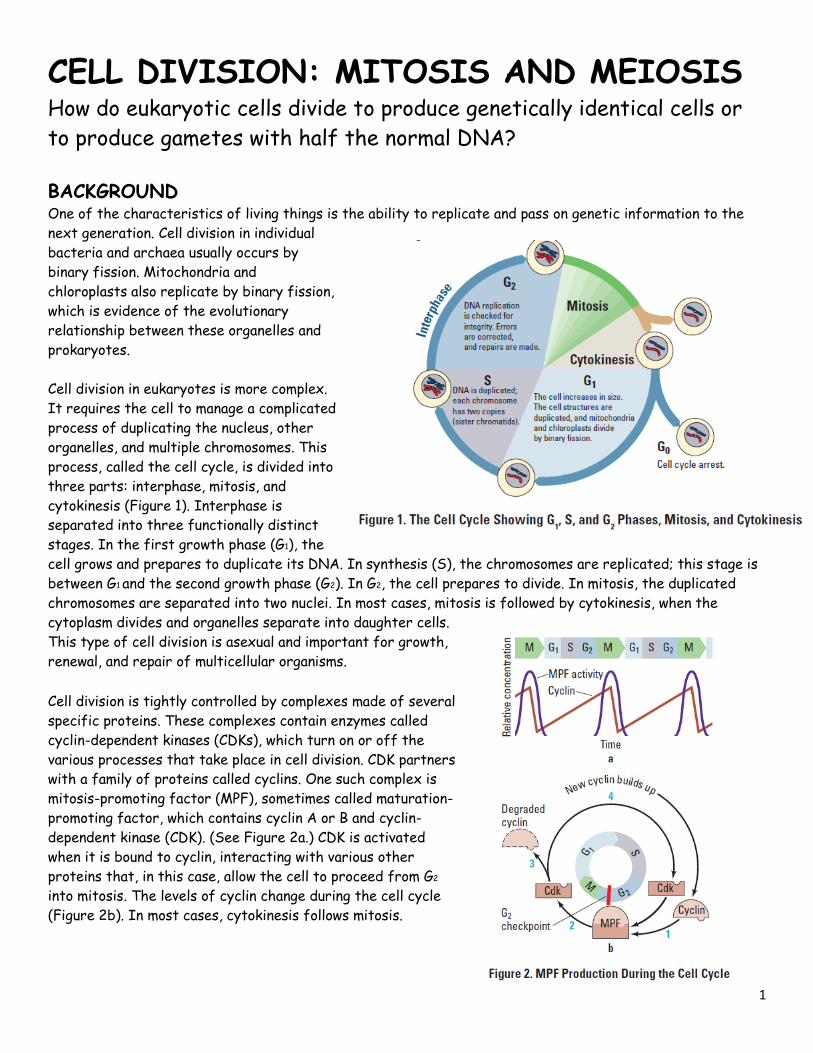
CELL DIVISION: MITOSIS AND MEIOSIS How do eukaryotic cells divide to produce genetically identical cells or
to produce gametes with half the normal DNA?
BACKGROUND One of the characteristics of living things is the ability to replicate and pass on genetic information to the
next generation. Cell division in individual
bacteria and archaea usually occurs by
binary fission. Mitochondria and
chloroplasts also replicate by binary fission,
which is evidence of the evolutionary
relationship between these organelles and
prokaryotes.
Cell division in eukaryotes is more complex.
It requires the cell to manage a complicated
process of duplicating the nucleus, other
organelles, and multiple chromosomes. This
process, called the cell cycle, is divided into
three parts: interphase, mitosis, and
cytokinesis (Figure 1). Interphase is
separated into three functionally distinct
stages. In the first growth phase (G1), the
cell grows and prepares to duplicate its DNA. In synthesis (S), the chromosomes are replicated; this stage is
between G1 and the second growth phase (G2). In G2, the cell prepares to divide. In mitosis, the duplicated
chromosomes are separated into two nuclei. In most cases, mitosis is followed by cytokinesis, when the
cytoplasm divides and organelles separate into daughter cells.
This type of cell division is asexual and important for growth,
renewal, and repair of multicellular organisms.
Cell division is tightly controlled by complexes made of several
specific proteins. These complexes contain enzymes called
cyclin-dependent kinases (CDKs), which turn on or off the
various processes that take place in cell division. CDK partners
with a family of proteins called cyclins. One such complex is
mitosis-promoting factor (MPF), sometimes called maturation-
promoting factor, which contains cyclin A or B and cyclin-
dependent kinase (CDK). (See Figure 2a.) CDK is activated
when it is bound to cyclin, interacting with various other
proteins that, in this case, allow the cell to proceed from G2
into mitosis. The levels of cyclin change during the cell cycle
(Figure 2b). In most cases, cytokinesis follows mitosis.
2
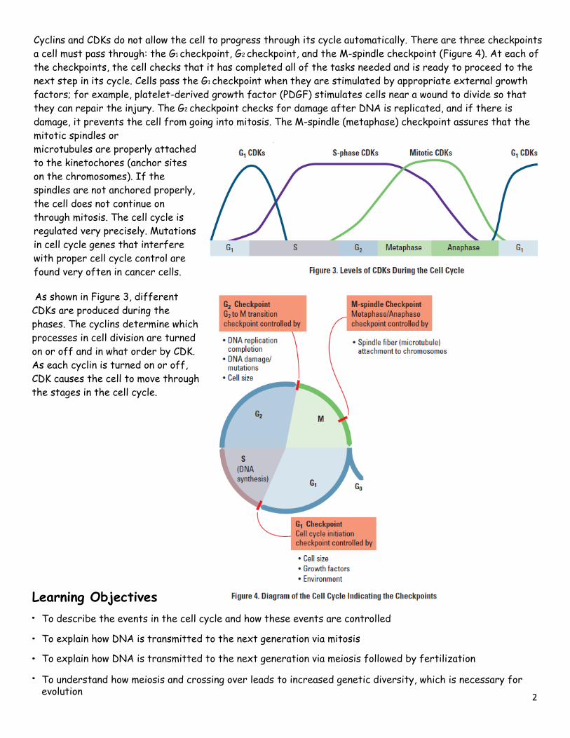
Cyclins and CDKs do not allow the cell to progress through its cycle automatically. There are three checkpoints
a cell must pass through: the G1 checkpoint, G2 checkpoint, and the M-spindle checkpoint (Figure 4). At each of
the checkpoints, the cell checks that it has completed all of the tasks needed and is ready to proceed to the
next step in its cycle. Cells pass the G1 checkpoint when they are stimulated by appropriate external growth
factors; for example, platelet-derived growth factor (PDGF) stimulates cells near a wound to divide so that
they can repair the injury. The G2 checkpoint checks for damage after DNA is replicated, and if there is
damage, it prevents the cell from going into mitosis. The M-spindle (metaphase) checkpoint assures that the
mitotic spindles or
microtubules are properly attached
to the kinetochores (anchor sites
on the chromosomes). If the
spindles are not anchored properly,
the cell does not continue on
through mitosis. The cell cycle is
regulated very precisely. Mutations
in cell cycle genes that interfere
with proper cell cycle control are
found very often in cancer cells.
As shown in Figure 3, different
CDKs are produced during the
phases. The cyclins determine which
processes in cell division are turned
on or off and in what order by CDK.
As each cyclin is turned on or off,
CDK causes the cell to move through
the stages in the cell cycle.
Learning Objectives
• To describe the events in the cell cycle and how these events are controlled
• To explain how DNA is transmitted to the next generation via mitosis
• To explain how DNA is transmitted to the next generation via meiosis followed by fertilization
• To understand how meiosis and crossing over leads to increased genetic diversity, which is necessary for evolution
3
THE INVESTIGATIONS These questions are designed to see how well you understand and can explain the key concepts related to cell
division before you begin your investigations.
1. How did you develop from a single-celled zygote to an organism with trillions of cells? How many mitotic
cell divisions would it take for one zygote to grow into an organism with 100 trillion cells?
__________________________________________________________________________________
__________________________________________________________________________________
2. How is cell division important to a single-celled organism? ________________________________
__________________________________________________________________________________
3. What must happen to ensure successful cell division? ______________________________________
__________________________________________________________________________________
4. How does the genetic information in one of your body cells compare to that found in other body cells?
__________________________________________________________________________________
__________________________________________________________________________________
5. What are some advantages of asexual reproduction in plants? _________________________________
__________________________________________________________________________________
6. Why is it important for DNA to be replicated prior to cell division? _____________________________
__________________________________________________________________________________
7. How do chromosomes move inside a cell during cell division? ________________________________
__________________________________________________________________________________
8. How is the cell cycle controlled? What would happen if the control were defective? _________________
__________________________________________________________________________________
4
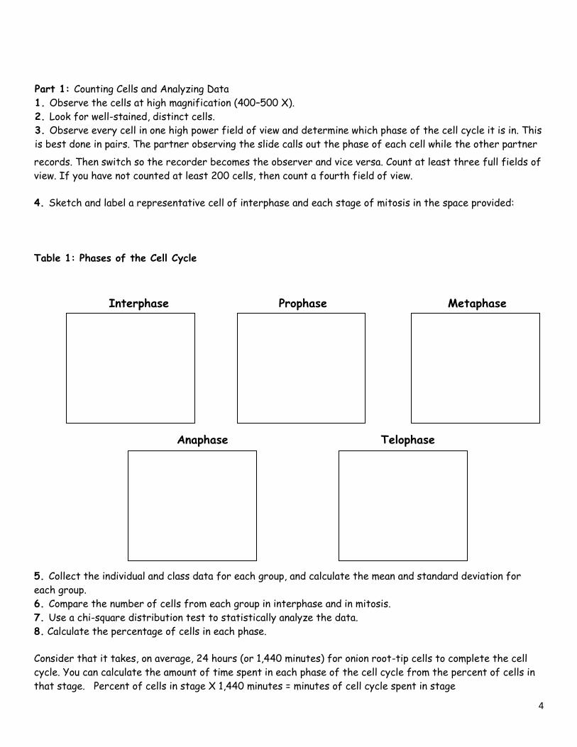
records. Then switch so the recorder becomes the observer and vice versa. Count at least three full fields of
view. If you have not counted at least 200 cells, then count a fourth field of view.
4. Sketch and label a representative cell of interphase and each stage of mitosis in the space provided:
Table 1: Phases of the Cell Cycle
5. Collect the individual and class data for each group, and calculate the mean and standard deviation for
each group.
6. Compare the number of cells from each group in interphase and in mitosis.
7. Use a chi-square distribution test to statistically analyze the data.
8. Calculate the percentage of cells in each phase.
Consider that it takes, on average, 24 hours (or 1,440 minutes) for onion root-tip cells to complete the cell
cycle. You can calculate the amount of time spent in each phase of the cell cycle from the percent of cells in
that stage. Percent of cells in stage X 1,440 minutes = minutes of cell cycle spent in stage
Anaphase Telophase
Interphase Prophase Metaphase
Part 1: Counting Cells and Analyzing Data
1. Observe the cells at high magnification (400–500 X).
2. Look for well-stained, distinct cells.
3. Observe every cell in one high power field of view and determine which phase of the cell cycle it is in. This
is best done in pairs. The partner observing the slide calls out the phase of each cell while the other partner
5
Prophase
Metaphase
Anaphase
Telophase
Total cells:
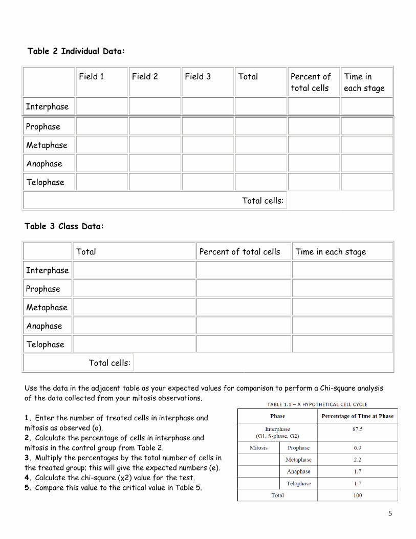
Table 3 Class Data:
Total Percent of total cells Time in each stage
Interphase
Prophase
Metaphase
Anaphase
Telophase
Total cells:
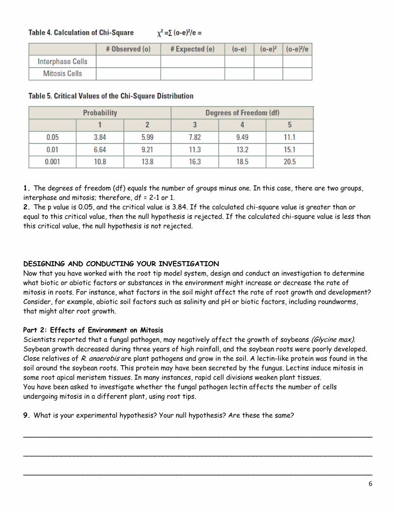
Use the data in the adjacent table as your expected values for comparison to perform a Chi-square analysis
of the data collected from your mitosis observations.
1. Enter the number of treated cells in interphase and
mitosis as observed (o).
2. Calculate the percentage of cells in interphase and
mitosis in the control group from Table 2.
3. Multiply the percentages by the total number of cells in
the treated group; this will give the expected numbers (e).
4. Calculate the chi-square (χ2) value for the test.
5. Compare this value to the critical value in Table 5.
Table 2 Individual Data:
Field 1 Field 2 Field 3 Total Percent of
total cells
Time in
each stage
Interphase
6
1. The degrees of freedom (df) equals the number of groups minus one. In this case, there are two groups,
interphase and mitosis; therefore, df = 2-1 or 1.
2. The p value is 0.05, and the critical value is 3.84. If the calculated chi-square value is greater than or
equal to this critical value, then the null hypothesis is rejected. If the calculated chi-square value is less than
this critical value, the null hypothesis is not rejected.
DESIGNING AND CONDUCTING YOUR INVESTIGATION
Now that you have worked with the root tip model system, design and conduct an investigation to determine
what biotic or abiotic factors or substances in the environment might increase or decrease the rate of
mitosis in roots. For instance, what factors in the soil might affect the rate of root growth and development?
Consider, for example, abiotic soil factors such as salinity and pH or biotic factors, including roundworms,
that might alter root growth.
Part 2: Effects of Environment on Mitosis
Scientists reported that a fungal pathogen, may negatively affect the growth of soybeans (Glycine max). Soybean growth decreased during three years of high rainfall, and the soybean roots were poorly developed.
Close relatives of R. anaerobis are plant pathogens and grow in the soil. A lectin-like protein was found in the
soil around the soybean roots. This protein may have been secreted by the fungus. Lectins induce mitosis in
some root apical meristem tissues. In many instances, rapid cell divisions weaken plant tissues.
You have been asked to investigate whether the fungal pathogen lectin affects the number of cells
undergoing mitosis in a different plant, using root tips.
9. What is your experimental hypothesis? Your null hypothesis? Are these the same?
__________________________________________________________________________________
__________________________________________________________________________________
__________________________________________________________________________________
7
10. How would you design an experiment with onion bulbs to test whether lectins increase the number of
cells in mitosis?
__________________________________________________________________________________
__________________________________________________________________________________
__________________________________________________________________________________
__________________________________________________________________________________
Independent variable: ____________________________________________________________
Dependent variable: ____________________________________________________________
11. What would you measure, and how would you measure it?
__________________________________________________________________________________
__________________________________________________________________________________
12. What appropriate controls for your experiment?
Controlled experiment: ____________________________________________________________
__________________________________________________________________________________
Controlled variables; ____________________________________________________________
__________________________________________________________________________________
Review from Part 1 & 2 13. How is the cell cycle controlled in normal cells?
__________________________________________________________________________________
14. What are cyclins and cyclin-dependent kinases? What do these proteins do in a cell?
__________________________________________________________________________________
15. What was the importance of collecting the class data?
__________________________________________________________________________________
16. Does an increased number of cells in mitosis mean that these cells are dividing faster than the cells in
the roots with a lower number of cells in mitosis?
__________________________________________________________________________________
17. What other way could you determine how fast the rate of mitosis is occurring in root tips?
__________________________________________________________________________________
__________________________________________________________________________________
8
18. If your observations had not been restricted to the area of the root tip that is actively dividing, how
would your results have been different?
__________________________________________________________________________________
19. Why is it more accurate to call mitosis “nuclear replication” than cell division?
__________________________________________________________________________________
Part 3: Loss of Cell Cycle Control in Cancer
Many of us have family members who have or have had cancer. Cancer can occur when cells lose control of
their cell cycle and divide abnormally. This happens when tumor suppressor genes, such as p53 or Rb
(retinoblastoma), are mutated. There are many questions you should consider before beginning your
investigation.
20. How are normal cells and cancer cells different from each other?
__________________________________________________________________________________
__________________________________________________________________________________
21. What are the main causes of cancer?
__________________________________________________________________________________
22. What goes wrong during the cell cycle in cancer cells?
__________________________________________________________________________________
23. What makes some genes responsible for an increased risk of certain cancers?
__________________________________________________________________________________
__________________________________________________________________________________
24. Do you think that the chromosomes might be different between normal and cancer cells?
__________________________________________________________________________________
The last question is the focus of this part of the lab. With your group, form a hypothesis as to how the
chromosomes of a cancer cell might appear in comparison to a normal cell and how those differences are
related to the behavior of the cancer cell.
25. Hypothesis: _____________________________________________________________________
9
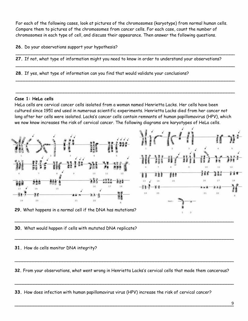
Case 1: HeLa cells
HeLa cells are cervical cancer cells isolated from a woman named Henrietta Lacks. Her cells have been
cultured since 1951 and used in numerous scientific experiments. Henrietta Lacks died from her cancer not
long after her cells were isolated. Lacks’s cancer cells contain remnants of human papillomavirus (HPV), which
we now know increases the risk of cervical cancer. The following diagrams are karyotypes of HeLa cells.
29. What happens in a normal cell if the DNA has mutations?
__________________________________________________________________________________
30. What would happen if cells with mutated DNA replicate?
__________________________________________________________________________________
31. How do cells monitor DNA integrity?
__________________________________________________________________________________
32. From your observations, what went wrong in Henrietta Lacks’s cervical cells that made them cancerous?
__________________________________________________________________________________
33. How does infection with human papillomavirus virus (HPV) increase the risk of cervical cancer?
__________________________________________________________________________________
For each of the following cases, look at pictures of the chromosomes (karyotype) from normal human cells.
Compare them to pictures of the chromosomes from cancer cells. For each case, count the number of
chromosomes in each type of cell, and discuss their appearance. Then answer the following questions.
26. Do your observations support your hypothesis?
__________________________________________________________________________________
27. If not, what type of information might you need to know in order to understand your observations?
__________________________________________________________________________________
28. If yes, what type of information can you find that would validate your conclusions?
__________________________________________________________________________________
__________________________________________________________________________________
10
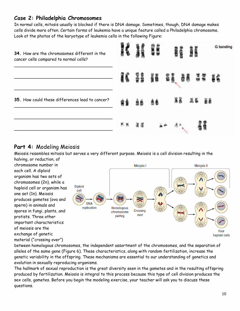
Case 2: Philadelphia Chromosomes In normal cells, mitosis usually is blocked if there is DNA damage. Sometimes, though, DNA damage makes
cells divide more often. Certain forms of leukemia have a unique feature called a Philadelphia chromosome.
Look at the photos of the karyotype of leukemia cells in the following Figure:
34. How are the chromosomes different in the
cancer cells compared to normal cells?
______________________________________
______________________________________
______________________________________
35. How could these differences lead to cancer?
______________________________________
______________________________________
______________________________________
Part 4: Modeling Meiosis Meiosis resembles mitosis but serves a very different purpose. Meiosis is a cell division resulting in the
halving, or reduction, of
chromosome number in
each cell. A diploid
organism has two sets of
chromosomes (2n), while a
haploid cell or organism has
one set (1n). Meiosis
produces gametes (ova and
sperm) in animals and
spores in fungi, plants, and
protists. Three other
important characteristics
of meiosis are the
exchange of genetic
material (“crossing over”)
between homologous chromosomes, the independent assortment of the chromosomes, and the separation of
alleles of the same gene (Figure 6). These characteristics, along with random fertilization, increase the
genetic variability in the offspring. These mechanisms are essential to our understanding of genetics and
evolution in sexually reproducing organisms.
The hallmark of sexual reproduction is the great diversity seen in the gametes and in the resulting offspring
produced by fertilization. Meiosis is integral to this process because this type of cell division produces the
sex cells, gametes. Before you begin the modeling exercise, your teacher will ask you to discuss these
questions.
11
36. How do sexually reproducing organisms produce gametes from diploid progenitors?
__________________________________________________________________________________
__________________________________________________________________________________
37. How does the process increase gamete diversity? ________________________________________
__________________________________________________________________________________
38. What are the outcomes from independent assortment and crossing over?
__________________________________________________________________________________
__________________________________________________________________________________
39.How does the distance between two genes or a gene and a centromere affect crossover frequencies
__________________________________________________________________________________
__________________________________________________________________________________
40. When is the DNA replicated during meiosis? _____________________________________________
41. Are homologous pairs of chromosomes exact copies of each other? _____________________________
42. What is crossing over? _____________________________________________________________
43. What physical constraints control crossover frequencies? ___________________________________
44. What is meant by independent assortment?
__________________________________________________________________________________
__________________________________________________________________________________
45. How can you calculate the possible number of different kinds of gametes? ______________________
__________________________________________________________________________________
46. What happens if a homologous pair of chromosomes fails to separate, and how might this contribute to
genetic disorders such as Down syndrome and cri du chat syndrome? ______________________________
__________________________________________________________________________________
__________________________________________________________________________________
47. How are mitosis and meiosis fundamentally different?
__________________________________________________________________________________
__________________________________________________________________________________
12
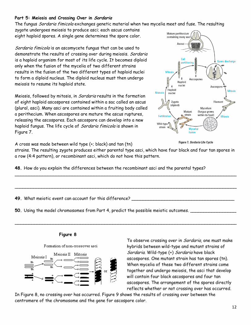
Part 5: Meiosis and Crossing Over in Sordaria The fungus Sordaria fimicola exchanges genetic material when two mycelia meet and fuse. The resulting
zygote undergoes meiosis to produce asci; each ascus contains
eight haploid spores. A single gene determines the spore color.
Sordaria fimicola is an ascomycete fungus that can be used to
demonstrate the results of crossing over during meiosis. Sordaria is a haploid organism for most of its life cycle. It becomes diploid
only when the fusion of the mycelia of two different strains
results in the fusion of the two different types of haploid nuclei
to form a diploid nucleus. The diploid nucleus must then undergo
meiosis to resume its haploid state.
Meiosis, followed by mitosis, in Sordaria results in the formation
of eight haploid ascospores contained within a sac called an ascus
(plural, asci). Many asci are contained within a fruiting body called
a perithecium. When ascospores are mature the ascus ruptures,
releasing the ascospores. Each ascospore can develop into a new
haploid fungus. The life cycle of Sordaria fimicola is shown in
Figure 7.
A cross was made between wild type (+; black) and tan (tn)
strains. The resulting zygote produces either parental type asci, which have four black and four tan spores in
a row (4:4 pattern), or recombinant asci, which do not have this pattern.
48. How do you explain the differences between the recombinant asci and the parental types?
__________________________________________________________________________________
__________________________________________________________________________________
49. What meiotic event can account for this difference? ______________________________________
50. Using the model chromosomes from Part 4, predict the possible meiotic outcomes. _________________
__________________________________________________________________________________
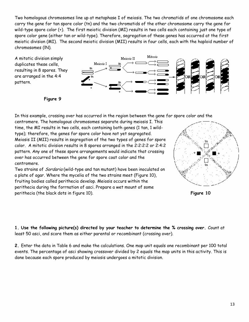
Figure 8 To observe crossing over in Sordaria, one must make
hybrids between wild-type and mutant strains of
Sordaria. Wild-type (+) Sordaria have black
ascospores. One mutant strain has tan spores (tn).
When mycelia of these two different strains come
together and undergo meiosis, the asci that develop
will contain four black ascospores and four tan
ascospores. The arrangement of the spores directly
reflects whether or not crossing over has occurred.
In Figure 8, no crossing over has occurred. Figure 9 shows the results of crossing over between the
centromere of the chromosome and the gene for ascospore color.
13
Two homologous chromosomes line up at metaphase I of meiosis. The two chromatids of one chromosome each
carry the gene for tan spore color (tn) and the two chromatids of the other chromosome carry the gene for
wild-type spore color (+). The first meiotic division (MI) results in two cells each containing just one type of
spore color gene (either tan or wild-type). Therefore, segregation of these genes has occurred at the first
meiotic division (MI). The second meiotic division (MII) results in four cells, each with the haploid number of
chromosomes (lN).
A mitotic division simply
duplicates these cells,
resulting in 8 spores. They
are arranged in the 4:4
pattern.
Figure 9
In this example, crossing over has occurred in the region between the gene for spore color and the
centromere. The homologous chromosomes separate during meiosis I. This
time, the MI results in two cells, each containing both genes (1 tan, 1 wild-
type); therefore, the genes for spore color have not yet segregated.
Meiosis II (MII) results in segregation of the two types of genes for spore
color. A mitotic division results in 8 spores arranged in the 2:2:2:2 or 2:4:2
pattern. Any one of these spore arrangements would indicate that crossing
over has occurred between the gene for spore coat color and the
centromere.
Two strains of Sordaria (wild-type and tan mutant) have been inoculated on
a plate of agar. Where the mycelia of the two strains meet (Figure 10),
fruiting bodies called perithecia develop. Meiosis occurs within the
perithecia during the forrnation of asci. Prepare a wet mount of some
perithecia (the black dots in figure 10). Figure 10
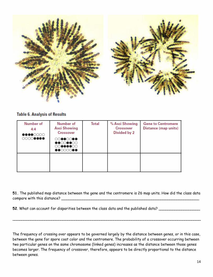
1. Use the following picture(s) directed by your teacher to determine the % crossing over. Count at
least 50 asci, and score them as either parental or recombinant (crossing over).
2. Enter the data in Table 6 and make the calculations. One map unit equals one recombinant per 100 total
events. The percentage of asci showing crossover divided by 2 equals the map units in this activity. This is
done because each spore produced by meiosis undergoes a mitotic division.
14
51. The published map distance between the gene and the centromere is 26 map units. How did the class data
compare with this distance? ____________________________________________________________
52. What can account for disparities between the class data and the published data? __________________
__________________________________________________________________________________
The frequency of crossing over appears to be governed largely by the distance between genes, or in this case,
between the gene for spore coat color and the centromere. The probability of a crossover occurring between
two particular genes on the same chromosome (linked genes) increases as the distance between those genes
becomes larger. The frequency of crossover, therefore, appears to be directly proportional to the distance
between genes.
15
A map unit is an arbitrary unit of measure used to describe relative distances between linked genes. The
number of map units between two genes or between a gene and the centromere is equal to the percentage of
recombinants. Customary units cannot be used because we cannot directly visualize genes with the light
microscope. However, due to the relationship between distance and crossover frequency, we may use the map
unit.
Evaluating Results
53. Why did you divide the percentage of asci showing crossover (recombinant) by 2? _________________
__________________________________________________________________________________
54. The published map distance between the spore color gene and the centromere is 26 map units. How did
the class data compare with this distance? _________________________________________________
__________________________________________________________________________________
55. How can you account for any disparities between the class data and the published data? ____________
__________________________________________________________________________________
__________________________________________________________________________________
56. Illustrate what happened during meiosis to produce the results you found.
57. Do you think the Philadelphia chromosome is a result of crossing over as seen in this part of the
investigation or some other chromosomal abnormality? Explain your answer. _________________________
__________________________________________________________________________________
__________________________________________________________________________________
58. Do you think the cell cycle described for mitosis could be applied to meiosis as well? Explain your answer.
__________________________________________________________________________________
__________________________________________________________________________________
__________________________________________________________________________________