Embed Size (px)
Citation preview
Cell Cycle-Mediated Cardiac Regeneration in the Mouse Heart
Arash Eghbali1, Austin Dukes1, Karl Toischer2, Gerd Hasenfuss2 and Loren J. Field1
(1) Krannert Institute of Cardiology and Wells Center for Pediatric Research, Indiana University
School of Medicine, Indianapolis, USA, and (2) Department of Cardiology and Pneumology,
Heart Center, Georg-August-University, Goettingen, Germany and DZHK (German Center for
Cardiovascular Research) Partnersite Goettingen
Corresponding author:
Loren J. Field
Krannert Institute of Cardiology and the Herman B Wells Center for Pediatric Research
Indiana University School of Medicine
1044 W. Walnut Street
Indianapolis, IN 46202, USA
Tel.: +1-317-274-5085
Fax: +1-317-278-9298
Authors:
Arash Eghbali
Austin Dukes
_______________________________________________
This is the author's manuscript of the article published in final edited form as:
Eghbali, A., Dukes, A., Toischer, K., Hasenfuss, G., & Field, L. J. (2019). Cell Cycle-Mediated Cardiac Regeneration in the Mouse Heart. Current Cardiology Reports, 21(10), 131. https://doi.org/10.1007/s11886-019-1206-9
Karl Toischer
Gerd Hasenfuss
Loren J. Field
Abstract
Many forms of heart disease result in the essentially irreversible loss of
cardiomyocytes. The ability to promote cardiomyocyte renewal may be a promising approach
to reverse injury in diseased hearts.
Purpose of Review: To describe the impact of cardiomyocyte cell cycle activation on cardiac
function and structure in several different models of myocardial disease.
Recent Findings: Transgenic mice expressing cyclin D2 (D2 mice) exhibit sustained
cardiomyocyte renewal in the adult heart. Earlier studies demonstrated that D2 mice exhibited
progressive myocardial regeneration in experimental models of myocardial infarction, and that
cardiac function was normalized to values seen in sham operated litter mates by 180 days
post-injury. D2 mice also exhibited markedly improved atrial structure in a genetic model of
atrial fibrosis. More recent studies revealed that D2 mice were remarkably resistant to heart
failure induced by chronic elevated afterload as compared to their wild type (WT siblings), with
a 6-fold increase in median survival as well as retention of relatively normal cardiac function.
Finally, D2 mice exhibited a progressive recovery in cardiac function to normal levels and a
concomitant reduction in adverse myocardial remodeling in an anthracycline cardiotoxicity
model.
Summary: The studies reviewed here make a strong case for the potential utility of inducing
cardiomyocyte renewal as a means to treat injured hearts. Several challenges which must be
met to develop a viable therapeutic intervention based on these observations are discussed.
Keywords: cardiac regeneration; cardiomyocyte renewal; cell cycle regulation
Introduction
It is largely believed that cardiomyocyte renewal in the adult mammalian heart occurs
via proliferation of pre-existing cardiomyocytes [1, 2], as opposed to the activity of
cardiomyogenic stem cells [3, 4]. The intrinsic renewal capacity of adult mammalian
cardiomyocytes is quite limited, with similarly low rates reported for rodents and humans [5-8].
Consequently, injuries causing cardiomyocyte death, as for example myocardial infarction,
result in reparative fibrosis as opposed to overt tissue regeneration [9]. Fibrosis results in
stiffening of the myocardial wall, which in turn increases cardiomyocyte stress leading to
additional cell loss, additional fibrosis and ultimately pump failure. Treatments have
traditionally relied on interventions aimed at restoring perfusion following acute ischemic
events, interventions aimed at reducing the vulnerability of at-risk myocardium (as for example
blocking apoptosis or enhancing angiogenesis), interventions aimed at enhancing contractility
of the surviving myocardium, and interventions aimed at modulating fibrosis. Advances in our
understanding of the molecular regulation of cell cycle activity suggest an additional approach,
namely promoting myocardial regeneration by activating cardiomyocyte renewal. This review
will focus on a model developed in our laboratory which exhibits increased rates of
cardiomyocyte cell cycle activity in the adult myocardium. These animals exhibit regenerative
cardiac growth, with a concomitant improvement in both cardiac architecture and function
following cardiac injury in a variety of experimental models.
Cyclin D2 expression results in sustained cardiomyocyte cell cycle activity in adult mice
The notion of inducing cardiomyocyte cell cycle with the hope of driving regeneration of
the myocardium is not new. Early studies relied on stimulating cell cycle regulatory pathways in
cultured cardiomyocytes via pharmacologic or genetic intervention [10]. The advent of
technologies to generate genetically modified animal models harboring either gain-of-function
or loss-of-functions alterations facilitated studies demonstrating that relatively simple genetic
changes can result in profound increases in cardiomyocyte cell cycle activity. For example,
early experiments demonstrated that expression of a powerful cell cycle regulatory molecule,
the SV40 Large T Antigen protein, was sufficient to induce proliferation of atrial [11, 12] or
ventricular [13] cardiomyocytes. Subsequently, numerous genetic modifications have been
shown to impact cardiomyocyte cell cycle activity to varying degrees.
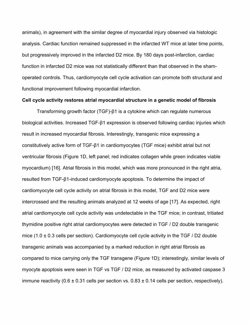
One such model utilized the alpha cardiac myosin heavy chain transcriptional regulatory
sequences to target expression of sequences encoding cyclin D2 (Figure 1A, upper panel
shows a schematic diagram of the transgene) [14]. Cyclin D2 (subsequently designated
CCND2) is a cofactor which regulates the activity of cyclin-dependent kinase 4 (CDK4), which
mediates transit through the restriction point of the cell cycle. Targeted expression of cyclin D2
in adult cardiomyocytes was sufficient to induce expression of endogenous CDK4 (Figure 1A,
lower left panels). Adult transgenic mice (designated D2 mice) exhibited an approximately 400-
fold increase in the level of ventricular cardiomyocyte S-phase activity as compared to their
wildtype (WT) siblings, as determined by tritiated thymidine incorporation using a transgenic
reporter to identify cardiomyocyte nuclei (Figure 1A, lower right panel; thymidine incorporation
levels were roughly 0.0005% vs. 0.2% for WT vs. D2 mice, respectively, following a single
injection of isotope).
Cell cycle activity restores ventricular cardiac structure and function following
myocardial infarction
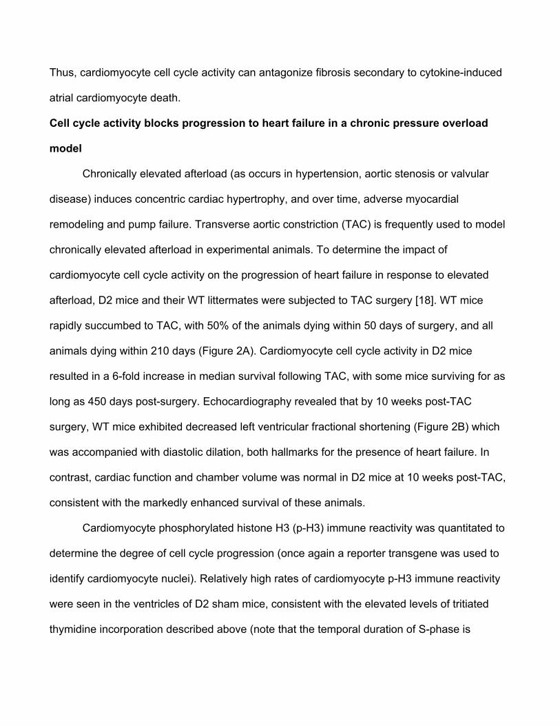
To determine the impact of cardiomyocyte cell cycle activity following ischemic injury to
the heart, D2 mice and their WT siblings were subjected to permanent coronary artery ligation
[15]. WT hearts examined at 7 days or 180 days post-infarction exhibited transmural defects
from the apex to the base (Figure 1B, left; Azan staining, grey indicates scar while magenta
indicates viable myocardium). D2 hearts harvested at 7 days post-infarction exhibited a similar
transmural defect as observed in WT mice, indicating that cyclin D2 expression did not bestow
any cardioprotective activity to acute injury. At 180 days post-infarction, D2 hearts exhibited
myocardial tissue along the endocardial and epicardial surface of the scar, from the apex to
the base (Figure 1B, right, see arrows). Because these structures were not apparent in hearts
at 7 days post-infarction, their presence is indicative of de novo myocardial growth.
To determine the impact of cardiomyocyte cell cycle activity on cardiac function, D2
mice and their WT siblings underwent permanent coronary artery ligation or sham surgery, and
were then subjected to hemodynamic analyses using intracardiac pressure-volume catheters.
dP/dtmax/EDV, a functional parameter which is not influenced by differences in heart rate, was
similarly suppressed in infarcted WT and D2 mice as compared to their sham-operated
controls (Figure 1C; y-axis indicates the percentage normalized to the sham-operated
animals), in agreement with the similar degree of myocardial injury observed via histologic
analysis. Cardiac function remained suppressed in the infarcted WT mice at later time points,
but progressively improved in the infarcted D2 mice. By 180 days post-infarction, cardiac
function in infarcted D2 mice was not statistically different than that observed in the sham-
operated controls. Thus, cardiomyocyte cell cycle activation can promote both structural and
functional improvement following myocardial infarction.
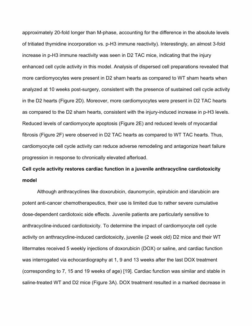
Cell cycle activity restores atrial myocardial structure in a genetic model of fibrosis
Transforming growth factor (TGF)-β1 is a cytokine which can regulate numerous
biological activities. Increased TGF-β1 expression is observed following cardiac injuries which
result in increased myocardial fibrosis. Interestingly, transgenic mice expressing a
constitutively active form of TGF-β1 in cardiomyocytes (TGF mice) exhibit atrial but not
ventricular fibrosis (Figure 1D, left panel; red indicates collagen while green indicates viable
myocardium) [16]. Atrial fibrosis in this model, which was more pronounced in the right atria,
resulted from TGF-β1-induced cardiomyocyte apoptosis. To determine the impact of
cardiomyocyte cell cycle activity on atrial fibrosis in this model, TGF and D2 mice were
intercrossed and the resulting animals analyzed at 12 weeks of age [17]. As expected, right
atrial cardiomyocyte cell cycle activity was undetectable in the TGF mice; in contrast, tritiated
thymidine positive right atrial cardiomyocytes were detected in TGF / D2 double transgenic
mice (1.0 ± 0.3 cells per section). Cardiomyocyte cell cycle activity in the TGF / D2 double
transgenic animals was accompanied by a marked reduction in right atrial fibrosis as
compared to mice carrying only the TGF transgene (Figure 1D); interestingly, similar levels of
myocyte apoptosis were seen in TGF vs TGF / D2 mice, as measured by activated caspase 3
immune reactivity (0.6 ± 0.31 cells per section vs. 0.83 ± 0.14 cells per section, respectively).
Thus, cardiomyocyte cell cycle activity can antagonize fibrosis secondary to cytokine-induced
atrial cardiomyocyte death.
Cell cycle activity blocks progression to heart failure in a chronic pressure overload
model
Chronically elevated afterload (as occurs in hypertension, aortic stenosis or valvular
disease) induces concentric cardiac hypertrophy, and over time, adverse myocardial
remodeling and pump failure. Transverse aortic constriction (TAC) is frequently used to model
chronically elevated afterload in experimental animals. To determine the impact of
cardiomyocyte cell cycle activity on the progression of heart failure in response to elevated
afterload, D2 mice and their WT littermates were subjected to TAC surgery [18]. WT mice
rapidly succumbed to TAC, with 50% of the animals dying within 50 days of surgery, and all
animals dying within 210 days (Figure 2A). Cardiomyocyte cell cycle activity in D2 mice
resulted in a 6-fold increase in median survival following TAC, with some mice surviving for as
long as 450 days post-surgery. Echocardiography revealed that by 10 weeks post-TAC
surgery, WT mice exhibited decreased left ventricular fractional shortening (Figure 2B) which
was accompanied with diastolic dilation, both hallmarks for the presence of heart failure. In
contrast, cardiac function and chamber volume was normal in D2 mice at 10 weeks post-TAC,
consistent with the markedly enhanced survival of these animals.
Cardiomyocyte phosphorylated histone H3 (p-H3) immune reactivity was quantitated to
determine the degree of cell cycle progression (once again a reporter transgene was used to
identify cardiomyocyte nuclei). Relatively high rates of cardiomyocyte p-H3 immune reactivity
were seen in the ventricles of D2 sham mice, consistent with the elevated levels of tritiated
thymidine incorporation described above (note that the temporal duration of S-phase is
approximately 20-fold longer than M-phase, accounting for the difference in the absolute levels
of tritiated thymidine incorporation vs. p-H3 immune reactivity). Interestingly, an almost 3-fold
increase in p-H3 immune reactivity was seen in D2 TAC mice, indicating that the injury
enhanced cell cycle activity in this model. Analysis of dispersed cell preparations revealed that
more cardiomyocytes were present in D2 sham hearts as compared to WT sham hearts when
analyzed at 10 weeks post-surgery, consistent with the presence of sustained cell cycle activity
in the D2 hearts (Figure 2D). Moreover, more cardiomyocytes were present in D2 TAC hearts
as compared to the D2 sham hearts, consistent with the injury-induced increase in p-H3 levels.
Reduced levels of cardiomyocyte apoptosis (Figure 2E) and reduced levels of myocardial
fibrosis (Figure 2F) were observed in D2 TAC hearts as compared to WT TAC hearts. Thus,
cardiomyocyte cell cycle activity can reduce adverse remodeling and antagonize heart failure
progression in response to chronically elevated afterload.
Cell cycle activity restores cardiac function in a juvenile anthracycline cardiotoxicity
model
Although anthracyclines like doxorubicin, daunomycin, epirubicin and idarubicin are
potent anti-cancer chemotherapeutics, their use is limited due to rather severe cumulative
dose-dependent cardiotoxic side effects. Juvenile patients are particularly sensitive to
anthracycline-induced cardiotoxicity. To determine the impact of cardiomyocyte cell cycle
activity on anthracycline-induced cardiotoxicity, juvenile (2 week old) D2 mice and their WT
littermates received 5 weekly injections of doxorubicin (DOX) or saline, and cardiac function
was interrogated via echocardiography at 1, 9 and 13 weeks after the last DOX treatment
(corresponding to 7, 15 and 19 weeks of age) [19]. Cardiac function was similar and stable in
saline-treated WT and D2 mice (Figure 3A). DOX treatment resulted in a marked decrease in
cardiac function in WT mice at 1 week after the last DOX treatment, and function remained
suppressed over the duration of the study. Cardiac function was also decreased in D2 mice at
1 week after the last DOX treatment, but function progressively improved over the course of
the study and was indistinguishable from saline treated animals by 13 weeks after the last
DOX injection.
Histologic analyses were performed at 1 week (designated the acute stage) and 13
weeks (designated the late stage) after the last DOX treatment. Cardiomyocyte apoptosis was
quantitated via activated caspase 3 immune reactivity, and cardiomyocyte cell cycle activity
was quantitated via tritiated thymidine incorporation. To facilitate comparison of cardiomyocyte
drop-out and cardiomyocyte renewal, the data was normalized to events per mm2 (Figure 3B).
DOX treatment of WT mice resulted in cardiomyocyte apoptosis during the acute stage, which
increased by approximately 3-fold in late stage hearts, in agreement with previous studies
using this juvenile cardiotoxicity model [20]. Essentially no cardiomyocyte cell cycle activity
was detected in DOX-treated WT hearts in the acute or late stage. Although DOX treatment
resulted in a similar level of cardiomyocyte apoptosis in acute stage D2 hearts, no increases
were observed in late stage D2 DOX-treated hearts. As expected, cardiomyocyte cell cycle
activity was observed in DOX-treated D2 hearts in both the acute and late stage. Of note, the
number of cell cycle events per mm2 was greater than the number of apoptotic events per mm2
in the D2 DOX-treated hearts in both the acute and late stages, suggesting a trend toward
myocardial regeneration despite the presence of on-going cardiomyocyte apoptosis.
Consistent with this, myocardial fibrosis was markedly reduced in late stage DOX-treated D2
hearts as compared to late stage DOX-treated WT hearts (Figure 3C). Thus, cardiomyocyte
cell cycle activity can antagonize adverse remodeling and restore cardiac function following
anthracycline-induced cardiotoxicity.
Future considerations
The studies reviewed above indicate that targeted expression of cyclin D2 is sufficient to
drive cardiomyocyte proliferation, and more importantly, that the presence of cardiomyocyte
cell cycle activity has a positive impact on myocardial function and/or structure in a variety of
injury models. The observation of progressive functional and structural improvement over the
long-term (i.e., 6 months post-infarct, 10 weeks post-TAC, and 13 weeks post-cardiotoxic drug
exposure), supports the notion that recovery results from the presence of active proliferation as
opposed to transgene-mediated cardioprotective activity. Indeed, when considering the
magnitude of the cell cycle response in D2 mice, one would a priory predict that a long-term
follow-up would be required to detect a beneficial effect with this model. A caveat of the D2
model is that transgene expression in the ventricle is activated during the temporal window of
cardiomyocyte cell cycle withdrawal which occurs in early postnatal life (i.e., expression may
activate prior to cardiomyocyte terminal differentiation). However, unpublished studies from our
group demonstrate that de novo activation of cyclin D2 in adult cardiomyocytes using a
tetracycline-regulated conditional system gives rise to a similar induction of cell cycle activity.
Moreover, de novo expression of a number of other genes has been shown to activate
cardiomyocyte cell cycle activity in adults. Thus, there is reasonable evidence supporting the
general approach of promoting cardiomyocyte renewal as a means to effect repair of injured
hearts.
An interesting observation from the experiments reviewed here is that when
cardiomyocyte renewal rates are similar to drop-out rates (as was seen in the atrial fibrosis,
TAC and cardiotoxicity studies), the net effect is a marked reduction or absence of overt
fibrosis. This suggests that the fibrosis normally observed in these injury models arise as a
compensatory response to cardiomyocyte loss, as opposed to injury-induced activation of
fibroblast signaling. Interestingly, this view is supported in cases where there is a massive
acute loss of cardiomyocytes (as was seen in the coronary artery ligation model); although
overt scar tissue persists, only minimal fibrosis is observed in the de novo myocardium.
Persistence of an overt scar may reflect the need to retain wall integrity to prevent cardiac
rupture at early stages post-injury. Although higher rates of cell cycle renewal may reduce
persistent scar content, it may come at the cost of greater vulnerability to cardiac rupture. The
potential contribution of a large number of proliferating cells to arrhythmia vulnerability must
also be considered. Thus, it remains to be seen if slow vs. fast rates of cardiomyocyte renewal
are ultimately better tolerated when effecting regenerative growth of the myocardium.
Another consideration for developing cardiomyocyte renewal-based approaches to
effect cardiac regeneration is the likely need to incorporate cell-type specificity into the
intervention. Most of the published studies rely on manipulating genetic pathways which can
induce proliferation in a wide variety of cell types, including cardiomyocytes. It is possible that
the used of vector systems with a tropism for targeting expression of cardiomyocytes will effect
some degree of cell-type preference. Use of non-vector based approaches (as for example,
delivery of RNA which has been modified for enhanced stability) may also be viable, provided
a sufficient number of cardiomyocytes can be transduced. As work in the field progresses, it is
possible that regulatory pathways which are intrinsic to and specific for cardiomyocyte renewal
will be identified. Such pathways could likely be exploited to develop cell-type specific
interventions.
In conclusion, the studies reviewed here, as well as many other examples in the
literature, make a strong case for the potential utility of inducing cardiomyocyte renewal as a
means to treat injured hearts. Although a number of formidable obstacles must be overcome,
advances in the field are accelerating and with it the hope of attaining a viable cardiomyocyte
renewal-based intervention.
Acknowledgment
Arash Eghbali and Austin Dukes contributed equally to this work.
Compliance with Ethical Standards
Conflict of Interest
Arash Eghbali, Austin Dukes, Karl Toischer, and Loren J. Field declare that they have no
conflict of interest.
Gerd Hasenfuss reports personal fees from Corvia, Servier, Impulse Dynamics, Novartis,
AstraZeneca, Vifor Pharma, Berlin Chemie, and Springer.
Human and Animal Rights and Informed Consent
All reported studies/experiments with human or animal subjects performed by the authors have
been previously published and complied with all applicable ethical standards (including the
Helsinki declaration and its amendments, institutional/national research committee standards,
and international/national/institutional guidelines).
References
Papers of particular interest, published recently, have been highlighted as:
• Of importance
•• Of major importance
1. Ali SR, Hippenmeyer S, Saadat LV, Luo L, Weissman IL, Ardehali R. Existing cardiomyocytes generate cardiomyocytes at a low rate after birth in mice. Proc Natl Acad Sci U S A. 2014;111(24):8850‐5. doi:10.1073/pnas.1408233111. 2. Senyo SE, Steinhauser ML, Pizzimenti CL, Yang VK, Cai L, Wang M et al. Mammalian heart renewal by pre‐existing cardiomyocytes. Nature. 2013;493(7432):433‐6. doi:10.1038/nature11682. 3. •• Eschenhagen T, Bolli R, Braun T, Field LJ, Fleischmann BK, Frisen J et al. Cardiomyocyte Regeneration: A Consensus Statement. Circulation. 2017;136(7):680‐6. doi:10.1161/CIRCULATIONAHA.117.029343. This consensus statement delineates the lack of critical data supporting a direct cardiomyogenic role for purported cardiac resident stem cells, and thus underscores the need to develop alternative strategies for promoting myocardial regeneration. 4. Madonna R, Van Laake LW, Davidson SM, Engel FB, Hausenloy DJ, Lecour S et al. Position Paper of the European Society of Cardiology Working Group Cellular Biology of the Heart: cell‐based therapies for myocardial repair and regeneration in ischemic heart disease and heart failure. Eur Heart J. 2016;37(23):1789‐98. doi:10.1093/eurheartj/ehw113. 5. Soonpaa MH, Field LJ. Assessment of cardiomyocyte DNA synthesis in normal and injured adult mouse hearts. Am J Physiol. 1997;272(1 Pt 2):H220‐6. 6. Soonpaa MH, Field LJ. Survey of studies examining mammalian cardiomyocyte DNA synthesis. Circ Res. 1998;83(1):15‐26. 7. Bergmann O, Bhardwaj RD, Bernard S, Zdunek S, Barnabe‐Heider F, Walsh S et al. Evidence for cardiomyocyte renewal in humans. Science. 2009;324(5923):98‐102. doi:10.1126/science.1164680. 8. Bergmann O, Zdunek S, Felker A, Salehpour M, Alkass K, Bernard S et al. Dynamics of Cell Generation and Turnover in the Human Heart. Cell. 2015;161(7):1566‐75. doi:10.1016/j.cell.2015.05.026. 9. Swynghedauw B. Molecular mechanisms of myocardial remodeling. Physiol Rev. 1999;79(1):215‐62. 10. Pasumarthi KB, Field LJ. Cardiomyocyte cell cycle regulation. Circ Res. 2002;90(10):1044‐54. 11. Field LJ. Atrial natriuretic factor‐SV40 T antigen transgenes produce tumors and cardiac arrhythmias in mice. Science. 1988;239(4843):1029‐33. 12. Behringer RR, Peschon JJ, Messing A, Gartside CL, Hauschka SD, Palmiter RD et al. Heart and bone tumors in transgenic mice. Proc Natl Acad Sci U S A. 1988;85(8):2648‐52. 13. Katz EB, Steinhelper ME, Delcarpio JB, Daud AI, Claycomb WC, Field LJ. Cardiomyocyte proliferation in mice expressing alpha‐cardiac myosin heavy chain‐SV40 T‐antigen transgenes. Am J Physiol. 1992;262(6 Pt 2):H1867‐76. 14. Pasumarthi KB, Nakajima H, Nakajima HO, Soonpaa MH, Field LJ. Targeted expression of cyclin D2 results in cardiomyocyte DNA synthesis and infarct regression in transgenic mice. Circ Res. 2005;96(1):110‐8. 15. Hassink RJ, Pasumarthi KB, Nakajima H, Rubart M, Soonpaa MH, de la Riviere AB et al. Cardiomyocyte cell cycle activation improves cardiac function after myocardial infarction. Cardiovasc Res. 2008;78(1):18‐25. 16. Nakajima H, Nakajima HO, Salcher O, Dittie AS, Dembowsky K, Jing S et al. Atrial but not ventricular fibrosis in mice expressing a mutant transforming growth factor‐beta(1) transgene in the heart. Circ Res. 2000;86(5):571‐9. 17. Nakajima H, Nakajima HO, Dembowsky K, Pasumarthi KB, Field LJ. Cardiomyocyte cell cycle activation ameliorates fibrosis in the atrium. Circ Res. 2006;98(1):141‐8.
18. • Toischer K, Zhu W, Hunlich M, Mohamed BA, Khadjeh S, Reuter SP et al. Cardiomyocyte proliferation prevents failure in pressure overload but not volume overload. J Clin Invest. 2017;127(12):4285‐4296. doi:10.1172/JCI81870. Findings from this study suggest that the presence of cardiomyocyte cell cycle activity can block the onset of heart failure resulting from chronic increases in cardiac after‐load. 19. • Zhu W, Reuter S, Field LJ. Targeted expression of cyclin D2 ameliorates late stage anthracycline cardiotoxicity. Cardiovasc Res. 2019;115(5):960‐5. doi:10.1093/cvr/cvy273. Findings from this study demonstrate that the presence of cardiomyocyte cell cycle activity can reverse heart failure in a chronic juvenile doxorubicin cardiotoxicity model. 20. Zhu W, Shou W, Payne RM, Caldwell R, Field LJ. A mouse model for juvenile doxorubicin‐induced cardiac dysfunction. Pediatric research. 2008;64(5):488‐94.
Figure Legends
Figure 1. Targeted expression of cyclin D2 results in cardiomyocyte cell cycle activity which
can promote structural and functional recovery following myocardial injury. A. Schematic
diagram of the MHC-cycD2 transgene (upper panel), and documentation of transgene
expression (lower left panel, anti-cyclin D2 immune histology, brown signal), endogenous
CDK4 expression (lower middle panel, anti-CDK4 immune histology, brown signal) and
cardiomyocyte S-phase activity (lower right panel, tritiated thymidine incorporation in
cardiomyocyte nuclei, black punctate signal over blue nuclear signal). (Reproduced from:
Pasumarthi KB, Nakajima H, Nakajima HO, Soonpaa MH, Field LJ: Circ Res. 2005;96(1):110-
8, with permission from Wolters Kluwer Health, Inc.) [14] B. Histologic analysis of WT and D2
hearts at 7 and 180 days post-infarction, sections sampled at 1 mm intervals from apex to
base and stained with Azan (magenta indicates viable myocardium, grey indicates scar).
(Reproduced from: Hassink RJ, Pasumarthi KB, Nakajima H, Rubart M, Soonpaa MH, de la
Riviere AB et al.: Cardiovasc Res. 2008;78(1):18-25., with permission from Oxford University
Press) [15] C. Cardiac function (as quantitated by dP/dtmax/EDV) in WT (open bars) and D2
(solid bars) at 7, 60 and 180 days post-infarction; data is normalized to the percent activity
observed in sham-operated littermates. (Reproduced from: Hassink RJ, Pasumarthi KB,
Nakajima H, Rubart M, Soonpaa MH, de la Riviere AB et al.: Cardiovasc Res. 2008;78(1):18-
25., with permission from Oxford University Press) [15] D. Histologic analysis of right atria from
12 week old TGF (left panel) and TGF / D2 double transgenic (right panel) mice; sections
were stained with Sirius Red and Fast Green (green indicates viable myocardium, red
indicates collagen deposition). (Reproduced from: Nakajima H, Nakajima HO, Dembowsky K,
Pasumarthi KB, Field LJ: Circ Res. 2006;98(1):141-8, with permission from Wolters Kluwer
Health, Inc.) [17].
Figure 2. Cyclin D2 expression blocks progression to heart failure in a chronic pressure
overload model. A. Survival of WT (blue trace) and D2 (red trace) mice following TAC surgery
(black trace shows survival of sham operated animals). B. Fractional shortening (FS) of WT
and D2 mice 10 weeks after sham or TAC surgery as measured by echocardiography. C.
Detection of p-H3 immune reactivity in cardiomyocyte nuclei (left panel, black punctate signal
over blue nuclear signal) and quantitation of levels in D2 mice 10 weeks after sham or TAC
surgery. D. Cardiomyocyte number determined from dispersed cell preparations of WT and D2
hearts 10 weeks after sham or TAC surgery. E. Activated caspase 3-positive cardiomyocytes
per mm2 tissue in WT and D2 mice at 3 weeks after sham or TAC surgery. F. Histologic
analysis of WT (left panel) and D2 (right panel) mice 10 weeks after TAC; sections were
stained with Sirius Red and Fast Green (green indicates viable myocardium, red indicates
collagen deposition). (Reproduced from: Toischer K, Zhu W, Hunlich M, Mohamed BA,
Khadjeh S, Reuter SP et al.: J Clin Invest. 2017;127(12):4285-4296, with permission from
American Society for Clinical Investigation) [18].
Figure 3. Cyclin D2 expression restores cardiac function in a juvenile anthracycline
cardiotoxicity model. A. Cardiac function in saline-treated WT (black trace), saline-treated D2
(magenta), DOX-treated WT (red trace) and DOX-treated D2 (blue trace) mice at various time
points in the study; vertical arrows indicate the time of saline or DOX injections. B. Quantitation
of tritiated thymidine incorporation and activated caspase 3 immune reactivity in DOX-treated
WT and D2 mice at the acute and late stages; results are plotted as events per mm2 to allow
direct comparison of the results. C. Histologic analysis of acute and late stage WT and D2
hearts 13 weeks after DOX treatment; sections were stained with Sirius Red and Fast Green
(green indicates viable myocardium, red indicates collagen deposition). (Reproduced from:
Zhu W , Reuter S, Field LJ.: Cardiovasc Res. 2019;115(5):960-5, with permission from Oxford
University Press) [19].
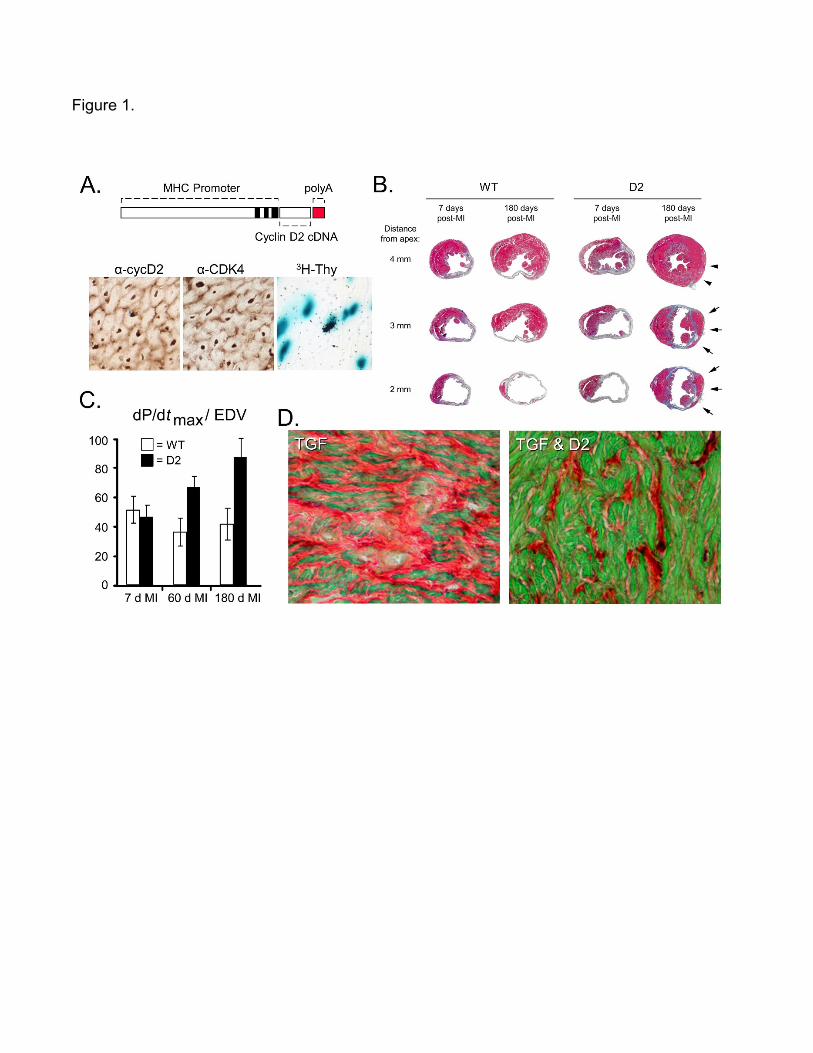
Figure 1.
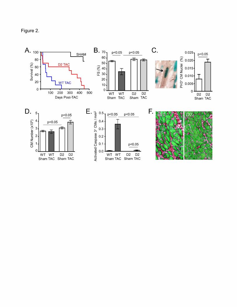
Figure 2.
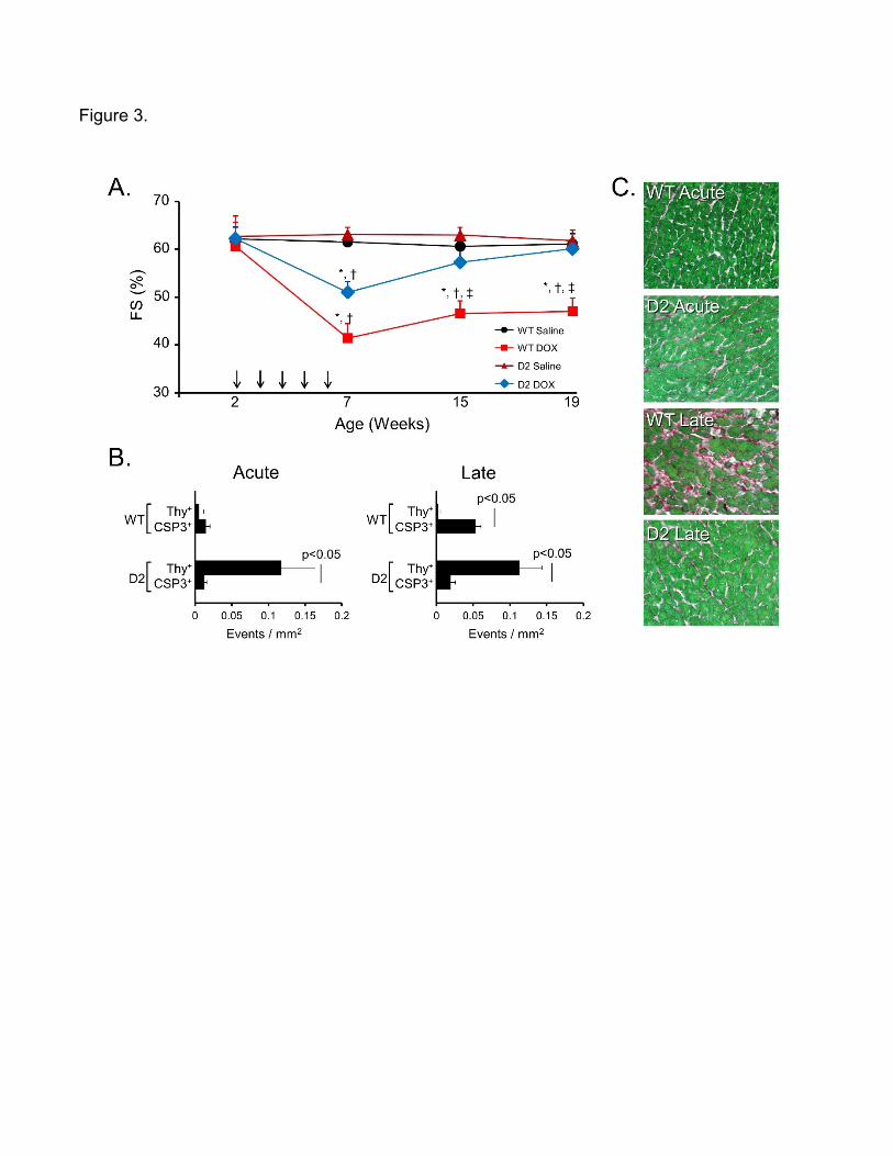
Figure 3.