Embed Size (px)
Citation preview
Molecular Biology of the CellVol. 6, 1397-1410, October 1995
Cell Cycle-dependent Phosphorylation andMicrotubule Binding of Tau Protein StablyTransfected into Chinese Hamster Ovary CellsUte Preuss, Frank Doring, Susanne Illenberger, and Eva-Maria Mandelkow*
Max-Planck-Unit for Structural Molecular Biology, c/o DESY, Notkestrasse 85,D-22603 Hamburg, Germany
Submitted June 15, 1995; Accepted August 11, 1995Monitoring Editor: J. Richard McIntosh
Tau protein, a neuronal microtubule-associated protein, is phosphorylated in situ andhyperphosphorylated when aggregated into the paired helical filaments of Alzheimer'sdisease. To study the phosphorylation of tau protein in vivo, we have stably transfectedhtau4O, the largest human tau isoform, into Chinese hamster ovary cells. The distributionand phosphorylation of tau was monitored by gel shift, autoradiography, immunofluo-rescence, and immunoblotting, using the antibodies Tau-1, AT8, AT180, and PHF-1,which are sensitive to the phosphorylation of Ser2O2, Thr2O5, Thr231, Ser235, Ser396, andSer4O4 and are used in the diagnosis of Alzheimer tau. In interphase cells, tau becomesphosphorylated to some extent, partly at these sites; most of the tau is associated withmicrotubules. In mitosis, the above Ser/Thr-Pro sites become almost completely phos-phorylated, causing a pronounced shift in Mr and an antibody reactivity similar to thatof Alzheimer tau. Moreover, a substantial fraction of tau is found in the cytoplasmdetached from microtubules. Autoradiographs of metabolically labeled Chinese hamsterovary cells in interphase and mitosis confirmed that tau protein is more highly phos-phorylated during mitosis. The understanding of tau phosphorylation under physiolog-ical conditions might help elucidate possible mechanisms for the hyperphosphorylationin Alzheimer's disease.
INTRODUCTION
Microtubule-associated proteins (MAPs)l are involvedin the rearrangement of the microtubular network,and their tissue- and development-specific stabiliza-tion of microtubules is presumably one of their mostimportant functions (Drubin and Kirschner, 1986;Chapin and Bulinski, 1992; Lee, 1993; Hirokawa, 1994).Phosphorylation of MAPs appears to modulate theaffinity for microtubules (Brugg and Matus, 1991;Buendia et al., 1992; Biernat et al., 1993; Lieuvin et al.,1994) but in vivo it is still poorly understood. Oneclass of mammalian brain MAPs is tau, which is spe-cifically localized in axons of neuronal cells. In Alz-
* Corresponding author.Abbreviations used: AD, Alzheimer's disease; CHO, Chinesehamster ovary; ECL, enhanced chemoluminescence; FCS, fetalcalf serum; MAP, microtubule-associated protein; PHF, pairedhelical filaments.
heimer's disease tau protein becomes hyperphospho-rylated and forms the paired helical filaments (PHFs).Hyperphosphorylation might therefore play a majorrole in the onset of neuronal degeneration by causingdisorganization of the microtubule cytoskeleton,blocking axonal transport, and allowing deposition ofhyperphosphorylated tau into PHFs. Most of the iden-tified aberrant phosphorylation sites in PHF-tau areSer/Thr-Pro motifs (Ishiguro et al., 1991; Lee et al.,1991; Biernat et al., 1992; Gustke et al., 1992; Hasegawaet al., 1992; Morishima-Kawashima et al., 1995). How-ever, one cannot conclude that the kinases phospho-rylating tau protein in vitro such as MAP kinase(Drewes et al., 1992), GSK-3 (Hanger et al., 1992;Mandelkow et al., 1992), cdc2 kinase (Ledesma et al.,1992; Vulliet et al., 1992), or cdk5 (Baumann et al.,1993; Paudel et al., 1993) are necessarily the onesthat have a strong influence on microtubule stabilityin vivo. A good candidate for regulating the dynam-
© 1995 by The American Society for Cell Biology 1397
U. Preuss et al.
ics and rearrangement of microtubules in cells viaphosphorylation of tau and other MAPs is the novelmicrotubule-associated protein/microtubule affini-ty-regulating kinase (pllO mark) (Drewes et al.,1995). This kinase phosphorylates tau on its KIGSand KCGS motifs in the repeat domain, a regionwithin the carboxy-terminal half of the protein thatcontains three or four imperfect repeats of 31 or 32residues and binds to microtubules (Lee et al., 1988;Himmler et al., 1989). In particular, phosphorylationof Ser262 in the first repeat abolishes tau's bindingto microtubules and makes the microtubules dy-namically unstable (Biernat et al., 1993).One way to identify phosphorylation sites of tau
protein is to use antibodies with known epitopes thatdiscriminate between "normal" tau and the hyper-phosphorylated "pathological" forms (Binder et al.,1985; Greenberg and Davies, 1990; Lee et al., 1991;Hasegawa et al., 1992; Mercken et al., 1992). Most ofthese antibodies recognize epitopes containing Ser-Proor Thr-Pro motifs. For example, AT8, a monoclonalantibody raised against PHF-tau, recognizes phos-phorylated Ser2O2/Thr2O5 (Biernat et al., 1992; Zheng-Fischhofer, 1994) while the monoclonal antibodyTau-1 reacts with almost the same region of tau, butonly if it is unphosphorylated (Kosik et al., 1988; Bier-nat et al., 1992; Szendrei et al., 1993). Therefore, theincrease of AT8 staining and the decrease of Tau-1staining can be used to mark the appearance of PHF-like phosphorylation in tau protein.Many studies have demonstrated that the dynamics
of microtubules varies markedly during the cell cycle(McIntosh and Hering, 1991). During interphase, inwhich microtubules are responsible for intracellulartransport and the maintenance of cellular architecture,the dynamics of growth and shrinkage is relativelyslow, but increase abruptly at the entry to mitosiswhere microtubules form the spindle apparatus that isessential for chromosome segregation (Belmont et al.,1990). Protein phosphorylation is a common mecha-nism utilized to regulate these cellular processes(Nigg, 1993), and in the case of microtubule dynamicsit is likely to operate at the level of MAP phosphory-lation (Buendia et al., 1992). In the present study, wehave stably transfected Chinese hamster ovary (CHO)cells, which normally do not express tau protein, toinvestigate the phosphorylation and dephosphoryla-tion of tau in the cell. This has two advantages. First,it ensures a homogeneous population of tau protein inthe cells, compared with the various isoforms found inthe brain. Secondly, the behavior of tau is likely to becomparable to the endogenous MAP4 because theseproteins have similar microtubule-interacting do-mains (Chapin and Bulinski, 1991; West et al., 1991).We observed that antibodies directed against phos-
phorylated Ser/Thr-Pro-motifs found in PHF-tau alsolabeled tau protein in transfected cells during mitosis.
These findings imply that kinases belonging to thefamily of proline-directed Ser/Thr-kinases can phos-phorylate tau during cell division and induce a mo-bility shift reminiscent of Alzheimer PHF tau. Thus,CHO cells stably transfected with a human tau iso-form provide a useful system for observing the influ-ence of phosphorylation on the interaction of tau pro-tein with microtubules in vivo (Bramblett et al., 1993).
MATERIALS AND METHODS
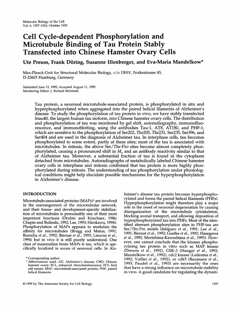
AntibodiesSeveral monoclonal antibodies were generous gifts of the followingcolleagues: Tau-1 clone from Dr. L. Binder (Northwestern Univer-sity, Evanstown, IL); AT8 from Dr. A. Vandevoorde (Innogenetics,Ghent, Belgium); PHF-1 from Drs. S. Greenberg and P. Davies(Albert Einstein College of Medicine, Bronx, NY); T46 from Dr.V.M.-Y. Lee (University of Pennsylvania, Philadelphia, PA). Ratmonoclonal anti-tubulin antibody YL1/2 (recognizing the C-termi-nus of tyrosylated a-tubulin; Kilmartin et al., 1982) was obtainedfrom Sera Lab (Sussex, England). The antibody epitopes on tauprotein are shown in Figure 1. The tau antibodies Tau-1 (Binder etal., 1985) and T46 (Kosik et al., 1988) are directed against unphos-phorylated epitopes; AT8 (Mercken et al., 1992) and PHF-1 (Green-berg and Davies, 1990) recognize phosphorylated epitopes. The sitesof Tau-1 and AT8 are nearly complementary to one another aroundresidue 200; Tau-1 reacts when there is no phosphorylation in thisregion (Kosik et al., 1988), and AT8 reacts when there are twophosphorylation sites, Ser202 and Thr2O5 in full-length tau (Biernatet al., 1992; Goedert et al., 1993; Szendrei et al., 1993; Zheng-Fischhofer, 1994). PHF-1 reacts with phosphorylated Ser396 (Lang etal., 1992) but preferably when Ser404 is also phosphorylated (Otvoset al., 1994); in this regard the antibody is similar to SM131 (Licht-enberg-Kraag et al., 1992). The polyclonal rabbit anti-tau antibody(Dako, Hamburg, Germany), which was used for immunoprecipi-tation, recognizes the C-terminal half of phosphorylated and non-phosphorylated tau protein.
Construction of a Vector Containing the cDNAof htau4OThe methods used here were mostly described previously (Biernatet al., 1992). Recombinant human tau protein (htau40) was derivedfrom the cDNA clones of Goedert et al. (1989) and expressed inEscherichia coli using pNG2, a variant of the pET expression vector(Studier et al., 1990). For direct subcloning of htau40 into the eu-karyotic expression vector, pRc/CMV (Invitrogen, San Diego, CA)was modified, introducing a new multiple cloning site. The 1.3-kbcDNA of htau4O was inserted into the NdeI/BglII sites of the mul-tiple cloning site. This plasmid containing the coding sequence ofhtau40 was used for stable transfection.
Cell Culture and TransfectionCHO cells were grown in HAM's F12 medium supplemented with10% fetal calf serum (FCS; Biochrom, Berlin, Germany). Approxi-mately 60% confluent cells were plated on 35-mm dishes for trans-fection or on coverslips for immunofluorescence analysis and incu-bated at 37°C with 5% CO2. The cells were transfected with 2 ,ug ofDNA and 9 ,ul of Lipofectamine according to the manufacturer'srecommendations (Life Technologies, Eggenstein, Germany). Stablytransfected cells were selected by growing them in the presence of800 jig/ml of Geneticin (G-418). After incubation for an additional2-3 wk, cells were cloned by limiting dilution and screened forhtau40-expressing cells by immunofluorescence and polymerasechain reaction.
Molecular Biology of the Cell1398
Tau Phosphorylation during Cell Cycle
ImmunofluorescenceCells were washed in a stabilizing buffer [80 mM piperazine-N,N'-bis(2-ethanesulfonic acid), 1 mM MgCl2, 1 mM EGTA, 4% polyeth-ylene glycol, pH 6.8] and fixed with either methanol at -20'C for 5min or with 2% paraformaldehyde for 20 min following permeabi-lization with 0.2% Triton X-100 for 5 min. The fixed cells weretreated with 5% nonfat dry milk for 1 h and incubated with theprimary monoclonal mouse antibodies T46, Tau-1, AT8, and PHF-1at 1:600,1:50, 1:2000, and 1:2000 dilutions, respectively, and with theanti-tubulin antibody YL1/2 at 1:200 for 1 h at 37°C. For the sec-ondary antibodies, fluorescein-conjugated goat anti-mouse or rhoda-mine-conjugated goat anti-rat antibodies (Dianova, Hamburg, Ger-many) were used at 1:300 dilution and incubated at 37°C for 30 min.The cells were examined with an Axioplan fluorescence microscope(Zeiss, Oberkochen, Germany) using filters optimized for double-label experiments and a 63x fluorescence objective.
Cell ExtractsTotal cell extracts of CHO cells transfected with htau40 or with thevehicle only were prepared by lysing subconfluent cells on ice inbuffer A containing 50 mM Tris (pH 7.4), 1% Nonidet P-40, 1 mMMgCl2, 5 mM EGTA, 5 mM dithiothreitol, 120 mM NaCl, 20 mMNaF, 1 mM vanadate, 0.1 mM phenylmethylsulfonyl fluoride, 1,Lg/ml leupeptin, 1 gg/ml aprotinin, 1 ,ug/ml pepstatin, and 10mMbenzamidine. Extracts were centrifuged immediately at 15800 x gfor 10 min and the supernatants were used directly for sodiumdodecyl sulfate-polyacrylamide gel electrophoresis (SDS-PAGE).Extracts were also prepared from mitotically arrested cells follow-ing treatment with 0.4 ,ug/ml nocodazole (Sigma, Deisenhofen,Germany) for 5 h. Cells were detached by mechanical shake-off andlysed on ice in buffer A containing 500 mM NaCl. Extracts wereboiled for 10 min and centrifuged at 15800 x g for 10 min. Thesoluble fraction containing heat-stable tau was applied to SDS-PAGE. Protein concentrations were determined by the method ofBradford (1976).
SDS-PAGE and ImmunoblottingExtract samples of both total and nocodazole-treated CHO cellswere electrophoresed on 10% SDS-polyacrylamide gels (12 ,ug pro-tein per lane) and electrophoretically transferred to polyvinylidenedifluoride membranes (Millipore, Eschborn, Germany). Residualprotein-binding sites on the membrane were blocked with 5% non-fat dry milk in Tris-buffered saline following incubation with themonoclonal antibodies T46 (1:6000), Tau-1 (1:500), AT8 (1:4000), andPHF-1 (1:400). Bound antibody was detected with a peroxidase-conjugated goat anti-mouse antibody (Dianova, Hamburg, Ger-many) using diaminobenzidine as substrate, or the immunostainingwas visualized using enhanced chemoluminescence (ECL) accord-ing to the manufacturer's instructions (Amersham, Braunschweig,Germany). For immunoblot analysis recombinant htau40 from E.coli was isolated by fast protein liquid Mono S (Pharmacia, Freiburg,Germany) chromatography, on the basis of its heat stability (fordetails see Hagestedt et al., 1989). Phosphorylation reaction wascarried out as described by Drewes et al. (1995).
32Phosphate LabelingStably transfected CHO cells grown in a 75-cm2 culture flask wereincubated for 30 min in phosphate-free DMEM medium (Life Tech-nologies) containing 5% FCS. The following incubation was carriedout with phosphate-free DMEM supplemented with 10% dialyzedFCS, 25 mM N-2-hydroxyethylpiperazine-N'-2-ethanesulfonic acid,and [V2P]orthophosphate (0.7 mCi/ml). After 1 h of preincubation,the cells were mitotically arrested and lysed as described above.
ImmunoprecipitationMitotic and nonmitotic cell-extracts labeled with [32P]orthophos-phate were boiled for 10 min and centrifuged immediately at 15800x g for 10 min. First, 5-10 ,ug of polyclonal rabbit anti-Tau antibody(Dako, Hamburg, Germany) was added to the supernatants, andthen after 4 h at 4°C, 40 ,ui of protein-A/G-Sepharose beads (Di-anova, Hamburg, Germany) was added. After an overnight incuba-tion at 4'C the immune complexes were recovered by centrifugationand rinsed three times in immunoprecipitation buffer. The immu-noprecipitated tau protein was resolubilized in SDS-sample bufferand boiled for 5 min. Electrophoresis was carried out on a 10% SDS-polyacrylamide gel and subjected to autoradiography. Before addi-tion of the polyclonal anti-Tau antibody, protein concentrations ofthe mitotic and interphase cells were determined by the method ofBradford (1976). Equal amounts of antibody were added, and afterimmunoprecipitation the same amounts of protein (5.5 jig per lane)were loaded onto a 10% SDS gel. The gel was silver stained, dried,and subjected to autoradiography. Furthermore, the tau bands werecut out of the dried gel, and radioactivity was quantified by Ceren-kov counting.
RESULTS
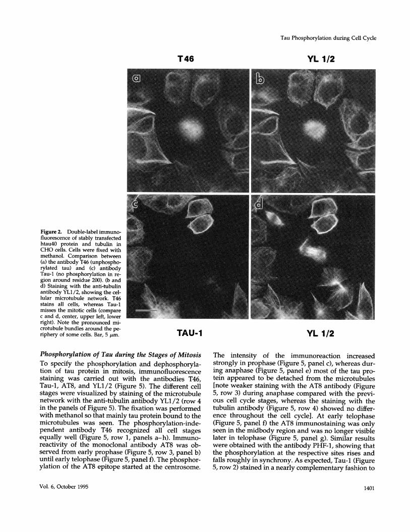
Proline-directed Phosphorylation of tauStably transfected CHO cells were examined bydouble-immunofluorescence for the uniformity ofexpression of htau40, the largest human isoform oftau (441 residues) using several anti-tau antibodies(Figure 1). The cells were fixed 36-48 h after platingon coverslips and stained with antibodies to tauprotein and tubulin. As seen in Figure 2a, the mono-clonal antibody T46, which reacts independently oftau's phosphorylation state, recognized all trans-fected cells equally well. The typical microtubularpattern was observed after staining with the anti-body YL1/2 (Figure 2b). No influence on the viabil-ity of these cells was noticed, although the expres-sion of tau slightly changed the morphology to amore rounded shape compared with the elongatedform of the control cells. Nontransfected CHO cells,which do not contain endogenous tau protein,showed no reaction with the tau antibodies exceptthe typical microtubular staining pattern with theantibody YL1/2 (our unpublished data). The anti-body Tau-1 (which binds optimally when the regionaround residue 200 is completely unphosphory-lated) recognized most transfected cells well (Figure2c). However, we also noted that some of the trans-fected cells showed no discernible staining with theantibody Tau-1. Comparison with the microtubulenetwork (Figure 2d) showed that this was correlatedwith the cell cycle stage: Tau-1 staining was neverobserved with transfected cells during mitosis (com-pare Figure 2, c and d, center), suggesting that thetransfected tau became phosphorylated at the Tau-1site during mitosis.A notable effect of transfection with tau is the for-
mation of microtubule bundles, often around the pe-riphery of the cell (e.g. Figure 2, a and c). This effecthas been observed with several MAPs (MAP2,
Vol. 6, October 1995 1399
U. Preuss et al.
N
- dephosphoryl. " "SP 202 SP 396
+ phosphoryl. TP 205 SP 404
AT$ + PHF-I +
MAP2c, and tau; Kanai et al., 1989; Weisshaar et al.,1992; Brandt and Lee, 1993; Berling et al., 1994) and canbe explained by the increased concentration and nu-cleation capacity of tau in the cytoplasm.To identify in vivo phosphorylation sites of tau pro-
tein, antibodies raised against Alzheimer PHFs thatrecognize specific phosphorylated epitopes of the tauprotein were used. The antibody AT8 is directedagainst phosphorylated Ser202/Thr205 and PHF-1recognizes phosphorylated Ser396/404. As a control,the cellular microtubules were visualized during allcell stages by staining with the anti-tubulin anti-body YL1 /2 (Figure 3, b and d). In contrast to Tau-1immunoreactivity, both phosphorylation-depen-dent antibodies AT8 and PHF-1 reacted stronglywith mitotic cells (compare Figure 3, a and b, with cand d). In nonmitotic cells, AT8 labeling was absentwhile PHF-1 showed a weak reaction (Figure 3c).These results indicate that the residues 202, 205, 396,and 404 of tau become preferentially phosphory-lated during mitosis, all of them within Ser/Thr-Promotifs, and that Ser396/Ser404 are phosphorylatedto a small extent even during interphase. Becausethe longest human tau isoform (htau40, 441 resi-dues) contains 17 Ser-Pro or Thr-Pro sites, many ofwhich are readily phosphorylatable, it is likely thateven more sites become phosphorylated that couldbe visualized by other antibodies (preliminary re-sults point in this direction).
Phosphorylation and Microtubule Binding of TauTau is normally thought to bind tightly to microtu-bules. However, in transfected cells this need not bethe case; moreover, phosphorylation is thought to re-duce the affinity of tau to microtubules. To check thispoint, we compared two fixation procedures. In thefirst set of experiments we used paraformaldehydefollowed by detergent permeabilization, whereby all
J Figure 1. Diagram of the largesthuman tau isoform (htau4O, 441residues) showing the epitopes oftau antibodies Tau-1 (unphospho-rylated region Prol89-Gly207),AT8 (phosphorylated Ser2O2,Thr2O5), PHF-1 (phosphory-
C lated Ser396, Ser4O4), and T46(Thr4O4-Leu441, phosphoryla-tion independent). This humantau isoform contains four imper-fect repeats near the carboxy ter-minus (shaded boxes, numbered1-4), which constitute the mi-crotubule-interacting domainand two 29 mer amino-terminalinserts (hatched boxes).
soluble and unsoluble proteins are cross-linked withinthe cells (Figure 3). In the second set, using methanolfixation (Figure 4), proteins were precipitated and res-olubilized during the following washing steps withaqueous solutions, whereby proteins can be partlyextracted (Fujiwara and Pollard, 1980; Melan andSluder, 1992). Because MAPs are thought to be lostfrom cells during detergent extraction (Schliwa et al.,1981; Chapin and Bulinski, 1994), we decided to usemethanol fixation to clearly demonstrate the colocal-ization of tau protein to microtubules in the trans-fected cells.The comparison allows one to distinguish between
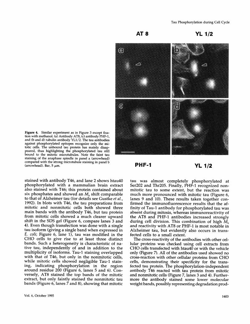
the two pools of tau: cytosolic and bound to microtu-bules. In cells fixed with paraformaldehyde (Figure 3),the bound fraction is largely concealed by the abun-dant unbound tau protein. Methanol fixation revealsmostly the colocalization of the bound protein withcellular microtubules (compare Figure 4, a and c).However, both of the fixation methods had no influ-ence on tau phosphoepitopes in general, becausebound and cytosolic tau protein showed similar reac-tivity with the phosphorylation-dependent antibodies.There was no Tau-1 staining observed in mitotic cells,while AT8 stained only mitotic cells and PHF-1stained mostly mitotic cells (Figure 4, a and c). Bothphosphorylation-dependent antibodies recognizedtau still bound to the mitotic spindle. This phenome-non occurred in all mitotic cells investigated. Thismeans that the phosphorylation of tau by proline-directed kinases does not necessarily lead to thedetachment of tau from microtubules (consistentwith the binding studies of Biernat et al., 1993). Thisobservation may be important with regard to Alz-heimer PHFs, which are thought to form via phos-phorylation of tau and release from microtubules(see DISCUSSION).
Molecular Biology of the Cell1400
Tau Phosphorylation during Cell Cycle
YL 1/2
Figure 2. Double-label immuno-fluorescence of stably transfectedhtau40 protein and tubulin inCHO cells. Cells were fixed withmethanol. Comparison between(a) the antibody T46 (unphospho-rylated tau) and (c) antibodyTau-1 (no phosphorylation in re-gion around residue 200). (b andd) Staining with the anti-tubulinantibody YL1/2, showing the cel-lular microtubule network. T46stains all cells, whereas Tau-1misses the mitotic cells (comparec and d, center, upper left, lowerright). Note the pronounced mi-crotubule bundles around the pe-riphery of some cells. Bar, 5 ,um.
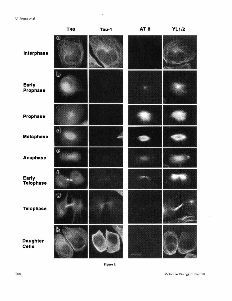
Phosphorylation of Tau during the Stages of MitosisTo specify the phosphorylation and dephosphoryla-tion of tau protein in mitosis, immunofluorescencestaining was carried out with the antibodies T46,Tau-1, AT8, and YL1/2 (Figure 5). The different cellstages were visualized by staining of the microtubulenetwork with the anti-tubulin antibody YLI /2 (row 4in the panels of Figure 5). The fixation was performedwith methanol so that mainly tau protein bound to themicrotubules was seen. The phosphorylation-inde-pendent antibody T46 recognized all cell stagesequally well (Figure 5, row 1, panels a-h). Immuno-reactivity of the monoclonal antibody AT8 was ob-served from early prophase (Figure 5, row 3, panel b)until early telophase (Figure 5, panel f). The phosphor-ylation of the AT8 epitope started at the centrosome.
TAU-1 YL 1/2
The intensity of the immunoreaction increasedstrongly in prophase (Figure 5, panel c), whereas dur-ing anaphase (Figure 5, panel e) most of the tau pro-tein appeared to be detached from the microtubules[note weaker staining with the AT8 antibody (Figure5, row 3) during anaphase compared with the previ-ous cell cycle stages, whereas the staining with thetubulin antibody (Figure 5, row 4) showed no differ-ence throughout the cell cycle]. At early telophase(Figure 5, panel f) the AT8 immunostaining was onlyseen in the midbody region and was no longer visiblelater in telophase (Figure 5, panel g). Similar resultswere obtained with the antibody PHF-1, showing thatthe phosphorylation at the respective sites rises andfalls roughly in synchrony. As expected, Tau-1 (Figure5, row 2) stained in a nearly complementary fashion to
Vol. 6, October 1995
T46
1401
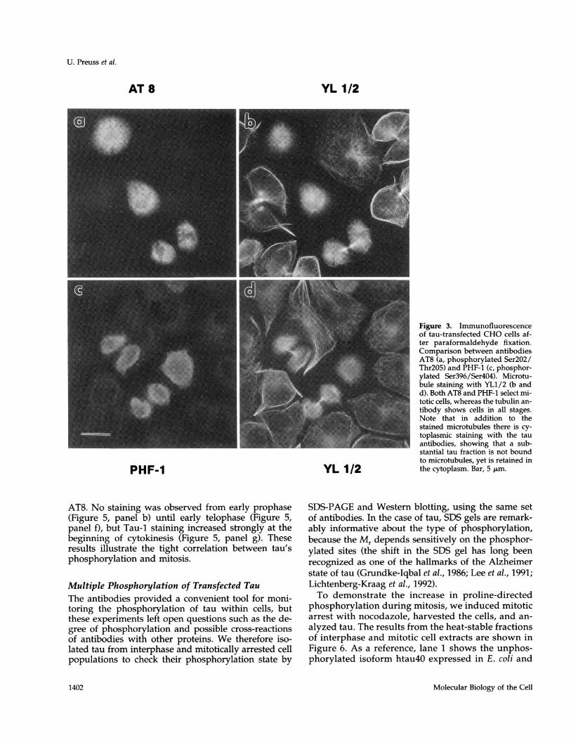
Figure 3. Immunofluorescenceof tau-transfected CHO cells af-ter paraformaldehyde fixation.Comparison between antibodiesAT8 (a, phosphorylated Ser2O2/Thr2O5) and PHF-1 (c, phosphor-ylated Ser396/Ser4O4). Microtu-bule staining with YL1/2 (b andd). Both AT8 and PHF-1 select mi-totic cells, whereas the tubulin an-tibody shows cells in all stages.Note that in addition to thestained microtubules there is cy-toplasmic staining with the tauantibodies, showing that a sub-stantial tau fraction is not boundto microtubules, yet is retained inthe cytoplasm. Bar, 5 ,um.
AT8. No staining was observed from early prophase(Figure 5, panel b) until early telophase (Figure 5,panel f), but Tau-1 staining increased strongly at thebeginning of cytokinesis (Figure 5, panel g). Theseresults illustrate the tight correlation between tau'sphosphorylation and mitosis.
Multiple Phosphorylation of Transfected TauThe antibodies provided a convenient tool for moni-toring the phosphorylation of tau within cells, butthese experiments left open questions such as the de-gree of phosphorylation and possible cross-reactionsof antibodies with other proteins. We therefore iso-lated tau from interphase and mitotically arrested cellpopulations to check their phosphorylation state by
SDS-PAGE and Western blotting, using the same setof antibodies. In the case of tau, SDS gels are remark-ably informative about the type of phosphorylation,because the Mr depends sensitively on the phosphor-ylated sites (the shift in the SDS gel has long beenrecognized as one of the hallmarks of the Alzheimerstate of tau (Grundke-Iqbal et al., 1986; Lee et al., 1991;Lichtenberg-Kraag et al., 1992).To demonstrate the increase in proline-directed
phosphorylation during mitosis, we induced mitoticarrest with nocodazole, harvested the cells, and an-alyzed tau. The results from the heat-stable fractionsof interphase and mitotic cell extracts are shown inFigure 6. As a reference, lane 1 shows the unphos-phorylated isoform htau4O expressed in E. coli and
Molecular Biology of the Cell
U. Preuss et al.
AT 8 YL 1/2
PHF-1 YL 1/2
1402
Tau Phosphorylation during Cell Cycle
Figure 4. Similar experiment as in Figure 3 except fixa-tion with methanol. (a) Antibody AT8, (c) antibody PHF-1,and (b and d) tubulin antibody YL1/2. The tau antibodiesagainst phosphorylated epitopes recognize only the mi-totic cells. The unbound tau protein has mainly disap-peared, thus highlighting the phosphorylated tau stillbound to the mitotic microtubules. Note the faint taustaining of the anaphase spindle in panel a (arrowhead)compared with the strong microtubule staining in panel b(arrowhead). Bar, 5 ,um.
stained with antibody T46, and lane 2 shows htau40phosphorylated with a mammalian brain extractalso stained with T46; this protein contained aboutsix phosphates and showed an Mr shift comparableto that of Alzheimer tau (for details see Gustke et al.,1992). In blots with T46, the tau preparations frommitotic and nonmitotic cells both showed threemain bands with the antibody T46, but tau proteinfrom mitotic cells showed a much clearer upwardshift in the SDS gel (Figure 6, compare lanes 3 and4). Even though transfection was done with a singletau isoform (giving a single band when expressed inE. coli; Figure 6, lane 1), tau was modified in theCHO cells to give rise to at least three distinctbands. Such a heterogeneity is characteristic of na-tive tau, independently of and in addition to themultiplicity of isoforms. Tau-1 staining overlappedwith that of T46, but only in the nonmitotic cells,while mitotic cells showed negligible Tau-1 stain-ing, indicating phosphorylation in the regionaround residue 200 (Figure 6, lanes 5 and 6). Con-versely, AT8 stained the top bands of the mitoticextract, but only faintly stained the nonmitotic taubands (Figure 6, lanes 7 and 8), showing that mitotic
tau was almost completely phosphorylated atSer2O2 and Thr2O5. Finally, PHF-1 recognized non-mitotic tau to some extent, but the reaction wasmuch more pronounced with mitotic tau (Figure 6,lanes 9 and 10). These results taken together con-firmed the immunofluorescence results that the af-finity of Tau-1 antibody for phosphorylated tau wasabsent during mitosis, whereas immunoreactivity ofthe AT8 and PHF-1 antibodies increased stronglyduring cell division. This combination of high Mrand reactivity with AT8 or PHF-1 is most notable inAlzheimer tau, but evidently also occurs in trans-fected cells to a small extent.The cross-reactivity of the antibodies with other cel-
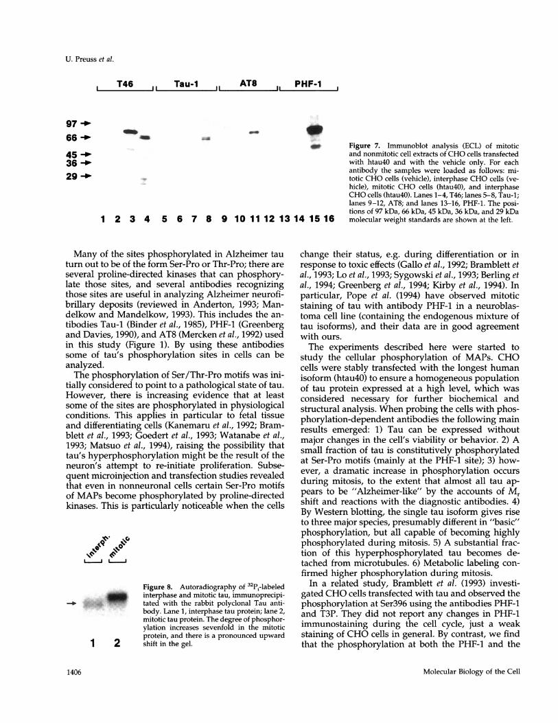
lular proteins was checked using cell extracts fromCHO cells transfected with htau40 or with the vehicleonly (Figure 7). All of the antibodies used showed nocross-reaction with other cellular proteins from CHOcells, demonstrating their specificity for the trans-fected tau protein. The phosphorylation-independentantibody T46 reacted with tau protein from mitoticand nonmitotic cells (Figure 7, lanes 3 and 4). Further-more the antibody stained some lower molecularweight bands, possibly representing degradation prod-
Vol. 6, October 1995
AT 8 YL 1/2
PHF-1 YL 1/2
1403
U. Preuss et al.
T46 Tau-1 AT 8
Interphase
EarlyProphase
Prophase
Metaphase
Anaphase
EarlyTelophase
Telophase
DaughterCells
YLI/2
1I
Figure 5.
Molecular Biology of the Cell1404

Tau Phosphorylation during Cell Cycle
ucts of the tau protein. However, no reaction was seenwith cellular proteins from mitotic and nonmitoticcells transfected with the vehicle only (Figure 7, lanes1 and 2). Tau-1 reacted as shown in Figure 6; there wasno Tau-1 staining in tau-transfected mitotic cells (Fig-ure 7, lane 7), whereas tau protein from transfectedinterphase cells was recognized (Figure 7, lane 8). NoTau-1 reaction was observed with vehicle-transfectedmitotic and nonmitotic cell extracts (Figure 7, lanes 5and 6). The phosphorylation-dependent antibodiesAT8 and PHF-1 reacted strongly with the tau fractionfrom mitotically arrested cells, showing the lowestelectrophoretic mobility (highest Mr) (Figure 7, lanes11 and 15). The antibody PHF-1 also stained variousbands of lower and higher molecular weight besidestau protein, the latter still being the most prominentband, however. Because no PHF-1 reactivity was ob-served in mitotic vehicle-transfected CHO cells (Fig-ure 7, lane 13), we assume that the additional lowmolecular bands represent degradation products ofthe transfected tau protein. The uppermost band ofapproximately 100 kDa probably corresponds to thedimerization of tau protein (Wille et al., 1992). AT8showed no reaction (Figure 7, lane 12) and PHF-1showed only a hardly discernible reaction (Figure 7,lane 16) with tau protein from transfected interphasecells.To see whether the increase in immunoreactivity of
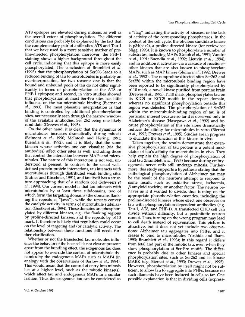
the phosphorylation-dependent antibodies AT8 andPHF-1 during mitosis correlates with a higher state ofphosphorylation of tau protein, interphase and mitot-ically arrested cells were incubated for several hoursin the presence of 32Pi. Labeled tau protein was iso-lated by immunoprecipitation and equal amounts ofinterphase and mitotic tau protein were loaded on anSDS gel. Figure 8 shows the autoradiography of thepolyacrylamide gel. Clear radioactive bands are seenin interphase and mitotic cells that correspond tohtau40 isolated from interphase and mitotic cells(compare Figure 7). Furthermore the labeled mitotictau protein showed a pronounced upward shift in thegel. To quantify the labeled interphase and mitotic tauprotein, bands were cut out of the gel and the radio-activity was determined by Cerenkov counting. Theobserved incorporation of 32Pi into tau protein was
Figure 5 (cont). Immunofluorescence staining of transfected CHOcells during mitosis. The fixation was done with methanol. Firstrow: antibody T46; second row: antibody Tau-1; third row: antibodyAT8; and fourth row: anti-tubulin antibody YL1/2. (a-h) Correlationbetween the appearance and disappearance of Tau-1 and AT8 stain-ing during mitosis. Note that the phosphorylation-independent an-tibody T46 stained all cells equally well throughout the cell cycle.Phosphoepitopes of tau begin to appear at early prophase at thecentrosome (b) and disappear at telophase (g). Bright AT8 stainingis seen during prophase and metaphase (c and d), whereas duringanaphase most tau seems to be detached from microtubules (noteweaker staining with AT8 in e compared with c and d). Bar, 2 ,um.
T46I ..
_~-
1 3 45 6
1 2 3 4 5 6
,, Tau-1 AT8 PHF-_.4-
7 8 9 10
Figure 6. Phosphorylation of tau in mitotic and nonmitotic-trans-fected CHO cells. The heat-stable fractions of the cell extracts wereincubated with different phosphorylation-dependent and indepen-dent antibodies. Lanes 1 and 2: recombinant htau40 before and afterphosphorylation with mouse brain extract. Lanes 3, 5, 7, and 9:interphase extracts of transfected CHO cells; lanes 4, 6, 8, and 10mitotic extracts of transfected CHO cells. Lanes 1-4 were probedwith antibody T46; lanes 5-6 with antibody Tau-1; lanes 7-8 withantibody AT8; and lanes 9-10 with antibody PHF-1. The arrowsindicate the shift in electrophoretic mobility of tau protein.
sevenfold higher in mitotic cells than in interphasecells.
DISCUSSION
The onset of mitosis correlates with a wave of phos-phorylation that affects, among others, MAPs andstructures involved in microtubule nucleation and dy-namics (Vandre et al., 1991; Buendia et al., 1992; Nigg,1993). Because the normal function of MAPs is tostabilize microtubules, the phosphorylation of MAPsis believed to be the mechanism by which their bind-ing to microtubules is weakened so that they becomemore dynamic and ready for the rearrangement of thecytoskeleton. The details of this hypothesis still haveto be worked out; at present, the MAPs involved, thephosphorylation sites affected, and the kinases areknown only in part or not at all. This illustrates theneed for a model system. Our approach was to use tauprotein transfected into CHO cells, for the followingreasons: 1) Tau is by far the best characterized MAP interms of phosphorylation sites and corresponding ki-nases and phosphatases; 2) a set of phosphorylation-dependent antibodies is available that enables one tofollow the state of phosphorylation in cells; 3) tau isdistinct from other MAPs yet shares homologous re-gions with some, especially the ubiquitous MAP4,implying similarity of function, e.g. in mitosis; 4) fi-nally, tau is interesting in itself because it forms thebasis for the PHFs of Alzheimer's disease (for reviewssee Mandelkow and Mandelkow, 1993; Kosik andGreenberg, 1994; Trojanowski and Lee, 1994). Thus, bystudying tau as an exogenous MAP in a dividing cellone hopes to gain information not only on the role ofMAP phosphorylation during the cell cycle, but alsoon the mechanisms underlying the pathology ofAlzheimer's disease.
Vol. 6, October 1995 1405
,, Tau-1 , AT8
97
66 --
45 -f36 -4'
29 -*
1 2 3 4 5 6 7 8 9 10 1112 1314 1516
Many of the sites phosphorylated in Alzheimer tauturn out to be of the form Ser-Pro or Thr-Pro; there are
several proline-directed kinases that can phosphory-late those sites, and several antibodies recognizingthose sites are useful in analyzing Alzheimer neurofi-brillary deposits (reviewed in Anderton, 1993; Man-delkow and Mandelkow, 1993). This includes the an-tibodies Tau-1 (Binder et al., 1985), PHF-1 (Greenbergand Davies, 1990), and AT8 (Mercken et al., 1992) usedin this study (Figure 1). By using these antibodiessome of tau's phosphorylation sites in cells can beanalyzed.The phosphorylation of Ser/Thr-Pro motifs was ini-
tially considered to point to a pathological state of tau.However, there is increasing evidence that at leastsome of the sites are phosphorylated in physiologicalconditions. This applies in particular to fetal tissueand differentiating cells (Kanemaru et al., 1992; Bram-blett et al., 1993; Goedert et al., 1993; Watanabe et al.,1993; Matsuo et al., 1994), raising the possibility thattau's hyperphosphorylation might be the result of theneuron's attempt to re-initiate proliferation. Subse-quent microinjection and transfection studies revealedthat even in nonneuronal cells certain Ser-Pro motifsof MAPs become phosphorylated by proline-directedkinases. This is particularly noticeable when the cells
L J L |J
Figure 8. Autoradiography of 32Pi-labeledinterphase and mitotic tau, immunoprecipi-
_. . tated with the rabbit polyclonal Tau anti-body. Lane 1, interphase tau protein; lane 2,mitotic tau protein. The degree of phosphor-ylation increases sevenfold in the mitoticprotein, and there is a pronounced upward
1 2 shift in the gel.
Figure 7. Immunoblot analysis (ECL) of mitoticand nonmitotic cell extracts of CHO cells transfectedwith htau4O and with the vehicle only. For eachantibody the samples were loaded as follows: mi-totic CHO cells (vehicle), interphase CHO cells (ve-hicle), mitotic CHO cells (htau4O), and interphaseCHO cells (htau4O). Lanes 1-4, T46; lanes 5-8, Tau-1;lanes 9-12, AT8; and lanes 13-16, PHF-1. The posi-tions of 97 kDa, 66 kDa, 45 kDa, 36 kDa, and 29 kDamolecular weight standards are shown at the left.
change their status, e.g. during differentiation or inresponse to toxic effects (Gallo et al., 1992; Bramblett etal., 1993; Lo et al., 1993; Sygowski et al., 1993; Berling etal., 1994; Greenberg et al., 1994; Kirby et al., 1994). Inparticular, Pope et al. (1994) have observed mitoticstaining of tau with antibody PHF-1 in a neuroblas-toma cell line (containing the endogenous mixture oftau isoforms), and their data are in good agreementwith ours.
The experiments described here were started tostudy the cellular phosphorylation of MAPs. CHOcells were stably transfected with the longest humanisoform (htau40) to ensure a homogeneous populationof tau protein expressed at a high level, which was
considered necessary for further biochemical andstructural analysis. When probing the cells with phos-phorylation-dependent antibodies the following mainresults emerged: 1) Tau can be expressed withoutmajor changes in the cell's viability or behavior. 2) Asmall fraction of tau is constitutively phosphorylatedat Ser-Pro motifs (mainly at the PHF-1 site); 3) how-ever, a dramatic increase in phosphorylation occurs
during mitosis, to the extent that almost all tau ap-pears to be "Alzheimer-like" by the accounts of Mrshift and reactions with the diagnostic antibodies. 4)By Western blotting, the single tau isoform gives riseto three major species, presumably different in "basic"phosphorylation, but all capable of becoming highlyphosphorylated during mitosis. 5) A substantial frac-tion of this hyperphosphorylated tau becomes de-tached from microtubules. 6) Metabolic labeling con-
firmed higher phosphorylation during mitosis.In a related study, Bramblett et al. (1993) investi-
gated CHO cells transfected with tau and observed thephosphorylation at Ser396 using the antibodies PHF-1and T3P. They did not report any changes in PHF-1immunostaining during the cell cycle, just a weakstaining of CHO cells in general. By contrast, we findthat the phosphorylation at both the PHF-1 and the
Molecular Biology of the Cell
U. Preuss et al.
T46 I PHF-1
1406
Tau Phosphorylation during Cell Cycle
AT8 epitopes are elevated during mitosis, as well asthe overall extent of phosphorylation. The differentconclusions can probably be explained by the fact thatthe complementary pair of antibodies AT8 and Tau-1that we have used is a more sensitive marker of pro-line-directed phosphorylation; moreover, the PHF-1staining shows a higher background throughout thecell cycle, indicating that this epitope is more easilyphosphorylated. The conclusion of Bramblett et al.(1993) that the phosphorylation of Ser396 leads to areduced binding of tau to microtubules is probably anoverinterpretation, for two reasons: one is that thebound and unbound pools of tau do not differ signif-icantly in terms of phosphorylation at the AT8 orPHF-1 epitopes; and second, in vitro studies showedthat phosphorylation at most Ser-Pro sites has littleinfluence on the tau-microtubule binding (Biernat etal., 1993). The most plausible interpretation is thatbinding is controlled by additional phosphorylationsites, not necessarily seen through the narrow windowof the available antibodies, Ser 262 being one likelycandidate (Drewes et al., 1995).On the other hand, it is clear that the dynamics of
microtubules increases dramatically during mitosis(Belmont et al., 1990; McIntosh and Hering, 1991;Buendia et al., 1992), and it is likely that the samekinases whose activities one can visualize (via theantibodies) affect other sites as well, including thosethat control the interaction between MAPs and micro-tubules. The nature of this interaction is not well un-derstood at present. In contrast to the usual well-defined protein-protein interactions, tau interacts withmicrotubules through distributed weak binding sites(Butner and Kirschner, 1991), and tau itself has a struc-ture approaching that of a random coil (Schweers etal., 1994). Our current model is that tau interacts withmicrotubules by at least three subdomains, two ofwhich form the targeting domains (the domains flank-ing the repeats as "jaws"), while the repeats conveythe catalytic activity in terms of microtubule stabiliza-tion (Gustke et al., 1994). These domains are phosphor-ylated by different kinases, e.g., the flanking regionsby proline-directed kinases, and the repeats by pllOmark. It therefore is likely that regulation could occuron the level of targeting and/or catalytic activity. Therelationship between these functions still needs fur-ther clarification.Whether or not the transfected tau molecules influ-
ence the behavior of the host cell is not clear at present;apart from the bundling effect, the exogenous tau doesnot appear to override the control of microtubule dy-namics by the endogenous MAPs such as MAP4 (inanalogy with the observations of Barlow et al., 1994).This would mean that the control of entry into mitosislies at a higher level, such as the mitotic kinase(s),which affect tau and endogenous MAPs in a similarfashion. Thus the exogenous tau can be considered as
a "flag" indicating the activity of kinases, or the lackof activity of the corresponding phosphatases. In thecontext of the cell cycle, the obvious candidate kinaseis p34(cdc2), a proline-directed kinase (for review seeNigg, 1993). It is known to phosphorylate a number ofsubstrates, including MAPs (Gotoh et al., 1991; Vandreet al., 1991; Buendia et al., 1992; Lieuvin et al., 1994),and in addition it activates-via a cascade of reactions-other kinases that are also known to phosphorylateMAPs, such as MAP kinase (Shiina et al., 1992; Dreweset al., 1992). The nonproline-directed sites Ser262 andSer356 within the microtubule binding region havebeen reported to be significantly phosphorylated bypl10 mark, a novel kinase purified from porcine brain(Drewes et al., 1995). P110 mark phosphorylates tau onits KIGS or KCGS motifs in the repeat domain,whereas no significant phosphorylation outside thisregion was detected. The phosphorylation of Ser262within the microtubule-binding region of tau is ofparticular interest because so far it is observed only inAlzheimer's disease (Hasegawa et al., 1992) and be-cause phosphorylation of this site alone dramaticallyreduces the affinity for microtubules in vitro (Biernatet al., 1992; Drewes et al., 1995). Studies are in progressto elucidate the function of this kinase in vivo.Taken together, the results demonstrate that exten-
sive phosphorylation of tau protein is a potent mod-ulator of tau's affinity for microtubules in vivo. Theyhelp explain the high degree of phosphorylation offetal tau (Bramblett et al., 1993) because during embry-ogenesis nerve cells still undergo mitosis. Further-more, this study supports a hypothesis stating that thepathological phosphorylation of Alzheimer tau maybe the result of the neuron's attempt to respond tosome insult, such as oxidative stress, ischaemia,3-amyloid toxicity, or another factor. The neuron be-haves as if it wanted to divide, thus turning on theappropriate phosphorylation cascades, including theproline-directed kinases whose effect one observes ontau with phosphorylation-dependent antibodies (e.g.Tau-1, AT8, and PHF-1). A transfected CHO cell candivide without difficulty, but a postmitotic neuroncannot. Thus, turning on the wrong program may leadto cell death instead of rejuvenation. This picture isattractive, but it does not yet include two observa-tions: Alzheimer tau aggregates into PHFs, and itceases to bind to microtubules (Yoshida and Ihara,1993; Bramblett et al., 1993); in this regard it differsfrom fetal and part of the mitotic tau, even when theyshow phosphorylation at Ser-Pro motifs. The differ-ence is probably due to other kinases and specialphosphorylation sites, such as Ser262 and its kinaseMARK (e.g. Biernat et al., 1993; Drewes et al., 1995).However, phosphorylation by itself might not be suf-ficient to allow tau to aggregate into PHFs, because nosuch filaments have been induced in cells so far. Onepossible explanation is that in dividing cells (express-
Vol. 6, October 1995 1407
U. Preuss et al.
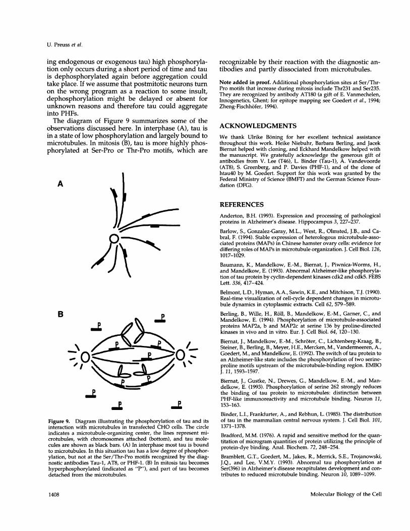
ing endogenous or exogenous tau) high phosphoryla-tion only occurs during a short period of time and tauis dephosphorylated again before aggregation couldtake place. If we assume that postmitotic neurons turnon the wrong program as a reaction to some insult,dephosphorylation might be delayed or absent forunknown reasons and therefore tau could aggregateinto PHFs.The diagram of Figure 9 summarizes some of the
observations discussed here. In interphase (A), tau isin a state of low phosphorylation and largely bound tomicrotubules. In mitosis (B), tau is more highly phos-phorylated at Ser-Pro or Thr-Pro motifs, which are
A
ID
-op
Figure 9. Diagram illustrating the phosphorylation of tau and itsinteraction with microtubules in transfected CHO cells. The circleindicates a microtubule-organizing center, the lines represent mi-crotubules, with chromosomes attached (bottom), and tau mole-cules are shown as black bars. (A) In interphase most tau is boundto microtubules. In this situation tau has a low degree of phosphor-ylation, but not at the Ser/Thr-Pro motifs recognized by the diag-nostic antibodies Tau-1, AT8, or PHF-1. (B) In mitosis tau becomeshyperphosphorylated (indicated as "P"), and part of tau becomesdetached from the microtubules.
recognizable by their reaction with the diagnostic an-tibodies and partly dissociated from microtubules.
Note added in proof. Additional phosphorylation sites at Ser/Thr-Pro motifs that increase during mitosis include Thr231 and Ser235.They are recognized by antibody AT180 (a gift of E. Vanmechelen,Innogenetics, Ghent; for epitope mapping see Goedert et al., 1994;Zheng-Fischhofer, 1994).
ACKNOWLEDGMENTSWe thank Ulrike Boning for her excellent technical assistancethroughout this work. Heike Niebuhr, Barbara Berling, and JacekBiemat helped with cloning, and Eckhard Mandelkow helped withthe manuscript. We gratefully acknowledge the generous gift ofantibodies from V. Lee (T46), L. Binder (Tau-1), A. Vandevoorde(AT8), S. Greenberg, and P. Davies (PHF-1), and of the clone ofhtau40 by M. Goedert. Support for this work was granted by theFederal Ministry of Science (BMFT) and the German Science Foun-dation (DFG).
REFERENCESAnderton, B.H. (1993). Expression and processing of pathologicalproteins in Alzheimer's disease. Hippocampus 3, 227-237.
Barlow, S., Gonzalez-Garay, M.L., West, R., Olmsted, J.B., and Ca-bral, F. (1994). Stable expression of heterologous microtubule-asso-ciated proteins (MAPs) in Chinese hamster ovary cells: evidence fordiffering roles of MAPs in microtubule organization. J. Cell Biol. 126,1017-1029.
Baumann, K., Mandelkow, E.-M., Biemat, J., Piwnica-Worms, H.,and Mandelkow, E. (1993). Abnormal Alzheimer-like phosphoryla-tion of tau protein by cyclin-dependent kinases cdk2 and cdk5. FEBSLett. 336, 417-424.
Belmont, L.D., Hyman, A.A., Sawin, K.E., and Mitchison, T.J. (1990).Real-time visualization of cell-cycle dependent changes in microtu-bule dynamics in cytoplasmic extracts. Cell 62, 579-589.
Berling, B., Wille, H., Roll, B., Mandelkow, E.-M., Gamer, C., andMandelkow, E. (1994). Phosphorylation of microtubule-associatedproteins MAP2a, b and MAP2c at serine 136 by proline-directedkinases in vivo and in vitro. Eur. J. Cell Biol. 64, 120-130.
Biemat, J., Mandelkow, E.-M., Schroter, C., Lichtenberg-Kraag, B.,Steiner, B., Berling, B., Meyer, H.E., Mercken, M., Vandermeeren, A.,Goedert, M., and Mandelkow, E. (1992). The switch of tau protein toan Alzheimer-like state includes the phosphorylation of two serine-proline motifs upstream of the microtubule-binding region. EMBOJ. 11, 1593-1597.
Biernat, J., Gustke, N., Drewes, G., Mandelkow, E.-M., and Man-delkow, E. (1993). Phosphorylation of serine 262 strongly reducesthe binding of tau protein to microtubules: distinction betweenPHF-like immunoreactivity and microtubule binding. Neuron 11,153-163.
Binder, L.I., Frankfurter, A., and Rebhun, L. (1985). The distributionof tau in the mammalian central nervous system. J. Cell Biol. 101,1371-1378.
Bradford, M.M. (1976). A rapid and sensitive method for the quan-titation of microgram quantities of protein utilizing the principle ofprotein-dye binding. Anal. Biochem. 72, 248-254.
Bramblett, G.T., Goedert, M., Jakes, R., Merrick, S.E., Trojanowski,J.Q., and Lee, V.M.Y. (1993). Abnormal tau phosphorylation atSer(396) in Alzheimer's disease recapitulates development and con-tributes to reduced microtubule binding. Neuron 10, 1089-1099.
Molecular Biology of the Cell
0000-0.
00000-
1408
Tau Phosphorylation during Cell Cycle
Brandt, R., and Lee, G. (1993). Functional organization of microtu-bule-associated protein tau: identification of regions which affectmicrotubule growth, nucleation, and bundle formation in vitro. J.Biol. Chem. 268, 3414-3419.
Brugg, B., and Matus, A. (1991). Phosphorylation determines thebinding of microtubule-associated protein-2 (MAP2) to microtu-bules in living cells. J. Cell Biol. 114, 735-743.
Buendia, B., Draetta, G., and Karsenti, E. (1992). Regulation of themicrotubule nucleating activity of centrosomes in Xenopus egg ex-tracts: role of cyclin A-associated protein kinase. J. Cell Biol. 116,1431-1442.
Butner, K.A., and Kirschner, M.W. (1991). Tau-protein binds tomicrotubules through a flexible array of distributed weak sites. J.Cell Biol. 115, 717-730.
Chapin, S.J., and Bulinski, J.C. (1991). Non-neuronal 210 kD Mrmicrotubule-associated protein (MAP4) contains a domain homol-ogous to the microtubule-binding domains of neuronal MAP2 andtau. J. Cell Sci. 98, 27-36.
Chapin, S.J., and Bulinski, J.C. (1992). Microtubule stabilization byassembly-promoting microtubule-associated proteins: a repeat per-formance. Cell Motil. Cytoskeleton 23, 236-243.
Chapin, S.J., and Bulinski, J.C. (1994). Cellular microtubules heter-ogenous in their content of microtubule-associated protein-4(MAP4). Cell Motil. Cytoskeleton 27, 133-149.
Drewes, G., Lichtenberg-Kraag, B., Doring, F., Mandelkow, E.-M.,Biernat, J., Goris, J., Doree, M., and Mandelkow, E. (1992). Mitogen-activated protein (MAP) kinase transforms tau protein into anAlzheimer-like state. EMBO J. 11, 2131-2138.
Drewes, G., Trinczek, B., Illenberger, S., Biernat, J., Schmitt-Ulms,G., Meyer, H.E., Mandelkow, E.-M., and Mandelkow, E. (1995).MAP/microtubule affinity-regulating kinase (pllOmark): a novelprotein kinase that regulates tau-microtubule interactions and dy-namic instability by phosphorylation at the Alzheimer-specific siteserine 262. J. Biol. Chem. 270, 7679-7688.
Drubin, D., and Kirschner, M. (1986). Tau protein function in livingcells. J. Cell Biol. 103, 2739-2746.
Fujiwara, K., and Pollard, T.D. (1980). Techniques for localizingcontractile proteins with fluorescent antibodies. In: Current Topicsin Developmental Biology, vol. 14, ed. M. Friedlander, New York:Academic Press, 271-296.
Gallo, J., Hanger, D., Twist, E., Kosik, K., and Anderton, B.H. (1992).Expression and phosphorylation of a 3-repeat isoform of tau intransfected nonneuronal cells. Biochem. J. 286, 399-404.
Goedert, M., Spillantini, M., Jakes, R., Rutherford, D., and Crowther,R.A. (1989). Multiple isoforms of human microtubule-associatedprotein-tau: sequences and localization in neurofibrillary tangles ofAlzheimer's disease. Neuron 3, 519-526.
Goedert, M., Jakes, R., Crowther, R.A., Six, J., Lubke, U., Vander-meeren, M., Cras, P., Trojanowski, J.Q., and Lee, V.M.Y. (1993). Theabnormal phosphorylation of tau protein at Ser202 in Alzheimer'sdisease recapitulates phosphorylation during development. Proc.Natl. Acad. Sci. USA 90, 5066-5070.
Goedert, M., Jakes, R., Crowther, R.A., Cohen, P., Vanmechelen, E.,Vandermeeren, M., and Cras, P. (1994). Epitope mapping of mono-clonal antibodies to the paired helical filaments of Alzheimers dis-ease: identification of phosphorylation sites in tau protein. Biochem.J. 301, 871-877.
Gotoh, Y., Nishida, E., Matsuda, S., Shiina, N., Kosako, H., Shio-kawa, K., Akiyama, T., Ohta, K., and Sakai, H. (1991). In vitro effectson microtubule dynamics of purified Xenopus M-phase-activatedMAP kinase. Nature 349, 251-254.
Greenberg, S.G., and Davies, P. (1990). A preparation of Alzheimerpaired helical filaments that displays distinct tau-proteins by poly-acrylamide-gel electrophoresis. Proc. Natl. Acad. Sci. USA 87, 5827-5831.
Greenberg, S.M., Koo, E.H., Selkoe, D.J., Qiu, W.Q., and Kosik, K.S.(1994). Secreted beta-amyloid precursor protein stimulates mitogen-activated protein kinase and enhances tau phosphorylation. Proc.Natl. Acad. Sci. USA 91, 7104-7108.
Grundke-Iqbal, I., Iqbal, K., Tung, Y., Quinlan, M., Wisniewski, H.,and Binder, L. (1986). Abnormal phosphorylation of the microtu-bule-associated protein tau in Alzheimer cytoskeletal pathology.Proc. Natl. Acad. Sci. USA 83, 4913-4917.
Gustke, N., Steiner, B., Mandelkow, E.-M., Biemat, J., Meyer, H.E.,Goedert, M., and Mandelkow, E. (1992). The Alzheimer-like phos-phorylation of tau protein reduces microtubule binding and in-volves Ser-Pro and Thr-Pro motifs. FEBS Lett. 307, 199-205.
Gustke, N., Trinczek, B., Biernat, J., Mandelkow, E.-M., and Man-delkow, E. (1994). Domains of tau protein and interactions withmicrotubules. Biochemistry 33, 9511-9522.
Hagestedt, T., Lichtenberg, B., Wille, H., Mandelkow, E.-M., andMandelkow, E. (1989). Tau protein becomes long and stiff uponphosphorylation: correlation between paracrystalline structure anddegree of phosphorylation. J. Cell Biol. 109, 1643-1651.
Hanger, D., Hughes, K., Woodgett, J., Brion, J., and Anderton, B.(1992). Glycogen-synthase kinase-3 induces Alzheimer's disease-like phosphorylation of tau: generation of paired helical filamentepitopes and neuronal localization of the kinase. Neurosci. Lett. 147,58-62.
Hasegawa, M., Morishima-Kawashima, M., Takio, K., Suzuki, M.,Titani, K., and Ihara, Y. (1992). Protein sequence and mass spectro-metric analyses of tau in the Alzheimer's disease brain. J. Biol.Chem. 267, 17047-17054.
Himmler, A., Drechsel, D., Kirschner, M., and Martin, D. (1989). Tauconsists of a set of proteins with repeated C-terminal microtubule-binding domains and variable N-terminal domains. Mol. Cell Biol.9, 1381-1388.
Hirokawa, N. (1994). Microtubule organization and dynamics de-pendent on microtubule-associated proteins. Curr. Opin. Cell Biol.6, 74-81.
Ishiguro, K., Omori, A., Sato, K.,'Tomizawa, K., Imahori, K., andUchida, T. (1991). A serine threonine proline kinase-activity is in-cluded in the tau-protein kinase.fraction forming a paired helicalfilament epitope. Neurosci.-Lett. 128, 195-198.
Kanai, Y., Takemura, R., Oshima, T., Mori, H., Ihara, Y., Yanagi-sawa, M., Masaki, T., and Hirokawa, N. (1989). Expression of mul-tiple tau isoforms and microtubule bundle formation in fibroblaststransfected with a single tau cDNA. J. Cell Biol. 109, 1173-1184.
Kanemaru, K., Takio, K., Miura, R., Titani, K., and Ihara, Y. (1992).Fetal-type phosphorylation of the tau in paired helical filaments. J.Neurochem. 58, 1667-1675.
Kilmartin, J.V., Wright, B., and Milstein, C. (1982). Rat monoclonalantitubulin antibodies derived by using a new nonsecreting rat cellline. J. Cell Biol. 93, 576-582.
Kirby, B.A., Merril, C.R., Ghanbari, H., and Wallace, W.C. (1994).Heat-shock proteins protect against stress-related phosphorylationof tau in neuronal PC12 cells that have acquired thermotolerance. J.Neurosci. 14, 5687-5693.
Kosik, K., Orecchio, L., Binder, L., Trojanowski, J., Lee, V., and Lee,G. (1988). Epitopes that span the tau molecule are shared withpaired helical filaments. Neuron 1, 817-825.
Vol. 6, October 1995 1409
U. Preuss et al.
Kosik, K.S., and Greenberg, S.M. (1994). Tau protein and Alzheimerdisease. In: Alzheimer Disease, ed. R. Terry, R. Katzman, and K.Bick, New York: Raven Press, 335-344.Lang, E., Szendrei, G.I., Lee, V.M.Y., and Otvos, L. (1992). Immu-nological and conformation characterization of a phosphorylatedimmunodominant epitope on the paired helical filaments found inAlzheimer's disease. Biochem. Biophys. Res. Commun. 187, 783-790.Ledesma, M.D., Correas, I., Avila, J., and Diaz-Nido, J. (1992). Im-plication of brain cdc2 and MAP2 kinases in the phosphorylation oftau protein in Alzheimer's disease. FEBS Lett. 308, 218-224.Lee, G., Cowan, N., and Kirschner, M. (1988). The primary structureand heterogeneity of tau protein from mouse brain. Science 239,285-288.Lee, V.M.Y., Balin, B.J., Otvos, L., and Trojanowski, J.Q. (1991). A68:a major subunit of paired helical filaments and derivatized forms ofnormal tau. Science 251, 675-678.Lee, G. (1993). Non-motor microtubule-associated proteins. Curr.Opin. Cell Biol. 5, 88-94.Lichtenberg-Kraag, B., Mandelkow, E.-M., Biernat, J., Steiner, B.,Schroter, C., Gustke, N., Meyer, H.E., and Mandelkow, E. (1992).Phosphorylation-dependent interaction of neurofilament antibodieswith tau protein: epitopes, phosphorylation sites, and relationshipwith Alzheimer tau. Proc. Natl. Acad. Sci. USA 89, 5384-5388.Lieuvin, A., Labbe, J.-C., Doree, M., and Job, D. (1994). Intrinsicmicrotubule stability in interphase cells. J. Cell Biol. 124, 985-996.Lo, M.M.S., Fieles, A.W., Norris, T.E., Dargis, P.G., Caputo, C.B.,Scott, C.W., Lee, V.M.Y., and Goedert, M. (1993). Human tau iso-forms confer distinct morphological and functional properties tostably transfected fibroblasts. Mol. Brain Res. 20, 209-220.Mandelkow, E.-M., Drewes, G., Biernat, J., Gustke, N., Van Lint, J.,Vandenheede, J.R., and Mandelkow, E. (1992). Glycogen synthasekinase-3 and the Alzheimer-like state of microtubule-associatedprotein tau. FEBS Lett. 314, 315-321.Mandelkow, E.-M., and Mandelkow, E. (1993). Tau as a marker forAlzheimer's disease. Trends Biol. Sci. 18, 480-483.Matsuo, E.S., Shin, R.W., Billingsley, M.L., Vandevoorde, A., Ocon-nor, M., Trojanowski, J.Q., and Lee, V.M.Y. (1994). Biopsy-derivedadult human brain tau is phosphorylated at many of the same sitesas Alzheimer's disease paired helical filament tau. Neuron 13, 989-1002.McIntosh, J.R., and Hering, G.E. (1991). Spindle fiber action andchromosome movement. Annu. Rev. Cell Biol. 7, 403-426.Melan, M.A., and Sluder, G. (1992). Redistribution and differentialextraction of soluble proteins in permeabilized cultured cells. J. CellSci. 101, 731-743.Mercken, M., Vandermeeren, M., Lubke, U., Six, J., Boons, J., Van deVoorde, A., Martin, J.-J., and Gheuens, J. (1992). Monoclonal anti-bodies with selective specificity for Alzheimer tau are directedagainst phosphatase-sensitive epitopes. Acta Neuropathol. 84, 265-272.Morishima-Kawashima, M., Hasegawa, M., Takio, K., Suzuki, M.,Yoshida, H., Titani, K., and Ihara, Y. (1995). Proline-directed andnon-proline-directed phosphorylation of PHF-tau. J. Biol. Chem.270, 823-829.Nigg, E. (1993). Cellular substrates of p34(cdc2) and its companioncyclin-dependent kinases. Trends Cell Biol. 3, 296-301.Otvos, L., Feiner, L., Lang, E., Szendrei, G., Goedert, M., and Lee,V.M.Y. (1994). Monoclonal antibody PHF-1 recognizes tau proteinphosphorylated at serine residue 396 and residue 404. J. Neurosci.Res. 39, 669-673.
Paudel, H., Lew, J., Ali, Z., and Wang, J. (1993). Brain proline-directed protein kinase phosphorylates tau on sites that are abnor-mally phosphorylated in tau associated with Alzheimer's pairedhelical filaments. J. Biol. Chem. 268, 23512-23518.
Pope, W.B., Lambert, M.P., Leypold, B., Seupaul, R., Sletten, L.,Krafft, G., and Klein, W.L. (1994). Microtubule-associated protein-tau is hyperphosphorylated during mitosis in the human neuroblas-toma cell-line SH-SY5Y. Exp. Neurol. 126, 185-194.
Schliwa, M., Euteneuer, U., Bulinski, J.C., and Izant, J. (1981). Cal-cium lability of cytoplasmic microtubules and their modulation bymicrotubule-associated proteins. Proc. Natl. Acad. Sci. USA 78,1037-1041.Schweers, O., Schonbrunn-Hanebeck, E., Marx, A., and Mandelkow,E. (1994). Structural studies of tau protein and Alzheimer pairedhelical filaments show no evidence for ,B structure. J. Biol. Chem.269, 24290-24297.
Shiina, N., Moriguchi, T., Ohta, K., Gotoh, Y., and Nishida, E. (1992).Regulation of a major microtubule-associated protein by MPF andMAP kinase. EMBO J. 11, 3977-3984.
Studier, W.F., Rosenberg, A.H., Dunn, J.J., and Dubendorff, J.W.(1990). Use of T7 RNA polymerase to direct the expression of clonedgenes. Methods Enzymol. 185, 60-89.
Sygowski, L.A., Fieles, A.W., Lo, M.M.S., Scott, C.W., and Caputo,C.B. (1993). Phosphorylation of tau protein in tau-transfected 3T3cells. Mol. Brain Res. 20, 221-228.
Szendrei, G.I., Lee, V.M.-Y., and Otvos, L. (1993). Recognition of theminimal epitope of monoclonal antibody Tau-1 depends upon thepresence of a phosphate group but not its location. J. Neurosci. Res.34, 243-249.
Trojanowski, J.Q., and Lee, V.M.Y. (1994). Paired helical filament tauin Alzheimer's disease: the kinase connection. Am. J. Pathol. 144,449-453.
Vandre, D., Centonze, V., Peloquin, J., Tombes, R., and Borisy, G.G.(1991). Proteins of the mammalian mitotic spindle: phosphoryla-tion-dephosphorylation of MAP-4 during mitosis. J. Cell Sci. 98,577-588.
Vulliet, R., Halloran, S., Braun, R., Smith, A., and Lee, G. (1992).Proline-directed phosphorylation of human tau protein. J. Biol.Chem. 267, 22570-22574.
Watanabe, A., Hasegawa, M., Suzuki, M., Takio, K., Morishima-Kawashima, M., Titani, K., Arai, T., Kosik, K.S., and Ihara, Y. (1993).In vivo phosphorylation sites in fetal and adult rat tau. J. Biol.Chem. 268, 25712-25717.Weisshaar, B., Doll, T., and Matus, A. (1992). Reorganization of themicrotubular cytoskeleton by embryonic microtubule-associatedprotein 2 (MAP2c). Development 116, 1151-1161.
West, R.R., Tenbarge, K.M., and Olmsted, J.B. (1991). A model formicrotubule-associated protein-4 structure: domains defined bycomparisons of human, mouse, and bovine sequences. J. Biol. Chem.266, 21886-21896.
Wille, H., Drewes, G., Biernat, J., Mandelkow, E.-M., and Man-delkow, E. (1992). Alzheimer-like paired helical filaments and anti-parallel dimers formed from microtubule-associated protein tau invitro. J. Cell Biol. 118, 573-584.
Yoshida, H., and Ihara, Y. (1993). Tau in paired helical filaments isfunctionally distinct from fetal tau: assembly incompetence ofpaired helical filament tau. J. Neurochem. 61, 1183-1186.
Zheng-Fischhofer, Q. (1994). Mapping of epitopes of phosphoryla-tion-dependent antibodies on tau protein. Diploma Thesis. Ham-burg, Germany: University of Hamburg.
Molecular Biology of the Cell1410