Embed Size (px)
Citation preview
Cell Communication
Teresa Burke Westford Academy
30 Patten Road Westford, MA 01886
[email protected] Mentored by William Cruikshank
Boston University Medical School-Immunology Funded through the American Association of Immunologists
“Can You Hear Me Now?” Cell communication
Dawn Martell Wilmington High School
159 Church Street Wilmington, MA 01887
[email protected] Mentored by Dr. William Cruikshank,
Boston University Medical School-Immunology Funded through the American Association of Immunologists
2
Table of Contents Teacher Preparation Section Overview……………………………………………………………………………………….. 4 Massachusetts Curriculum Frameworks……………………………………………………….. 5 Science Background Column Chromatography………………………………………………………………. 6 Ouchterlony Technique………………………………………………………………… 7 Immune Response……………………………………………………………………… 7 Student Outcomes……………………………………………………………………………… 8 Learning Objectives..................................................................................................................... 9 Time Requirements…………………………………………………………………………...... 9 Advance Preparation and Materials……………………………………………………………. 9 Total Costs……………………………………………………………………………………....12 Possible Substitutions……………………………………………………………………….......13 Safety…………………………………………………………………………………………....13 Student Prior Knowledge and Skills Misconception of the Immune System………………………………………………… 13 Student Expectation……………………………………………………………………………. 14 Anticipated Results Immunoassay…………………………………………………………………………... 14 Results of Plate A & Plate B……………………………..…………..………………… 15 Classroom Discussion………………………………………………………………………….. 16 Assessment……………………………………………………………………………………... 17 Student Section “Can You Hear Me Now” Laboratory Procedure Column Chromatography………………………………………………………………. 18 Ouchterlony Plates…………………………………….……………………………….. 19 Animated Drawing……………………………………………………………………………... 23 Animated Drawing Example…………………………………………………………………… 24 Animated Drawing Rubric……………………………………………………………………... 25 Biology Laboratory Rubric…………………………………………………………………….. 26 Pre/Post Quiz……………………………………………………………………………..……. 27 CD Review Questions………………………………………………………………………….. 31 References……………………………………………………………………….……………... 33
3
Teacher Preparation
Section
4
Overview
Cell communication and homeostasis is an integral process by which the body maintains functions. Sustaining life depends on a quick and effective response from the immune system in recognition and reaction to foreign invaders. In order to maintain homeostasis of the immune system, several positive and negative feedback loops are constantly regulating the human body. In this laboratory investigation, students will have the opportunity to learn how the immune system builds its arsenal to defend itself from foreign invaders. Students will also gain the understanding of how the lymphatic System facilitates immune responses. A particular focus in this unit will include the origin and role of APC (Antigen-Presenting Cells) and chemokines. Specific chemokines attract, activate, and promote the proliferation of helper T cells. These newly produced helper T class in turn proliferate into memory T cells through a positive feedback loop. Moreover, the activation process also helps stimulate other lymphocytes called B cells to manufacture antibodies which can locate and help to destroy foreign antigens. Students will observe and analyze the effects of chemokines on cell migration. Students will understand cell interaction and learn how the immune system may be activated through various pathogenic processes. The unit will provide students with the opportunity to better understand the importance of a healthy life style. Working in groups of two, students will build a size exclusion chromatography column to separate simulated blood suspension and then collect the elusion. Students will also prepare and run their own Ouchterlony plates and become more proficient in various other laboratory skills. These skills include micropipetting and volume measurements. The lessons in this unit can be adapted for any level of biology. We have specifically designed the lessons to be used in AP biology and mainstream biology classrooms. This unit has also been designed to address the established guidelines for the AP biology program and the Massachusetts Curriculum Frameworks (MCAS) in biology.
5
Massachusetts Curriculum Frameworks: Content Standard Standard 4-Anatomy and Physiology: 2.6 Describe the cell cycle and the process of Mitosis in the formation of new cells.
• A brief informational session on the cloning, proliferation of new and the activation of APCs/macrophages and helper T cells.
4.7 Recognize that communication among cells is required for coordination of body functions.
• Discussion session on cell signaling 4.8 Recognize that the body’s systems interact to maintain homeostasis. Describe the basic function of a physiological feedback loops.
• Discussion on the importance of feedback loops to maintain homeostasis in the body will take place
Scientific Inquiry Skills Standard*: SIS1. Make observations, raise questions, and formulate hypotheses. SIS2. Design and conduct scientific investigations. SIS3. Analyze and interpret results of scientific investigations. SIS4. Communicate and apply the results of scientific investigations. *For specifics see Massachusetts Curriculum Frameworks Learning Standards
6
Science Background: Chromatography is a process used to separate molecules. There are many types of chromatography, such as gas, ion exchange, electrophoresis, or paper. Below is a brief description of the more common types of chromatography techniques used. These descriptions can be used as an introduction for the students before beginning the laboratory process of chromatography in the separation of simulated human blood. Gas chromatography is usually the separation of a liquid or polymer using an un-reactive gas, which is contained inside a glass or metal tube (column). The gas is separated by different rates due to its chemical and physical properties and how these gases interact with the stationary phase (column). These interactions cause the gases to exit the column at different rates, which is electronically detected determining the amount of time each component reaches the outlet. Ion-exchange chromatography is used for the separation of the most charged particles or ions such as such as in proteins, nucleic acids or amino acids. A resin (stationary phase) is used with this type of chromatography, depending on the resin properties, the ion molecules will either stick to the resin or elude from the column. Ion exchange is very useful for environmental studies and water quality analysis. Paper chromatography is the separation of mixtures, such as, pigments, inks, marker, pens or even amino acids into their constituents. The mixtures are placed within a solvent and move by capillary action up the paper either due to their solubility of the solvent or paper. Measurements are taken for the distance each component has traveled and calculations are done finding the Rf (rate of flow) value. Another type of process to separate molecules is electrophoresis. Electrophoresis uses an electrical current to separate molecules of different electrical charge and size. These molecules are located within an agarose gel. These molecules migrate through the gel matrix from the negatively charge electrode towards the positively charged electrode. Those molecules that are smaller and have a greater positive charge will migrate faster than those that are larger and have less of a positive charge. The different molecules are seen as bands in the gel matrix. The type of chromatography used in this exploration is size exclusion chromatography. The size exclusion process uses a column, which is packed with a polyester medium in which particles filter through a column according to their size. As the molecules travel over the polyester fiber the smaller molecules get “caught up” in this medium and the larger molecules travel over the length and elude out of the column together.
7
Ouchterlony Technique The Ouchterlony technique is an immunoprecipitation assay in which chemicals migrate through a medium and when these chemicals react with one another form a precipitation band at the point of contact. This exploration is to determine if the chemoattractant-interleukin 16 (IL-16) and macrophage inflammatory proteins (MIP) influence the migration of CD4 Helper T cells, cheek cells and an unknown sample. CD4 Helper T cells become Th1 and Th2 cells in mediated responses. One of the following cells, either helper T cells, cheek cells or the unknown will migrate through the plate towards a specific chemokine, the point where the cells and the chemokine meet, a precipitation band will form. Students will be able to observe the precipitation band that represents the interaction of cytokines (chemokines) acting on target cells by binding to their specific membrane receptors in a simulated inflammation response. Students will be able to identify the unknown cell solution by matching the band to the known cell type demonstrating cell-specific chemokine to cell migration and cell signaling. Immune Response When a foreign invader enters the body, the immune system is activated. Lymphocytes, T cells, are derived in the bone marrow. Then, T cells travel to the thymus gland where they mature and acquire the antigen specificity. This enables them to respond during the inflammatory process. T cells are mainly involved in cell-mediated immunity. Their function is to attack cells that are infected by microbes and viruses or cells that are recognized as foreign such as cancer cells. After T-cells have matured, they move into the blood and lymph tissue and circulate throughout the body looking for invaders. T-cells generate cytokines, also called lymphokines, which are proteins that are important in cell communication. During an immune response, T cells produce a number of different lymphkines that function to activate other T cells, B-cells, NK-Natural Killer cells, or macrophages. Some of the lymphokines, such as IL-16 and MIP act to recruit the other cells to the site of inflammation. Other lymphokines activate the cells to facilitate their role in the inflammatory process. T-cell activation of B lymphocytes results in production of antibodies. This can further enhance the immune response and serve to act as an early response mechanism for future encounters with the same pathogen. Some of the cytokines produced by the T cells also act to regulate the immune response and help to resolve inflammation once the pathogen has been destroyed. Recruitment of immune cells is a selective process. Each chemoattractant protein, such as IL-16 or MIP, binds to a specific receptor. Cells can only respond if they express the appropriate receptor. Therefore, at sits of inflammation where only IL is produced, only cells that express the IL-16 receptor will be recruited. Cells that express the MIP receptor will not be recruited. In this fashion, the types of recruiting

8
cytokines produced influence the type of cells that are recruited. Therefore, different types of inflammation can occur. The two primary types of T-cells are CD8T and CD4T. CD8T cells are cytotoxic T cells and killer cells. They respond to foreign antigens on cell surfaces and eliminates those cells by directly killing them. Examples of these cells are cancer, foreign cells introduced following transplantation and cells that have been infected with pathogens or viruses. CD4T (helper T cell) secrete chemical signals called cytokines (chemokines) that activate or enhance the immune system. In turn, two types of helper T-cells are produced. Th1 cells are involved in cell-mediated immunity and Th2 cells are involved in antibody-mediated immunity which also induces B cells to divide and produce antibodies. In addition to activation by cytokines, T-cells are also activated through their antigen receptors which are called T cell receptor (TCR). TCR recognize foreign antigens presented on the surface of antigen-presenting-cells (APC). APC cells engulf the antigen and present the antigen on its cell surface in association with a protein called the Major-Histocompatability complex (MHC). The TCR on the helper T cell binds to the antigen and MHC on the APC which results in activation of the T cell. Macrophages, dendritic cells, B cells or epithelial cells can activate T lymphocytes by presenting foreign antigen on their surface, and therefore are considered APCs. The activated T cell produces cytokines that in turn activate other immune cells including B cells. The activated B cell then proliferates into cloned competent B cells called plasma cells which all produce the same antibody. Student Outcomes The skills students will obtain from performing these laboratory procedures and activities will help them connect the “hands on” laboratory activities with the knowledge they have gained. The new knowledge will include learning more about the immune system deficiencies, cures for the future and how cell signaling is essential in maintaining a long and healthy life. To help reinforce the concepts of cell signaling, homeostasis and feedback loops the Interactive Study Partner for Biology, Fifth Edition, Campbell, Reece, Mitchell, 1999, Benjamin/Cummings, Addison, Wesley, Longman, Chap. 11.2 will be used. Students will also create cartoon drawings of cell communication of the immune system to demonstrate their knowledge.
9
ur embers each. Material and supplies may be modified based on class size and needs.
) n (58.8g CaCl/200 ml distilled water)
3 ml of 70% isopropyl alcohol
• .25M NaOH/25ml = .25M NaOH
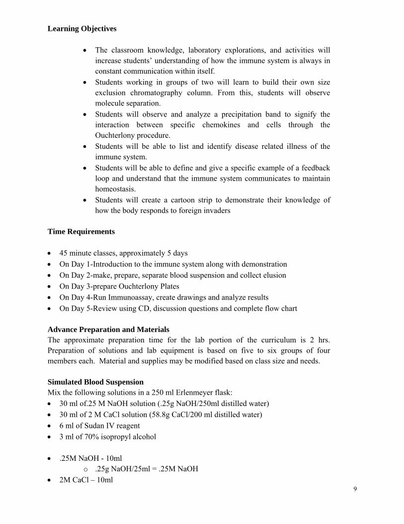
Learning Objectives
• The classroom knowledge, laboratory explorations, and activities will increase students’ understanding of how the immune system is always in constant communication within itself.
• Students working in groups of two will learn to build their own size exclusion chromatography column. From this, students will observe molecule separation.
• Students will observe and analyze a precipitation band to signify the interaction between specific chemokines and cells through the Ouchterlony procedure.
• Students will be able to list and identify disease related illness of the immune system.
• Students will be able to define and give a specific example of a feedback loop and understand that the immune system communicates to maintain homeostasis.
• Students will create a cartoon strip to demonstrate their knowledge of how the body responds to foreign invaders
Time Requirements • 45 minute classes, approximately 5 days • On Day 1-Introduction to the immune system along with demonstration • On Day 2-make, prepare, separate blood suspension and collect elusion • On Day 3-prepare Ouchterlony Plates • On Day 4-Run Immunoassay, create drawings and analyze results On Day 5-Review using CD, discussion questions and complete flow chart •
Advance Preparation and Materials The approximate preparation time for the lab portion of the curriculum is 2 hrs. Preparation of solutions and lab equipment is based on five to six groups of fom Simulated Blood Suspension Mix the following solutions in a 250 ml Erlenmeyer flask: • 30 ml of.25 M NaOH solution (.25g NaOH/250ml distilled water• 30 ml of 2 M CaCl solutio• 6 ml of Sudan IV reagent •
aOH - 10ml o .25g N
• 2M CaCl – 10ml
10
ohol mples
h2 cell sample .4M NaOH, 30 drops=.5ml
asic (3 drops) P, phenolphthalein (3 drops)
r
H and the CaCl solutions. They will both be used in the lab.
the
ion
o 100ml of distilled
microtube and bring the microtubes back to their workbench.
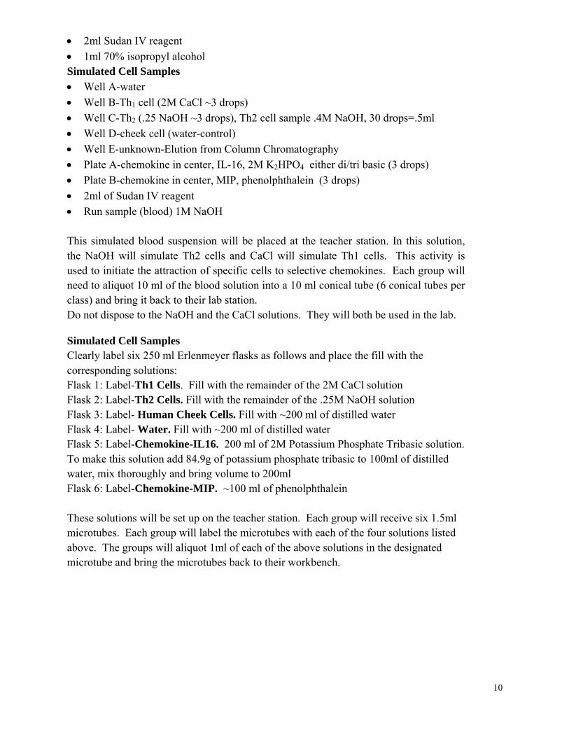
• 2ml Sudan IV reagent • 1ml 70% isopropyl alcSimulated Cell Sa• Well A-water • Well B-Th1 cell (2M CaCl ~3 drops) • Well C-Th2 (.25 NaOH ~3 drops), T• Well D-cheek cell (water-control) • Well E-unknown-Elution from Column Chromatography • Plate A-chemokine in center, IL-16, 2M K2HPO4 either di/tri b• Plate B-chemokine in center, MI 2ml of Sudan IV reagent •• Run sample (blood) 1M NaOH This simulated blood suspension will be placed at the teacher station. In this solution, the NaOH will simulate Th2 cells and CaCl will simulate Th1 cells. This activity is used to initiate the attraction of specific cells to selective chemokines. Each group will need to aliquot 10 ml of the blood solution into a 10 ml conical tube (6 conical tubes peclass) and bring it back to their lab station. Do not dispose to the NaO
Simulated Cell Samples Clearly label six 250 ml Erlenmeyer flasks as follows and place the fill withcorresponding solutions: Flask 1: Label-Th1 Cells. Fill with the remainder of the 2M CaCl solution
NaOH solutFlask 2: Label-Th2 Cells. Fill with the remainder of the .25MFlask 3: Label- Human Cheek Cells. Fill with ~200 ml of distilled water Flask 4: Label- Water. Fill with ~200 ml of distilled water Flask 5: Label-Chemokine-IL16. 200 ml of 2M Potassium Phosphate Tribasic solution. To make this solution add 84.9g of potassium phosphate tribasic t
ater, mix thoroughly and bring volume to 200ml wFlask 6: Label-Chemokine-MIP. ~100 ml of phenolphthalein These solutions will be set up on the teacher station. Each group will receive six 1.5ml microtubes. Each group will label the microtubes with each of the four solutions listed above. The groups will aliquot 1ml of each of the above solutions in the designated
11
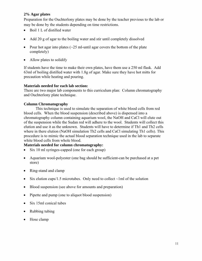
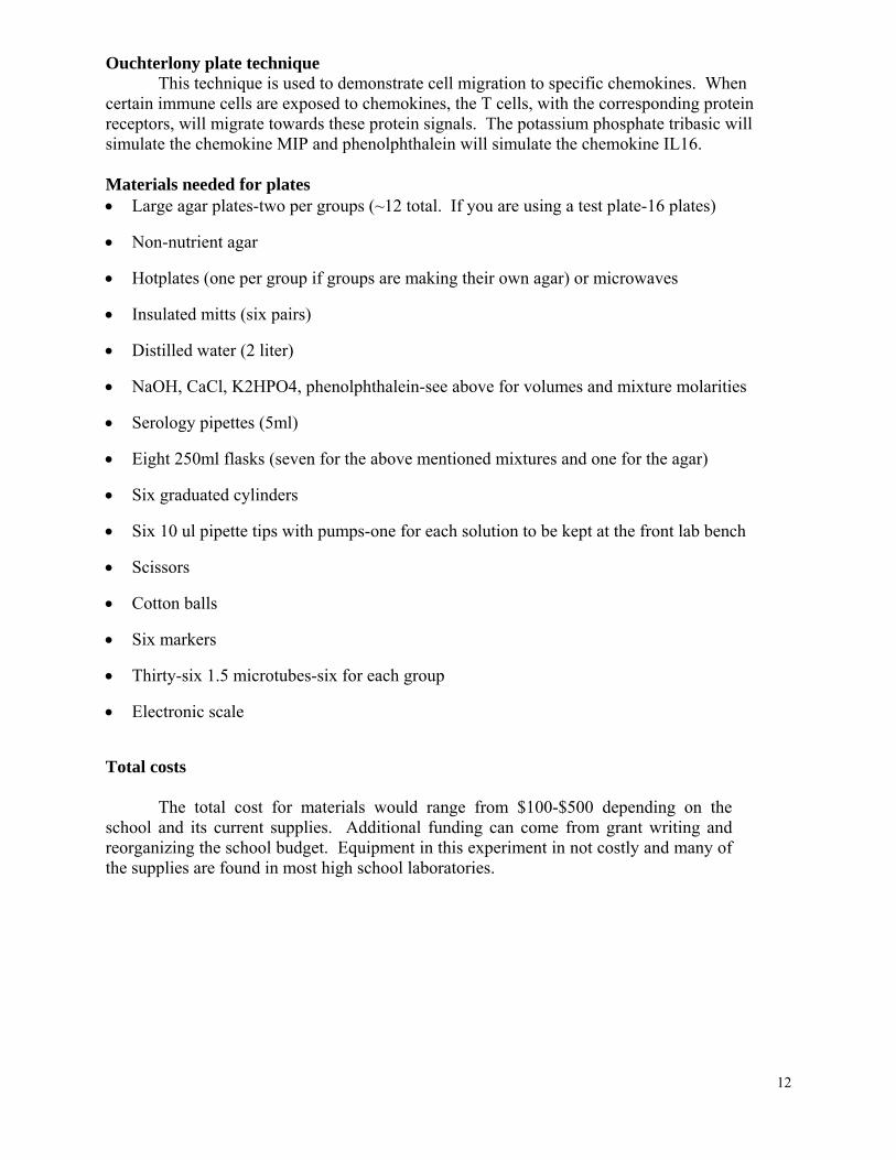
• Hose clamp
2% Agar plates Preparation for the Ouchterlony plates may be done by the teacher previous to the lab or may be done by the students depending on time restrictions. • Boil 1 L of distilled water
• Add 20 g of agar to the boiling water and stir until completely dissolved
• Pour hot agar into plates (~25 ml-until agar covers the bottom of the plate completely)
• Allow plates to solidify
If students have the time to make their own plates, have them use a 250 ml flask. Add 63ml of boiling distilled water with 1.8g of agar. Make sure they have hot mitts for precaution while heating and pouring. Materials needed for each lab section: There are two major lab components to this curriculum plan: Column chromatography and Ouchterlony plate technique. Column Chromatography This technique is used to simulate the separation of white blood cells from red blood cells. When the blood suspension (described above) is dispensed into a chromatography column containing aquarium wool, the NaOH and CaCl will elute out of the suspension while the Sudan red will adhere to the wool. Students will collect this elution and use it as the unknown. Students will have to determine if Th1 and Th2 cells where in there elution (NaOH simulation Th2 cells and CaCl simulating Th1 cells). This procedure is to mimic the actual blood separation technique used in the lab to separate white blood cells from whole blood. Materials needed for column chromatography: • Six 10 ml syringes-capped (one for each group)
• Aquarium wool-polyester (one bag should be sufficient-can be purchased at a pet store)
• Ring-stand and clamp
• Six elution cups/1.5 microtubes. Only need to collect ~1ml of the solution
• Blood suspension (see above for amounts and preparation)
• Pipette and pump (one to aliquot blood suspension)
• Six 15ml conical tubes
• Rubbing tubing
12
• Six markers
• Thirty-six 1.5 microtubes-six for each group
• Electronic scale
otal costs
iment in not costly and many of the supplies are found in most high school laboratories.
Ouchterlony plate technique This technique is used to demonstrate cell migration to specific chemokines. When certain immune cells are exposed to chemokines, the T cells, with the corresponding protein receptors, will migrate towards these protein signals. The potassium phosphate tribasic will simulate the chemokine MIP and phenolphthalein will simulate the chemokine IL16. Materials needed for plates • Large agar plates-two per groups (~12 total. If you are using a test plate-16 plates)
• Non-nutrient agar
• Hotplates (one per group if groups are making their own agar) or microwaves
• Insulated mitts (six pairs)
• Distilled water (2 liter)
• NaOH, CaCl, K2HPO4, phenolphthalein-see above for volumes and mixture molarities
• Serology pipettes (5ml)
• Eight 250ml flasks (seven for the above mentioned mixtures and one for the agar)
• Six graduated cylinders
• Six 10 ul pipette tips with pumps-one for each solution to be kept at the front lab bench
• Scissors
• Cotton balls
T The total cost for materials would range from $100-$500 depending on the school and its current supplies. Additional funding can come from grant writing and reorganizing the school budget. Equipment in this exper
13
Possible substitutions For chemicals, most chemicals that can react in a double-replacement reaction will be suitable. Potassium Phosphate tribasic can be substituted for monobasic or dibasic. Small Petri dishes can be used in replace of larger ones. Instead of plastic disposable pipettes for creating holes in the agar, plastic straws can be used. Safety Normal safety precautions should be taken including: goggles, aprons, and insulated mitts. All chemicals can be disposed of in the trash receptacles and in the drain with dilutions of water to follow. Student Knowledge and Skills After this laboratory investigation, students will have a greater understanding of how the immune system functions, be able to explain the sequence of events in response to foreign invaders and how the body maintains of homeostasis. Students will be familiar with measurements of volumes, such as the skills needed to micropipette. They will understand chromatography column and be able to contrast the difference between types of chromatography. Students will be able to perform and understand the Ochterlony essay. Misconceptions of immune system The following are a few misconceptions of the immune system which students may be unaware of, these misconceptions may be used for an opening discussion between teacher and students or may be used as a group activity to evaluate students preknowledge on their understanding of the immune system. Does receiving the flu shot give you the flu? When one receives the flu shot, they receive an inactive part or dead virus. The part of the flu that makes you sick is “turned off”. The inactive part or dead virus stimulates antibody production in the body. When one does encounter this virus antibodies are already present to “fight” this virus. A person who has severe allergies to eggs should not receive the flu shot because eggs are used to create the flu vaccine. Does cold weather cause one to catch a cold? Cold weather does not cause a viral infection. There are over 200 different known viruses. Viruses are transmitted from an infected person usually via airborne, shaking of hands, contact with a phone or door handles.
14
Can one get arthritis from cracking their knuckles? Cracking the knuckles does not give you arthritis. Cracking the knuckles causes the two joints to be pulled apart. The synovial fluid located within this joint fills more of the space. This decreases the fluid and dissolved gases, these gases leave the joint as tiny bubbles. It is the popping of the gas bubbles that one hears. Continuous cracking of the knuckles can injure the joint and weaken the fingers. Student Expectations • Students will research the functions of Th1, Th2 cells, chemokines IL-16 and MIP.
This information will be placed within the introduction of their laboratory report. • Students will write a scientific hypothesis for both procedures, column
chromatography and Ouchterlony Plates • Students will complete a formal laboratory report. • Students will draw and interpret the results from Ouchterlony plates and complete
analysis questions from laboratory exploration • Students will create a cartoon drawing of cell communication within the immune
system. • Students will complete the CD activity sheet. • Students will take an assessment test. Anticipated Results During the column chromatography process the simulated red blood cells will adhere to the polyester fiber located in the column. The simulated white cells will elude from the column. The migration of the solutions in the Ochterlony plates takes approximately 30-40 minutes before results will appear. Immunoassay Results: • All the cells and chemokines will permeate through the agar. Only specific cells that
are attracted to their exact chemokines will react causing a precipitation band. • Results of Plate A: As the chemokine-IL-16 (Potassium Phosphate Tribasic solution)
and Th1 cells (CaCl) make contact a color change will take place and a white precipitation band will form.
• Results of Plate B: As the chemokine-MIP (phenolphthalein) and Th2 cells (NaOH) make contact a color change will take place and a pink precipitation band will form, see diagrams below.
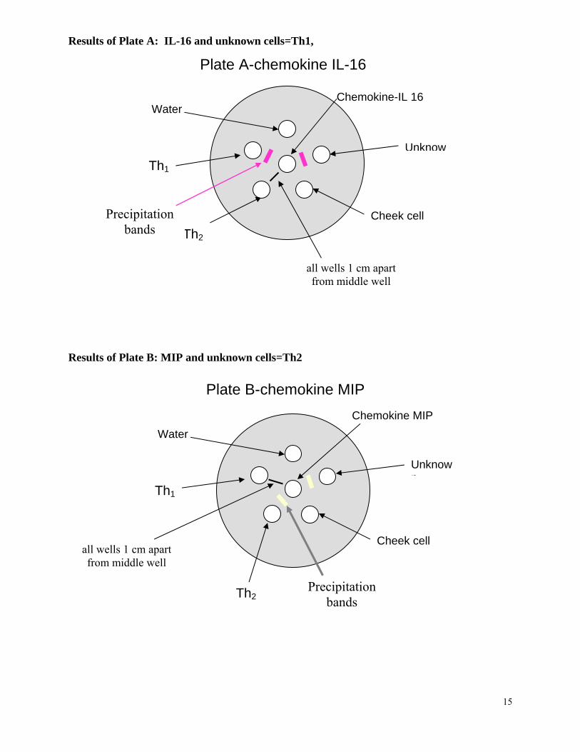
Results of Plate A: IL-16 and unknown cells=Th1, Plate A-chemokine IL-16
Results of Plate B: MIP and unknown cells=Th2
Water
Th1
Cheek cell
Unknow
Th2
all wells 1 cm apart from middle well
Chemokine-IL 16
Plate B-chemokine MIP
Water
Th1
Cheek
Th2
all wells 1 cm apart from middle well
Chemokine MIP
Precipitation bands
Precipitation bands
Unknow
cell
n
15
16
Classroom Discussion • Demonstration of the activation of APC, chemokines and helper T cells • Function of the immune system • Review terminology
o Specifically macrophage, helper T cells and chemokines • Discuss the differences in the precipitation bands from Plate A and Plate B
o Different chemicals represent simulated chemokines and cells, as each react with a specific chemical causing a color change take place
• Discuss specific chemokine functions and their attachment to specific cell surface receptors
• Discuss the advantages and disadvantages of having a healthy and unhealthy immune system
Assessment • Classroom discussion and, feedback from students during question and answer
sessions during daily lessons and after laboratory exploration • Formal laboratory report:
o Introduction-research on function of the immune system, including chemokines-MIP and IL-16, macrophages, APC cells and helper T 1&2 cells
o A final statement that includes data collection and laboratory results Analysis questions
o• Animated Drawing that models the response of the immune system to a foreign
invader. procedures • Teacher observations of students during preparation and laboratory
to follow along using the CD on the immune system • Activity sheet• Student Test
StudentSection
17
Name:___________________________________Date:_______________Class:______
“Can You Hear Me Now?” Laboratory Procedures
Chromatography Column Introduction Molecules can be separated using a variety of different chromatography methods. Paper, ion and gas are a few types of methods. In this laboratory investigation you will make a chromatography column. You will use it to separate a simulated blood suspension by applying the technique of size exclusion column chromatography. The chromatography column is packed with a polyester fiber medium. The blood suspension will be placed into the top of the column. It will travel and then separate over the polyester fiber. The red blood cells will adhere to the polyester fiber and the larger white blood cells will elude from the column. You will collect the elusion from the column and use it in the next procedure of this laboratory exercise. Part 1 Materials:
18
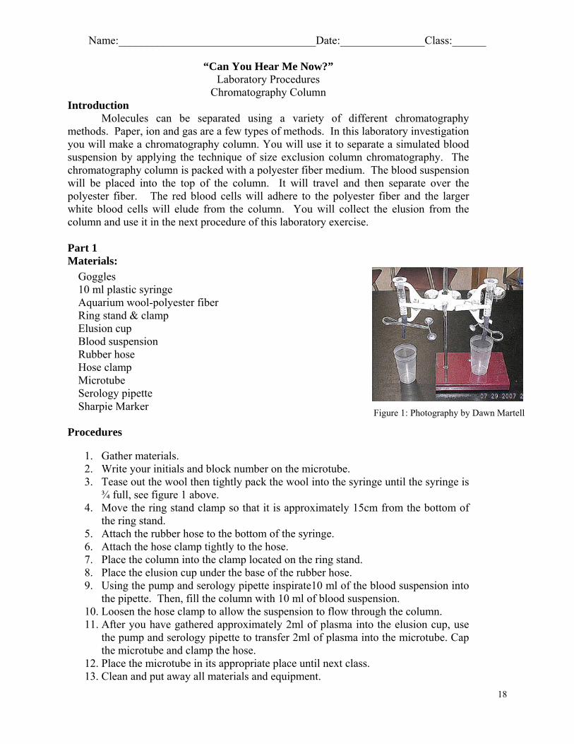
Goggles 10 ml plastic syringe Aquarium wool-polyester fiber Ring stand & clamp Elusion cup Blood suspension Rubber hose Hose clamp Microtube Serology pipette Sharpie Marker Figure 1: Photography by Dawn Martell
Procedures
1. Gather materials. 2. Write your initials and block number on the microtube. 3. Tease out the wool then tightly pack the wool into the syringe until the syringe is
¾ full, see figure 1 above. 4. Move the ring stand clamp so that it is approximately 15cm from the bottom of
the ring stand. 5. Attach the rubber hose to the bottom of the syringe. 6. Attach the hose clamp tightly to the hose. 7. Place the column into the clamp located on the ring stand. 8. Place the elusion cup under the base of the rubber hose. 9. Using the pump and serology pipette inspirate10 ml of the blood suspension into
the pipette. Then, fill the column with 10 ml of blood suspension. 10. Loosen the hose clamp to allow the suspension to flow through the column. 11. After you have gathered approximately 2ml of plasma into the elusion cup, use
the pump and serology pipette to transfer 2ml of plasma into the microtube. Cap the microtube and clamp the hose.
12. Place the microtube in its appropriate place until next class. 13. Clean and put away all materials and equipment.
Name:___________________________________Date:_______________Class:______
“Can You Hear Me Now?” Laboratory Procedure
Ouchterlony Plate Introduction Cell communication and homeostasis is an integral part of maintaining normal body function. Sustaining life depends on a quick response from the immune system in the recognition and reaction to foreign invaders. Continual enhancement of the immune system returns the body to homeostasis via feedback loops.
Through this laboratory investigation you will have a better understanding of how the lymphatic system facilitates immune responses. Lymphocyte T cells chemically communicate between specific cells to destroy cells that contain foreign invaders by releasing cytotoxins. You will describe the role of Antigen Presenting Cells (APC), the role and the origin of chemokines and that specific chemokines that attract and activate certain helper T cells. These helper T cells in return stimulate self-growth and activate the division of more helper T cells. The newly produced helper T cells proliferate memory cells through a positive feedback loop. The activation of helper T cells also helps to trigger B cells. The B cells manufacture antibodies which in turn activate Cytotoxic T cells during the immune response. You will observe and analyze the migration of specific chemokines to determine the interattraction of certain cells.
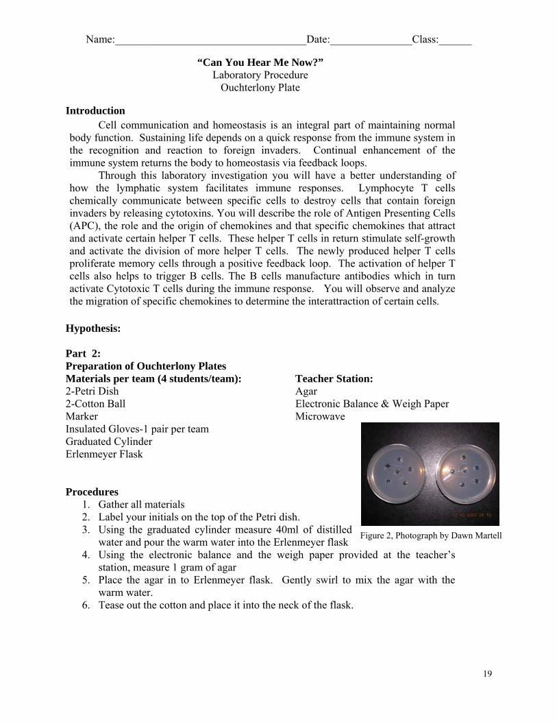
Hypothesis: Part 2: Preparation of Ouchterlony Plates Materials per team (4 students/team): Teacher Station: 2-Petri Dish Agar 2-Cotton Ball Electronic Balance & Weigh Paper Marker Microwave Insulated Gloves-1 pair per team Graduated Cylinder Erlenmeyer Flask Procedures
1. Gather all materials 2. Label your initials on the top of the Petri dish. 3. Using the graduated cylinder measure 40ml of distilled
water and pour the warm water into the Erlenmeyer flask 4. Using the electronic balance and the weigh paper provided at the teacher’s
station, measure 1 gram of agar
Figure 2, Photograph by Dawn Martell
5. Place the agar in to Erlenmeyer flask. Gently swirl to mix the agar with the warm water.
6. Tease out the cotton and place it into the neck of the flask.
19
20
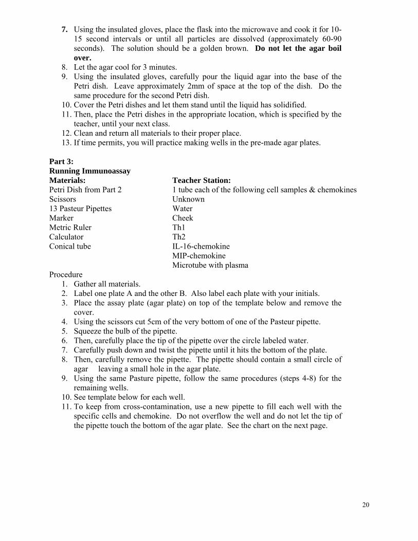
7. Using the insulated gloves, place the flask into the microwave and cook it for 10-15 second intervals or until all particles are dissolved (approximately 60-90 seconds). The solution should be a golden brown. Do not let the agar boil over.
8. Let the agar cool for 3 minutes. 9. Using the insulated gloves, carefully pour the liquid agar into the base of the
Petri dish. Leave approximately 2mm of space at the top of the dish. Do the same procedure for the second Petri dish.
10. Cover the Petri dishes and let them stand until the liquid has solidified. 11. Then, place the Petri dishes in the appropriate location, which is specified by the
teacher, until your next class. 12. Clean and return all materials to their proper place. 13. If time permits, you will practice making wells in the pre-made agar plates.
Part 3: Running Immunoassay Materials: Teacher Station: Petri Dish from Part 2 1 tube each of the following cell samples & chemokines Scissors Unknown 13 Pasteur Pipettes Water Marker Cheek Metric Ruler Th1 Calculator Th2 Conical tube IL-16-chemokine MIP-chemokine Microtube with plasma Procedure
1. Gather all materials. 2. Label one plate A and the other B. Also label each plate with your initials. 3. Place the assay plate (agar plate) on top of the template below and remove the
cover. 4. Using the scissors cut 5cm of the very bottom of one of the Pasteur pipette. 5. Squeeze the bulb of the pipette. 6. Then, carefully place the tip of the pipette over the circle labeled water. 7. Carefully push down and twist the pipette until it hits the bottom of the plate. 8. Then, carefully remove the pipette. The pipette should contain a small circle of
agar leaving a small hole in the agar plate. 9. Using the same Pasture pipette, follow the same procedures (steps 4-8) for the
remaining wells. 10. See template below for each well. 11. To keep from cross-contamination, use a new pipette to fill each well with the
specific cells and chemokine. Do not overflow the well and do not let the tip of the pipette touch the bottom of the agar plate. See the chart on the next page.
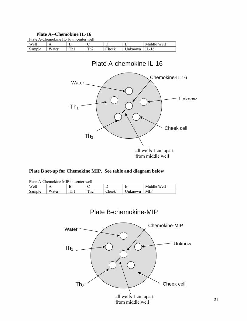
Plate A--Chemokine IL-16
Plate A-Chemokine IL-16 in center well Well A B C D E Middle Well Sample Water Th1 Th2 Cheek Unknown IL-16
Plate A-chemokine IL-16
Water
Cheek cell
Unknow
Chemokine-IL 16
Th2
all wells 1 cm apart from middle well
Th1
Plate B set-up for Chemokine MIP. See table and diagram below Plate A-Chemokine MIP in center well Well A B C D E Middle Well Sample Water Th1 Th2 Cheek Unknown MIP Plate B-chemokine-MIP
Chemokine-MIP Water
Che
all wells 1 cm apart from middle well
w
Th1
Th2
Unkno
21
ek cell
22
12. Place the covers on Petri dishes. 13. Observe, draw, and record results. 14. Clean your lab station and replace all materials to their proper place. Place drawing below:
Analysis Questions:
1. What is the purpose of using the water?
2. Was there any reaction between the chemokine and cells? If so explain this reaction. 3. Which type of cell represents the unknown for Plate A? For Plate B? How do know?
23
Animated Drawing of an Immune Response
Name:___________________________________Date:_______________Class:______
Introdcution As we begin the study of cell communication, feedback loops and homeostasis of the immune system, you and a partner will create an animation drawing. As cartoonists you will create a drawing that simulates an inflammation response to a foreign invader that has entered your body. Part I Pre-drawing For the first part of the assignment, you will have 20 minutes to brainstorm some ideas that you may use for your first drawing. List these ideas below. Then using the list you created, you are to sketch or draw, and label your drawing. Then, give a brief explanation about the drawing. White drawing paper and colored pencils will be provided. After finishing your drawing place your names on the back and tape the drawing on the board. See an example on the next page. _______________________________ ____________________________________ _______________________________ ____________________________________ _______________________________ ____________________________________ _______________________________ ____________________________________ _______________________________ ____________________________________ _______________________________ ____________________________________ Part II Post-drawing Immunology Animated Drawing Cell communication and homeostasis is an integral part of maintaining normal body function. Sustaining life depends on a quick response from the immune system in the recognition and reaction to foreign invaders. This along with continual enhancement returns the body to homeostasis via feedback loops. Using the knowledge you have acquired from classroom discussions and the laboratory investigations, you and your partner will create at least a three-frame animated drawing to show how the immune system simulates an inflammation response to a foreign invader, and how the immune system responds to this foreign invader. Your animated drawing must demonstrates the roles and steps involved using Antigen Presenting Cells (APC), helper T cells, B cells and cytokines in an immune response, which has been activated by a foreign invader
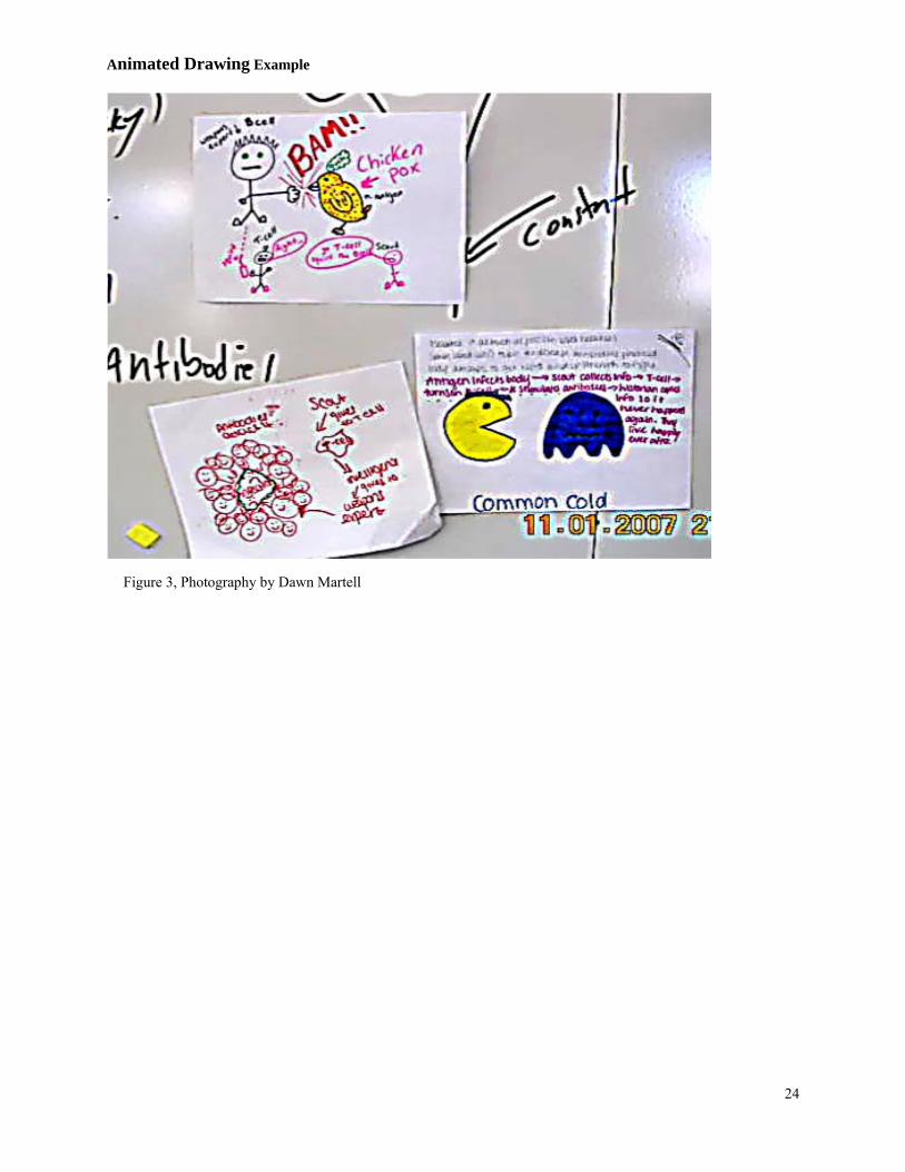
Animated Drawing Example
Figure 3, Photography by Dawn Martell
24
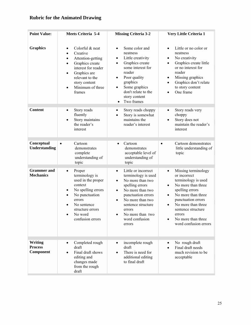
Rubric for the Animated Drawing
Point Value: Graphics
Meets Criteria 5-4
• Colorful & neat • Creative • Attention-getting • Graphics create
interest for reader • Graphics are
relevant to the story content
• Minimum of three frames
Missing Criteria 3-2
• Some color and neatness
• Little creativity • Graphics create
some interest for reader
• Poor quality graphics
• Some graphics don't relate to the story content
• Two frames
Very Little Criteria 1
• Little or no color or neatness
• No creativity • Graphics create little
or no interest for reader
• Missing graphics • Graphics don’t relate
to story content • One frame
Content • Story reads fluently
• Story maintains the reader’s interest
• Story reads choppy • Story is somewhat
maintains the reader’s interest
• Story reads very choppy
• Story does not maintain the reader’s interest
Conceptual Understanding
• Cartoon demonstrates complete understanding of topic
• Cartoon demonstrates acceptable level of understanding of topic
• Cartoon demonstrates little understanding of topic
Grammer and Mechanics
• Proper terminology is used in the proper context
• No spelling errors • No punctuation
errors • No sentence
structure errors • No word
confusion errors
• Little or incorrect terminology is used
• No more than two spelling errors
• No more than two punctuation errors
• No more than two sentence structure errors
• No more than two word confusion errors
• Missing terminology or incorrect terminology is used
• No more than three spelling errors
• No more than three punctuation errors
• No more than three sentence structure errors
• No more than three word confusion errors
Writing Process Component
• Completed rough draft
• Final draft shows editing and changes made from the rough draft
• incomplete rough draft
• There is need for additional editing to final draft
• No rough draft • Final draft needs
much revision to be acceptable
25
26
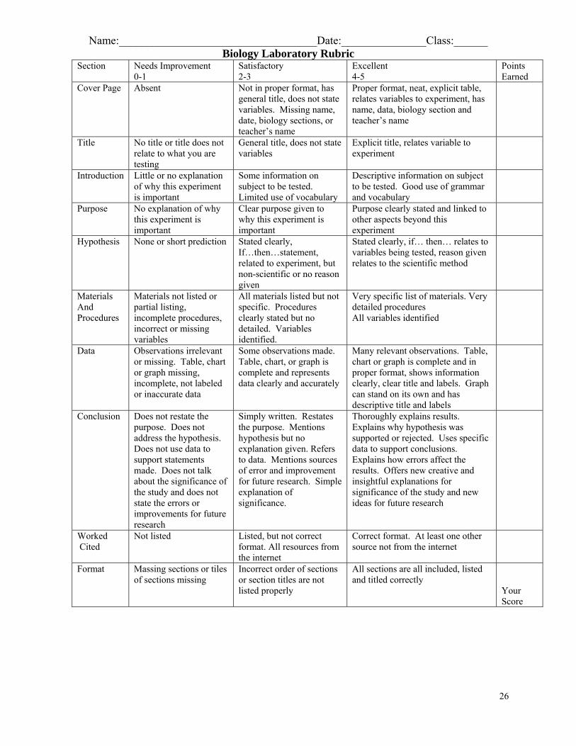
Name:___________________________________Date:_______________Class:______ Biology Laboratory Rubric
Section Needs Improvement 0-1
Satisfactory 2-3
Excellent 4-5
Points Earned
Cover Page Absent Not in proper format, has general title, does not state variables. Missing name, date, biology sections, or teacher’s name
Proper format, neat, explicit table, relates variables to experiment, has name, data, biology section and teacher’s name
Title No title or title does not relate to what you are testing
General title, does not state variables
Explicit title, relates variable to experiment
Introduction Little or no explanation of why this experiment is important
Some information on subject to be tested. Limited use of vocabulary
Descriptive information on subject to be tested. Good use of grammar and vocabulary
Purpose No explanation of why this experiment is important
Clear purpose given to why this experiment is important
Purpose clearly stated and linked to other aspects beyond this experiment
Hypothesis None or short prediction Stated clearly, If…then…statement, related to experiment, but non-scientific or no reason given
Stated clearly, if… then… relates to variables being tested, reason given relates to the scientific method
Materials And Procedures
Materials not listed or partial listing, incomplete procedures, incorrect or missing variables
All materials listed but not specific. Procedures clearly stated but no detailed. Variables identified.
Very specific list of materials. Very detailed procedures All variables identified
Data Observations irrelevant or missing. Table, chart or graph missing, incomplete, not labeled or inaccurate data
Some observations made. Table, chart, or graph is complete and represents data clearly and accurately
Many relevant observations. Table, chart or graph is complete and in proper format, shows information clearly, clear title and labels. Graph can stand on its own and has descriptive title and labels
Conclusion Does not restate the purpose. Does not address the hypothesis. Does not use data to support statements made. Does not talk about the significance of the study and does not state the errors or improvements for future research
Simply written. Restates the purpose. Mentions hypothesis but no explanation given. Refers to data. Mentions sources of error and improvement for future research. Simple explanation of significance.
Thoroughly explains results. Explains why hypothesis was supported or rejected. Uses specific data to support conclusions. Explains how errors affect the results. Offers new creative and insightful explanations for significance of the study and new ideas for future research
Worked Cited
Not listed Listed, but not correct format. All resources from the internet
Correct format. At least one other source not from the internet
Format Massing sections or tiles of sections missing
Incorrect order of sections or section titles are not listed properly
All sections are all included, listed and titled correctly
Your Score
Name:___________________________________Date:_______________Class:______
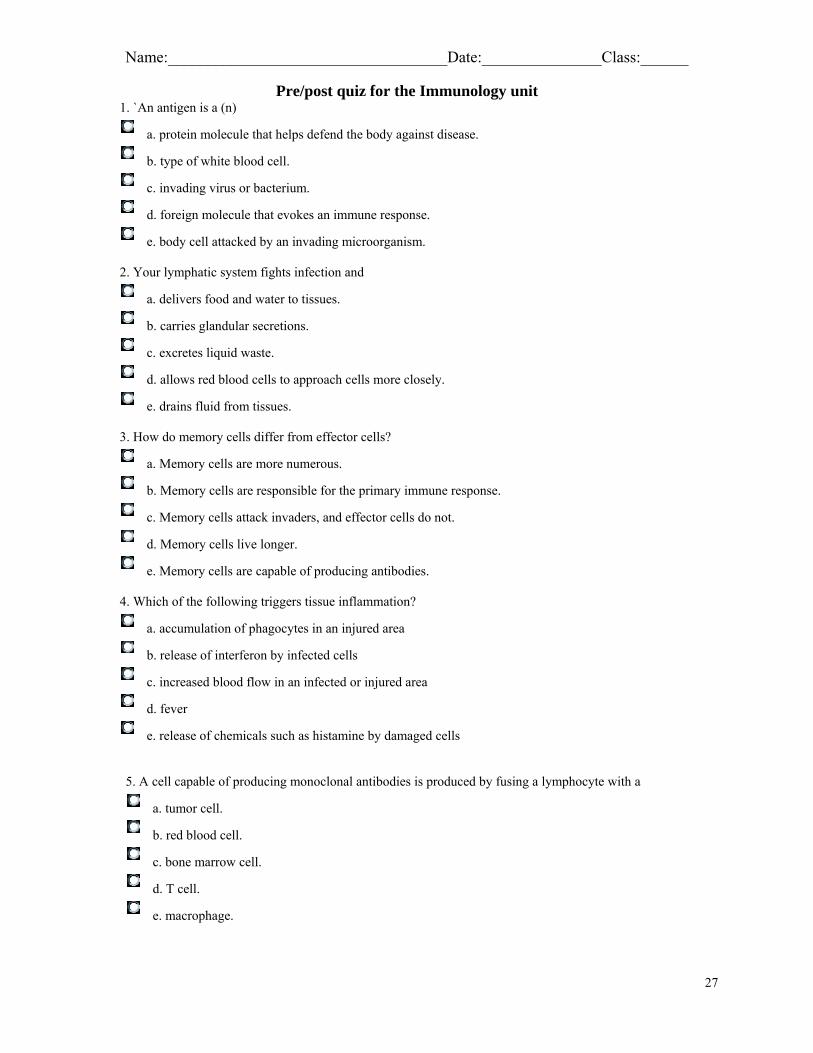
Pre/post quiz for the Immunology unit 1. `An antigen is a (n)
a. protein molecule that helps defend the body against disease.
b. type of white blood cell.
c. invading virus or bacterium.
d. foreign molecule that evokes an immune response.
e. body cell attacked by an invading microorganism. 2. Your lymphatic system fights infection and
a. delivers food and water to tissues.
b. carries glandular secretions.
c. excretes liquid waste.
d. allows red blood cells to approach cells more closely.
e. drains fluid from tissues. 3. How do memory cells differ from effector cells?
a. Memory cells are more numerous.
b. Memory cells are responsible for the primary immune response.
c. Memory cells attack invaders, and effector cells do not.
d. Memory cells live longer.
e. Memory cells are capable of producing antibodies. 4. Which of the following triggers tissue inflammation?
a. accumulation of phagocytes in an injured area
b. release of interferon by infected cells
c. increased blood flow in an infected or injured area
d. fever
e. release of chemicals such as histamine by damaged cells
5. A cell capable of producing monoclonal antibodies is produced by fusing a lymphocyte with a
a. tumor cell.
b. red blood cell.
c. bone marrow cell.
d. T cell.
e. macrophage.
27
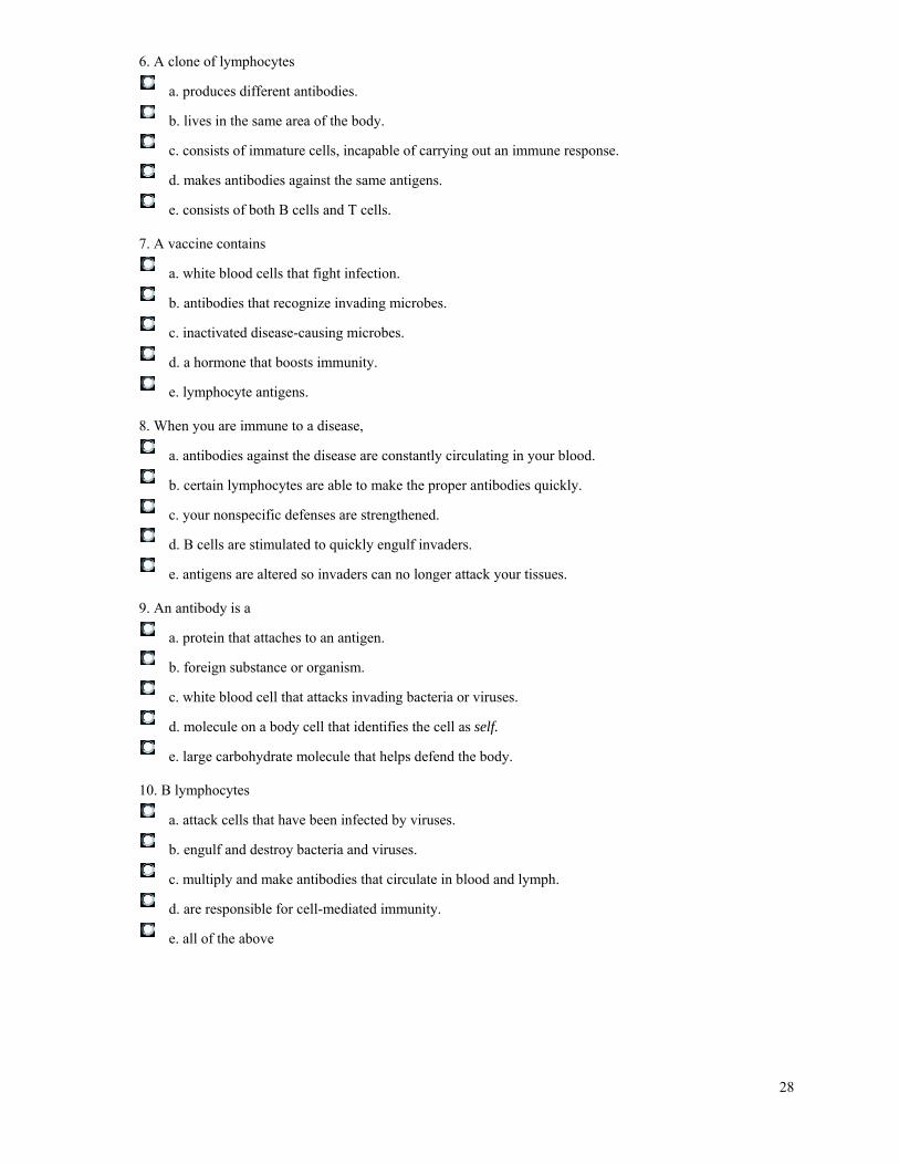
6. A clone of lymphocytes
a. produces different antibodies.
b. lives in the same area of the body.
c. consists of immature cells, incapable of carrying out an immune response.
d. makes antibodies against the same antigens.
e. consists of both B cells and T cells. 7. A vaccine contains
a. white blood cells that fight infection.
b. antibodies that recognize invading microbes.
c. inactivated disease-causing microbes.
d. a hormone that boosts immunity.
e. lymphocyte antigens. 8. When you are immune to a disease,
a. antibodies against the disease are constantly circulating in your blood.
b. certain lymphocytes are able to make the proper antibodies quickly.
c. your nonspecific defenses are strengthened.
d. B cells are stimulated to quickly engulf invaders.
e. antigens are altered so invaders can no longer attack your tissues. 9. An antibody is a
a. protein that attaches to an antigen.
b. foreign substance or organism.
c. white blood cell that attacks invading bacteria or viruses.
d. molecule on a body cell that identifies the cell as self.
e. large carbohydrate molecule that helps defend the body. 10. B lymphocytes
a. attack cells that have been infected by viruses.
b. engulf and destroy bacteria and viruses.
c. multiply and make antibodies that circulate in blood and lymph.
d. are responsible for cell-mediated immunity.
e. all of the above
28
11. Bacteria in body fluids are attacked by
a. antibodies from B cells.
b. cytotoxic T cells.
c. interferons.
d. helper T cells.
e. antigens. 12. What do the antibodies secreted by plasma cells (the effector cells of humoral immunity) do to attack their targets?
a. activate complement to punch holes in them
b. clump cells together so that phagocytes can ingest them
c. cause antigen molecules to settle out of solution
d. attach to antigens and detoxify them
e. all of the above
13. Which of the following is not present until after the primary immune response occurs?
a. memory cells
b. macrophages
c. helper T cells
d. complement proteins
e. antigens 14. The body produces antibodies complementary to foreign antigens. The process by which the body comes up with the correct antibodies to a given disease is most like
a. going to a tailor and having a suit made to fit you.
b. ordering the lunch special at a restaurant without looking at the menu.
c. going to a shoe store and trying on shoes until you find a pair that fits.
d. picking out a video that you haven't seen yet.
e. selecting a lottery prize winner by means of a random drawing. 15. Rhonda has been diagnosed as suffering from an immunodeficiency disease. Her doctor suspected Rhonda might have an immunodeficiency because
a. Rhonda strongly rejected an organ transplant.
b. Rhonda suffered from numerous allergies.
c. Rhonda's blood showed high levels of numerous antibodies.
d. Rhonda seemed to be immune to her own self molecules.
e. Rhonda suffered from repeated, prolonged infections.
29
16. The idea behind vaccination is to induce _____ without the vaccinated individual having to get sick.
a. passive immunity
b. the primary immune response
c. anaphylactic shock
d. nonspecific defenses
e. inflammation
17. Which of the following would be effective in eliminating bacteria but ineffective against viruses? (Hint: Viruses are not cells.)
a. activation of complement proteins
b. secretion of interferon by infected cells
c. neutralization by antibodies
d. agglutination by antibodies
e. perforin secretion by cytotoxic T cell 18. An allergen acts like
a. an antigen.
b. histamine.
c. interferon.
d. an antibody.
e. complement.
Pre/post quiz answer sheet. 1. D 2. E 3. E 4. E 5. A 6. D 7. C 8. A 9. A 10. C 11. B 12. E 13. A 14. A 15. D 16. B 17. C 18. A
Campbell, Reece, Mitchell, Interactive Study Partner for Biology, The Immune System, Fifth Edition, CD-ROM, San Francisco, CA, Pearson, Benjamin Cummings, 2006.
30
31
Name:___________________________________Date:_______________Class:______
CD Review question sheet
As you watch chapter 24a on the interactive CD, answer the following questions. 1. What are the dual defense mechanisms of the immune system?
2. How do B-cells work?
3. What is the main role of antibodies made by the plasma cells?
4. What are the four effector mechanisms of the humoral immune response?
5. Briefly describe each of the above responses.
6. How does the immune system recognize foreign invaders?
7. What cells act as antigen presenting cells and how does these cells become activated?
8. What are the roles of helper T-cells?
32
9. What chemical signal enhances the activation of T- cells and what type of cells secrete these signals?
10. What does IL-2 stimulate?
11. How do cytotoxic T-cells work?
12. What is the purpose of perforin?
Campbell, Reece, Mitchell, Interactive Study Partner for Biology, The Immune System, Fifth Edition, CD-ROM, San Francisco, CA, Pearson, Benjamin Cummings, 2006.
33
References
Banerjee, Ena Ray, et. al., “α4 and β2 intergins have nonredundant roles for asthma development, but for optimal allergen sensitization only α4 is critical.” Experimental Hematology, 35(2007): 605-617. Bernhagen, Jurgen; et.al, “MIF is a noncognate ligand of CXC chemokine receptors in inflammatory and atherogenic cell recruitment.” Nature Medicine, 13(2007): 587-596. Campbell, Reece, Mitchell, Interactive Study Partner for Biology, The Immune System, Fifth Edition, CD-ROM, San Francisco, CA, Pearson, Benjamin Cummings, 2006. Center, David M., Kornfeld, Hardy, Ryan, Thomas C., Cruickshank, William W., “Interleukin 16: implications for CDE4 function and HIV-1 progression.” Immunology Today, 21(2000): 273-279. Desmetz, Caroline; et.al., “Rapid Communication Cell surface CCR5 density determines the intensity of T cell migration towards rheumatoid arthritis synoviocytes.” ScienceDirect, Clinical Immunology, 123(2007):148-154. www.sciencedirect.com Gallagher, Richard B., “Immunology Today.” Elsevier Trends Journals, 13(1992): 469- 516. Gordon, Dan, ed. The Dana Sourcebook of Immunology: Resources for Secondary and Post-Secondary Teachers and Students, Washington, DC: Dana Press, 2005. www.dana.org/books/press Herlyn, Dr. Dorothee, Ph.D., Simulation of Protein Identification Through Immunoassay: Immunoassay, Shoestring Biotech, The Wistar Institute, Philadelphia, PA, 1-16. Lynch, E.L., Little, F.F., Wilson, K.C., Center, D.M., Cruikshank, W.W., “Immunomodulatory cytokines in asthmatic inflammation.” Cytokine & Growth Factor Review, Science Direct, 14(2003): 489-498. Massachusetts Department of Education, Biology, Massachusetts Science and Technology/Engineering Curriculum High School Standards, October 2006. www.mde.edu “Rubric Studio.” Rcampus.com. 15, Jan. 2007. Online. Available http://www.rcampus.com/rubricshowc.cfm?code=G84W5&sp=yes 9, March, 2007 Siewert, Christine; Menning, Astrid; Dudda, Jan; Siegmund, Kerstin; Lauer, Uta; Floess, Stefan, Campbell, Daniel J.; Hamann, Alf; Huehn, Jochen; “Induction of Organ- Selective CD4+ regulatory T cell homing.” European Journal of Immunology, 37(2007): 978-989. www.Eji-journal.eu