Embed Size (px)
Citation preview
ORIGINAL ARTICLE
Cell-cell interactions in spheroids maintained in suspension
BOZIDAR DJORDJEVIC1 & CHRISTOPHER S. LANGE1
1Department of Radiation Oncology, State University of New York, Downstate Medical Center, Brooklyn, New York, USA
AbstractWe have developed a system of mixed aggregates of cultured cells, to model in situ cell interactions. This three-dimensional(3D) system of floating cell aggregates, termed spheroids for their round shape, enables one to monitor their growth in bothsize and number of constituent clonogens and to measure survival curves for cells having 3D cell-cell interactions. Thissystem was used to measure the three-dimensional cell-cell interactions on growth, and clonogenicity of either AG1522fibroblasts, or HeLa cervical cancer cells (pure spheroids, or if both feeder and test cells are the same type, pseudohybridspheroids), and/or of mixtures of both (hybrid spheroids). By following the increase or decrease in size of, or number ofclonogens per, spheroid over time, one obtains growth or inhibition curves. By relating these clonogen numbers, oneobtains, after a suitable growth period, relative survival. The system allows one to score the effects of irradiation and of othertreatments, as well as the effect of interaction of the constituent cells on their survival. Floating pure, or pseudohybrid(composed of 10% live fibroblasts and 90% supralethally irradiated fibroblast feeder cells) spheroids, shrank to about10�20% of their volume in three days and then remained at that size for up to six days. In contrast, pure spheroidscomposed of live HeLa cells increased their volume by an order of magnitude over the same period. Survival of cells inspheroids was measured by the ability of individual spheroids to grow beyond a size implying a ten-fold increase. A caveat tobe observed is to correct survival for cellular multiplicity, i.e. reduce survival values to compensate for more than one colonyformer at the time of irradiation. The system of spheroids floating and growing in nutrient medium provides a selectivesystem for evaluating growth of HeLa, and by implication, other neoplastic cells, without interference from (overgrowth by)normal fibroblasts. Thus it is possible to discriminate between normal and neoplastic cells by virtue of whether or not cellsgrow in suspension. Such a system seems ideal for testing novel strategies (radiation in combination with chemicals), in an invivo-like environment.
Tumor cells from surgical samples or biopsies have
exceedingly low plating efficiencies, such that only a
miniscule fraction, probably unrepresentative of the
tumor as a whole, will grow in culture. However,
knowledge of the sensitivity of each patient’s tumor
to various chemotherapies and radiation treatments
is essential to treating each patient as an individual
rather than the average of all clinical experience. The
heterogeneity of tumors and of patient responses to
therapy is well-known, so the ability to individualize
patient treatment plans, taking into account the
inherent sensitivities of their tumor(s) could be an
important advance in improving the cure rates of
various cancers.
The first attempt to measure the clonogenic
survival of tumors in culture as an assay system
was that of Salmon and Hamburger [1]. Their
system of tumor cell growth in soft agar produced
sufficient cells to perform an assay in only about
25% of their patient samples and for each cell to
form a colony, 100 000 had to be plated. This low
plating efficiency (PE�/10�5) made it difficult to
believe that the results were representative of the
tumor in situ . An improvement on this system was
based on the Courtney et al. double layer soft-agar
method [2], used by West et al. [3] to measure the
sensitivity of cervical cancers to a single dose of 2 Gy.
This method obtained results from about three
quarters of patients and had a PE of about 0.1%
(10�3). A major benefit of the soft-agar methods
was supposed to have been that they prevented the
growth of stromal fibroblasts, which would otherwise
take over the culture, so that only the tumor cells
were thought to grow in agar. Similarly, the cell
adhesive matrix assay (CAM) was thought to be
selective for fibroblasts. However, Lawton et al. [4]
Correspondence: Bozidar Djordjevic, Department of Radiation Oncology, Box 1212, SUNY, Downstate Medical Center, 450 Clarkson Avenue, Brooklyn, NY
11203, USA. Tel: �/1 718 270 1250. Fax: �/1 718 270 1608. E-mail: [email protected]
Acta Oncologica, 2006; 45: 412�420
(Received 4 April 2005; accepted 6 December 2005)
ISSN 0284-186X print/ISSN 1651-226X online # 2006 Taylor & Francis
DOI: 10.1080/02841860500520743
Act
a O
ncol
Dow
nloa
ded
from
info
rmah
ealth
care
.com
by
Bro
wn
Uni
vers
ity L
ibra
ry o
n 06
/10/
12Fo
r pe
rson
al u
se o
nly.
and Stausbol-Gron et al. [5] showed that this
assumption was incorrect; stromal fibroblasts also
proliferate on the matrix plates [4] and in agar [5], so
that the results were for fibroblasts (or possibly both
mixed fibroblasts and tumor cells), and not the
tumors. Moreover, like all single-cell plating assays,
there is a fundamental problem that soft-agar assays
lack physiological relevance to in situ tumors,
because they lack the three-dimensional (3D) con-
tact seen in tumors. It may be for exactly this reason
that no one has yet correctly predicted individual
patient outcomes using these assays [3]. To alleviate
these problems, we have developed an in vivo-like
system, the hybrid spheroid assay [6�10], suitable
for testing primary tumor cells. Our system exhibits
a much higher PE�/1�10%, with almost all samples
producing sufficient colonies for assay [6]. Further-
more, in the hybrid spheroid system, cells are
enveloped in a 3D agglomerate of cells exhibiting
all the mutual influences on survival after treatment.
This is important, as it is becoming clear that the
survival of tumor cells and the functionality of
various tissues surrounding tumors are determined
not only by the direct impact of inactivating agents,
but also by the now well recognized Bystander Effect
(BE) (see review in [11]).
Hybrid spheroids were first shown to form from
mixtures of unirradiated HeLa test cells and supra-
lethally irradiated HeLa feeder cells [6]. Using this
system with test cells directly from human tumor
surgical samples, it was shown that the PE was
considerably higher than that in other clonogenicity-
based putative predictive assays for patient tumor
radiosensitivity [7]. Unlike monolayer assays, this
system appeared to maintain the G0 fraction in
chemosensitivity assays [8], and was the only one to
reliably provide radiation survival curves [6�10].
However, the radiosensitivity of HeLa test cells to
each daily dose fraction was shown to increase
exponentially with increasing number of fractions
[9], rather than remain constant, as previously
assumed. This was shown to be a form of BE [11].
Since test cell contact with supralethally irradiated
feeder cells produces an artifactually large BE effect
(rather than the much smaller one due to the feeder
cells receiving the same dose as the test cells),
this led us to examine the use of fibroblasts as feeder
cells in hybrid spheroids. This use of fibroblast
feeder cells also can block the growth of stromal
fibroblasts (which could otherwise overgrow the
culture). The results of some of the studies using
HeLa test cells (as a repeatable standard) are
presented here.
Until recently, the effects of ionizing radiations on
cells were considered to be independent for each cell,
with no interaction between neighbors. The analysis
of in vitro single cell survival curves was based on this
assumption, as were in vitro models for tumor
radiosensitivity. However, there is a phenomenon
of collective response to irradiation known by the
term ‘‘Bystander Effect’’ (BE, [11�20]). It deals
with the fact that cells in culture (and in whole
animals) which had not been traversed by a photon
or ionizing radiation track, are able to be affected by
their traversed neighbors traversed neighbors via
either a direct contact [12,15�17,19,20] or a med-
ium-transmitted effect [14,18]. From all appear-
ances, this phenomenon may be of considerable
significance in clinical situations, notably during
protracted treatment regimens. The text below
briefly summarizes the phenomenon, and highlights
possibilities of modulating clinical practices, with a
view to obtain a therapeutic gain in tumor control.
We became aware that BE may be operating in our
system of PseudoHybrid Spheroids composed of
agglomerated supralethally irradiated HeLa feeder
and live test HeLa cells, when we described expo-
nentially increased radiosensitivity in the course of
multifraction irradiation [8]. A much smaller radio-
sensitizing effect was seen when a similar combina-
tion of HeLa cells in monolayer, with or without
feeder cells, was repeatedly irradiated [9]. Since such
findings indicated an effect of close cellular contact
in BE, it was reasoned that three-dimensional cell-
cell contact maintained for longer periods of time,
could be more revealing of the effect of cellular
contact and more like the situation in situ . Conse-
quently, we have measured the importance of
cellular contact on test cell survival in long-term
spheroids composed of test and irradiated or uni-
rradiated feeder cells. This approach may provide a
better in vitro model for in vivo cellular radiation
responses.
Materials and methods
Types of spheroids
Three types of spheroids are used in this study: (1)
Pure spheroids, in which all the cells are of the same
type, as in pure fibroblast or pure HeLa spheroids;
(2) Hybrid spheroids, in which two cell types are
mixed (e.g., HeLa and fibroblasts), one type being
the test cells whose growth and viability are being
measured (1% or 10% of the initial spheroid cells),
while the other type consists of nonclonogenic feeder
cells (99% or 90% of the initial spheroid cells;
rendered such by irradiation or culture condition);
and (3) Pseudohybrid spheroids, in which all the
cells are of the same type, but the feeder cells (99%
or 90%) are supralethally irradiated.
Cell-cell interactions 413
Act
a O
ncol
Dow
nloa
ded
from
info
rmah
ealth
care
.com
by
Bro
wn
Uni
vers
ity L
ibra
ry o
n 06
/10/
12Fo
r pe
rson
al u
se o
nly.

Spheroid formation and maintenance
The method of spheroid formation has been de-
scribed previously [6�10]. Briefly, a mixed cell
suspension composed of cells to be tested (test cells)
and carrier cells (feeder cells) is co-incubated over-
night, in a bacteriological Petri dish, to which cells
do not adhere. Under these conditions, hybrid (or
pseudohybrid or pure, depending on the mixture of
cells used) spheroids are formed, the composition of
which closely corresponds to the input of the original
cell mixture [6].
In the present study, two cell lines were used:
AG1522 fibroblasts of human origin (early passage,
obtained from Coriell Cell Repositories, Camden,
NJ, and retrieved from liquid nitrogen storage
shortly before use), and HeLa cells (maintained in
this laboratory for more than a decade; periodically
removed from cryogenic storage to minimize genetic
drift).
To form hybrid spheroids with AG1522 feeder
cells and HeLa test cells, a mixture of 3�5�/106
AG1522 fibroblasts and 1/10th that of test cells for
cell growth assays, or 1/100th that for cell radio-
sensitivity assays were incubated in a 100 mm
bacteriological Petri dish (hydrophobic surface)
with 10 ml of Eagle’s Minimal Essential Medium
with glutamine and non-essential amino acids,
supplemented with 15% fetal bovine serum, strep-
tomycin (100 mg/ml), and sodium bicarbonate
(0.22% w/v) (complete MEM) (all Invitrogen Life
Technologies Gibco Products, Carlsbad, CA; below
noted as Gibco). The reason for the different average
numbers of test cells used for the growth and
radiosensitivity assays is that for the former, the
time to see changes in spheroid volume and test cell
number is shorter (the larger number of test cells
providing a head start), while for the latter, having
an average of about one test cell per spheroid makes
it easier to perform multiplicity corrections and
determine surviving fractions of test cells (if there
is only one test cell in a spheroid, there will be no
growth if that cell is sterilized or there will be growth
if it survives).
For spheroids made with HeLa feeder cells and
AG1522 test cells, the inverse of the above, the same
number of fibroblast test cells was used for both
viability and radiosensitivity assays. The test cell to
feeder cell ratio used was 1/10, because the fibro-
blasts have about a 10% plating efficiency (a 10
times higher concentration for growth curves would
mimic pure spheroids rather than pseudohybrid or
hybrid spheroids; see below). In a variant of this
procedure, spheroids made exclusively of fibroblasts,
or of HeLa cells (pure spheroids), were also used.
After agglomeration, spheroids of a desired size were
selected by passing the entire harvest of cell agglom-
erates through a system of nylon sieves, and spher-
oids were then eluted from a selected sieve. We have
found most satisfactory results with spheroids pas-
sing the 125 mm pore sieve, and arrested on the 88
mm pore sieve (with most of the harvested spheroids
falling in the range of 100�110 mm in diameter and
containing about 170 cells; this is almost twice the
number previously reported for HeLa feeder cells
grown with bromodeoxyuridine [6], presumably due
to the larger size of the analog-containing cells. We
took the time of harvesting spheroids as zero time for
our subsequent growth and survival measurements.
Spheroids which were selected in the 250 mm
diameter size range contained about 2 700 cells.
Principal features of the modified hybrid spheroid assay
The principal feature of our modified, improved,
hybrid spheroid assay, as used in the present study,
was to monitor the capacity of spheroids to grow
beyond an arbitrary chosen size (equivalent to 10
divisions by the test cells), assumed to denote the
ability of unlimited proliferation. The improved
assay also prevents fibroblasts (potentially emanating
from surgical specimens) from being scored in
survival experiments. In order to obtain growth of
neoplastic cells, hybrid (i.e., containing both fibro-
blastic feeder cells, as well as neoplastic HeLa test
cells) spheroids were incubated over a 10�12 day
period in bacteriological dishes, in which there is no
cell attachment. Under such conditions, unirra-
diated AG1522 fibroblastic feeder cells fail to grow
in floating spheroids; they could also be easily
distinguished from HeLa cells on the basis of
morphological appearance upon dispersal and re-
plating. This feature enabled the system of floating
hybrid spheroids to be used to monitor the radiation
response of test HeLa cells, whereby growth beyond
a certain size signifies survival, in a manner analo-
gous to colony formation.
Dispersal of spheroids for cellular content studies
To determine the number of cells per spheroid,
either in order to follow the growth pattern of
spheroids, or for the purpose of measuring cellular
multiplicity (see below), we trypsinized a known
number of spheroids. This was accomplished by
spinning spheroids in a centrifuge tube, aspirating
supernatant medium, washing spheroids in 0.05%
trypsin in EDTA (Gibco), and incubating them in
0.05% trypsin in EDTA for 10 min to disperse
spheroids into individual cells. After dispersal of
spheroids, the trypsin in the resultant cell suspension
was inactivated by the addition of an equal volume of
414 B. Djordjevic & C. S. Lange
Act
a O
ncol
Dow
nloa
ded
from
info
rmah
ealth
care
.com
by
Bro
wn
Uni
vers
ity L
ibra
ry o
n 06
/10/
12Fo
r pe
rson
al u
se o
nly.

complete MEM, the cells counted in a hemacyt-
ometer, and an appropriate dilution plated in tissue
culture Petri dishes or flasks for colony formation.
From these data (cell numbers, colonies formed per
spheroid) one obtains either the growth of spheroids
as a function of time, or the average colony forming
cell number per spheroid, at the time of irradiation,
the so called cellular multiplicity (M). One can
obtain the value of M in the original mixed cell
suspension by allowing a sufficient growth period for
constituent cells to form microcolonies. HeLa cells
in microcolonies are easily distinguished from
AG1522 cells in a culture dish in which both cell
types are attached and grow.
X-irradiation and measurement of survival of spheroids
For irradiation studies, spheroids eluted from the
selected sieve were distributed in equal aliquots into
a series of 60 mm bacteriological Petri dishes, and
irradiated with graded 2 Gy doses of 250 kVp X-rays
from a Philips RT 250 therapy machine (15 mA, 2
mm Al inherent filtration, HVL 0.475 mm Cu, 50
cm FSD, dose rate 2.7 Gy/min). At the end of the
incubation period allowed for scorable spheroid
formation (10�12 days), spheroids over 250 mm in
diameter were counted under a dissecting micro-
scope, using an underlying transparent sheet with a
grid to facilitate counting in the dish. These spher-
oids are now composed chiefly of HeLa test-cells;
reflect growth from one or a few clonogen cells to a
spheroid with a diameter more than double that of
the original. Since the 250 mm diameter spheroids
contained about 2 700 cells, the test cells in hybrid
or pseudohybrid spheroids which had grown from
100 mm to 250 mm from their test cells, must have
undergone about 10 to 11 doublings (divisions) per
original clonogen. Survival of whole spheroids was
determined by relating the number of such enlarged
spheroids in the treated (irradiated) dish, to that in
the control dish.
Survival curves were not measured for HeLa cells
in pure spheroids because, with about 170 clonogens
to start, very large doses (�/14 Gy) would have been
required to reduce the surviving fraction toB/1 cell/
spheroid and thus have no colony of surviving cells.
Thus, at doses comparable to those used for the
other spheroid conditions, almost all spheroids
would have produced colonies. If all 170 cells were
colongenic then only about 4 divisions would have
been necessary to produce spheroids of 250 mm
diameter, while the standard for survival of clono-
genicity is 10 divisions in 10 days. Spheroids much
larger than this could not be used since they have
hypoxic centers and no longer behave the same as
the 100�250 mm spheroids used in these studies.
Cellular multiplicity corrections of spheroid survival
To obtain single cell survival curves, fractional
survival of whole spheroids was corrected for cellular
multiplicity [6,21,22] by using the equation
SF(s:c)�1�(1�SF(spher))1=M (1)
where SF(s.c.) is the surviving fraction of single
clonogens, SF(spher) is the surviving fraction of whole
spheroids, and M is the average multiplicity of test
cells in the control (unirradiated) series at the
beginning of the irradiation procedure. In order to
obtain M, an aliquot of same size control spheroids
was dispersed by trypsinization at the time of
irradiation and plated for colony formation. The
cellular multiplicity, M, was determined as the ratio
of test-cell colony numbers obtained from dispersed
spheroids divided by the number of spheroids
counted in unirradiated Petri dishes at the end of
the time of trypsinization. Dispersal of spheroids
served only this limited purpose. It should be noted
that multiplicity denotes the clonogen content per
spheroid at the time of irradiation, and not at the
time of spheroid scoring at the end of the incubation
period for spheroid survival determination. It should
also be understood that the much smaller proportion
of test cells for survival studies was necessitated by
the method of multiplicity correction at the time of
irradiation, which would be impractical or impossi-
ble with the higher input hybrid spheroids.
X-Ray survival curves of monolayer cells and cells from
dispersed spheroids
Survival curves were obtained from dispersed cells
plated in the conventional manner, by relating
numbers of colonies formed 10�12 days following
irradiation (or Plating Efficiencies) in the treated
versus the control group; no multiplicity corrections
were needed in these cases, since M was near unity.
Spheroid growth measurements
This was achieved by measuring spheroid diameters
under a dissecting microscope using an ocular
reticle, with the individual spheroid volume calcu-
lated as a function of time. To assure the round state
of spheroids, we used conditions discouraging
attachment, such as growth in bacteriological dishes
or placing spheroids in 24 well plates coated with
0.5% agarose. In other situations, growth was
measured by dispersal of spheroids by trypsinization,
followed by counting cells in the dispersed cell
suspension.
Cell-cell interactions 415
Act
a O
ncol
Dow
nloa
ded
from
info
rmah
ealth
care
.com
by
Bro
wn
Uni
vers
ity L
ibra
ry o
n 06
/10/
12Fo
r pe
rson
al u
se o
nly.

Statistical methods
The data points shown in all figures are the means
with standard errors of the mean of the results of
multiple flasks/dishes, normalized to unity within
each experiment, prior to combining the data of
replicate experiments. The standard error is the
standard deviation divided by the square root of
the number of observations, and is properly used
when comparing mean values with each other rather
than data points with the mean (for which the
standard deviation should be used). The standard
errors of ratios (as in plating efficiencies and surviv-
ing fractions) are determined using the chain rule for
equipartition of variance, which takes into account
the proper weight of the uncertainty contributions of
both the numerator and the denominator [23,24].
Results
Growth characteristics of spheroids composed of
fibroblasts and/or HeLa cells
In order to verify the utility of AG1522 fibroblasts as
(non-growing) feeder cells in hybrid spheroids,
spheroids composed entirely of either live fibro-
blasts, or of supralethally irradiated fibroblasts,
were incubated in suspension over a period of several
days, and the growth (change in volume of spher-
oids) measured. The results in Figure 1 show that
both types of spheroids shrank to about 10�20% of
their original size within three days (day four after
creation), after which, they remain the same reduced
size for the rest of the period of observation (up to
two weeks). In contrast, pure spheroids of HeLa cells
grow, increasing in size (volume) by an order of
magnitude over a six day period. The latter is
consistent with the previously reported Gompertzian
growth of these cells in spheroids [10].
Figure 2 shows the increases in the relative
number of clonogens with time in culture for HeLa
cells in either attached or floating pure spheroids;
i.e., spheroids composed of HeLa cells only. For
comparison, a growth curve of HeLa cells in mono-
layer is also shown. Their respective doubling times
are 15.7, 13.4, and 18.1 hours. In all three cases, the
growth rates are similar.
The next two sets of data (triangles and black
stars) represent the growth of HeLa cells in hybrid
spheroids (with irradiated AG1522 fibroblast feeder
cells) and of unirradiated AG1522 fibroblasts grown
in monolayer. Their common doubling time is 31.6
hours, about half the rate of the HeLa cells in pure
spheroids or monolayer. For HeLa cells in hybrid
spheroids with unirradiated fibroblast feeder cells
(data not shown), we can deduce a HeLa cell
doubling time of 5/24 hours. This is based on the
survival curve data in Figure 5 and the survival
criterion that the test cells undergo at least 10
Days After Spheroid Harvest
0 2 4 6 8 10 12 14
Sph
eroi
d R
elat
ive
Vol
umes
0.1
1
10
Figure 1. Effects of time held in suspension on spheroid volume.
Relative volumes of spheroids composed exclusively of live (black
squares), and supralethally irradiated (20 Gy, open squares)
AG1522 fibroblasts are plotted for successive days after spheroid
selection. Both types of spheroids shrank to about 10�20% of
their original size within three days (day four after creation), after
which, they remain the same reduced size for the rest of the period
of observation (up to two weeks). In contrast, pure spheroids of
HeLa cells (black circles) grow, increasing in size (volume) by an
order of magnitude over a 6-day period. Data obtained from four
experiments. Error bars in this and in other graphs are standard
errors of the mean.
Days After Spheroid Harvest or Cell Plating0 1 2 3 4 5
Rel
ativ
e C
lono
gen
Num
ber
1
10
Figure 2. Increases in the relative number of clonogens with time
in culture. The top two curves with solid lines are for HeLa cells in
either attached (open squares) or floating (open circles) pure
spheroids. For comparison, a growth curve of HeLa cells in
monolayer is also shown (open stars, dashed line). The next two
curves represent the growth of HeLa cells in hybrid spheroids
(with irradiated AG1522 fibroblast feeder cells; open triangles)
and of unirradiated AG1522 fibroblasts grown in monolayer
(black stars). The bottom curve (black circles) shows the initial
stasis, and then growth, of AG1522 fibroblasts as test cells in
attached hybrid spheroids with irradiated HeLa feeder cells; the
final growth seen between days 3 and 4 is similar to that of the
HeLa cells in floating or attached pure spheroids or in monolayer.
416 B. Djordjevic & C. S. Lange
Act
a O
ncol
Dow
nloa
ded
from
info
rmah
ealth
care
.com
by
Bro
wn
Uni
vers
ity L
ibra
ry o
n 06
/10/
12Fo
r pe
rson
al u
se o
nly.
doublings (�/1024 cells) in 10 days with a dose-
dependent delay in the start of growth. Such a
doubling time is consistent with that observed for
the other three conditions.
The bottom curve (black circles) represents the
initial stasis and then growth of AG1522 fibroblasts
as test cells in attached hybrid spheroids with
irradiated HeLa feeder cells. The final growth seen
between days three and four has a doubling time of
16.3 hours, similar to that of the HeLa cells in
floating or attached pure spheroids or in monolayer.
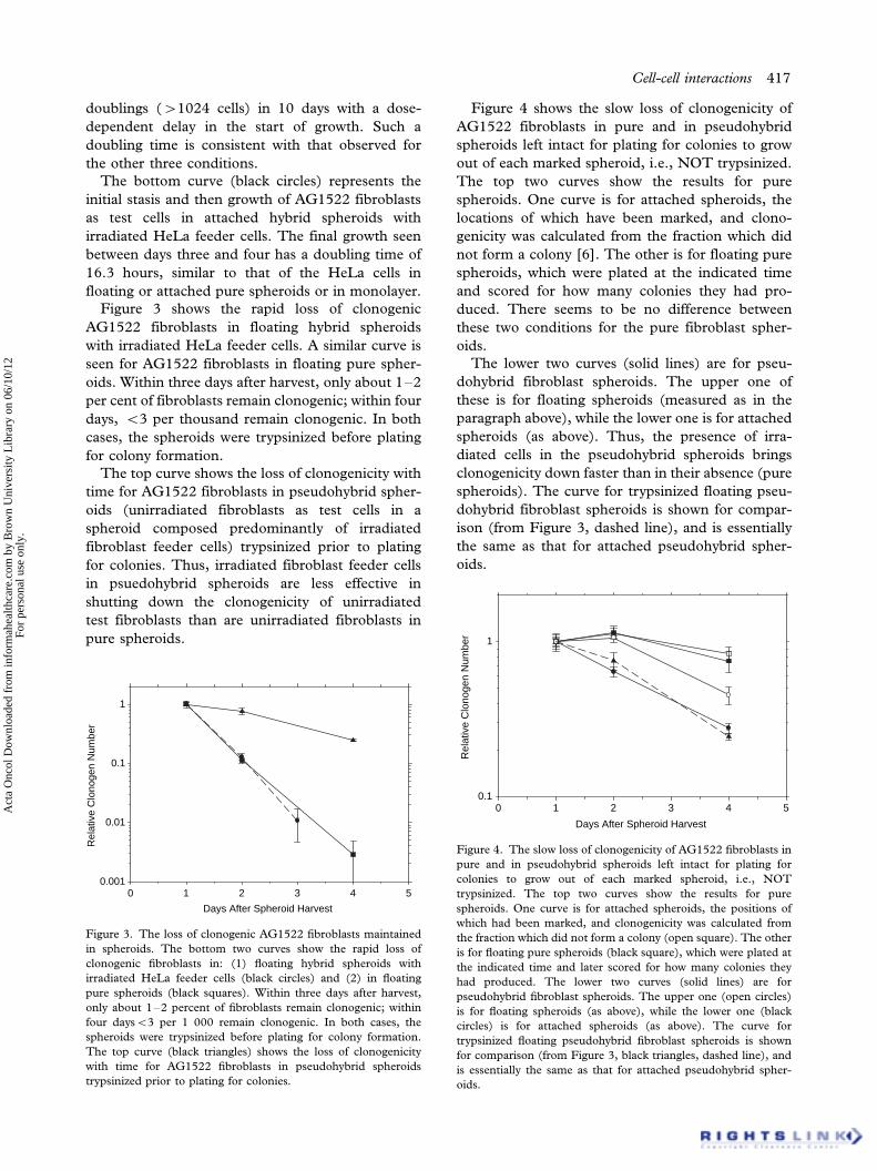
Figure 3 shows the rapid loss of clonogenic
AG1522 fibroblasts in floating hybrid spheroids
with irradiated HeLa feeder cells. A similar curve is
seen for AG1522 fibroblasts in floating pure spher-
oids. Within three days after harvest, only about 1�2
per cent of fibroblasts remain clonogenic; within four
days, B/3 per thousand remain clonogenic. In both
cases, the spheroids were trypsinized before plating
for colony formation.
The top curve shows the loss of clonogenicity with
time for AG1522 fibroblasts in pseudohybrid spher-
oids (unirradiated fibroblasts as test cells in a
spheroid composed predominantly of irradiated
fibroblast feeder cells) trypsinized prior to plating
for colonies. Thus, irradiated fibroblast feeder cells
in psuedohybrid spheroids are less effective in
shutting down the clonogenicity of unirradiated
test fibroblasts than are unirradiated fibroblasts in
pure spheroids.
Figure 4 shows the slow loss of clonogenicity of
AG1522 fibroblasts in pure and in pseudohybrid
spheroids left intact for plating for colonies to grow
out of each marked spheroid, i.e., NOT trypsinized.
The top two curves show the results for pure
spheroids. One curve is for attached spheroids, the
locations of which have been marked, and clono-
genicity was calculated from the fraction which did
not form a colony [6]. The other is for floating pure
spheroids, which were plated at the indicated time
and scored for how many colonies they had pro-
duced. There seems to be no difference between
these two conditions for the pure fibroblast spher-
oids.
The lower two curves (solid lines) are for pseu-
dohybrid fibroblast spheroids. The upper one of
these is for floating spheroids (measured as in the
paragraph above), while the lower one is for attached
spheroids (as above). Thus, the presence of irra-
diated cells in the pseudohybrid spheroids brings
clonogenicity down faster than in their absence (pure
spheroids). The curve for trypsinized floating pseu-
dohybrid fibroblast spheroids is shown for compar-
ison (from Figure 3, dashed line), and is essentially
the same as that for attached pseudohybrid spher-
oids.
Days After Spheroid Harvest
0 1 2 3 4 5
Rel
ativ
e C
lono
gen
Num
ber
0.001
0.01
0.1
1
Figure 3. The loss of clonogenic AG1522 fibroblasts maintained
in spheroids. The bottom two curves show the rapid loss of
clonogenic fibroblasts in: (1) floating hybrid spheroids with
irradiated HeLa feeder cells (black circles) and (2) in floating
pure spheroids (black squares). Within three days after harvest,
only about 1�2 percent of fibroblasts remain clonogenic; within
four daysB/3 per 1 000 remain clonogenic. In both cases, the
spheroids were trypsinized before plating for colony formation.
The top curve (black triangles) shows the loss of clonogenicity
with time for AG1522 fibroblasts in pseudohybrid spheroids
trypsinized prior to plating for colonies.
Days After Spheroid Harvest
0 1 2 3 4 5
Rel
ativ
e C
lono
gen
Num
ber
0.1
1
Figure 4. The slow loss of clonogenicity of AG1522 fibroblasts in
pure and in pseudohybrid spheroids left intact for plating for
colonies to grow out of each marked spheroid, i.e., NOT
trypsinized. The top two curves show the results for pure
spheroids. One curve is for attached spheroids, the positions of
which had been marked, and clonogenicity was calculated from
the fraction which did not form a colony (open square). The other
is for floating pure spheroids (black square), which were plated at
the indicated time and later scored for how many colonies they
had produced. The lower two curves (solid lines) are for
pseudohybrid fibroblast spheroids. The upper one (open circles)
is for floating spheroids (as above), while the lower one (black
circles) is for attached spheroids (as above). The curve for
trypsinized floating pseudohybrid fibroblast spheroids is shown
for comparison (from Figure 3, black triangles, dashed line), and
is essentially the same as that for attached pseudohybrid spher-
oids.
Cell-cell interactions 417
Act
a O
ncol
Dow
nloa
ded
from
info
rmah
ealth
care
.com
by
Bro
wn
Uni
vers
ity L
ibra
ry o
n 06
/10/
12Fo
r pe
rson
al u
se o
nly.

Thus, several factors affect the survival of test cells
in spheroids (Figures 1�4): (1) the type of test cells
(fibroblast vs HeLa) and the prior treatment (irra-
diated vs unirradiated) of the feeder cells in the
spheroids * where the proximity of different cells
may affect their survival differently, (2) the attach-
ment status of spheroids * floaters vs . attached, (3)
dispersal of spheroids * trypsinization diminished
survival of clonogens more in hybrid spheroids than
in pure spheroids, and (4) trypsinization diminished
clonogenicity much faster in floating spheroids than
it did in attached spheroids.
The negative growth characteristics (shrinkage) of
spheroids in suspension, composed of fibroblasts
(Figures 1�4), indicated that when hybrid spheroids
grew, it was due to the proliferation of HeLa test
cells, not of AG1522 fibroblast feeder cells. It follows
for hybrid spheroids, that fibroblasts did not make a
contribution to the growth of (hybrid) spheroids, as
further evidenced from the following experiment.
Upon termination of the spheroid growth experi-
ment (ten days), resulting spheroids were trypsi-
nized, plated in a tissue culture dish, and stained
after a suitable growth period (ten additional days),
as detailed in the Methods section. It was found that
the only colonies (�/99.9%) to grow from these
dispersed hybrid spheroids were HeLa cells, even
though the spheroids initially consisted predomi-
nantly of fibroblasts (data not shown).
Survival curves of X-irradiated hybrid spheroids
maintained in suspension
In the next experiment, two types of fibroblastic
feeder cells were used, unirradiated and supralethally
irradiated, encasing HeLa test cells. In order to
obtain survival curves using the improved spheroid
assay, hybrid spheroids with the two types of feeder
cells were placed in 60 mm bacteriological Petri
dishes (in which spheroids remain floating), X-
irradiated in 2 Gy increments up to a dose of 6
Gy, and maintained over a period of 10�12 days.
Survival was determined as described in Methods,
scoring only spheroids �/220 mm in diameter (which
corresponds to 10 divisions by a HeLa test cell in the
spheroid). The results of these measurements are
presented in Figure 5, after appropriate corrections
for cellular multiplicity as described in Methods.
Both survival curves were fitted to an exponential
equation, even when continuous bending of the
lower curve (with irradiated feeder cells) remained
a possibility [22,25]. The important observation is
that the radiosensitivities of the two series were
different, with the lower curve being steeper by a
factor of 1.549/0.13. Evidently, lethally irradiated
fibroblast feeder cells confer radiosensitivity to
tumor test cells in spheroids, with the difference in
survival between the two conditions (live vs. dead
feeders) most pronounced at the highest dose used
(6 Gy). [Unfortunately, it is not practical to deliver
higher radiation doses in the system presented here
in order to detect larger differences in survival levels,
for reasons of both a diminishing statistical accuracy
with higher doses (i.e., fewer scorable spheroids),
and of problems associated with spheroids over-
loaded with test cells (when occasional breakup of
spheroids could produce spurious carriers of clono-
gens).] Thus, the use of irradiated feeder cells carries
with it the problem of a Bystander effect on the test
cells, while the use of unirradiated fibroblasts as
feeder cells, which will not proliferate when in 3-D
contact as in spheroids, avoids this problem.
Survival of monolayer HeLa cells and cells from
dispersed spheroids
Unlike HeLa cells in floating spheroids, monolayer
HeLa cells, obtained either from conventional stock
cultures, or from cultures from dispersed hybrid
spheroids, displayed identical radiation responses
when tested for their colony forming ability 12
days after irradiation, irrespective of the kind of
feeder cells present (Figure 6). It appears therefore,
that for the radiosensitizing effect by supralethally
irradiated AG1522 fibroblasts, and a close 3-dimen-
sional contact with test cells must take place during
and for prolonged periods of time following irradia-
tion.
Dose [Gy]
0 1 2 3 4 5 6 7
Sur
vivi
ng F
ract
ion
0.01
0.1
1
Figure 5. Survival curves of HeLa cells irradiated in hybrid
spheroids, corrected for clonogen multiplicity. Hybrid spheroids
were maintained in suspension over a period of 10�12 days before
scoring surviving spheroids. Data obtained from four experiments
in each series. Black circles: live fibroblast feeder cells, Do�/
2.959/0.08 Gy (a�/0.3399/0.009 Gy�1). White circles: supra-
lethally (20 Gy) irradiated fibroblast feeder cells, Do�/1.929/0.15
Gy (a�/0.529/0.04 Gy�1). The ratio of D0 values�/1.549/0.13.
418 B. Djordjevic & C. S. Lange
Act
a O
ncol
Dow
nloa
ded
from
info
rmah
ealth
care
.com
by
Bro
wn
Uni
vers
ity L
ibra
ry o
n 06
/10/
12Fo
r pe
rson
al u
se o
nly.

Discussion
We were able to use unirradiated normal (diploid)
AG1522 human fibroblasts as feeder cells after
demonstrating that fibroblasts do not grow in
spheroids suspended in nutrient medium (see Fig-
ures 1�4). This non-growth feature of normal
fibroblasts is essential for scoring the response of
neoplastic cells to cytocidal agents: only the latter
will grow and respond to various treatments in our
system in an easily detectable fashion. Undoubtedly,
a suitable model is important; recall that the failure
of a number of predictive tests for tumor control
could be traced to their inability to differentiate
between normal and neoplastic cells [4,5,26,27].
Our system of floating hybrid spheroids is ideally
suited for the measurement of radiation effects in
HeLa cells, in a three-dimensional, in vivo-like
environment. We applied the same principle of
measuring survival as in the conventional method,
relying on an appropriate increase in the size of
proliferating spheroids after a 10�12 day incubation
period (i.e ., corresponding to �/10 divisions by an
initial HeLa clonogen). This was performed at a
higher stringency than in surface attached colonies
(10 vs. 5.5 divisions to form a 50 cell colony [25]).
As in conventional systems, cellular multiplicities
were obtained for spheroids at the time of irradia-
tion.
One of the interesting findings in the present
report is seen in the series of floating spheroids
with supralethally irradiated fibroblasts (Figure 2).
The introduction of irradiated fibroblastic feeder
cells to HeLa test cells significantly reduces growth,
when compared to HeLa cells growing free of feeder
cells (Figure 3). In addition, there seems to be a
direct effect of irradiated feeders on survival, as seen
in Figure 5. This strongly indicates a kind of
Bystander Effect, hinted at previously in conjunction
with incremental doses in a fractionated radiation
regimen [9]. Apparently, not all incremental doses
have the same effect; this effect increases with
the accumulation of irradiated neighbors. Since a
post-irradiation, possibly long term, cell contact is
required for this type of radiation response modifica-
tion; a Bystander Effect (BE) appears to be involved.
The mechanism of the BE remains to be deter-
mined, but several different mechanisms have been
proposed [12�20]. Some involve plasma-membrane
gap-junctional phenomena [15�17,19,20], and
others act through diffuse vectors [14,18]. Not all
radiation doses during a multi-fraction treatment
have the same efficacy: as we have shown previously
[9], the BE may critically increase with accumulated
dose and fraction number, with increased duration
of cell contact, and with increasing numbers of
sterilized neighbors. Thus, the mechanism of action
of genotoxic agents may not be as clear-cut as is
generally assumed, and survival may be modified at
several time-points after the initial damage.
One may speculate that the plating efficiency (PE)
may be influenced by autocrine and/or paracrine
mechanisms, as found in tumors in situ [28�30].
The same interactions may occur in spheroids too. It
is our hope that before long we may be able to solve
this problem. It is tempting to contemplate that
beneficial modifications of clinical practices may be
achieved through the predictive power of our Hybrid
Spheroid assay, after necessary modifications are
made to make the system compatible with fractio-
nated schedules extending over longer periods of
time. In this fashion, protocols which presently are
either difficult to compare, or were otherwise in-
compatible, could be tested in parallel and the best
variant selected for patient treatment. It is envisaged,
that when the system becomes fully operational, it
will be applied to surgical or biopsy tumor samples
and the best protocol selected in a clinical setting.
References
[1] Salmon SE, Hamburger AW. Primary bioassay of human
tumor stem cells. Science 1977;/197:/461�3.
Dose [Gy]
0 2 4 6 8
Sur
vivi
ng F
ract
ion
0.01
0.1
1
Figure 6. Fibroblast feeder cells (dead or alive) do not affect the
radiosensitivity of HeLa test cells in spheroids dispersed before
irradiation. HeLa cell survival curves were obtained for cells
dispersed, irradiated and then incubated for 10�12 days before
scoring colonies and fitting the Linear-Quadratic equation to the
data. Dispersed monolayer cultures with (squares) and without
(circles) irradiated feeder cells, dispersed hybrid spheroids con-
taining live fibroblast feeders (triangles), or dispersed hybrid
spheroids containing supralethally irradiated fibroblast feeders
(inverted triangles). Parameters from common curve, a�/0.189/
0.03 Gy�1, b�/0.0309/0.008 Gy�2.
Cell-cell interactions 419
Act
a O
ncol
Dow
nloa
ded
from
info
rmah
ealth
care
.com
by
Bro
wn
Uni
vers
ity L
ibra
ry o
n 06
/10/
12Fo
r pe
rson
al u
se o
nly.
[2] Courtney VD, Selby PJ, Smith JE, Mills J, Peckham MJ.
Growth of human tumor cell colonies from biopsies using
two soft-agar techniques. Brit J Cancer 1978;/38:/77�81.
[3] West CML, Davidson SE. The independence of intrinsic
radiosensitivity as a prognostic factor for patient response to
radiotherapy for carcinoma of the cervix. Br J Cancer 1997;/
76:/1184�90.
[4] Lawton PA, Hodgkiss RJ, Eyden BP, Joiner MC. Growth of
fibroblasts as a potential confounding factor in soft agar
clonogenic assays for tumor cell radiosensitivity. Radiother
Oncol 1994;/32:/218�25.
[5] Stausbol-Gron B, Nielsen OS, Moller Bentzen S, Overgaard
J. Selective assessment of in vitro radiosensitivity of tumor
cells and fibroblasts from single tumor biopsies using
immunocytochemical identification of colonies in the soft
agar clonogenic assay. Radiother Oncol 1995;/37:/87�99.
[6] Djordjevic B, Lange CS. Clonogenicity of mammalian cells
in hybrid spheroids: A new assay method. Radiat Environ
Biophys 1990;/29:/31�46.
[7] Lange CS, Djordjevic B, Brock WA. The hybrid spheroid
clonogenic assay for the intrinsic radio- and chemo-sensitiv-
ities of human tumors. Int J Radiat Oncol Biol Phys 1992;/24:/
511�8.
[8] Djordjevic B, Lange CS. Measurement of sensitivity to
adriamycin in hybrid spheroids. Cancer Invest 1991;/9:/
505�12.
[9] Djordjevic B, Lange CS, Rotman MZ, Torres C, Zheng Z.
Increasing radiosensitivity in the course of fractionated�/
irradiation: The effect of contact with dead and dying cells.
Radiat Res 1998;/150:/275�82.
[10] Djordjevic B, Lange CS. Hybrid spheroids as a tool for
prediction of radiosensitivity in tumor therapy. Indian J
Exper Biol 2004;/42:/443�7.
[11] Djordjevic B. Bystander effect: A concept in need of
clarification. BioEssays 2000;/22:/286�90.
[12] Azzam EI, Little JB. The radiation-induced bystander effect:
Evidence and significance. Hum Exp Toxicol 2004;/23:/61�5.
[13] Iyer R, Lehnert BE. Effects of ionizing radiation in targeted
and nontargeted cells. Arch Biochem Biophys 2000;/376:/14�25.
[14] Seymour CB, Mothersill C. Relative contribution of
bystander and targeted cell killing to the low-dose region of
the radiation dose-response curve. Radiat Res 2000;/153:/
508�11.
[15] Ishii K, Watanabe M. Participation of the gap-junctional
cell communication on the adaptive response in human cells
induced by low dose of X-rays. Int J Radiat Biol 1996;/69:/
291�9.
[16] Bishayee A, Rao DV, Howell RW. Evidence for pronounced
bystander effects caused by nonuniform distributions of
radioactivity using a novel three-dimensional tissue culture
model. Radiat Res 1999;/152:/88�97.
[17] Azzam EI, De Toledo SM, Gooding T, Little J. Intercellular
communication is involved in the bystander regulation of
gene expression in human cells exposed to very low fluences
of alpha particles. Radiat Res 1998;/150:/497�504.
[18] Hill HZ, Trizna Z, Hill GJ. A radiation resistance factor in
cultured Cloudman mouse melanoma cells. Radiat Res
1992;/129:/43�7.
[19] Iyer R, Lehnert BE. Factors underlying the cell growth-
related bystander response to a particles. Cancer Res 2000;/
60:/1290�8.
[20] Prise KM, Belyakov OV, Folkard M, Michael BD. Studies of
bystander effects in human fibroblasts using a charged
particle microbeam. Int J Radiat Biol 1998;/74:/793�8.
[21] Sinclair WK, Morton RA. Recovery following x-irradiation
of synchronized Chinese hamster cells. Nature 1964;/203:/
247�50.
[22] Elkind MM, Whitmore GF. In: The Radiobiology of
Cultured Mammalian Cells. New York: Gordon and Breach;
1967. p 69�74.
[23] Hald A. In: Statistical Theory with Engineering Applica-
tions. New York: John Wiley & Sons, Inc: 1952. See sections
5.7�5.9 and 5.16�5.17, esp. Eq.5.17.7.
[24] Lange CS, Liberman DF, Clark RW, Ferguson P. The
organization and repair of DNA in the mammalian chromo-
some. I. Calibration procedures and errors in the determina-
tion of the molecular weight of a native DNA. Biopolymers
1977;1063�81.
[25] Puck TT, Markus PI. Action of X-rays on mammalian cells.
J Exp Med 1956;/103:/653�66.
[26] Brock WA, Brown DW, Goepfert H, Peters LJ. In vitro
radiosensitivity of tumor cells and local tumor control by
radiotherapy. In: Dewey WC, Edington M, Fry RJM, Hall
EJ, Whitmore GF, editors. Radiation Research: A Twentieth
Century Perspective. San Diego: Academic Press; 1992.
p 696�9.
[27] Parkins CS, Steel GG. Growth and radiosensitivity testing of
human tumor cells using the adhesive tumor cell culture
system. Br J Cancer 1990;/62:/935�41.
[28] Edelman M, Gamara F, Kemp da Silva A, Hornung V,
Castro M, Passlick B, et al. Cell cycle effects of radiation on
human bronchial epithelium and lung carcinoma cells in
monolayer cultures and a three-dimensional co-culture
system. Radiat Res 2005;/164:/391�9.
[29] Muerkoster S, Wegehenkel K, Arlt A, Witt M, Bence S,
Kruse M, et al. Tumor stroma interactions induce chemore-
sistance in pancreatic ductal carcinoma cells involving
increased secretion and paracrine effects of nitric oxide and
interlukin-1b. Cancer Res 2004;/64:/1331�7.
[30] Sung SY, Chung LW. Prostate tumor�stroma interaction:
Molecular mechanisms and opportunities for molecular
targeting. Differentiation 2002;/70:/506�21.
420 B. Djordjevic & C. S. Lange
Act
a O
ncol
Dow
nloa
ded
from
info
rmah
ealth
care
.com
by
Bro
wn
Uni
vers
ity L
ibra
ry o
n 06
/10/
12Fo
r pe
rson
al u
se o
nly.


![Geometrical Modeling of Cell Division and Cell Remodeling ......et al. (2007)], multi-cell spheroids [Schaller and Meyer-Hermann (2005)], and Dictyostelium discoideum slug [Dallon](https://img.pdfslide.us/doc/110x75/60cbb2dde840504d8234c264/geometrical-modeling-of-cell-division-and-cell-remodeling-et-al-2007.jpg)