Embed Size (px)
Citation preview
Bucher Biotec AG Viaduktstrasse 42 4051 Basel Phone 061 269 1111 www.bucher.ch
June
‘11
ContentsCell Biosciences Inc. FluorChem® M
Next-Generation Platform for Multiplex Western Blot Detection
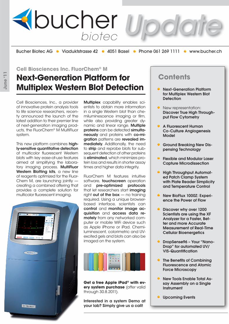
Cell Biosciences, Inc., a provider of innovative protein analysis tools to life science researchers, recen-ty announced the launch of the latest addition to their premier line of next-generation imaging prod-ucts, the FluorChem® M MultiFluor system.
This new platform combines high-ly-sensitive quantitative detection of multicolor fluorescent Western blots with key ease-of-use features aimed at simplifying the labora-tory imaging process. MultiFluor Western Blotting kits, a new line of reagents optimized for the Fluor-Chem M, are launching jointly — creating a combined offering that provides a complete solution for multicolor fluorescent imaging.
Multiplex capability enables sci-entists to obtain more information in a single Western blot than che-miluminescence imaging or film, while also providing greater dy-namic and linear range. Multiple proteins can be detected simulta-neously and proteins with co-mi-gration patterns are revealed im-mediately. Additionally, the need to strip and reprobe blots for sub-sequent detection of other proteins is eliminated, which minimizes pro-tein loss and results in shorter assay times and higher data integrity.
FluorChem M features intuitive software, touchscreen operation and pre-optimized protocols that let researchers start imaging right out of the box — no training required. Using a unique browser-based interface, scientists can control and monitor image ac-quisition and access data re-motely from any networked com-puter or mobile WiFi device such as Apple iPhone or iPad. Chemi-luminescent, colorimetric and UV-excited gels and blots can also be imaged on the system.
Get a free Apple iPad® with ev-ery system purchase (offer valid through 30.8.2011).
Interested in a system Demo at your lab? Simply give us a call!
Next-Generation Platform for Multiplex Western Blot Detection
New representation: Discover True High Through-
put Flow Cytometry
A Fluorescent Human Co-Culture Angiogenesis Model
Ground Breaking New Dis-pensing Technology
Flexible and Modular Laser Capture Microdissection
High Throughput Automat-ed Patch Clamp System with Plate Reader Simplicity and Temperature Control
New BioFlux 1000Z: Experi-ence the Power of Flow
Discover why over 1200 Scientists are using the XF Analyzer for a Faster, Bet-ter and more Accurate Measurement of Real-Time Cellular Bioenergetics
DropSense96 – Your “Nano-Drop” for automated UV/VIS-Quantification
The Benefits of Combining Fluorescence and Atomic Force Microscopy
New Tools Enable Total As-say Assembly on a Single Instrument
Upcoming Events
- 2 -
June 2011
Key Features 96 wells in < 3 min.
384 wells in <12 min.
No priming & No dead vol-ume
Analyze up to 5’000 cells/second
HyperCytPRO intelligent soft-ware for data management
Detects bright and dim cells in each well
Simple set up – User friendly
Brings cytometry into screen-ing labs
Simply call us (061 269 1111) for a system demonstration in your lab!
Bucher Biotec AG is very pleased to announce the appointment as the exclusive Swiss Distributor for the product portfolio of IntelliCyt™ Corporation.
IntelliCyt™ develops innovative high throughput cell screening solutions using flow cytometry technology for drug discovery and life science research.
The company offers innovative products that make it possible for researchers to handle large-scale cell-based assays faster than previously possible.
The New Benchtop Screening Solution for Suspension Cells
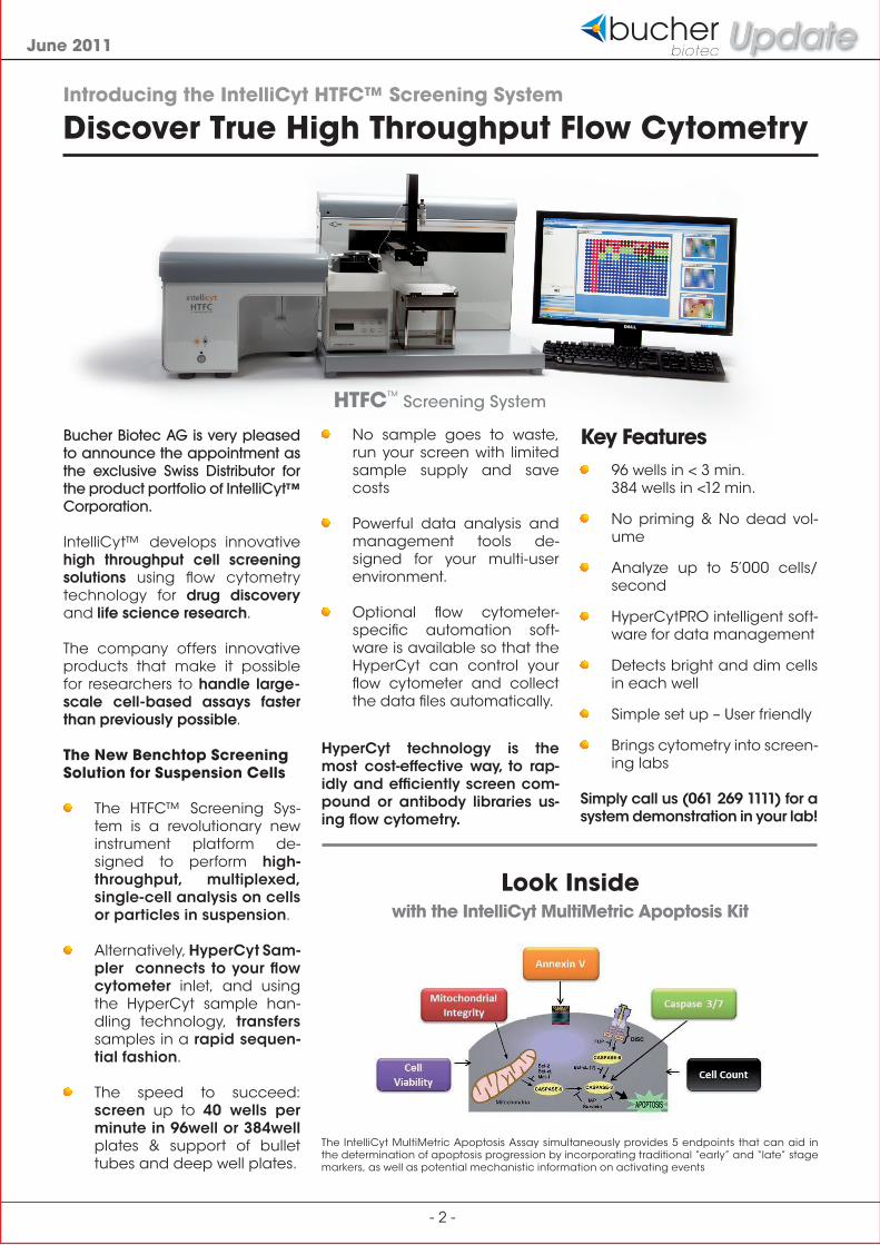
The HTFC™ Screening Sys-tem is a revolutionary new instrument platform de-signed to perform high-throughput, multiplexed, single-cell analysis on cells or particles in suspension.
Alternatively, HyperCyt Sam-pler connects to your flow cytometer inlet, and using the HyperCyt sample han-dling technology, transfers samples in a rapid sequen-tial fashion.
The speed to succeed: screen up to 40 wells per minute in 96well or 384well plates & support of bullet tubes and deep well plates.
The IntelliCyt MultiMetric Apoptosis Assay simultaneously provides 5 endpoints that can aid in the determination of apoptosis progression by incorporating traditional “early” and “late” stage markers, as well as potential mechanistic information on activating events
Look Insidewith the IntelliCyt MultiMetric Apoptosis Kit
HTFC™ Screening System
No sample goes to waste, run your screen with limited sample supply and save costs
Powerful data analysis and management tools de-signed for your multi-user environment.
Optional flow cytometer-specific automation soft-ware is available so that the HyperCyt can control your flow cytometer and collect the data files automatically.
HyperCyt technology is the most cost-effective way, to rap-idly and efficiently screen com-pound or antibody libraries us-ing flow cytometry.
Introducing the IntelliCyt HTFC™ Screening System
Discover True High Throughput Flow Cytometry
- 3 -
June 2011
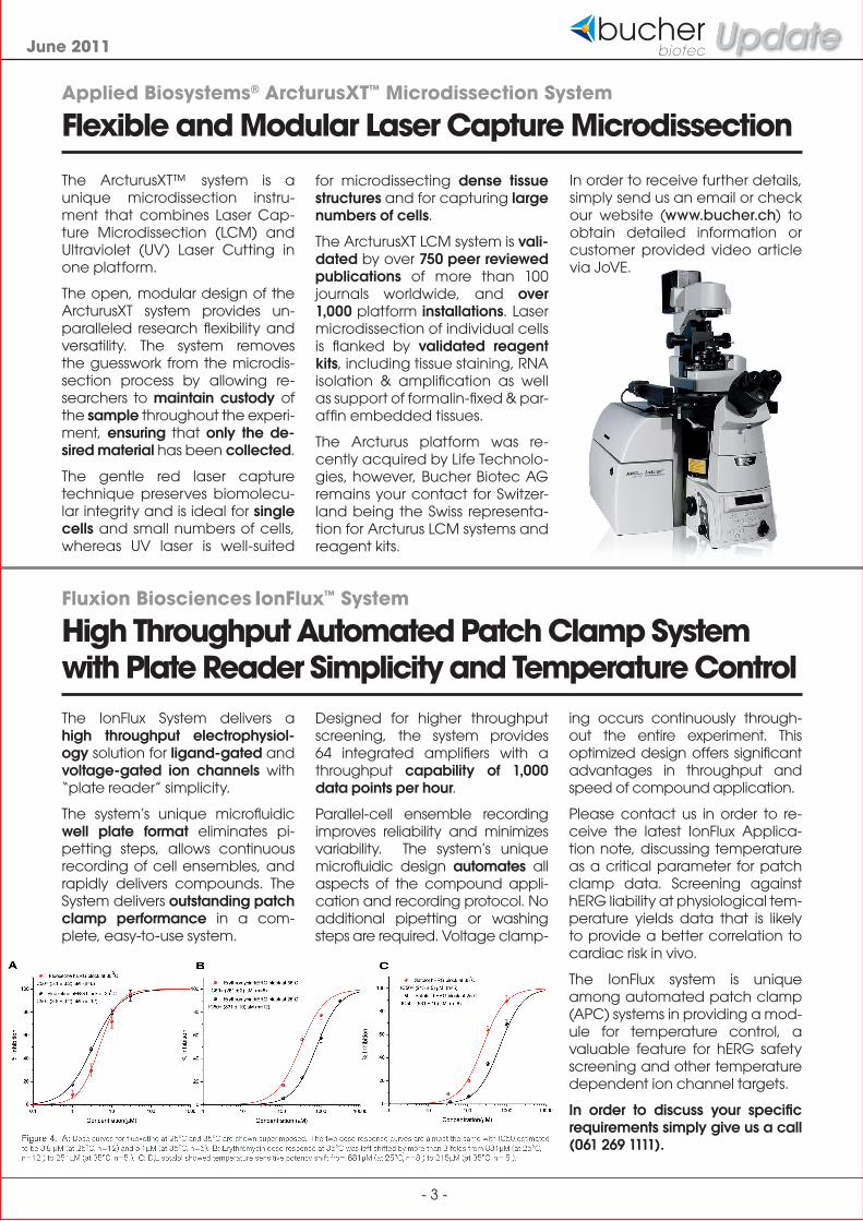
The ArcturusXT™ system is a unique microdissection instru-ment that combines Laser Cap-ture Microdissection (LCM) and Ultraviolet (UV) Laser Cutting in one platform.
The open, modular design of the ArcturusXT system provides un-paralleled research flexibility and versatility. The system removes the guesswork from the microdis-section process by allowing re-searchers to maintain custody of the sample throughout the experi-ment, ensuring that only the de-sired material has been collected.
The gentle red laser capture technique preserves biomolecu-lar integrity and is ideal for single cells and small numbers of cells, whereas UV laser is well-suited
Applied Biosystems® ArcturusXT™ Microdissection System
Flexible and Modular Laser Capture Microdissection
Fluxion Biosciences IonFlux™ System
High Throughput Automated Patch Clamp System with Plate Reader Simplicity and Temperature Control
for microdissecting dense tissue structures and for capturing large numbers of cells.
The ArcturusXT LCM system is vali-dated by over 750 peer reviewed publications of more than 100 journals worldwide, and over 1,000 platform installations. Laser microdissection of individual cells is flanked by validated reagent kits, including tissue staining, RNA isolation & amplification as well as support of formalin-fixed & par-affin embedded tissues.
The Arcturus platform was re-cently acquired by Life Technolo-gies, however, Bucher Biotec AG remains your contact for Switzer-land being the Swiss representa-tion for Arcturus LCM systems and reagent kits.
The IonFlux System delivers a high throughput electrophysiol-ogy solution for ligand-gated and voltage-gated ion channels with “plate reader” simplicity.
The system’s unique microfluidic well plate format eliminates pi-petting steps, allows continuous recording of cell ensembles, and rapidly delivers compounds. The System delivers outstanding patch clamp performance in a com-plete, easy-to-use system.
Designed for higher throughput screening, the system provides 64 integrated amplifiers with a throughput capability of 1,000 data points per hour.
Parallel-cell ensemble recording improves reliability and minimizes variability. The system’s unique microfluidic design automates all aspects of the compound appli-cation and recording protocol. No additional pipetting or washing steps are required. Voltage clamp-
ing occurs continuously through-out the entire experiment. This optimized design offers significant advantages in throughput and speed of compound application.
Please contact us in order to re-ceive the latest IonFlux Applica-tion note, discussing temperature as a critical parameter for patch clamp data. Screening against hERG liability at physiological tem-perature yields data that is likely to provide a better correlation to cardiac risk in vivo.
The IonFlux system is unique among automated patch clamp (APC) systems in providing a mod-ule for temperature control, a valuable feature for hERG safety screening and other temperature dependent ion channel targets.
In order to discuss your specific requirements simply give us a call (061 269 1111).
In order to receive further details, simply send us an email or check our website (www.bucher.ch) to obtain detailed information or customer provided video article via JoVE.

- 4 -
June 2011
Seahorse Bioscience XF Extracellular Flux Analyzer
Discover why over 1200 Scientists are using the XF Analyzer for a Faster, Better and more Accurate Measurement of Real-Time Cellular Bioenergetics
The XF Extracellular Flux assays al-low non-invasive and label-free measurements of cellular bioener-getics. Simultaneous recording of O2 consumption and H+ produc-tion in a fully integrated system us-ing 24-well or 96-well cell culture plates allows researchers easy and efficient access to the physiologi-cal state of primary cells, cell-lines, islets or isolated mitochondria.
By measuring the two major en-ergy producing pathways of the cell simultaneously, mitochon-drial respiration and glycolysis, scientists get the most physiologi-cally relevant bioenergetic as-say available, resulting in a bet-ter overall view of metabolism. The XF Analyzer addresses basal oxygen consumption, fatty acid
Cellular Bioenergetics Webinars On-Demand:Ready when you need them.
To View On-Demand Webinars, visit www.seahorsebio.com
Paolo BernardiUniversity of Padua
Mitochondrial Bioenergetics in Disease: Toward Mitochondrial Therapy
Martin BrandBuck Institute &MRC Dunn
Rate & Coupling Efficiencyof Oxidative Phosphorylationin Mitochondria & Cells
Craig BeesonMedical University of South Carolina
How Bioenergitics Drive Biological Function
Orian ShirihaiTufts School of Medicine
How to Measure Mitochondrial Function in Whole Islets
Antonio C. BiancoUniversity of Miami
Thyroid Hormone Deiodination: a Key Switch for Energy Expenditure
Gary L. WrightEast Tennessee State University
Protective Bioenergetic Responses of Cardiomyocytes to Hypoxic Stress
Vamsi MoothaHarvard Medical School
Human Disorders of Bioenergetics
Russell H. SwerdlowUniversity of Kansas School of Medicine
Cybrid Modeling of Sporadic Neurodegenerative Disorders
Yvonne WillPfizer, Inc.
Mitochondrial Toxicity: Designing Screens to Reduce NCE Attrition
James DykensPfizer, Inc.
Bioenergetic & Metabolic Profiling in Drug Discovery
John J. LeMastersMedical University of South Carolina
Mitochondrial Function in the Life & Death of Cells
David G. NichollsBuck Institute
Quantifying the Mitochondrial ProtonCircuit in Intact Cells
Brian PolsterUniversity of Maryland School of Medicine
Assessing Mitochondrial Dysfunction & the Role of Calpain Proteases in Vitro
Victor Darley-UsmarUniversity of Alabama
Response of the Mitochondria to Oxidative Stress in Cardiovascular Cells
Ben Van HoutenUniversity of Pittsburgh Cancer Institute
Oxidative Stress, Mitochondrial DNADamage & Disease
Metabolic Diseases, Obesity, & Diabetes
Mitochondrial Dysfunction & Toxicity
Neurodegeneration, Aging, & Oxidative Stress
oxidation and metabolism of glu-cose and amino acids for kinetic metabolic information.
We want to welcome you to the popular monthly Seahorse Biosci-ence Cellular Bioenergetics We-binar Series featuring key thought leaders discussing their areas of expertise and the Seahorse XF technology.
Please see www.seahorsebio.com which lists all of the upcoming and past webinars, also sorted by Re-search Area.
Seahorse Bioscience now allows studying mitochondrial function and dysfunction in living cells with less setup and clean up than tra-ditional techniques, such as Clark electrodes.
Fluxion Biosciences Inc BioFlux 1000Z System
New BioFlux 1000Z: Experience the Power of FlowThe new BioFlux 1000Z System is a fully-integrated high content screening platform for bridging the gap between in vitro and in vivo experiments. It integrates
the industry-leading Zeiss Axio-Observer microscopy worksta-tion with the BioFlux electro-pneumatic pumping system for controlling shear flow.
The BioFlux Montage software offers full hardware control over all motorized components and is the command center for design-ing flow experiments, acquiring images and analyzing data.
One System ... many applications
Oncology: Cell adhesion and rolling, Transmigration, Migra-tion and Invasion, Chemot-axis
Platelet Function: Platelet aggregation and adhesion, Thrombosis, Atherosclerosis models
Vascullar Biology: Special-ized Cell Culture, Cell ad-hesion, Atherosclerosis, Mechanical loading , Throm-bosis, Migration and Invasion
Immunology: Transmigration, Migration, Cell Adhesion and Rolling, Wound Healing
Stem Cells: Controlled Dif-ferentiation, Growth Condi-tions, Mechanical Loading, Bioproduction
Microbiology: Biofilm Growth, Mutant Screens, Antibacte-rial or antifungal Screening, Host-Pathogen Interactions, Adhesion Strength

- 5 -
June 2011
The Trinean DropSense96® is a new multichannel spectropho-tometer for quick and precise UV-VIS spectral analysis of micro-liter droplets of DNA/RNA, protein or small compounds.
The spectrophotometer has all the characteristics of a plate reader and can be used with standard 96-well Microplates and specific DropPlate16/96 chips with 1.5 -3 µl sample volume.
The unique features of the DropSense96 are
UV-VIS absorption spectrometry
Full spectrum analysis of 96 samples in less than 5 minutes (230 – 750 nm)
Integrated barcode scanner for sample tracking
Compatible with both Drop-Plate16/96 and classic 96well microplates
Predefined protocols for DNA, RNA and protein concentra-tion measurements
User-friendly software on a re-mote PC
Trinean DropSense96® Multichannel Spectrophotometer
DropSense96 – Your “NanoDrop” for automated UV/VIS-Quantification
Key applications:
Nucleic acids concentration measurements
Purity calculations
Measurement of dye labe-ling efficiency of micro-array samples
Direct protein quantifications
Color measurements (e.g. Bradford)
General UV/VIS measurements
The unique features of the Drop-Plate16 and 96 are:
1,5-3,0 µl samples
16 to 96 sample disposable technology
Compatible with liquid han-dling robots
No sample evaporation with-in 2 hours
Dynamic range: 5-3500 ng/µl dsDNA
No sample dilution needed
Trinean DropSense96® UV-VIS Spectrophotometer
- 6 -
June 2011
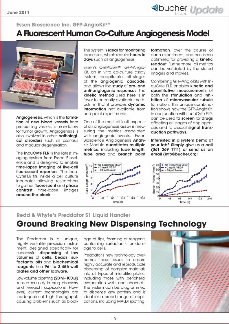
Angiogenesis, which is the forma-tion of new blood vessels from pre-existing vessels, is mandatory for tumor growth. Angiogenesis is also involved in other pathologi-cal disorders such as psoriasis and macular degeneration.
The IncuCyte FLR is the latest im-aging system from Essen Biosci-ence and is designed to enable time-lapse imaging of live-cell fluorescent reporters. The Incu-CyteFLR fits inside a cell culture incubator allowing researchers to gather fluorescent and phase contrast time-lapse images around-the-clock.
The Preddator is a unique, highly versatile precision instru-ment, designed specifically for successful dispensing of low volumes of cells, beads, sur-factants, oils and biochemical reagents into 96- to 3,456-well plates and other labware.
Low volume pipetting (20 nl - 100 μl) is used routinely in drug discovery and research applications. How-ever, current technologies are inadequate at high throughput, causing problems such as block-
Essen Bioscience Inc. GFP-AngioKit™
A Fluorescent Human Co-Culture Angiogenesis Model
Redd & Whyte’s Preddator S1 Liquid Handler
Ground Breaking New Dispensing Technology
The system is ideal for monitoring processes, which require hours to days such as angiogenesis.
Essen’s CellPlayer™ GFP-Angio-Kit, an in vitro co-culture assay system, recapitulates all stages of the angiogenic cascade, and allows the study of pro- and anti-angiogenic responses. The kinetic method used here is in favor to currently available meth-ods, in that it provides dynamic information not available from end point experiments.
One of the most difficult aspects of an angiogenesis assay is mea-suring the metrics associated with angiogenic events. Essen Bioscience Angiogenesis Analy-sis Module quantitates multiple metrics, including tube length, tube area and branch point
formation, over the course of each experiment, and has been optimized for providing a kinetic readout. Furthermore, all metrics can be validated by the stored images and movies.
Combining GFP-AngioKits with In-cuCyte FLR enables kinetic and quantitative measurements of both the stimulation and inhi-bition of microvascular tubule formation. This unique combina-tion shows how the GFP-AngioKit, in conjunction with IncuCyte FLR, can be used to screen for drugs affecting all stages of angiogen-esis and to dissect signal trans-duction pathways.
Interested in a system Demo at your lab? Simply give us a call (061 269 1111) or send us an email ([email protected])!
age of tips, foaming of reagents containing surfactants, or dam-age to cells.
Preddator’s new technology over-comes these issues to ensure highly accurate and reproducible dispensing of complex materials into all types of microtitre plates, including those with peripheral evaporation wells and channels. The system can be programmed to dispense any pattern and is ideal for a broad range of appli-cations, including MALDI spotting.

- 7 -
June 2011
Integration of optical and atomic force microscopy (AFM) provides a powerful tool to obtain comprehensive infor-mation on a variety of samples. Especially combining fluores-cence microscopy and the AFM technique provides com-plementary information: the fluorescence about the loca-tion of labelled molecules not detectable by transmission light microscopy, the AFM finally on the topology of the sample.
The design of the JPK Nano-Wizard® 3 AFM allows its integra-tion into inverted optical devices providing different optical tech-niques like epifluorescence. The use of the DirectOverlay™ fea-ture available for the JPK SPM software enables real optical integration, not only by detect-ing the position of the cantilever within the optical image but also by correcting optical distortions caused by optical lenses.
In this report the combination of sensitive fluorescence detec-tion and AFM, and the way they
JPK Instruments NanoWizard 3 BioAFM
The Benefits of Combining Fluorescence and Atomic Force Microscopy
are complementing each other are described by different ap-plications.
Imaging Cellular structures and extracellular matrix proteins
MC3T3 osteablasts (fig. 1) were stained for f-actin using FITC-phalloidin. The cells were grown on coverslips for use with the CoverslipHolder™. By applying phalloidin staining the actin stress fibers could be visualized using fluorescence microscopy. Additional topographical in-formation could be derived by AFM imaging revealing the fine structure of the filaments.
Another example where the AFM image reveals substruc-tures invisible by fluorescence imaging is D-periodic collagen. Collagen type-1 forms thick fi-bers (approx. 30 nm) if polym-erized on glass (Cisneros et al. 2006, J. Struct. Biol. 154: 232-245). The D-banding pattern can be nicely resolved using AFM imaging (fig. 2).
Fig. 1:Fluorescence image of MC3T3 fibroblasts stained for filamentous actin with inserted de-flection image. Cells were stained with FITC-phalloidin. The AFM image was taken in contact mode in liquid. The scan region was 15 µm x 15 µm, the height range around 300 nm.
Fig. 2: Right: Fluorescence Image of labelled collagen type 1 on glass and AFM image (box).Left: AFM Image of the collagen fibers displaying the D-banding. The scan region was 2 µm x 2 µm, the height range around 50 nm.
Conclusion
The DirectOverlay™ feature al-lows for real optical integra-tion and provides optical and topographic information with-in one region. Fluorescence la-belled cellular components like the cytoskeleton and traffick-ing molecules can be optically tagged and the morphology of the corresponding region char-acterized by AFM imaging. When investigating structures in the nanometer range that can-not be resolved optically, fluo-rescence can help to roughly detect the structures and thus serve as an orientation tool for the search for interesting re-gions to be finally resolved by AFM imaging.
In order to discuss your specific AFM requirements contact us ei-ther by email ([email protected]) or by using the attached info re-quest card.
- 8 -
June 2011
FEMS 2011 Palexpo Geneva
June 26 - 30, 2011
HUPO 2011 Human Proteome Organization Palexpo Geneva
September 4 - 7, 2011
Euro AFM Forum 2011 ETH Zürich
September 7 - 9, 2011
MipTec 2011 The Leading Event for Drug Discovery Congress Center Basel
September 20 - 22, 2011
1st. Intl. SystemsX.ch Conference on Systems Biology
Congress Center Basel October 24 - 26, 2011
Upcoming Events
Labcyte Inc. Echo® Acoustic Liquid Handling Systems
New Tools Enable Total Assay Assembly on a Single Instrument
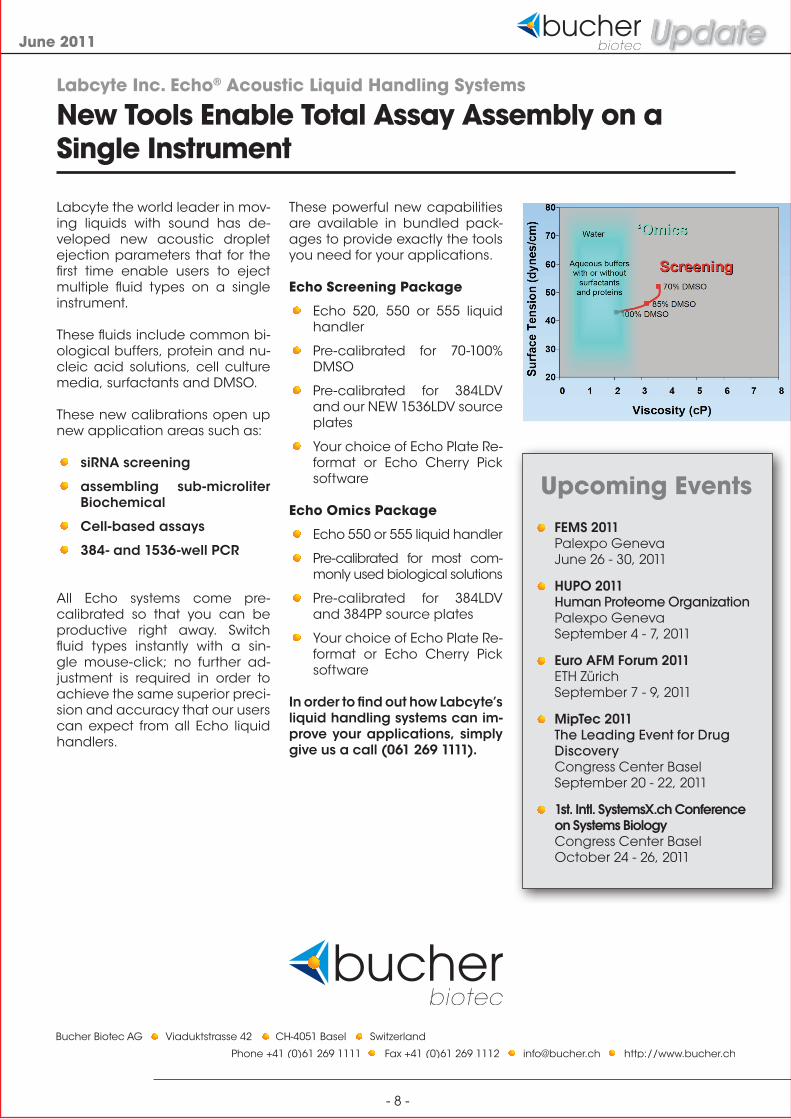
Labcyte the world leader in mov-ing liquids with sound has de-veloped new acoustic droplet ejection parameters that for the first time enable users to eject multiple fluid types on a single instrument.
These fluids include common bi-ological buffers, protein and nu-cleic acid solutions, cell culture media, surfactants and DMSO.
These new calibrations open up new application areas such as:
siRNA screening
assembling sub-microliter Biochemical
Cell-based assays
384- and 1536-well PCR
All Echo systems come pre-calibrated so that you can be productive right away. Switch fluid types instantly with a sin-gle mouse-click; no further ad-justment is required in order to achieve the same superior preci-sion and accuracy that our users can expect from all Echo liquid handlers.
These powerful new capabilities are available in bundled pack-ages to provide exactly the tools you need for your applications.
Echo Screening Package
Echo 520, 550 or 555 liquid handler
Pre-calibrated for 70-100% DMSO
Pre-calibrated for 384LDV and our NEW 1536LDV source plates
Your choice of Echo Plate Re-format or Echo Cherry Pick software
Echo Omics Package
Echo 550 or 555 liquid handler
Pre-calibrated for most com-monly used biological solutions
Pre-calibrated for 384LDV and 384PP source plates
Your choice of Echo Plate Re-format or Echo Cherry Pick software
In order to find out how Labcyte’s liquid handling systems can im-prove your applications, simply give us a call (061 269 1111).