Embed Size (px)
Citation preview

Curriculum Unit 13.04.03 1 of 16
Curriculum Units by Fellows of the Yale-New Haven Teachers Institute2013 Volume IV: Asking Questions in Biology: Discovery versus Knowledge
Cell Biology: From HeLa Cells to the Polio VaccineCurriculum Unit 13.04.03by Lindsey Flanick
Rationale
As a high school science teacher in New Haven it is often challenging to develop units that are engaging andrelevant to my students. When I was given the opportunity to join the Yale New Haven Teachers Institute andparticipate in the "Asking Questions in Biology" seminar I knew that I wanted to develop a unit that involved acase study that was both interesting and relevant to my students. As a result of this seminar, I decided thatusing The Immortal Life of Henrietta Lacks and the discovery of HeLa cells as a focus was the perfect way forme to interest students in cell biology, and to teach them the importance of asking questions in science.
HeLa cells are one of the oldest and most commonly used immortalized cell lines in scientific research. Whencervical cancer cells were taken from Henrietta Lacks in 1951, doctors, researchers and scientists had no ideathe impact they would have on the understanding of cell biology and treatment of health/disease. HeLa cellshave been used to develop vaccines, in cancer and AIDS research, and in countless genetic studies. In thisunit students will use the discovery of HeLa cells and their use in biomedical research to study cell biologytopics such as: cell growth, cell division, virus-cell interactions and vaccine development.
This unit will be centered on the discovery of HeLa cells by using Rebecca Skloot's, "The Immortal Life ofHenrietta Lacks" as an anchor text and backdrop for the content of the unit. With the discovery of HeLa cellsas the central focus of the unit, students will learn about the importance of scientific discovery and the impactit has on future research questions and studies. Students will also analyze the Rolling Stone article, "TheDouble-Edged Helix," which covers the initial challenges of using HeLa cells in research and the impact theyhad on science. The unit will meet both content and inquiry standards as students learn not only about cellbiology, but also about questioning in science and the importance of asking good questions in biology.
The inquiry standards that will be addressed in this unit focus on identifying and developing scientificquestions that can be answered through scientific investigation. Students will learn what it means to ask aquestion and what the criteria are for good questions. As students learn the importance of question-asking inthis unit, they will develop their own question for a scientific investigation that they will carry out as aculminating performance task.
The content standards that will be addressed in this unit are related to cell biology. Students will learn about
Curriculum Unit 13.04.03 2 of 16
the basic structure of cells and cell division as they read excerpts from "The Immortal Life of Henrietta Lacks"and then will move on to studying the differences between cells and viruses, and the development ofvaccines. The use of HeLa cells has been monumental in the development of vaccines, most notably the poliovaccine. Students will see how the accidental discovery of this immortal cell line led scientists to ask morequestions and to develop the vaccines and treatments that we use today. Additionally, students will look athow HeLa cells are still being used in research to answer questions related to cancer and AIDS research.
The use of HeLa cells has been a controversial topic in science. During the time when Henrietta Lacks wasbeing treated for cervical cancer it was common practice for doctors to take samples of cells for use in thelaboratory. A sample of cells was taken from Henrietta Lacks without her knowledge, and it was these cellsthat became the first immortalized human cell line. Henrietta and her family were not made aware that hercells were being used all over the country and world and therefore they never received and compensation.The ethics regarding the use of human cells without the consent or knowledge of the individual they weretaken from remains an interesting topic to explore. Since it is such an engaging topic, students may also raisequestions related to the ethics of using HeLa cells and the impact this discovery had on Henrietta Lacks andher family. Although these questions may not be about biology content, students will be practicing askingquestions and using curiosity to develop their own ideas and understanding.
The Inquiry-Based Classroom: The Importance of Asking Questions
The current trend in science education is to promote inquiry-based learning and student-centered classrooms.These are classrooms where students are asking questions about topics they are interested in and designinginvestigations to develop new knowledge. This idea of the inquiry-based classroom teaches students to "think"like scientists by having them make observations about the world around them and develop questions thatthey can answer through research and laboratory investigations 1 . After all, asking questions is how themajority of the scientific discoveries throughout history were made. Although this idea of scientific discoveryand inquiry-based learning is a trend in education, current lab exercises in high school and even at the collegelevel provide step-by-step directions with a desired and already known outcome 1 . These lab exercises do notteach students to become scientists, they simply teach them to follow instructions. These laboratoryexperiments do not even truly teach the scientific process or scientific method because they are carried outwith adesired result in mind and a "correct answer." True learning and understanding of science occurs whenstudents are able to develop their own experiments without knowing the results so that they can learnsomething new. Recent studies show that inquiry-based classrooms also have increased student engagementand students have a greater understanding of content and material 1 .
So what does an inquiry-based classroom have to do with asking questions in biology? According to Polacekand Keeling, "asking questions is a critical aspect of thinking about science" 1 . Students are more likely to"think" and learn about science when they are asking questions about how discoveries were made that led tothe facts that they are often asked to memorize and recall in class. Students should be asking questions aboutbiology, then carrying out laboratory investigations, and finally reflecting on their data to stimulate theirthinking about new questions and new experiments 1 . This is true science in action, an inquiry-basedclassroom with "question-asking" at its core. In fact, the core of science classrooms should be a laboratory
Curriculum Unit 13.04.03 3 of 16
setting to fully engage students in their learning and understanding of science content and to promotescientific thinking. Biology, specifically, is the study of life and the natural phenomena of living things and thecourse content itself should be thought provoking enough to stimulate students' curiosity and question asking2 . But in a traditional classroom setting where a teacher lectures, students' curiosity can be stifled and theycan fall into the pattern of simply memorizing material without having any interest as to why these naturalphenomena occur or how the world works. In classrooms today, the skill of asking questions to discoverknowledge must be specifically taught and developed in an inquiry classroom 2 .
Not only does asking questions in science have an impact on inquiry-based learning in the laboratory setting,it can also have a significant impact on scientific literacy and understanding. Scientific literacy is the ability toread a scientific article or watch a newscast about a science topic and have an understanding of the materialthat is being discussed and be able to be skeptical and judge whether it is good science 3 . The root ofunderstanding science and scientific literacy is being able to ask a good question. Once a student asks aquestion, they are able to make some observations and begin the research process 3 . By asking questions andmaking observations, students' curiosity about a topic is piqued and it is the beginning of scientific researchand understanding.
Henrietta Lacks and the Discovery of HeLa Cells
"The Immortal Life of Henrietta Lacks" was written by Rebecca Skloot and chronicles the events that led to thediscovery of HeLa cells and the subsequent research in science that eventually led to the development of thepolio vaccine, different medicines, gene mapping, cancer research, and many other important scientificdiscoveries. This accidental, or not-so-accidental, discovery in science is perhaps one of the most important incell biology.
Henrietta Lacks was a poor 30-year-old black woman who died of cervical cancer at Johns Hopkins Hospital inBaltimore, Maryland on October 4, 1951 4 . Before her death, a sample of cervical cells was taken from Lacks,which became the immortalized cell line known as HeLa cells 4 . HeLa was the name given to the human tissuecell culture that was able to thrive in a glass-bound container in the laboratory, given the proper nutrients forcell growth 5 . Essentially, tissue cultures have been artfully convinced that the glass walls that surround themare part of the warm body that they came from and they are able to grow and reproduce like normal cells toform a tissue 5 . Today, trillions upon trillions of HeLa cells are used throughout the world for cell biologyresearch and every single HeLa cell today derived from the one original sample taken from Henrietta Lacksnearly 60 years ago 4 . Tissue cell cultures have allowed scientists to observe cellular processes, withouthaving to do tests on actual human beings 5 . Scientists have seen cellular processes in action, looked atbacterial and viral infections, cancer research, and even studied nutrition 5 . In her book, Skloot searches foranswers about how Lack's cells were obtained and why her family wasn't informed until years later about theamazing contributions these cells made to science.
When Henrietta was diagnosed with cervical cancer in the 1950's it was standard procedure for doctors toobtain a sample of cells and not even notify the patient. Dr. George Otto Gey at Johns Hopkins was trying tocreate an immortalized cell line that could be kept alive in the laboratory, at the time that Henrietta came to

Curriculum Unit 13.04.03 4 of 16
Johns Hopkins for treatment. When Gey was given a sample of Lacks' cells, he was amazed to find that theycould be kept alive and grown, unlike any human cell that he or other scientists had previously tried to culture4 . Previous cell samples would die after just a couple of days. Gey was able to isolate one specific cell fromLacks' sample, multiply it, and start the HeLa cell line. This cell line can be grown in the lab and could be usedfor many experiments. This unexpected discovery was monumental in cell biology and HeLa cells havebecome the most widely studied cultured cells in science 6 .
"The Immortal Life of Henrietta Lacks" would be used in this unit as a hook to interest students in cell biologyand eventually to introduce infectious diseases and vaccinations. The story of Henrietta Lacks and thediscovery of HeLa cells is a compelling one, with many ethical issues to pique the interest of students. Inaddition to having students read excerpts from the book, an article in Rolling Stone magazine, "The Double-Edged Helix" would be used in class to discuss the ethical issues regarding the use of HeLa cells.
Cell Structure: An Overview
Prokaryotic Cells
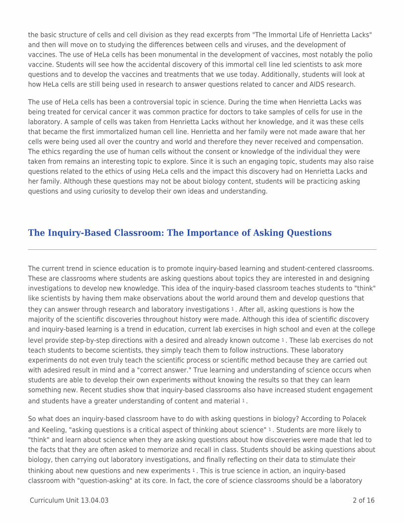
Prokaryotic cells do not house their DNA within a nucleus, and lack certain organelles such as mitochondria.Prokaryotic cells are generally smaller than eukaryotic cells (cells with a nucleus); however, these single cellscan perform all of the basic functions associated with living organisms. Even though these cells do not containa nucleus, they do have genetic material called DNA that floats freely in the cell 7 . This DNA does bundletogether in the center of the cell and is called a nucleoid, however it is not compartmentalized in a nucleuslike eukaryotic cells. Along with their DNA, these cells contain ribosomes, a cell wall, cell membrane, andcytoplasm. Figure 1 is a drawing of a typical prokaryotic cell with the DNA and other organelles labeled.Bacteria are an example of prokaryotic cells.
Curriculum Unit 13.04.03 5 of 16
Figure 1. Prokaryotic Cell
http://upload.wikimedia.org/wikipedia/commons/5/5a/Average_prokaryote_cell-_en.svg
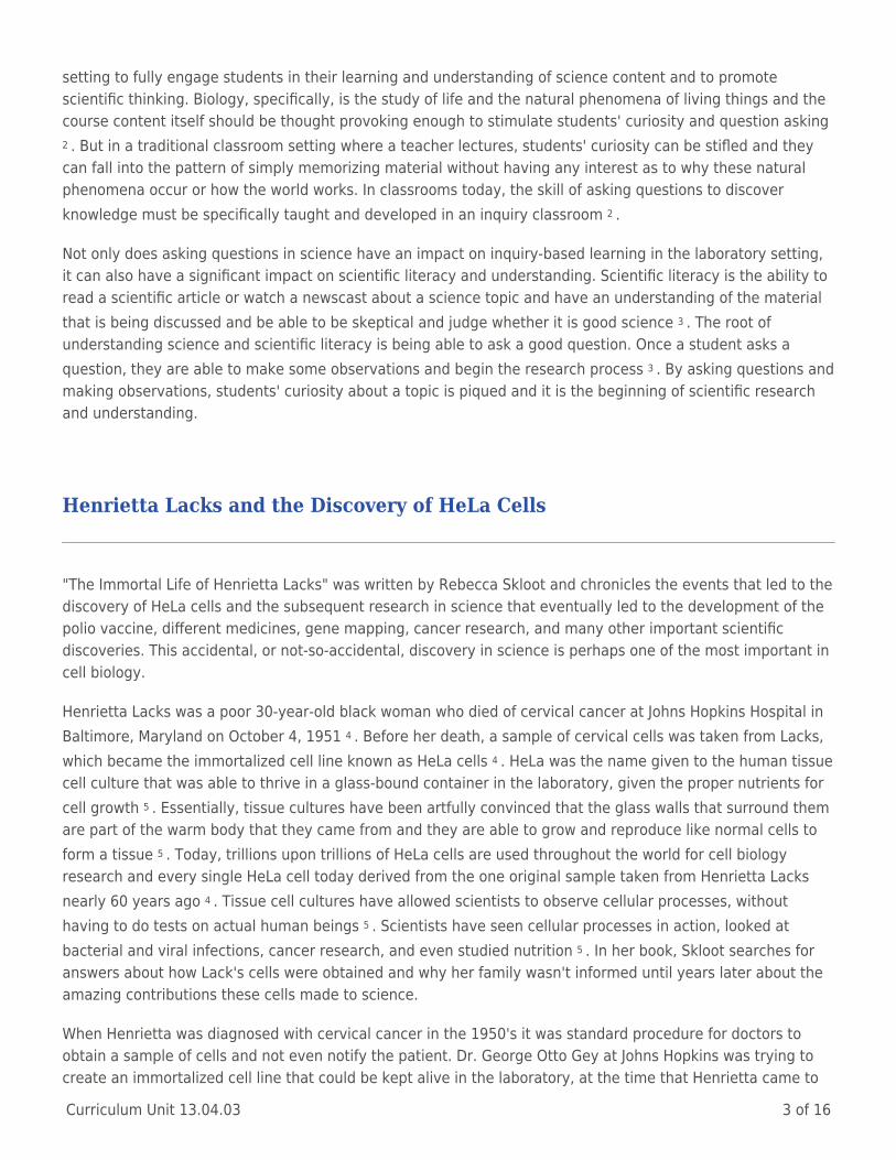
Eukaryotic Cells
Eukaryotic cells are usually much larger and more complex than prokaryotic cells. There are two types ofeukaryotic cells that are taught in high school biology: plant and animal. Plant and animal cells are verysimilar and contain many of the same structures, but a few key differences are used to tell them apart, suchas the presence of chloroplasts and a cell wall in plant cells. All eukaryotic cells contain DNA enclosed in anucleus and also have many different organelles to carry out their cellular functions 7 . These cellularorganelles are like "little organs" that are specialized for the function that they carry out in the cell and theyare all found within the cytoplasm of the cell 7 . The nucleus of the cell holds the DNA, which contains all of thegenetic material to direct the cell to make proteins and reproduce. The nucleus is often referred to as theheadquarters of the cell because it controls all of the cell's activities.
Curriculum Unit 13.04.03 6 of 16
Figure 2. Eukaryotic Animal Cell
http://upload.wikimedia.org/wikipedia/commons/4/48/Animal_cell_structure_en.svg
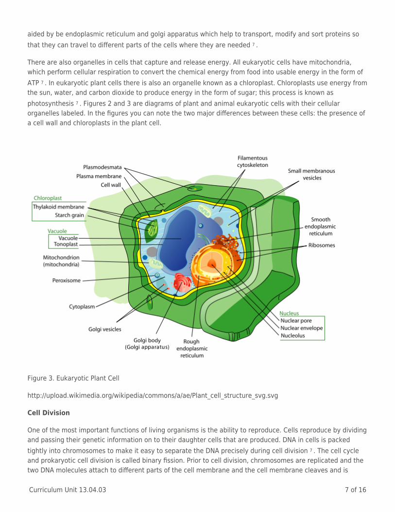
Outside of the nucleus, the remaining organelles float in the cytoplasm and are surrounded by a cellmembrane or plasma membrane. This membrane is made up of a double-layer of phospholipids and is called alipid bilayer 7 . Some membrane proteins also are in the lipid bilayer and help to move materials in and out ofthe cell. The lipid bilayer is considered a selectively permeable membrane because only certain materials areallowed to move in and out of the cell, depending on their size, solubility, and charge. All eukaryotic cells havea cell membrane, and some also have a strong cell wall outside of the membrane to support, shape, andprotect the cell 7 . The existence of a cell wall is one of the major differences between eukaryotic plant andanimal cells; plant cells have a cell wall and animal cells do not.
Within the cytoplasm there are several organelles that are used for storage, clean up and support. Vacuolesare large membrane sacs that store materials such as water, salts, proteins and sugars 7 . Also within the cellare lysosomes, organelles responsible for digesting materials within the cell. These lysosomes containdigestive enzymes that are able to break down lipids, sugars and proteins into smaller molecules that can beused by the cell 7 . Within all eukaryotic cells there is a web of proteins known as the cytoskeleton that givesthe cells their shape 7 .
There are also a group of organelles within eukaryotic cells with the specific job to build proteins. Ribosomesare organelles in cells that are composed of RNA and their job is to build proteins 7 . Protein construction is
Curriculum Unit 13.04.03 7 of 16
aided by be endoplasmic reticulum and golgi apparatus which help to transport, modify and sort proteins sothat they can travel to different parts of the cells where they are needed 7 .
There are also organelles in cells that capture and release energy. All eukaryotic cells have mitochondria,which perform cellular respiration to convert the chemical energy from food into usable energy in the form ofATP 7 . In eukaryotic plant cells there is also an organelle known as a chloroplast. Chloroplasts use energy fromthe sun, water, and carbon dioxide to produce energy in the form of sugar; this process is known asphotosynthesis 7 . Figures 2 and 3 are diagrams of plant and animal eukaryotic cells with their cellularorganelles labeled. In the figures you can note the two major differences between these cells: the presence ofa cell wall and chloroplasts in the plant cell.
Figure 3. Eukaryotic Plant Cell
http://upload.wikimedia.org/wikipedia/commons/a/ae/Plant_cell_structure_svg.svg
Cell Division
One of the most important functions of living organisms is the ability to reproduce. Cells reproduce by dividingand passing their genetic information on to their daughter cells that are produced. DNA in cells is packedtightly into chromosomes to make it easy to separate the DNA precisely during cell division 7 . The cell cycleand prokaryotic cell division is called binary fission. Prior to cell division, chromosomes are replicated and thetwo DNA molecules attach to different parts of the cell membrane and the cell membrane cleaves and is
Curriculum Unit 13.04.03 8 of 16
pinched to divide into two cells 7 . The result is two identical daughter cells with the same DNA.
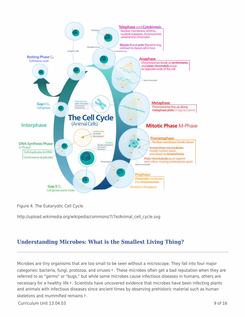
The eukaryotic cell cycle is relatively more complex and has four major phases: G1, S, G2, and M phase 7 .Figure 4 gives an overview of the eukaryotic cell cycle and cell division. A eukaryotic cell spends most of its"life" in interphase, which is composed of G1, S and G2. G1 and G2 are the growth phases where the cellgrows and prepares for cell division. The "S" phase is the synthesis phase, where DNA is being replicated forcell division. After the cell goes through interphase, it enters the mitotic phase, which is composed of mitosisand cytokinesis. Mitosis can be broken down into a series of events that lead to cell division: prophase,metaphase, anaphase and telophase 7 . During prophase the cell "prepares" for cell division as DNA condensesinto chromosomes and the nuclear envelope dissolves to allow the chromosomes to migrate throughout thecell 7 . The next step, metaphase, is where chromosomes migrate to the "middle" of the cell and spindle fibersthat have formed attach to the centromere at the center of the chromosomes and to opposite ends of thecells. During anaphase the spindle fibers begin to pull the replicated chromosomes "apart" and towardsopposite ends so that a complete set of chromosomes is on each side 7 . The last step in mitosis is telophase.This is where chromosomes spread back out and a nuclear envelope reforms around the two identical sets ofDNA at opposite ends of the cell. At this point mitosis is complete, but cell division has not yet occurred 7 . Theactual process of cell division is called cytokinesis. During cytokinesis the cell membrane begins to cleave inthe center and eventually the cytoplasm is pulled into two parts. The result is two identical daughter cells withthe same DNA. The cell cycle occurs in both eukaryotic plant and animal cells to promote cell growth.
The cell cycle is closely controlled by regulatory proteins. There are both internal regulators and externalregulators. External regulators are proteins that respond to events outside of the cell 7 . Examples of externalregulators are growth factors, which are proteins that control the cell cycle when you have an injury or duringthe development of an embryo 7 . Internal regulators are proteins that respond to events inside of the cell andmake sure that the cell cycle is controlled and that the timing of mitosis is regulated 7 . When cell growthbecomes uncontrolled it results in cancer. Cancer develops when a cell becomes aberrant and ignores checkson its own growth 7 . In the case of Henrietta Lacks, cervical cells were growing without regulation and a tumorformed in her cervix. A tumor is a mass of cancer cells that grows and divides rapidly 7 . As the cancer cellsgrow and divide they use nutrients from nearby healthy cells and can prevent organs and tissues fromfunctioning normally. Cancer is caused by a mutation in the DNA that regulates cell growth and division 7 .
Once students are introduced to the basic cell structure of prokaryotic and eukaryotic cells it their divisionprocess, this unit moves towards infectious diseases and how HeLa cells were used to develop vaccines forprevention. It is important for students to have a basic understanding of both bacteria and viruses beforelearning about infectious diseases and how they affect the human body.
Curriculum Unit 13.04.03 9 of 16
Figure 4. The Eukaryotic Cell Cycle
http://upload.wikimedia.org/wikipedia/commons/7/7e/Animal_cell_cycle.svg
Understanding Microbes: What is the Smallest Living Thing?
Microbes are tiny organisms that are too small to be seen without a microscope. They fall into four majorcategories: bacteria, fungi, protozoa, and viruses 8 . These microbes often get a bad reputation when they arereferred to as "germs" or "bugs," but while some microbes cause infectious diseases in humans, others arenecessary for a healthy life 8 . Scientists have uncovered evidence that microbes have been infecting plantsand animals with infectious diseases since ancient times by observing prehistoric material such as humanskeletons and mummified remains 8 .
Curriculum Unit 13.04.03 10 of 16
Bacteria
The group of microbes known as bacteria is single-celled organisms that fall into the category of life known asthe prokaryotes. Prokaryotic cells do not contain a nucleus, but bacterial cells do contain DNA as their geneticmaterial. Like other prokaryotic cells, bacteria often have a rough cell wall, plasma membrane, and ribosomessuspended in their cytoplasm. Many bacteria require oxygen and need food for energy 8 . Bacteria are amongthe oldest living things on Earth, with scientists having found fossilized remains that date back more than 3.5billion years 8 . Bacteria can also survive in the most extreme conditions, from extremely cold to extremely hotareas. There are thousands of species of bacteria but a majority of bacteria fall into three basic shapes: rod-shaped bacilli, ball-shaped cocci, and spiral-shaped 8 . Of the thousands of species of bacteria, less than onepercent causes diseases in humans 8 .
Viruses
It is often debated whether or not a virus is considered a living thing. Most notably, these functions includemetabolism and reproduction. While viruses do contain their own genetic material (DNA or RNA), a virus is notmade up of cells and cannot perform the basic functions of living things without hijacking a cell and takingcontrol of its metabolism to reproduce 8 . So is it alive? Or is a virus this strange particle that straddles a thinline between a living and nonliving thing? The question continues to be asked by scientists today. Viruses arebasically small bundles of genetic material, either DNA or RNA, covered in a protein coat called a capsid 8 .They can be rod-shaped, sphere-shaped, or icosahedral 8 . Like other microbes, viruses can be found almosteverywhere on Earth. They are the smallest microbe, and are basically found anywhere that there is a cell toinfect; from bacterial cells to human cells. There are different viruses, each one behaving differently and veryparticular about what type of cell it hijacks and infects 8 . When they attack, viruses attach to the outside of acell, enter the cell or inject their genetic material, and then take over the metabolism of the cell and direct itto make more copies of the viruses that are released to infect other cells 8 .
Infectious Diseases
Tiny microbes, such as viruses, bacteria, fungi and protozoa, can cause infectious diseases in humans. Whilemany microbes do not harm us and some even help us, the disease-causing microbes that we refer to as"germs" can make us sick and can even be deadly 9 . Microbes are spread through food, water, the air, theenvironment, or physical contact between two people or sometimes contact with animals 9 .
The Immune Response and Germ Theory
When our bodies are infected with a microbe our immune system recognizes the foreign invader and launchesan immune response 10 . The main defense we have against foreign substances such as bacteria or viruses areour white blood cells. White blood cells are produced in the bone marrow and we make about a billion of themeach day 10 . White blood cells, called macrophages, will detect and destroy bacteria when they see them.When a viral infection begins our T and B-lymphocytes will work to fight off the infection 10 . B-cells areimportant in producing antibodies, which bind to viruses to stop them from replicating and tag them so that

Curriculum Unit 13.04.03 11 of 16
other cells can recognize them. Once an infection is cleared, some specialized B and T-cells stay in our bodiesand act as memory cells that will easily recognize a virus if we come in contact with it again 10 . Scientistshave been able to use this idea of acquired immunity and resistance with memory cells to create vaccines,which will be discussed later in this section.
Before the discovery of these disease-causing microbes, people blamed sickness on evil spirits or "bad blood"9 . It wasn't until the invention of the microscope that scientists were able to see microbes and began to studybacteria, viruses, protozoa and fungi. Louis Pasteur was the first scientist to establish the presence of germsby linking them to illnesses. Pasteur proved that fungi and bacteria were present in the air and that theycaused sickness in humans 9 . He was able to show that germs caused diseases and began to pioneer a way toget rid of them with a process called pasteurization; eventually he was able to develop vaccines for chickenpox, cholera, diphtheria, anthrax and rabies 9 . Pasteurization is the process of heating and then cooling aliquid food to remove microbes that may spoil that food (beer, wine, milk, etc.). Pasteur began by studyingalcohol fermentation and observed that wine would go sour and turned to vinegar unexpectedly 11 . Using amicroscope, Pasteur was able to see that small, rod-like microbes were present during fermentation and causethe contaminating the wine 11 . Pasteur was able to identify microbes that were responsible for contaminatingbeer, wine and milk and found that if he heated the liquids to a high temperature and then cooled them downhe could kill the microbes and sterilize the liquids 11 . This process became known as pasteurization, and wascoined to honor Louis Pasteur and his discovery. The identification of microbes that "spoiled" beer and wineled scientists to the understanding that diseases are caused by microorganisms that entered the body; similarto how microbes could contaminate the liquids Pasteur studied 11 . Today, scientists are still researchingcauses of various human diseases and ways to prevent or cure them.
Treatment and Prevention
Two of the most important tools for curing or preventing diseases are antibiotics and vaccines. Antibiotics area class of drugs that kill bacteria in the human body 9 . Antibiotics are targeted at bacterial infections andcome in the form of pills, liquids, injections, lotions, or creams; each type is used to attack a specific bacterialpathogen 9 .
Vaccines are another way to fight infectious diseases, but unlike antibiotics that are used to treat an infection,vaccines are used to prevent disease. Vaccines stimulate the immune system by creating antibodies to fightspecific infections. A vaccine usually contains a killed or weakened form of a microbe that causes a diseaseand your body is able to produce antibodies to kill that germ without getting sick 9 . When a person who hasbeen given a vaccine is later exposed to the illness, their body is already prepared to fight the infectionbecause of the antibodies that are already created. Edward Jenner developed the first real vaccine when heinoculated a young boy with the cowpox virus in 1796 9 . Jenner found that people exposed to the cowpoxvirus did not contract smallpox, a disease that was killing thousands of people. Edward Jenner began byobserving patients who had contracted cowpox while working on the farm. These patients had milddiscomfort, aching, pustules and swelling but the disease did not lead to death like smallpox 12 . Jenner notedthat the individuals who were infected with cowpox did not become infected with smallpox when they wereexposed to this. Jenner hypothesized that this was because the individuals who were infected with cowpox hadantibodies that made them immune to the smallpox virus. Jenner tested his hypothesis on an 8-year old boy,James Phipps, by vaccinating him with a fluid from pustules of cowpox-infected patients and then exposinghim to the smallpox virus 12 . When he came in contact with the disease Phipps did not contract smallpox, a
Curriculum Unit 13.04.03 12 of 16
result of the immunity he obtained from the cowpox injection. Jenner had successfully created the smallpoxvaccination by triggering the immune system to create antibodies for a pathogen that was closely related tosmallpox to fight the infection when exposed to it. This discovery would lead to the development of manyother vaccines throughout history.
Vaccine Development: A Focus on Polio
Polio is a disease caused by a virus that attacks the central nervous system and causes paralysis 13 . Thepoliovirus is spread by direct person-to-person contact by contact with infected mucus or feces and leads tothe disease, poliomyelitis 13 . Like all viruses, the poliovirus works by infecting human cells. The poliovirusenters the body through the mouth and nose where it travels through the digestive system and multiplies 13 .Once it reaches the intestinal tract, the virus is absorbed into the blood stream and spread to the lymphaticsystem 13 . While polio has been largely eradicated, lack of immunization and exposure to the poliovirus willcause a person to contract the disease. The people that are most likely to be infected by the poliovirus arechildren, pregnant women, and the elderly 13 .
Polio was a worldwide epidemic between 1840 and 1950, before the polio vaccine was developed. Thedevelopment of the vaccine was aided largely by the discovery of HeLa cells and the incidence of the diseasehas been greatly reduced 13 . In fact, the disease has been completely eradicated in some countries. In 1948,Dr. Jonas Salk began researching the poliovirus at a virology lab at the University of Michigan 14 . HeLa cellswere easily infected by the poliovirus and therefore were perfect for vaccine testing and development. SinceHeLa cells were easily grown and cultured in the laboratory, Salk was able to infect large amounts of HeLacells and develop a vaccine that was viable for human use. In 1950 he began working to create an inactivatedpoliovirus vaccine, and by 1952 Dr. Salk had created what he considered a safe and effective vaccine againstthe poliovirus 14 .
Teaching Strategies
The Cell City
Teaching students to memorize all of the organelles in prokaryotic cells and eukaryotic plant and animal cellscan be a tedious job. Instead, have students complete a project where they are asked to create an analogybetween a cell and a city. The cell can be thought of as its own miniature city: each organelle represents apart of a city that carries out a specific job or function so that the cell can survive. For example, the nucleuswould be like the town hall of a small city. The nucleus is in charge of controlling all of the actions of the cell,just as the town hall is responsible for controlling the activities. Additionally, the mitochondria can becompared to the power company in a town. The mitochondria are responsible for powering the cell throughcellular respiration and the formation of ATP. Similarly, the power company produces energy for the city.Students should be asked to come up with a comparison for all major cellular organelles to develop a

Curriculum Unit 13.04.03 13 of 16
complete cell city analogy. The final project would consist of a poster presentation or 3D model of the city thatis labeled with both the cell and city parts. A written summary explaining the analogy should also be included.Also, this activity can be extended by using the "cell city" as an example and then asking students to come upwith their own analogy such as a classroom, school bus, car, kitchen, house, etc.
Infectious Disease Case Studies
In this activity students are given the opportunity to be the doctor and diagnose patients that are presentingwith various infectious diseases. Provide the students with a list of patients and a brief summary of theirsymptoms. The goal of the activity is to have students correctly diagnose the patients using the Internet andtheir understanding of pathogens. Students should use websites such as Web MD and Mayo Clinic to assessthe symptoms of their patients. In addition to correctly diagnose their patients, students should determinewhat type of pathogen caused the infectious disease (virus, bacteria, protist or fungus) and should develop aplan for treatment. Some diseases that can be used are: athlete's foot (fungus), HIV/AIDS (virus), strep throat(bacteria), tetanus (bacteria), amebic dysentery (bacteria), and malaria (protist).
Spread of Disease Laboratory
A great way to demonstrate the transmission of disease is to have students complete the spread of diseaselaboratory experiment. In this lab, students are each given a cup with a clear, colorless solution and adisposable pipette. All students in the class, except for one, are given a cup with just water in it. One studentin class is the "index patient" and their cup has a few drops of sodium hydroxide in it. None of the studentsknow who the index patient is at the start of the lab. Students are instructed to "come in contact" with 3 otherpeople in the class by exchanging a pipette full of solution with each person. This should be completed inrounds and signaled by the instructor. Students should keep track of with whom they have come in contactwith in their data table. At the end of the third round a drop of phenolphthalein should be added to eachstudents' cup. Students who were infected by the disease will end up with a pink solution in their cup. As aclass, students should create a tree to determine how the disease was transmitted and to determine who theindex patient was. This activity promotes collaboration between all students in the classroom anddemonstrates how quickly a disease can be spread throughout a population. Additionally, this activity can berepeated with more than one index patient to make the determination of transmission more difficult for theclass to solve.
Pathogen Jigsaw
In this unit is important to understand that there are several pathogens that cause disease in humans:bacteria, fungi, protists, and viruses. An interactive way to get students involved in their learning and promotereading is to have students participate in a pathogen jigsaw. Students should be divided into groups of four forthis activity. Each member in a group will be assigned a different pathogen that they are responsible forlearning about. Students are given short articles with information about the pathogens and they are giventime to read the information and look for key points and facts. Students are instructed to become an "expert"on their specific pathogen. Then, each expert will teach the other three members in their group about thepathogen they read about. Students can be given a graphic organizer to take notes about each pathogen sothat at the end of the activity they have information about viruses, bacteria, fungi and protists.
Microscope Investigations
The use of light microscopes is essential for this unit. Students should be given several opportunities

Curriculum Unit 13.04.03 14 of 16
throughout the unit to observe life at the cellular level using a microscope. There are many different ways toincorporate microscopes into this unit, such as: the use of prepared slides to observe microscopic bacteria,fungi and protists, the use of the plant Elodea to observe chloroplasts, and the use of potato or cork toobserve plant cell walls.
HeLa Cells and Chromosome Spreads
CellServ produces a kit that will allow students to use HeLa cells in the lab to observe cells and chromosomesunder the microscope. Students are given microtubules containing HeLa cells that have been fixed in solutionprior to metaphase in mitosis. Students use pipettes to drop the cell solution onto clean microscope slides andthen stain the slides with stain that is included in the kit. Once the slides have dried, students are able to lookfor cells under the microscope. Using the oil immersion lens on a light microscope students should be able tosee chromosomes inside of the cells.
iPad Applications
There are several applications that are available on the iPad that give students a look at cellular life and thespread of disease. A list of a few of these applications and a short summary are below.
Powers of Minus Ten – Cells and Genetics: This application allows students to virtually zoom in through ahuman hand to observe skin and blood cells. The application can be used to show students different phases ofmitosis, differentiate between different types of cells, and identify cell parts.
Plague Inc.: This application is a simulation about how a virus can spread throughout the globe rapidly. A virusrapidly reproduces and mutates as students try to stop it by creating vaccines and ways to prevent the spreador cure the disease.
3D Cell Simulation and Stain Tool: This application allows students to virtually look at 3D plant and animalcells. The application has information about cellular organelles and students can stain the cell to focus on oneor more organelles at a time.
Appendix A: Standards
DINQ.1 Identify questions that can be answered through scientific investigation
DINQ. 2 Read, interpret and examine the credibility and validity of scientific claims in different sources ofinformation
DINQ. 4 Design and conduct appropriate types of scientific investigations to answer different questions
D.31 Describe the similarities and differences between bacteria and viruses
D.30 Explain the role of the cell membrane in supporting cell functions
D.27 Describe significant similarities and differences in the basic structure of plant and animal cells
Curriculum Unit 13.04.03 15 of 16
Appendix B: Materials for Students
National Institute of Allergy and Infectious Disease. "Understanding Microbes: in Sickness and in Health." NIH Publication , no.09-4914 (2009).This is a publication of the NIAID and covers basics about microbes such as: how microbes make us sick,immunity, types of microbes, infectious diseases, treatment, and prevention.
Rogers, Michael. "The Double-Edged Helix." Rolling Stone Magazine . March 23, 1976.This is one of the first articles published aboutHeLa cells and the controversy over the use of Henrietta Lacks' cells in science without the consent of her family.
"Smallest Thing." American Association for the Advancement of Science. Last modified 2013.http://sciencenetlinks.com/science-news/science-updates/smallest-thing/. This website has lesson plans that talk about microbes,specifically viruses and the question of whether they can be considered living things. The website includes articles, questions andlesson ideas.
Skloot, Rebecca. The Immortal Life of Henrietta Lacks. New York: Broadway Paperbacks, 2010.This book is used as the backdrop forthis unit. Skloot details the life of Henrietta Lacks and the contributions her cells have made to science, as well as the controversyher family has become a part of with the use of her cells without their knowledge.
Appendix C: Bibliography
1 Polacek, Kelly Myer and Keeling, Elena Levine. "Easy Ways to Promote Inquiry in a Laboratory Course." Journal of College ScienceTeaching (2005): 52-55. This resource is a research article about using inquiry in laboratory investigations at the high school andcollege level.
2 Zion, Michal and Sadeh, "Curiosity and Open Inquiry Learning." Journal of Biological Education 41, no. 4 (2007): 162-168. Thisresearch article talks about inquiry learning in the science classroom.
3 Brewer, Carol. Scientific Literacy in the Classroom .By ActionBioscience.org. AIBS Annual Meeting, 2007.This resource is aninterview by Carol Brewer about the use of scientific literacy to empower students in the classroom and improve their inquiry andcritical thinking skills.
4 Claiborne, Ron and Wright, Sidney. "How One Woman's Cells Changed Medicine." ABC World News , January 31, 2010.http://abcnews.go.com/WN/henrietta-lacks-woman-cells-polio-cancer-flu-research-medicine/story?id=9712579#.UaQiLpV3cwE. Thisarticle gives a general overview of HeLa cells and their use in the development of the polio vaccine and medicine.
5 Rogers, Michael. "The Double-Edged Helix." Rolling Stone Magazine . March 23, 1976. This is one of the first articles publishedabout HeLa cells and the controversy over the use of Henrietta Lacks' cells in science without the consent of her family.
6 Brumback, Roger A. "Book Reviews: The Immortal Life of Henrietta Lacks." Journal of Evidence-Based Complementary & AlternativeMedicine 16, no. 3 (2011).This resource is a book review of the Immortal Life of Henrietta Lacks by Rebecca Skloot.

Curriculum Unit 13.04.03 16 of 16
7 Miller, Kenneth R., and Levine, Joseph S. Biology: Foundation Edition . New Jersey: Pearson Education, Inc., 2010. A high schooltextbook with general information related to cell biology and infectious diseases. A great student resource as well.
8 National Institute of Allergy and Infectious Disease. "Understanding Microbes: in Sickness and in Health." NIH Publication , no.09-4914 (2009).This is a publication of the NIAID and covers basics about microbes such as: how microbes make us sick, immunity,types of microbes, infectious diseases, treatment, and prevention.
9 "Part 2: So, What's Changed." American Association for the Advancement of Science. Last modified 2013.http://sciencenetlinks.com/media/filer/2012/02/17/yourhealth9-21.pdf. An article by the AAAS about microbes and infectious disease.
10 "How Does the Body Fight Off a Virus?" BBC. Last modified 2013. http://www.bbc.co.uk/science/0/22028517. This resource is anarticle by BBC Science: Knowledge and Learning Beta. It reviews the immune response to a viral infection and the role macrophages,T-cells and B-cells play in immunity.
11 Cohn, Ph.D. David V. "Louis Pasteur." October 2004. http://pyramid.spd.louisville.edu/~eri/fos/interest1.html. This resource givessome background information about Louis Pasteur and the development of the pasteurization process for elimination microbes. Italso briefly describes how Pasteur contributed to the germ theory for the spread of disease.
12 Cohn, Ph.D. David V. "Edward Jenner." October 2004. http://pyramid.spd.louisville.edu/~eri/fos/jenner.html. This resource givessome background information about Edward Jenner and the development of the smallpox vaccine.
13 The University of Maryland: First Year Book Program. 2013. http://fyb.umd.edu/2011/henrietta-lacks.html.This website by theUniversity of Maryland covers some background information about HeLa cells, Rebecca Skloot's book and the development of thepolio vaccine at the Salk Institute.
14 The Salk Institute for Biological Studies. 2013. http://www.salk.edu/index.php.This resource gives a timeline for the development ofthe polio vaccine from the Salk Institute.
https://teachersinstitute.yale.edu©2019 by the Yale-New Haven Teachers Institute, Yale UniversityFor terms of use visit https://teachersinstitute.yale.edu/terms