Embed Size (px)
Citation preview
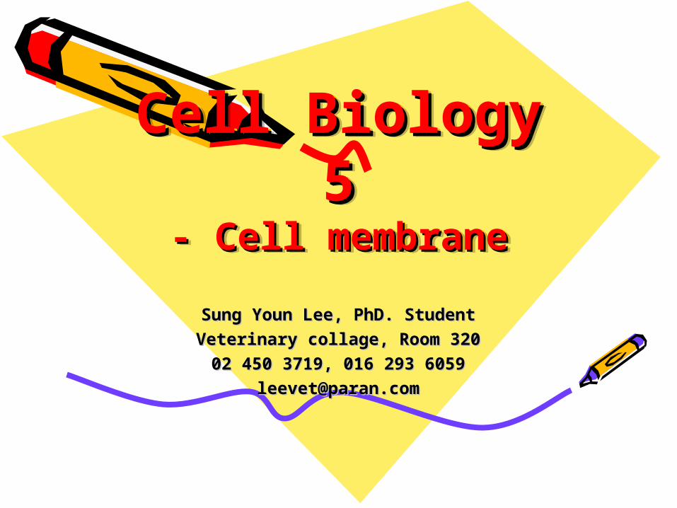
Cell Biology 5Cell Biology 5- Cell membrane- Cell membrane
Cell Biology 5Cell Biology 5- Cell membrane- Cell membrane
Sung Youn Lee, PhD. StudentSung Youn Lee, PhD. StudentVeterinary collage, Room 320Veterinary collage, Room 32002 450 3719, 016 293 605902 450 3719, 016 293 6059
[email protected]@paran.com
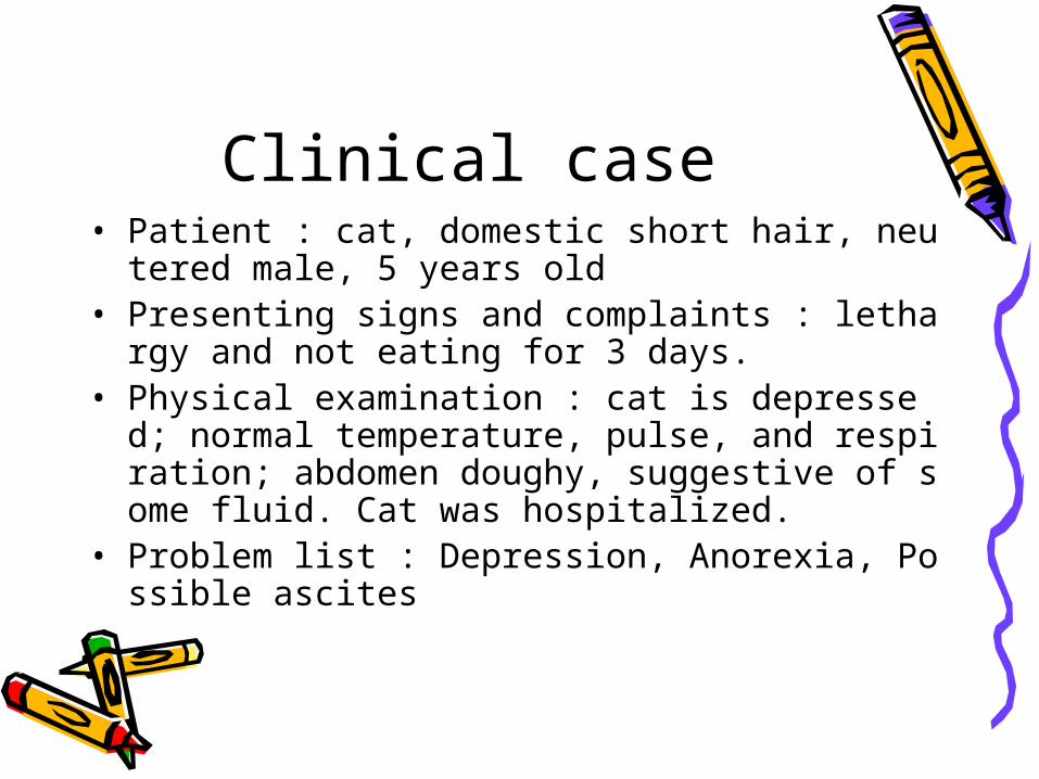
Clinical case• Patient : cat, domestic short hair, neutered male, 5 y
ears old• Presenting signs and complaints : lethargy and not e
ating for 3 days.• Physical examination : cat is depressed; normal temp
erature, pulse, and respiration; abdomen doughy, suggestive of some fluid. Cat was hospitalized.
• Problem list : Depression, Anorexia, Possible ascites
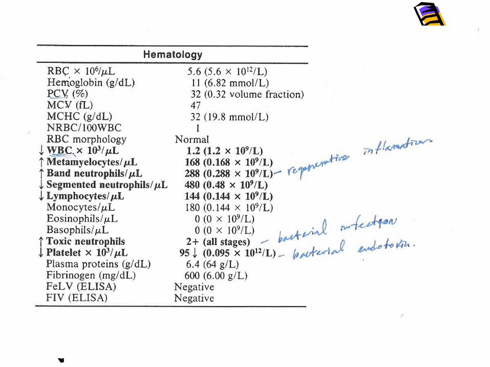
Hematogram• Neutropenia with a marked left shift and
toxic neutrophils is compatible with a degenerative shift and probably associated with septic exudate in the abdomen which, because of the excessive demand, results in depletion of the marrow storage and maturation pools.
Hematogram• This is in contrast to Case 1 in which there w
as also a septic condition, but the neutropenia was caused by bone marrow dysplasia resulting from FeLV infection. The presence of a degenerative left shift aids in differentiating between neutropenia caused by excessive tissue demand and ineffective myelopoiesis.
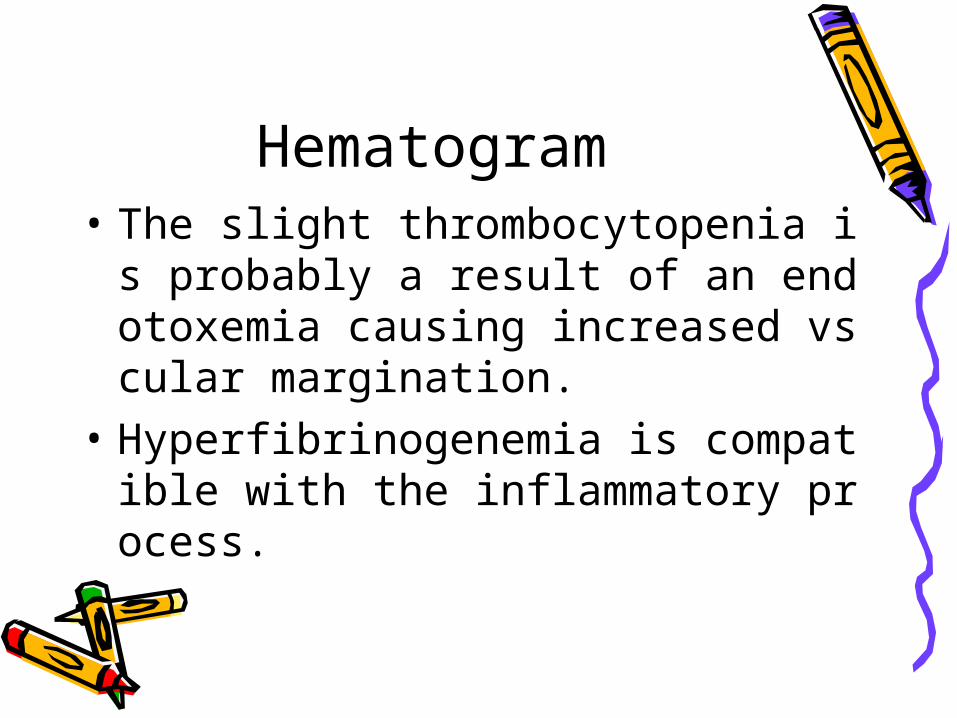
Hematogram• The slight thrombocytopenia is probably
a result of an endotoxemia causing increased vscular margination.
• Hyperfibrinogenemia is compatible with the inflammatory process.

Diagnosis• The patient was taken to surgery for a laparot
omy, and a small rupture in the duodenum was found leaking intestinal contents into the abdomen.
• A reason for the rupture of the viscus was not apparent. The defect was repaired, and the cat made an uneventful recovery.
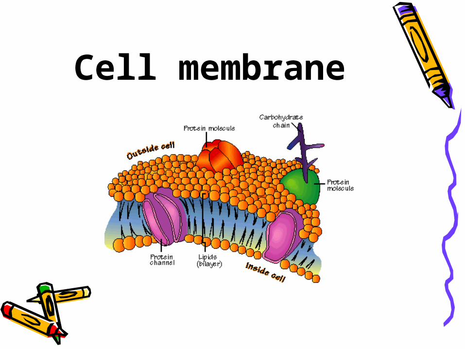
Cell membrane
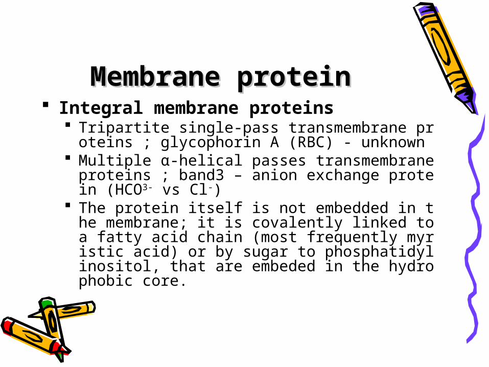
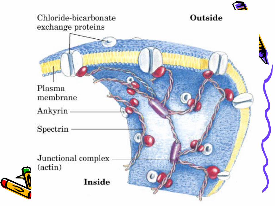
Membrane proteinMembrane protein Integral membrane proteins
Tripartite single-pass transmembrane proteins ; glycophorin A (RBC) - unknown
Multiple α-helical passes transmembrane proteins ; band3 – anion exchange protein (HCO3- vs Cl-)
The protein itself is not embedded in the membrane; it is covalently linked to a fatty acid chain (most frequently myristic acid) or by sugar to phosphatidylinositol, that are embeded in the hydrophobic core.
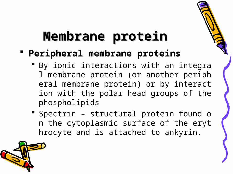
Membrane proteinMembrane protein Peripheral membrane proteins
By ionic interactions with an integral membrane protein (or another peripheral membrane protein) or by interaction with the polar head groups of the phospholipids
Spectrin – structural protein found on the cytoplasmic surface of the erythrocyte and is attached to ankyrin.
Integral and peripheral• Detergent – disrupt the membrane - integral• By shifting the ionic strength or pH of aqueou
s solution - dissociation of the ionic interaction of the peripheral protein with either phospholipid polar head groups or other membrane proteins– peripheral
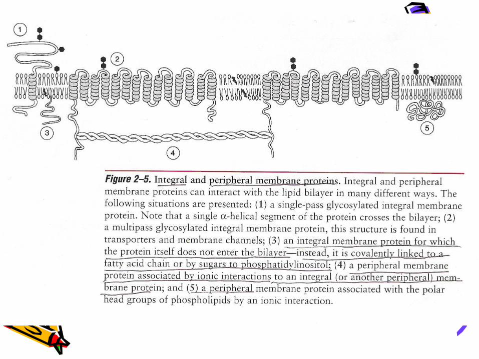
Figure 2-5. Integral and peripheral membrane proteins
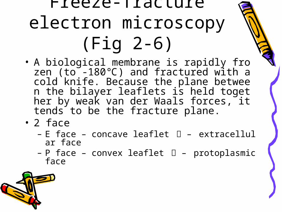
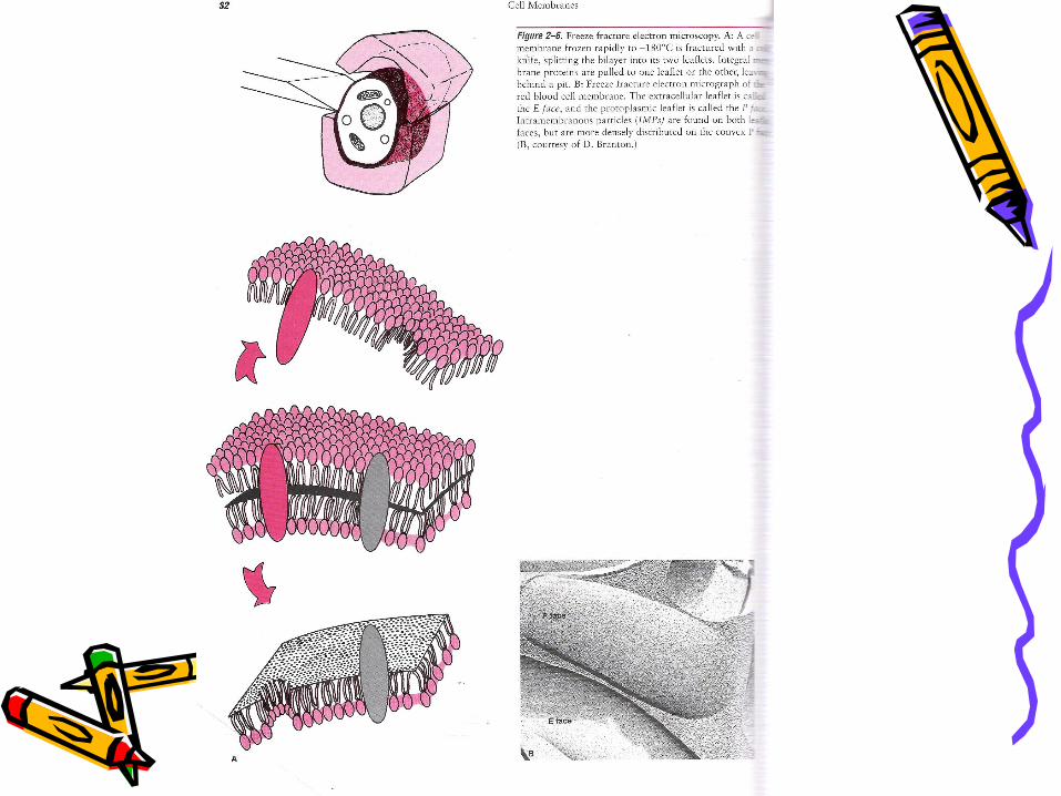
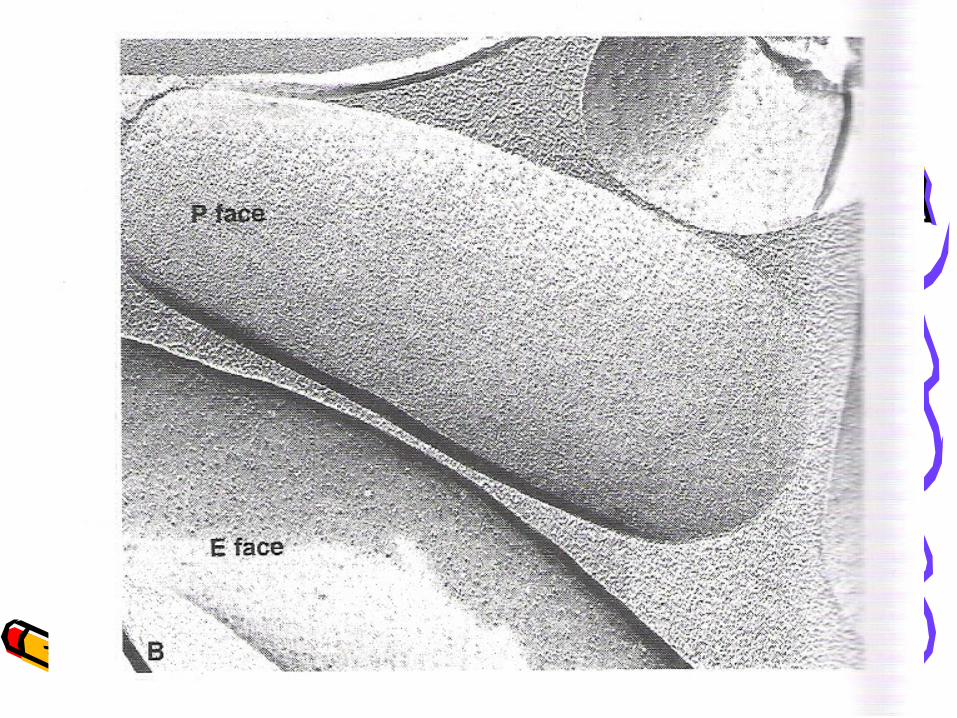
Freeze-fracture electron microscopy
(Fig 2-6)• A biological membrane is rapidly frozen (to -1
80℃) and fractured with a cold knife. Because the plane between the bilayer leaflets is held together by weak van der Waals forces, it tends to be the fracture plane.
• 2 face– E face – concave leaflet 凹 – extracellular face– P face – convex leaflet 凸 – protoplasmic face
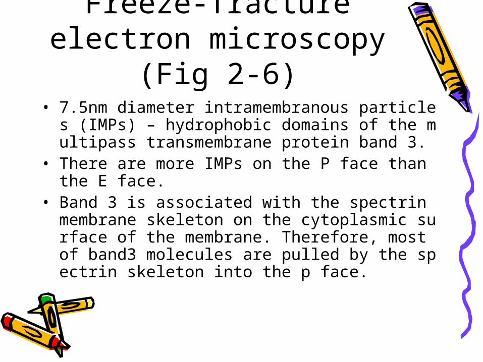
Freeze-fracture electron microscopy
(Fig 2-6)• 7.5nm diameter intramembranous particles (IMPs) –
hydrophobic domains of the multipass transmembrane protein band 3.
• There are more IMPs on the P face than the E face.• Band 3 is associated with the spectrin membrane sk
eleton on the cytoplasmic surface of the membrane. Therefore, most of band3 molecules are pulled by the spectrin skeleton into the p face.
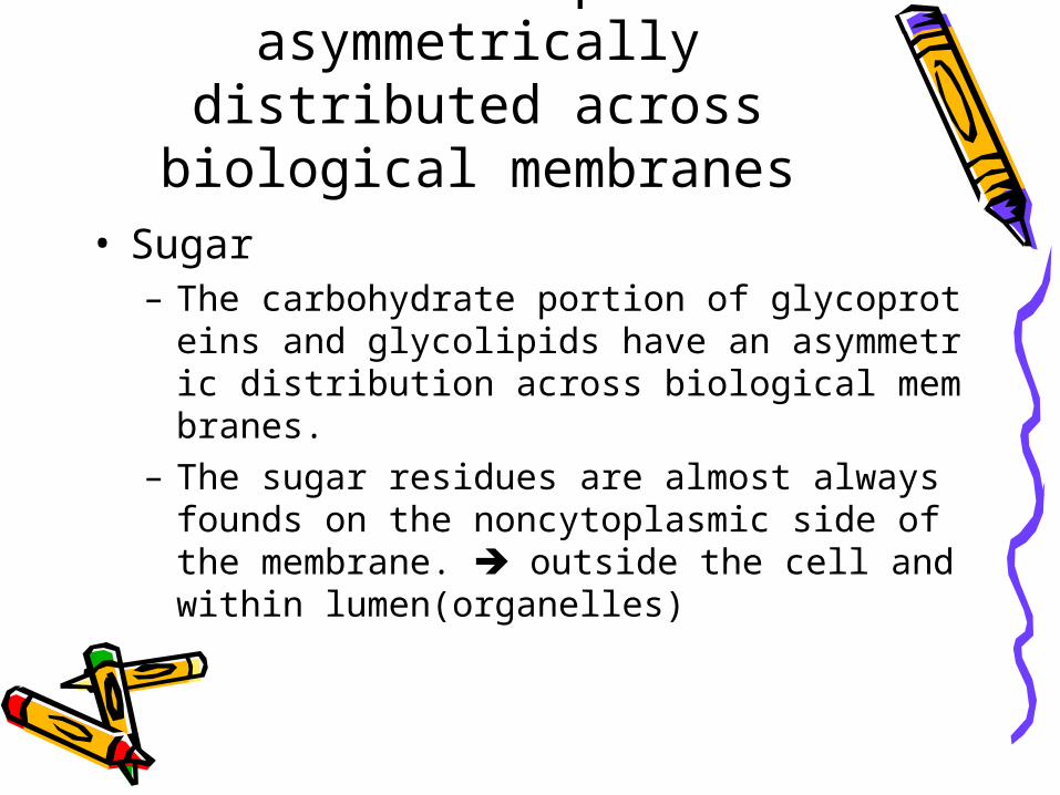
Protein and lipids are asymmetrically distributed
across biological membranes• Sugar
– The carbohydrate portion of glycoproteins and glycolipids have an asymmetric distribution across biological membranes.
– The sugar residues are almost always founds on the noncytoplasmic side of the membrane. outside the cell and within lumen(organelles)
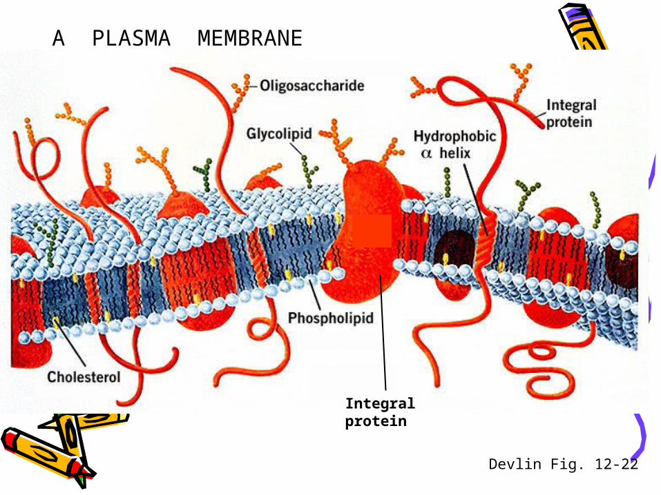
A PLASMA MEMBRANE
Integralprotein
Devlin Fig. 12-22
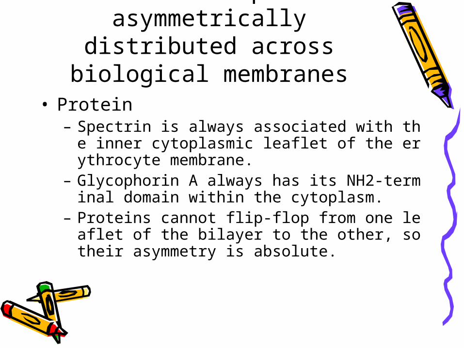
Protein and lipids are asymmetrically distributed
across biological membranes• Protein
– Spectrin is always associated with the inner cytoplasmic leaflet of the erythrocyte membrane.
– Glycophorin A always has its NH2-terminal domain within the cytoplasm.
– Proteins cannot flip-flop from one leaflet of the bilayer to the other, so their asymmetry is absolute.
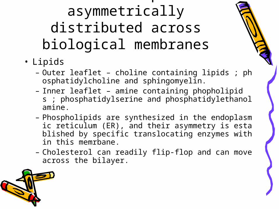
Protein and lipids are asymmetrically distributed
across biological membranes• Lipids
– Outer leaflet – choline containing lipids ; phosphatidylcholine and sphingomyelin.
– Inner leaflet – amine containing phopholipids ; phosphatidylserine and phosphatidylethanolamine.
– Phospholipids are synthesized in the endoplasmic reticulum (ER), and their asymmetry is established by specific translocating enzymes within this memrbane.
– Cholesterol can readily flip-flop and can move across the bilayer.

Protein and lipids are asymmetrically distributed
across biological membranes• What is the significance of the
asymmetric distribution?• Answer : It allows the two sides of
membrane to be functionally distinct.
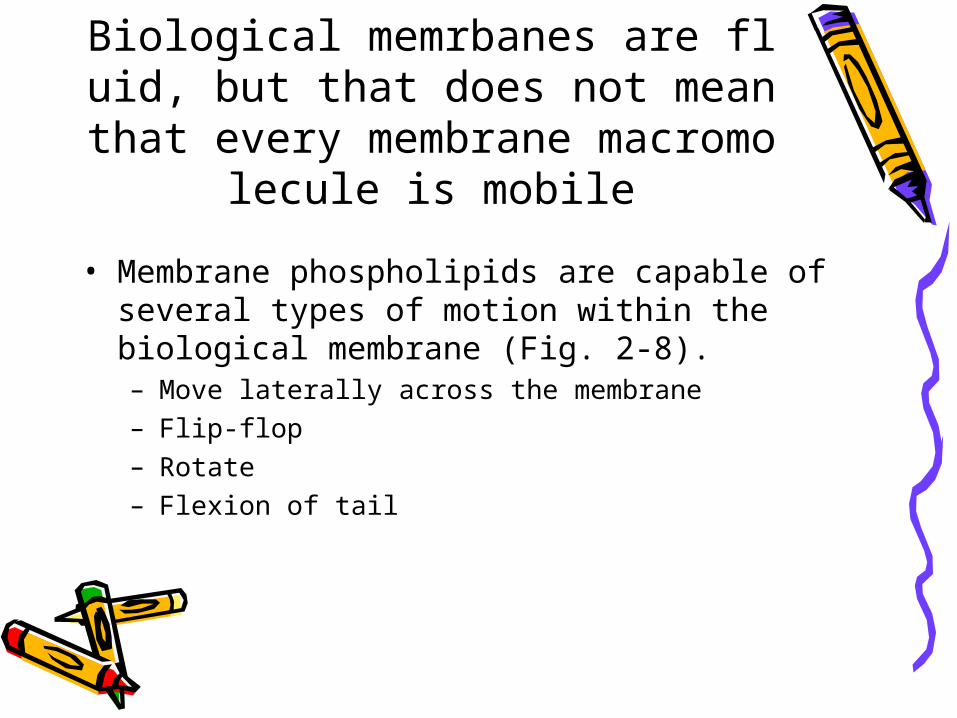
Biological memrbanes are fluid, but that does not mean that every membr
ane macromolecule is mobile
• Membrane phospholipids are capable of several types of motion within the biological membrane (Fig. 2-8).– Move laterally across the membrane– Flip-flop– Rotate– Flexion of tail
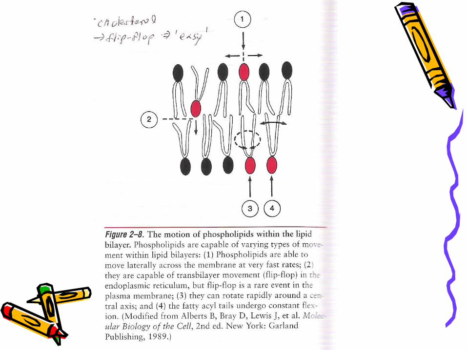
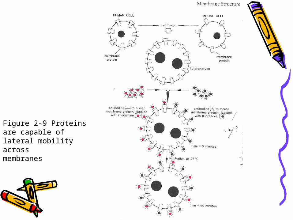
Figure 2-9 Proteins are capable of lateral mobility across membranes


Thank you for your attention ~Thank you for your attention ~