Embed Size (px)
Citation preview
Large intestine
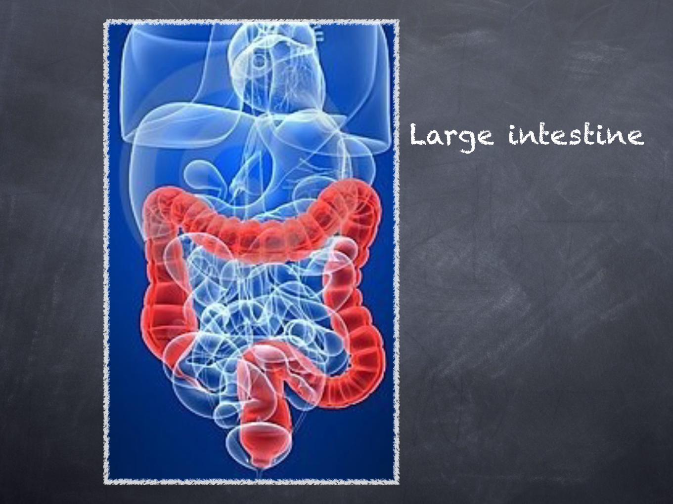
Functions
• Formation of faeces • Elimination of wastes • Re-absorption of water • Manufacture of nutrients • Inhibition of pathogenic organisms
Large intestine
Large intestine
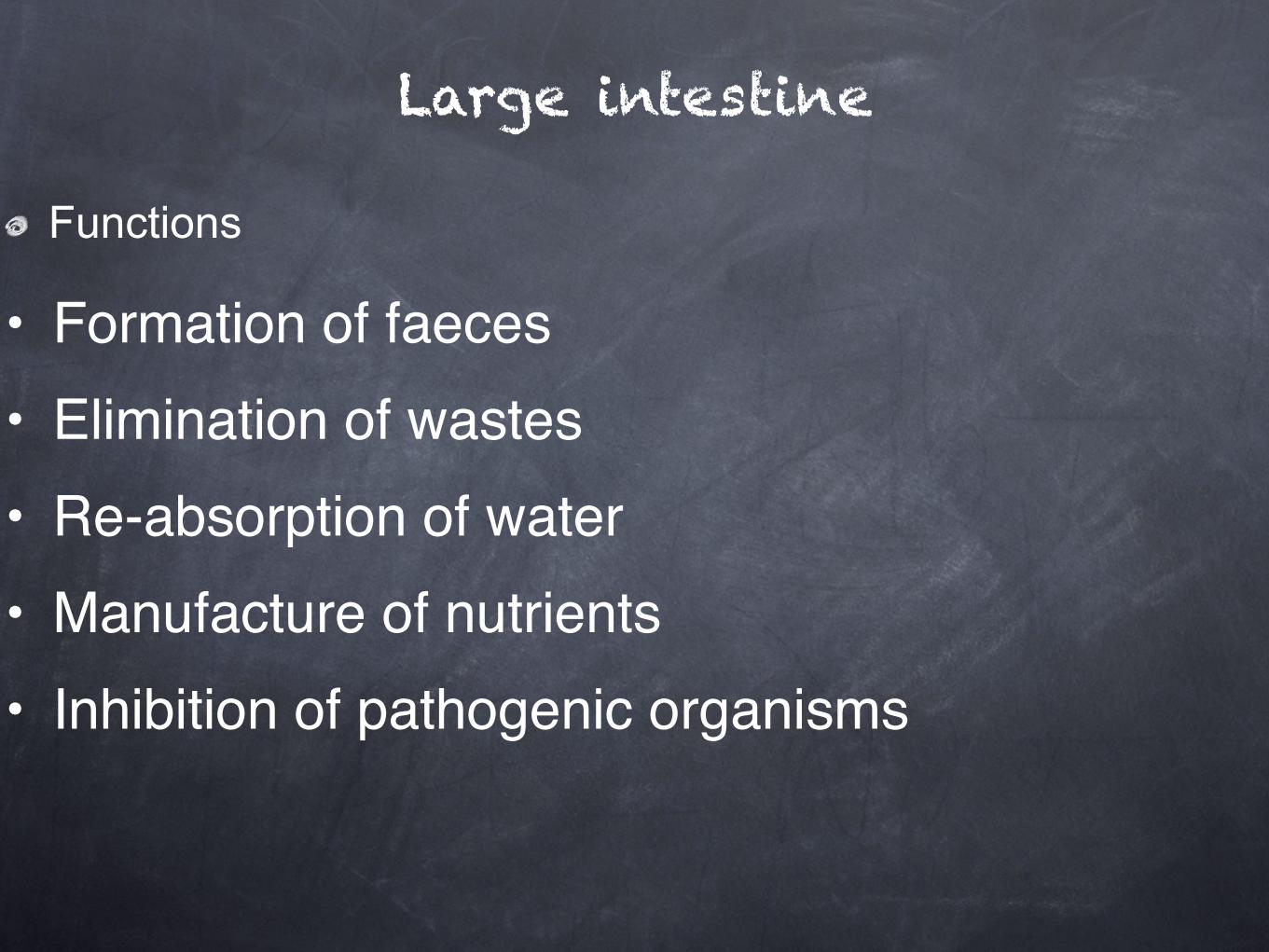
Anatomy: Component Parts:
From ICV to Anus Caecum Appendix Ascending Colon Transverse Colon Descending Colon Sigmoid Colon Rectum Anal Canal
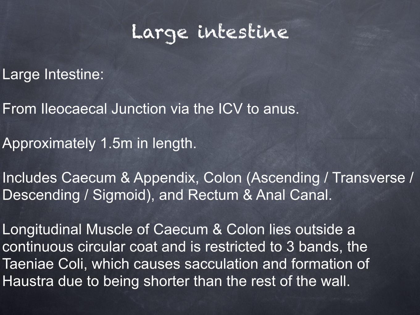
Large Intestine: From Ileocaecal Junction via the ICV to anus. Approximately 1.5m in length. Includes Caecum & Appendix, Colon (Ascending / Transverse / Descending / Sigmoid), and Rectum & Anal Canal. Longitudinal Muscle of Caecum & Colon lies outside a continuous circular coat and is restricted to 3 bands, the Taeniae Coli, which causes sacculation and formation of Haustra due to being shorter than the rest of the wall.
Large intestine

Fig. 27-9. The large intestine shown by a barium enema. Note
the different levels of the transverse colon. A shows the appendix. B shows the ileum
and the pattern of the colon. C, Oblique view of the colon
and rectum. D, The colon and rectum outlined by a double contrast enema. (A, Courtesy
of Maurice C. Howard, M.D., Omaha, Nebraska. C, Courtesy of Robert A. Powers, M.D., Palo Alto, California. D, Courtesy of
Eugene E. Ahern, M.D., Minneapolis, Minnesota. C and
D are from Medical Radiography and Photography.)
Large Intestine
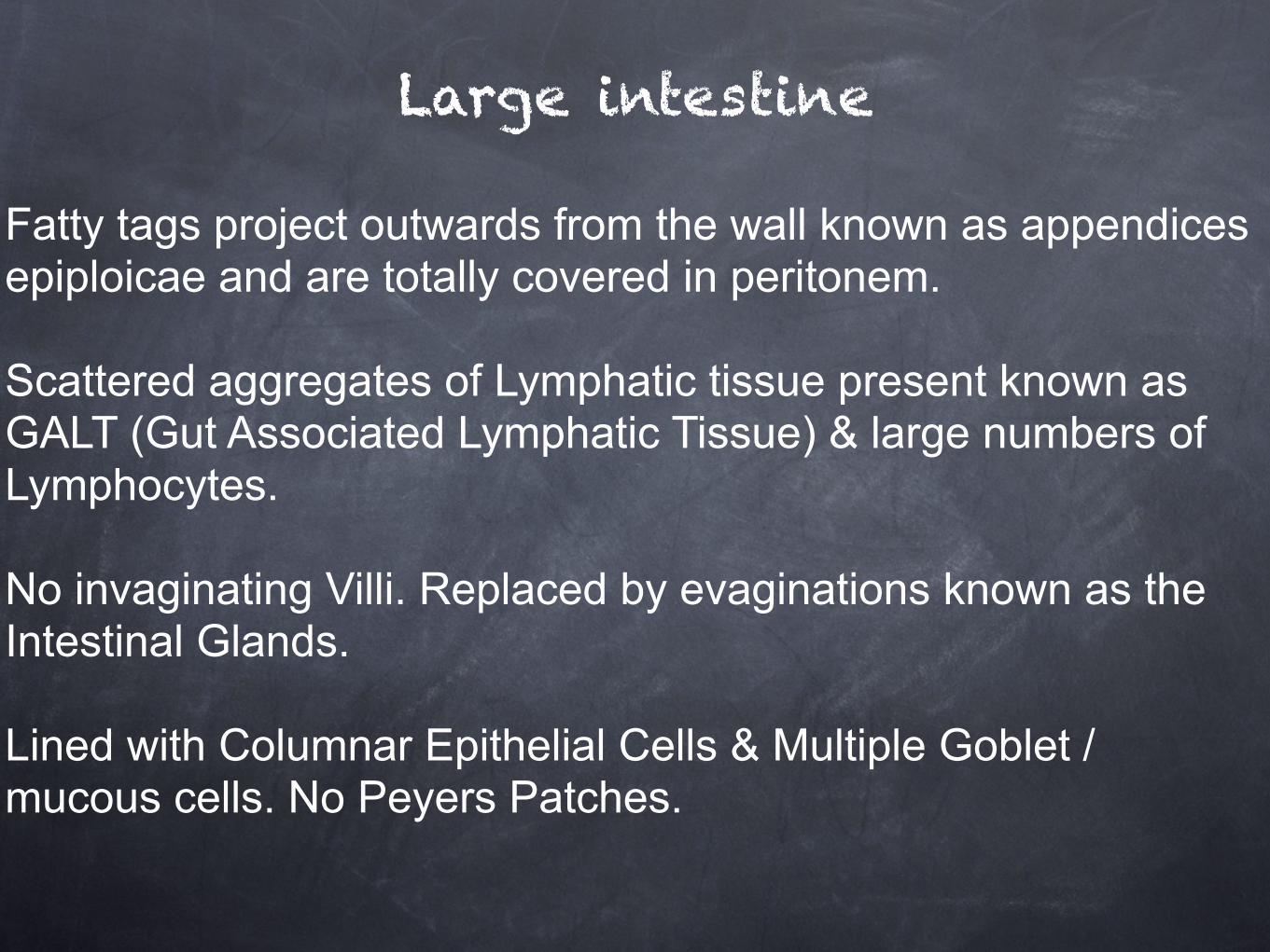
Fatty tags project outwards from the wall known as appendices epiploicae and are totally covered in peritonem. Scattered aggregates of Lymphatic tissue present known as GALT (Gut Associated Lymphatic Tissue) & large numbers of Lymphocytes. No invaginating Villi. Replaced by evaginations known as the Intestinal Glands. Lined with Columnar Epithelial Cells & Multiple Goblet / mucous cells. No Peyers Patches.
Large intestine
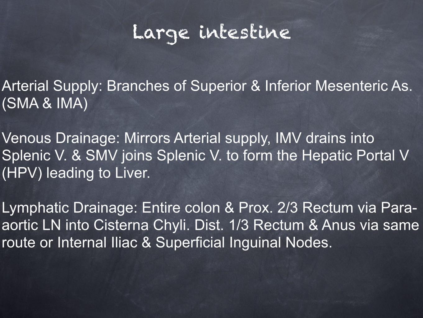
Arterial Supply: Branches of Superior & Inferior Mesenteric As. (SMA & IMA) Venous Drainage: Mirrors Arterial supply, IMV drains into Splenic V. & SMV joins Splenic V. to form the Hepatic Portal V (HPV) leading to Liver. Lymphatic Drainage: Entire colon & Prox. 2/3 Rectum via Para-aortic LN into Cisterna Chyli. Dist. 1/3 Rectum & Anus via same route or Internal Iliac & Superficial Inguinal Nodes.
Large intestine
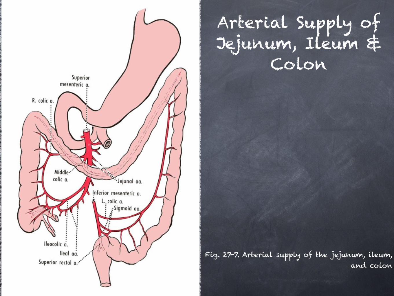
Fig. 27-7. Arterial supply of the jejunum, ileum, and colon
Arterial Supply of Jejunum, Ileum &
Colon
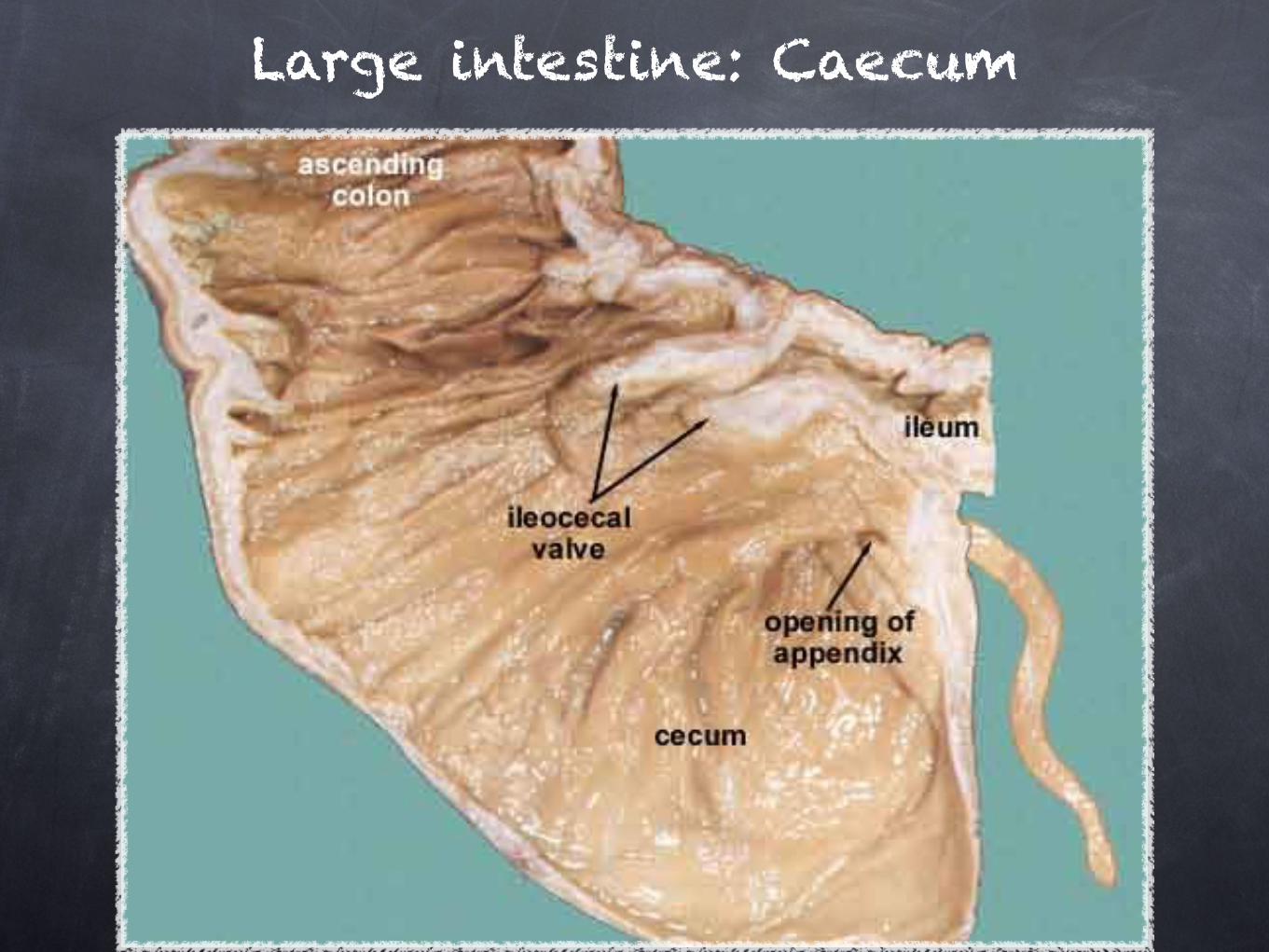
Large intestine: Caecum
Large intestine: Caecum
Caecum (L. Blind): A blind sac (8cmx8cm) continuous with small intestine. Location: R Iliac Fossa / LRQ, potentially reaching Pelvic Brim. Taeniae Coli converge posteromedially on the appendix. ICV connects ileum to caecum on medial wall. Connects to Ascending Colon via the cecocolic junction. Completely contained by peritoneum. No villi
Relations: Anterior: Distal Small Intestine & Anterior Abd. Wall. Posterior: Lies on ilacus & psoas muscles, Femoral & Lateral Femoral Cutaneous Nerve. Medial: Femoral N. & External Iliac vessels. Posteromedial: Testicular A & Genitofemoral N.
Large intestine: Caecum
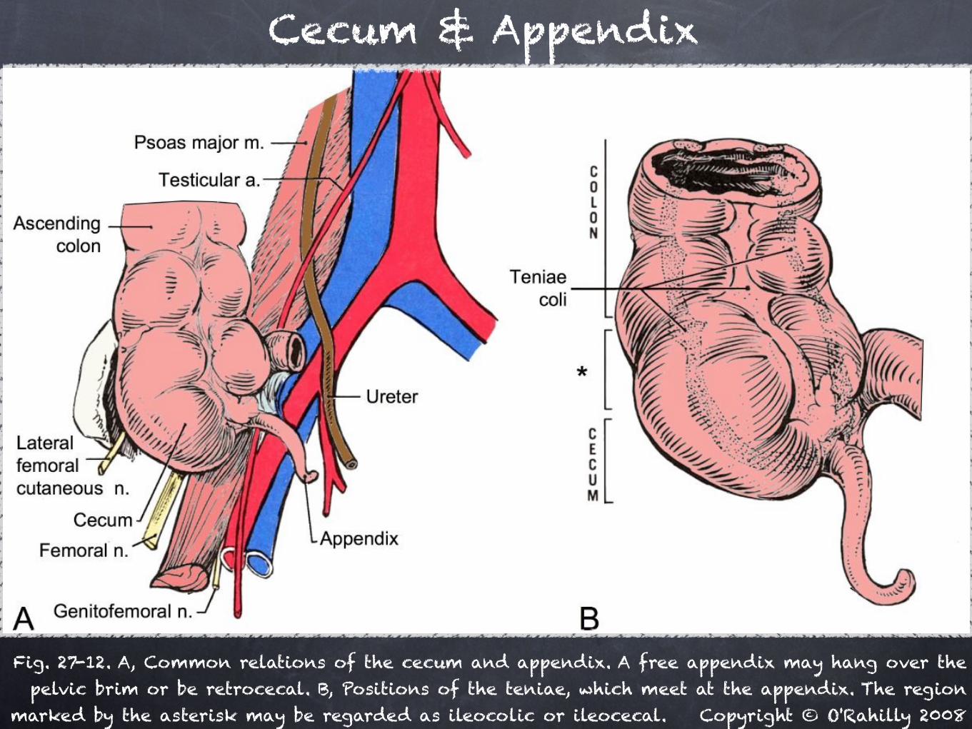
Cecum & Appendix
Fig. 27-12. A, Common relations of the cecum and appendix. A free appendix may hang over the pelvic brim or be retrocecal. B, Positions of the teniae, which meet at the appendix. The region
marked by the asterisk may be regarded as ileocolic or ileocecal. Copyright © O'Rahilly 2008
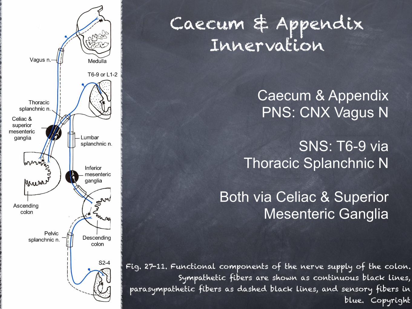
Fig. 27-11. Functional components of the nerve supply of the colon. Sympathetic fibers are shown as continuous black lines,
parasympathetic fibers as dashed black lines, and sensory fibers in blue. Copyright
Caecum & Appendix Innervation
Caecum & Appendix PNS: CNX Vagus N
SNS: T6-9 via
Thoracic Splanchnic N
Both via Celiac & Superior Mesenteric Ganglia
Function: Reservoir for remaining meal contents after small intestine digestion / absorption, prior to peristaltic movement along Colon. Allows bacteria from Appendix to be introduced to the Colon. Begins final processes of faecal formation and bacterial digestion / fermentation of food for production of B-SCFAs and nutrients e.g. vitamin K.
Large intestine: Caecum
Large intestine: Appendix
Appendix (L. Worm-shaped): Narrow Diverticulum, approx 8-10cm in length, arises where the 3 taniae coli converge, from the posteromedial wall of the caecum, 1-3cm inferior to ICV. Attached to terminal Ileum via the Mesoappendix, containing the apendicular artery (Banch of ileocolic a.) Mucosa layer has greater quantity of GALT vs GIT which can replace muscle coat entirely in places.
Large intestine: Appendix
Relations: Variable: Normally positioned Anteriorly or Posteriorly, may be “free” or “fixed”.
Anterior: Most common, “Ileal” or “Pelvic” Posterior: “Subcaecal”, “Retrocaecal” or Retrocolic”. Therefore may make contact with any structures related to
Caecum.
Large intestine: Appendix
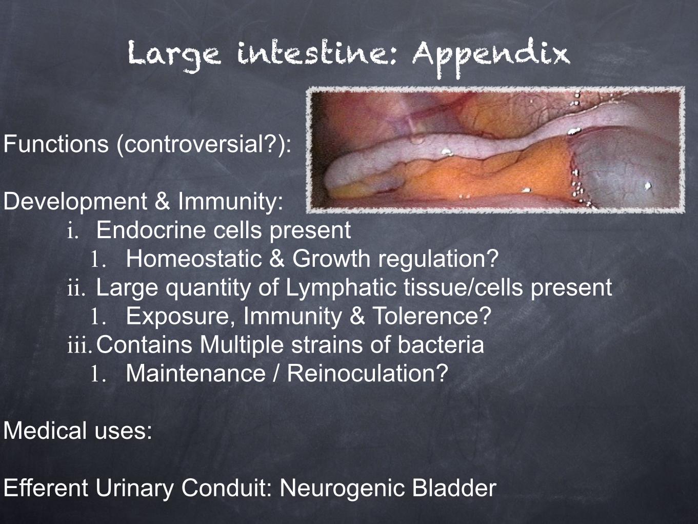
Functions (controversial?): Development & Immunity:
i. Endocrine cells present 1. Homeostatic & Growth regulation?
ii. Large quantity of Lymphatic tissue/cells present 1. Exposure, Immunity & Tolerence?
iii.Contains Multiple strains of bacteria 1. Maintenance / Reinoculation?
Medical uses: Efferent Urinary Conduit: Neurogenic Bladder
Large intestine: Appendix
Pathology: Appendicitis & Carcinoid Tumors
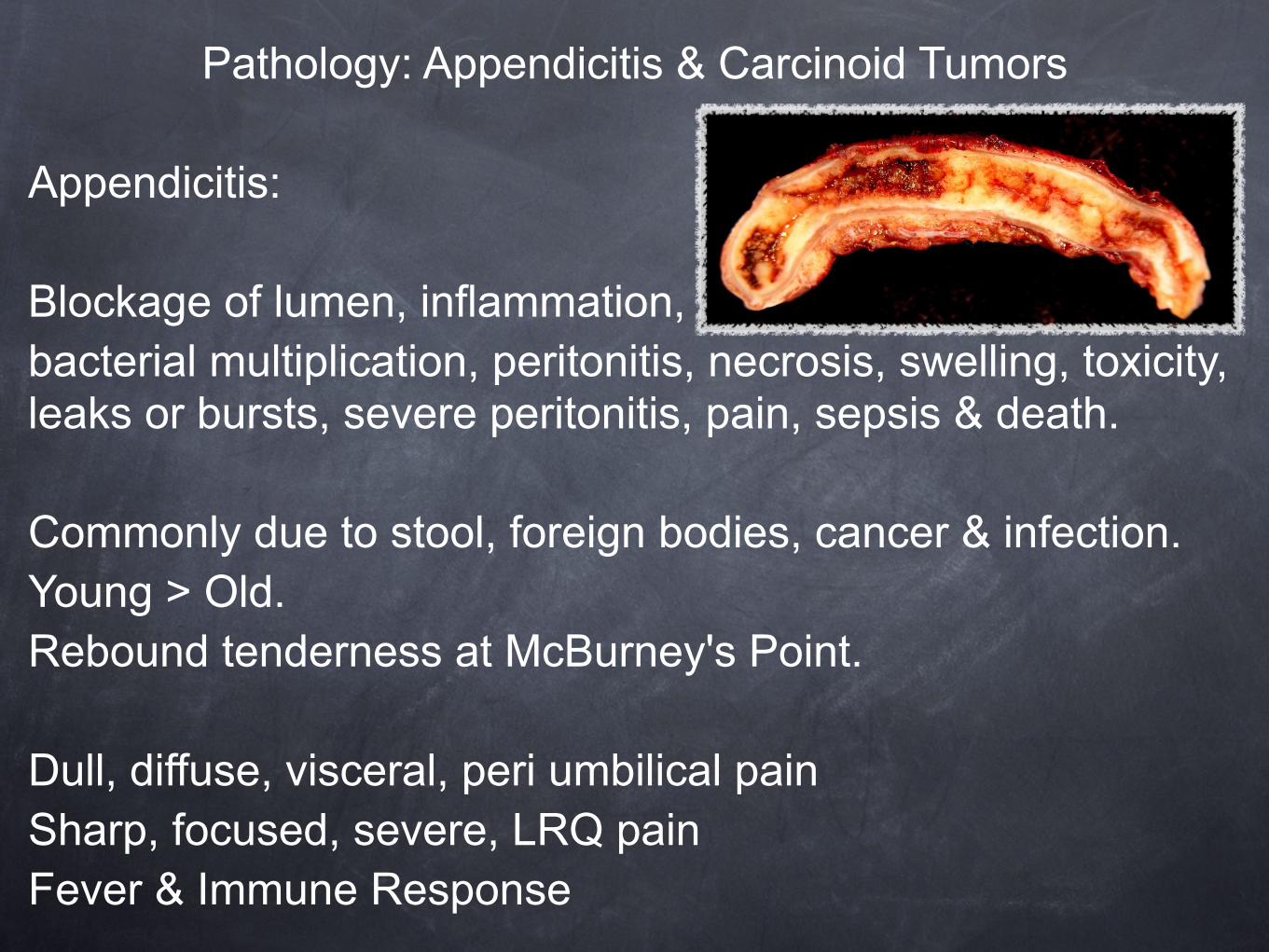
Appendicitis:
Blockage of lumen, inflammation, bacterial multiplication, peritonitis, necrosis, swelling, toxicity, leaks or bursts, severe peritonitis, pain, sepsis & death.
Commonly due to stool, foreign bodies, cancer & infection. Young > Old. Rebound tenderness at McBurney's Point.
Dull, diffuse, visceral, peri umbilical pain Sharp, focused, severe, LRQ pain Fever & Immune Response
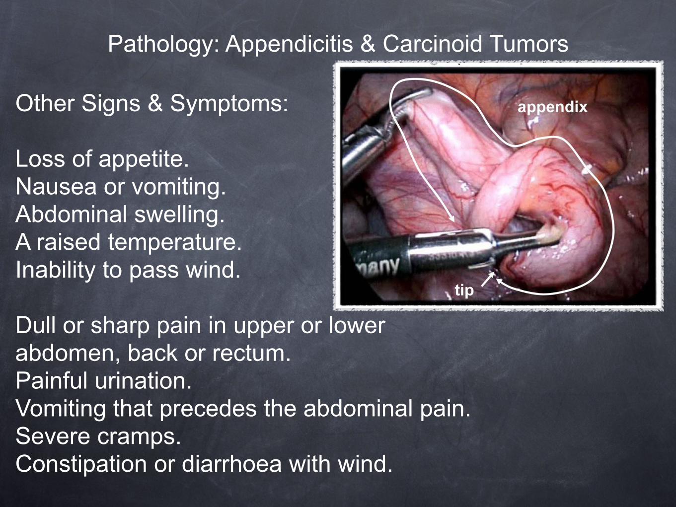
Pathology: Appendicitis & Carcinoid Tumors Other Signs & Symptoms: Loss of appetite. Nausea or vomiting. Abdominal swelling. A raised temperature. Inability to pass wind. Dull or sharp pain in upper or lower abdomen, back or rectum. Painful urination. Vomiting that precedes the abdominal pain. Severe cramps. Constipation or diarrhoea with wind.
Pathology: Appendicitis & Carcinoid Tumors
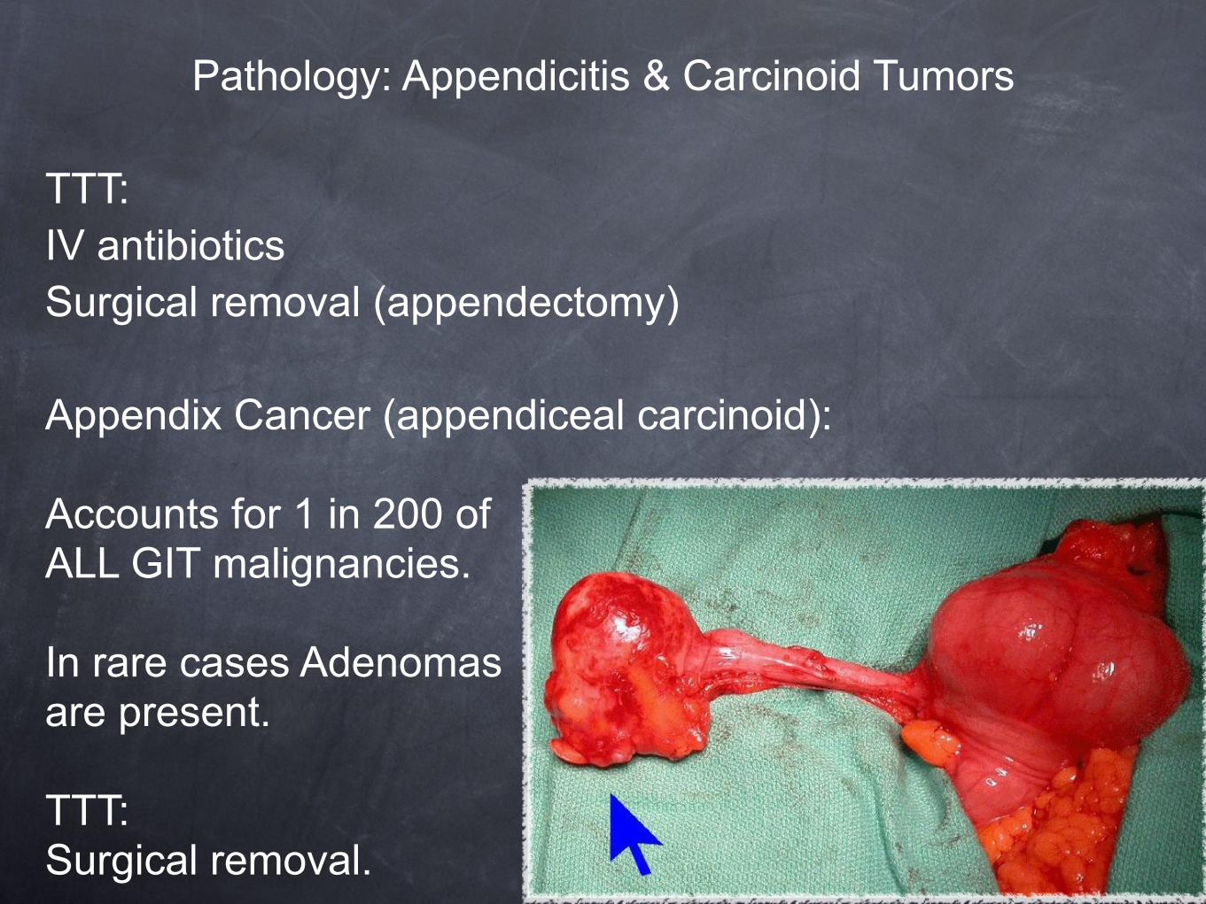
TTT: IV antibiotics Surgical removal (appendectomy)
Appendix Cancer (appendiceal carcinoid): Accounts for 1 in 200 of ALL GIT malignancies. In rare cases Adenomas are present. TTT: Surgical removal.
Large intestine: Ascending Colon
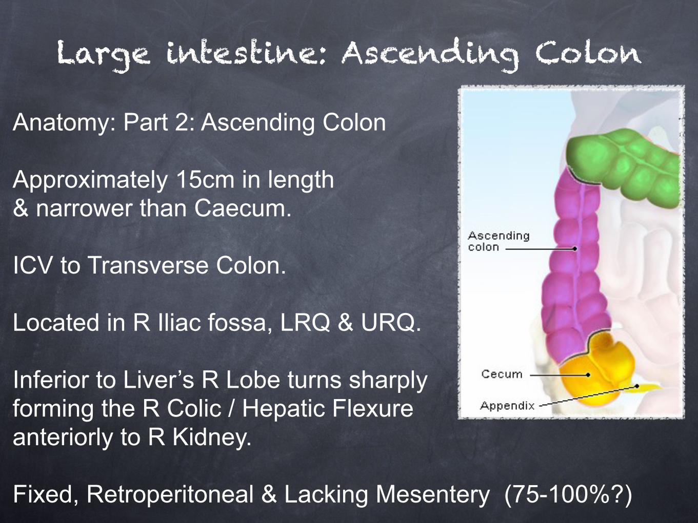
Anatomy: Part 2: Ascending Colon Approximately 15cm in length & narrower than Caecum. ICV to Transverse Colon. Located in R Iliac fossa, LRQ & URQ. Inferior to Liver’s R Lobe turns sharply forming the R Colic / Hepatic Flexure anteriorly to R Kidney. Fixed, Retroperitoneal & Lacking Mesentery (75-100%?)
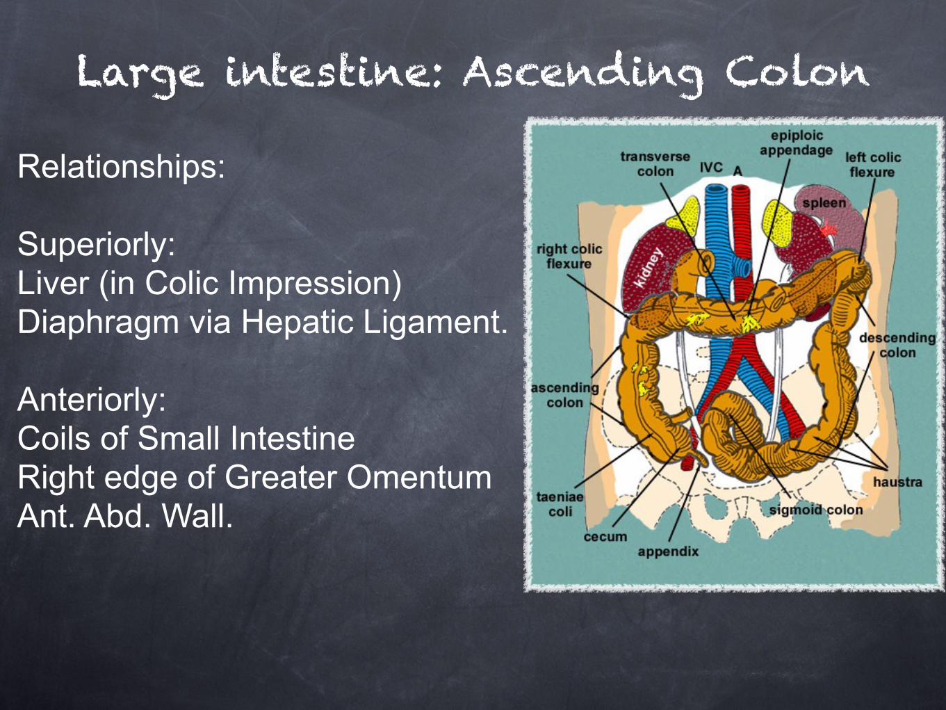
Relationships: Superiorly: Liver (in Colic Impression) Diaphragm via Hepatic Ligament. Anteriorly: Coils of Small Intestine Right edge of Greater Omentum Ant. Abd. Wall.
Large intestine: Ascending Colon
Posteriorly: Iliacus, Quadratus Lumborum, aponeurotic origin of Transversus Abdominus & Diaphragm at tip of rib 12 Iliolumbar Ligament Lateral Cutaneous, Ilioinguinal & Iliohypogastric Ns Iliac branches of Iliolumbar vessels & 4th Lumbar artery Lower Pole of Right Kidney. Medially: Galbladder (at Colic Flexure)
Large intestine: Ascending Colon

Fig. 27-11. Functional components of the nerve supply of the colon. Sympathetic fibers are shown as continuous black lines,
parasympathetic fibers as dashed black lines, and sensory fibers in blue. Copyright
Caecum & Appendix Innervation
Caecum & Appendix PNS: CNX Vagus N
SNS: T6-9 via
Thoracic Splanchnic N
Both via Celiac & Superior Mesenteric Ganglia