Embed Size (px)
Citation preview
Isolation and characterization of expressioncDNA clones encoding antigens of Onchocercavolvulus infective larvae.
T R Unnasch, … , K D Erttmann, B M Greene
J Clin Invest. 1988;82(1):262-269. https://doi.org/10.1172/JCI113581.
The isolation of recombinant cDNA clones expressing antigens found in Onchocercavolvulus infective larvae is described. To isolate such clones, an expression cDNA libraryconstructed from adult O. volvulus RNA was screened with antiserum raised againstinfective larvae. One clone, designated lambda RAL-1 was characterized further. Therecombinant antigen produced by lambda RAL-1 stimulates proliferation of peripheral bloodmononuclear cells from O. volvulus infected humans. Lambda RAL-1 is derived from a 1450bases message that encodes a protein with an apparent molecular weight of 42,000 in adultO. volvulus. The inserted DNA of lambda RAL-1 contains an open reading frame of 1008bp. The amino acid sequence predicted by this open reading frame contains three repeatsof the sequence KKPEDWD. The identification of clones such as lambda RAL-1 willprovide quantities of purified antigens sufficient to begin to study the immune response toand explore the development of immunity against the infectious form of the parasite.
Research Article
Find the latest version:
http://jci.me/113581-pdf
Isolation and Characterization of Expression cDNAClones Encoding Antigensof Onchocerca volvulus Infective LarvaeThomas R. Unnasch,* Michaela Y. Gallin, Peter T. Soboslay, Klaus D. Erttmann, and Bruce M. GreeneDivision of Geographic Medicine, Department of Medicine, and Department of Microbiology and Molecular Biology,Case Western Reserve University, Cleveland, Ohio 44106
Abstract
The isolation of recombinant cDNAclones expressing antigensfound in Onchocerca volvulus infective larvae is described. Toisolate such clones, an expression cDNA library constructedfrom adult 0. volvulus RNAwas screened with antiserumraised against infective larvae. One clone, designated XRAL-1was characterized further. The recombinant antigen producedby XRAL-1 stimulates proliferation of peripheral blood mono-nuclear cells from 0. volvulus infected humans. XRAL-1 isderived from a 1450 bases message that encodes a protein withan apparent molecular weight of 42,000 in adult 0. volvulus.The inserted DNAof XRAL-1 contains an open reading frameof 1008 bp. The amino acid sequence predicted by this openreading frame contains three repeats of the sequenceKKPEDWD.The identification of clones such as XRAL-1 willprovide quantities of purified antigens sufficient to begin tostudy the immune response to and explore the development ofimmunity against the infectious form of the parasite.
Introduction
Onchocerciasis, or river blindness, is a major cause of infec-tious blindness in the world. In severely affected areas, asmany as half of the adult males may be blinded by the disease(1), and mortality rates in such persons are increased as muchas fourfold (2). Attempts to control the disease have centeredon control of the vector for the parasite, the blackfly Simuliumsp., as well as on the development of chemotherapeutic agentswhich may be used to eliminate the parasite from infectedindividuals. Unfortunately, control of the vector has provenextremely difficult. Furthermore, to date no practical chemo-therapeutic agent has been identified that kills the adult formof the parasite.
Because neither vector control nor chemotherapy hasproven successful in controlling the disease, the developmentof a vaccine against infection with the parasite has become ahigh priority (3). In order to develop a vaccine, it is necessaryto first identify antigens that induce immunity against the par-asite. In the case of onchocerciasis, however, the identificationof such antigens is complicated by the fact that much of the
Address reprint requests to Dr. Unnasch, Division of Geographic Med-icine, Case Western Reserve University, University Hospitals, 2074Abington Road, Cleveland, OH44106.
Receivedfor publication 10 November 1987 and in revisedform 15January 1988.
pathology seen during the course of the disease may be causedby the induction of the hosts immune response to certain anti-gens of the parasite (4). Because of this concern, it is necessaryto delineate as fully as possible which parasite antigens areinvolved in inducing potentially beneficial as opposed toharmful aspects of the host immune response.
In order to undertake such a dissection of the immuneresponse, large quantities of purified parasite antigens will berequired. However, no animal host has been identified thatcan be used to provide the various stages of Onchocerca vol-vulus in quantity. As a consequence, the lack of parasite mate-rial has posed a major impediment to immunological studies.This problem has been particularly acute for the study of theinfectious form of the parasite, the third stage larva, or L3.Antigens from this stage may be especially important, since ithas been shown in other filarial infections that irradiated in-fective larvae can induce protective immunity (5, 6).
To circumvent the problem of the lack of parasite material,we have undertaken an alternative strategy to simply isolatingantigens directly from the limited amounts of parasite materialthat are available. This is based upon the identification ofcDNAclones that express antigens associated with 0. volvulusinfective larvae. To identify such clones, we have used antiseraproduced by introducing living infective larvae into a nonper-missive host to screen an 0. volvulus expression cDNAlibrary.Using this approach, six clones have been identified. Oneclone, designated XRAL- 1, a member of the class that appearsto react most strongly with the immune sera, was chosen fordetailed characterization. Based upon immunoaffinity puri-fied antibody probing of crude 0. volvulus protein extracts, themRNAthat gave rise to XRAL- 1 appeared to encode a proteinwith an apparent molecular weight of 42,000 in vivo. Therecombinant antigen produced by this clone stimulated T cellsfrom infected individuals to proliferate, suggesting that thenative antigen related to XRAL- 1 has relevance in natural in-fections. Sequence analysis of the cDNA insert of XRAL- 1revealed the presence of a single long open reading frame,which encodes three repeats of the polypeptide sequenceKKPEDWD.The isolation and characterization of XRAL-Idemonstrates that it will be possible to use clones expressingantigens found in 0. volvulus infective larvae to examine therole that such antigens may play in the development of a pro-tective immune response against the parasite.
MethodsIsolation of 0. volvulus RNA and DNA. RNAwas prepared fromfreshly excised nodules obtained from people infected with 0. volvulusresiding in Grand Bassa county, Liberia. The nodules were carefullytrimmed to remove as much host tissue as possible. The parasite mate-rial was immediately homogenized in a solution of 6 Mguanidiumthiocyanate, 5 mMsodium citrate (pH 7.0), 100 mM,-mercaptoeth-anol and 0.5% sarkosyl, using a mechanical tissue grinder. The extractswere frozen at -20'C, and transported to the United States where
262 Unnasch, Gallin, Soboslay. Erttmann, and Greene
J. Clin. Invest.© The American Society for Clinical Investigation, Inc.0021-9738/88/07/0262/08 $2.00Volume 82, July 1988, 262-269
RNAwas isolated by centrifugation through a CsC1 cushion, as de-scribed by Glisin et al. (7). Between 15 and 30 ,g of RNAwas obtainedper adult female worm.
0. volvulus genomic DNAwas prepared from adult worms thatwere freed from surrounding host tissue using the collagenase digestionprotocol described by Schulz-Key (8) and stored in liquid nitrogen.The DNAwas prepared using the method described by Shah et al. (9).Between 3 and 10 Mg of DNAwas obtained per adult female worm.Each parasite DNApreparation was tested for the presence of contami-nating host cell DNAby hybridization with a variety of probes fordifferent human repeated genes. No contaminating host cell DNAwasdetected in any of the preparations used in the experiments describedbelow.
In vitro translation ofparasite RNAand immunoprecipitation ofthetranslated polypeptides. Total RNA isolated from adult 0. volvulusand Dirofilaria immitis was translated in vitro in rabbit reticulocytelysates (Promega Biotech, Madison, WI), using the protocol providedby the manufacturer. Rabbit antisera were prepared for use in immu-noprecipitation of the translation products by heating to 60C for 20min. Immunoglobulins were precipitated by addition of ammoniumsulfate to a final saturation of 40%. Precipitated protein was resus-pended in 25 mMTris-HC1 (pH 7.5), 100 mMNaCI, and dialyzedagainst the same buffer overnight at 4VC. Aliquots of the in vitrotranslation products and the purified antisera were mixed together andincubated overnight at 4VC. The antibody-antigen complexes werethen bound to Protein A Sepharose beads (Pharmacia, Uppsala, Swe-den). The beads were washed repeatedly in a solution of 50 mMTris(pH 6.9), 150 mMNaCl, 5 mMEDTA, 1%NP-40, 1%sodium deoxy-cholate, 0.2% SDS, and 0.5% bovine serum albumin. The beads wereresuspended in a solution of 62.5 mMTris-glycine (pH 6.8), 3%SDS,0.5% 2-mercaptoethanol, and 10% glycerol, and heated for 3 min at100°C. Freed polypeptides were separated on a 10% polyacrylamide
gel, prepared as described by Laemmli (10). Following electrophoresis,the gel was treated with the autoradiography enhancer Enlightening(New England Nuclear), dried and subjected to autoradiography.
Preparation of 0. volvulus expression cDNA library. 0. volvulusRNA, prepared as described above, was passed over an oligo-dT cellu-lose column, and poly A' RNAeluted from the column as describedby Maniatis et al. (I 1). The poly A' fraction from the column was thenused to prepare first and second strand cDNA, using the RNAse Hprotocol of Gubler and Hoffman (12). The cDNAwas treated with EcoRI methylase (New England Biolabs, Beverly, MA), using the condi-tions described by the manufacturer. Following methylation, syntheticEco RI linkers (Collaborative Research, Lexington, MA), phosphory-lated as described by the manufacturer, were ligated to the ends of thecDNA. The preparation was then digested with Eco RI, and the freedlinkers removed by chromatography on Bio-Gel P-30 (Bio Rad Labo-ratories, Richmond, CA). The cDNAwas then ligated to Xgtl I arms(Stratagene, San Diego, CA). The resulting concatemers were packagedusing Gigapack packaging extracts (Stratagene), using the protocolprovided by the manufacturer. The library produced in this mannercontained - 500,000 independent recombinant clones.
Preparation of 0. volvulus antisera. To prepare antisera against 0.volvulus infective larvae, living L3s were isolated by dissection fromexperimentally infected flies which had been maintained as describedby Hamand Banya (13). A total of 164 living larvae suspended inRPMI tissue culture medium without adjuvant were injected subcuta-neously into a New Zealand white rabbit. At one-week intervals fol-lowing the first inoculation, the rabbit received two booster injectionsof - 100 living larvae. A final boost of 229 living infective larvae wasadministered five weeks after the initial innoculation. Eight weeks afterthe final injection of infective larvae, the rabbit was subjected to adetailed postmortum examination. No evidence of developing para-sites was found.
Antiserum against adult 0. volvulus was prepared by homogenizingcollagenase freed adult parasites in sterile saline in a Ten-Broeck glass-glass homogenizer. The extract was mixed 1:1 (vol/vol) with Freund'scomplete adjuvant, and used to immunize a NewZealand white rab-
bit. After the initial immunization, the rabbit was boosted twice atweekly intervals with the same antigen-adjuvant mixture.
Antibody screening of the cDNA library. Portions of the cDNAlibrary were plated at a density of 35,000 plaque forming units per135 mmPetri dish, and the plaques induced to produce fusion proteinsaccording to the protocol of Huynh et al. (14). Duplicate filters pre-pared from each plate were incubated overnight at room temperaturewith the appropriate antiserum. Antisera used in the screening proce-dure were diluted 1/20 in a solution of 20 mMTris-HCI (pH 7.5), 0.5MNaCl, and 3% BSA (TBS/BSA). Antibodies reacting with Esche-richia coli antigens were removed prior to use by the protocol ofHuynh et al. (14). Following incubation with the primary antiserum,the filters were washed in TBS/BSA, and exposed to Protein-A conju-gated to horseradish peroxidase (Bio-Rad Laboratories, Richmond,CA) for 2 h at room temperature. Positive signals were identified bydevelopment in a solution containing 66 mMTris-HCl, (pH 7.5), 20%methanol, 0.06% 4-chloro-I-napthol, and 0.02% hydrogen peroxide.
Lymphocyte blastogenesis assays. PBMCswere isolated by Ficoll-Hypaque gradient centrifugation of fresh heparinized blood, obtainedfrom 0. volvulus infected individuals living in Liberia. The cells weresuspended in a solution containing 40% RPMI 1640 tissue culturemedium, 40% DMSO,and 20% heat inactivated FCS, and frozen inliquid nitrogen. For the blastogenesis assays, the cells were thawed,washed twice in RPMI 1640 tissue culture medium and resuspended inRPMI 1640 containing 2 mML-glutamine, 100 U/ml penicillin, 100U/ml streptomycin, and 10% heat inactivated pooled human serum.The cells were then plated at a concentration of 106 cells/ml togetherwith extracts of induced lysogen cultures of XRAL-l. Extracts wereprepared as described by Huynh et al. (14), dialyzed against PBS, andfiltered through a 0.45-Mm filter before use. Extracts were used in afinal concentration of 15 jg/ml of protein. Cultures were incubated for78 h at 370C in an atmosphere containing 5%CO2. At this point, I MCiof [3H]thymidine (Amersham, Arlington Heights, IL) was added to thecultures, and the incubation continued for an additional 18 h. Cellswere harvested on filter paper, and [3H]thymidine incorporation as-sessed by counting the samples in a liquid scintillation counter. Resultswere expressed as the mean counts per minute of four culturewells±SEM.
Affinity purification of antibodies reactive with the recombinantantigen produced by XRAL-J. Lysates of induced lysogen cultures ofXgtl I and XRAL-I were prepared according to the protocol of Huynhet al. (14). Protein from these lysates was coupled to cyanogen bromideactivated Sepharose CL-6B (Pharmacia), using the protocol providedby the manufacturer. An aliquot of the antiserum raised against adult0. volvulus was diluted 1/20 in TBS/BSA. The antibody solution wasthen preabsorbed to remove antibodies that reacted with E. coli anti-gens following the protocol of Huynh et al. (14). The antiserum prepa-ration was then passed through a column containing the gel to whichthe Xgtl I protein had been bound. The eluate was collected from thiscolumn, and passed over a column containing the gel to which theXRAL-I lysate protein had been attached. Both columns were washedwith a solution of 50 mMTris-HCI (pH 7.5). 0.5 MNaCl, and boundantibodies eluted with a solution containing 0.2 MTris (pH 7.5) and 8M urea. The eluate was adjusted to 3% (wt/vol) BSA and dialyzedagainst TBS.
DNA sequencing. Isolated cDNA insert was subcloned into theplasmid vector pUC 13, and clones containing the inserted DNAinopposite orientations identified. Plasmids containing the insertedDNA in opposite orientations were linearized by digestion with therestriction enzyme Hind III, and nested deletions prepared using theexonuclease Bal 31. The deleted insert DNAwas liberated by digestionwith Eco Rl and purified from an agarose gel as described by Vogel-stein and Gillespie (15). The purified deleted insert DNAwas thendirectionally cloned into the single stranded bacteriophage vector M13Mpl 8. Clones containing deletions whose ends differed by 200 bpin length were isolated, and single stranded phage DNAprepared asdescribed by Schreier and Cortese (16). This DNAwas then sequencedusing the dideoxynucleotide termination method (17).
Onchocerca volvulus Larval Complementary DNAClones 263
Results
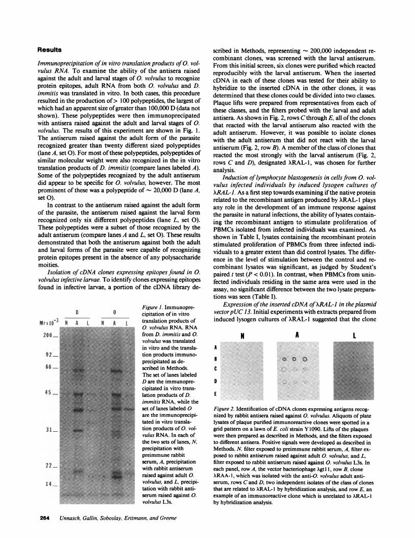
Immunoprecipitation of in vitro translation products of 0. vol-vulus RNA. To examine the ability of the antisera raisedagainst the adult and larval stages of 0. volvulus to recognizeprotein epitopes, adult RNA from both 0. volvulus and D.immitis was translated in vitro. In both cases, this procedureresulted in the production of> 100 polypeptides, the largest ofwhich had an apparent size of greater than 100,000 D (data notshown). These polypeptides were then immunoprecipatedwith antisera raised against the adult and larval stages of 0.volvulus. The results of this experiment are shown in Fig. 1.The antiserum raised against the adult form of the parasiterecognized greater than twenty different sized polypeptides(lane A, set 0). For most of these polypeptides, polypeptides ofsimilar molecular weight were also recognized in the in vitrotranslation products of D. immitis (compare lanes labeled A).Some of the polypeptides recognized by the adult antiserumdid appear to be specific for 0. volvulus, however. The mostprominent of these was a polypeptide of - 20,000 D (lane A,set 0).
In contrast to the antiserum raised against the adult formof the parasite, the antiserum raised against the larval formrecognized only six different polypeptides (lane L, set 0).These polypeptides were a subset of those recognized by theadult antiserum (compare lanes A and L, set 0). These resultsdemonstrated that both the antiserum against both the adultand larval forms of the parasite were capable of recognizingprotein epitopes present in the absence of any polysaccharidemoities.
Isolation of cDNA clones expressing epitopes found in 0.volvulus infective larvae. To identify clones expressing epitopesfound in infective larvae, a portion of the cDNA library de-
Figure 1. Immunopre-D 0 cipitation of in vitro
Mr3lO N A L N A L translation products of200_ 5. an,.v'''''',0volvulus RNA. RNA20 0 t from D. immitis and 0.
volvulus was translatedin vitro and the transla-
92 tion products immuno-precipitated as de-
66 scribed in Methods.The set of lanes labeledD are the immunopre-cipitated in vitro trans-lation products of D.immitis RNA, while theset of lanes labeled 0are the immunoprecipi-tated in vitro transla-
31. tion products of 0. vol-vulus RNA. In each ofthe two sets of lanes, N,precipitation with
-* ~~~~preimmune rabbit22 -&.: 1 .t serum, A, precipitation
with rabbit antiserumraised against adult 0.
14 ~~~~~~~~~volvulus, and L, precipi-tation with rabbit anti-
serum:raised against 0.volvulus LUs.
scribed in Methods, representing - 200,000 independent re-combinant clones, was screened with the larval antiserum.From this initial screen, six clones were purified which reactedreproducibly with the larval antiserum. When the insertedcDNA in each of these clones was tested for their ability tohybridize to the inserted cDNA in the other clones, it wasdetermined that these clones could be divided into two classes.Plaque lifts were prepared from representatives from each ofthese classes, and the filters probed with the larval and adultantisera. As shown in Fig. 2, rows Cthrough E, all of the clonesthat reacted with the larval antiserum also reacted with theadult antiserum. However, it was possible to isolate cloneswith the adult antiserum that did not react with the larvalantiserum (Fig. 2, row B). A member of the class of clones thatreacted the most strongly with the larval antiserum (Fig. 2,rows C and D), designated XRAL-1, was chosen for furtheranalysis.
Induction of lymphocyte blastogenesis in cells from 0. vol-vulus infected individuals by induced lysogen cultures ofXRAL-J. As a first step towards examining if the native proteinrelated to the recombinant antigen produced by XRAL- 1 playsany role in the development of an immune response againstthe parasite in natural infections, the ability of lysates contain-ing the recombinant antigen to stimulate proliferation ofPBMCsisolated from infected individuals was examined. Asshown in Table I, lysates containing the recombinant proteinstimulated proliferation of PBMCsfrom three infected indi-viduals to a greater extent than did control lysates. The differ-ence in the level of stimulation between the control and re-combinant lysates was significant, as judged by Student'spaired t test (P < 0.01). In contrast, when PBMCsfrom unin-fected individuals residing in the same area were used in theassay, no significant difference between the two lysate prepara-tions was seen (Table I).
Expression of the inserted cDNA of XRAL-I in the plasmidvector pUC13. Initial experiments with extracts prepared frominduced lysogen cultures of XRAL- 1 suggested that the clone
N A LA
B
C
D
E
0 0 0
Figure 2. Identification of cDNAclones expressing antigens recog-nized by rabbit antisera raised against 0. volvulus. Aliquots of platelysates of plaque purified immunoreactive clones were spotted in agrid pattern on a lawn of E. coli strain Y1090. Lifts of the plaqueswere then prepared as described in Methods, and the filters exposedto different antisera. Positive signals were developed as described inMethods. N, filter exposed to preimmune rabbit serum, A, filter ex-posed to rabbit antiserum raised against adult 0. volvulus, and L,filter exposed to rabbit antiserum raised against 0. volvulus L3s. Ineach panel, row A, the vector bacteriophage Xgt I1, row B, cloneXRAA- 1, which was isolated with the anti-C. volvulus adult anti-serum, rows Cand D, two independent isolates of the class of clonesthat are related to XRAL- I by hybridization analysis, and row E, anexample of an immunoreactive clone which is unrelated to XRAL- Iby hybridization analysis.
264 Unnasch, Gallin, Soboslay, Erttmann, and Greene
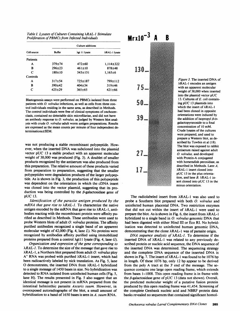
Table I. Lysates of Cultures Containing XRAL-J StimulateProliferation of PBMCsfrom Infected Individuals
Culture additions
Cell source Buffer Xgt 11 lysate ARAL-I lysate
PatientsA 379±74 472±60 1,114±322B 296±23 461±10 878±48C 180±10 543±151 1,165±6
ControlsA 317±54 725±187 799±112B 390±42 404±34 319±46C 425±29 365±65 42 1±66
Blastogenesis assays were performed on PBMCsisolated from threepatients with 0. volvulus infections, as well as cells from three con-trol individuals residing in the same area, as described in Methods.The control individuals were free of clinical symptoms of onchocer-ciasis, contained no detectable skin microfilariae, and did not havean antibody response to 0. volvulus, as judged by Western blot anal-ysis with crude 0. volvulus adult worm antigen preparations. Resultsare expressed as the mean counts per minute of four independent de-terminations±SEM.
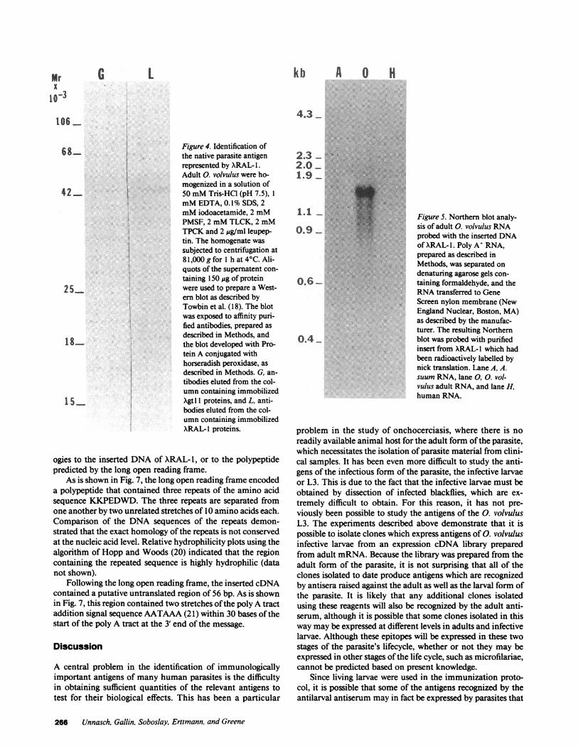
was not producing a stable recombinant polypeptide. How-ever, when the inserted DNAwas subcloned into the plasmidvector pUC 13 a stable product with an apparent molecularweight of 38,000 was produced (Fig. 3). A doublet of smallerproducts recognized by the antiserum was also produced fromthis preparation. The relative amount of these products variedfrom preparation to preparation, suggesting that the smallerpolypeptides were degradation products of the larger polypep-tide. As is shown in Fig. 3, the production of this polypeptidewas dependent on the orientation in which the cDNA insertwas cloned into the vector plasmid, suggesting that its pro-duction was being controlled by the fl-galactosidase gene ofpUC 13.
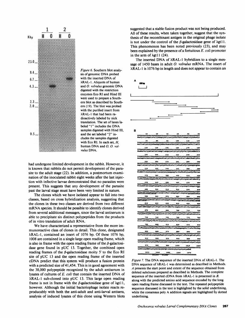
Identification of the parasite antigen produced by themRNAthat gave rise to XRAL-J. To characterize the nativeantigen encoded by the mRNAthat gave rise to ARAL- 1, anti-bodies reacting with the recombinant protein were affinity pu-rified as described in Methods. These antibodies were used toprobe Western blots of adult 0. volvulus proteins. The affinitypurified antibodies recognized a single band of an apparentmolecular weight of 42,000 (Fig. 4, lane L). No proteins wererecognized by antibodies affinity purified using immobilizedproteins prepared from a control Xgt 1 lysate (Fig. 4, lane G).
Organization and expression of the gene corresponding toXRAL-J. To determine the size of the message that gave rise toXRAL- 1, a Northern blot prepared from adult 0. volvulus ployA' RNAwas probed with purified XRAL-l insert, which hadbeen radioactively labeled by nick translation. As Fig. 5, laneo demonstrates, the inserted DNAfrom XRAL-l hybridizedto a single message of 1450 bases in size. No hybridization wasdetected to RNAisolated from uninfected human cells (Fig. 5,lane H). The results presented in Fig. 5 also suggest that anidentical message is not present in mRNAprepared from theintestinal helminthic parasite Ascaris suum. However, inoverexposed autoradiographs of the blot, a small amount ofhybridization to a band of 1650 bases is seen in A. suum RNA.
MrxlO-3 A B
Figure 3. The inserted DNAofXRAL-I encodes an antigenwith an apparent molecularweight of 38,000 when insertedinto the plasmid vector pUC13. Cultures of E. coli contain-ing pUC 13 plasmids intowhich the insert of XRAL-1had been cloned in oppositeorientations were induced bythe addition of isopropyl /l-D-galactopyranoside to a finalconcentration of 10 mM.Crude lysates of the cultureswere prepared, and used toprepare a Western blot, as de-scribed by Towbin et al (18).The blot was exposed to rabbitantiserum raised against adult0. volvulus, and developedwith Protein-A conjugatedwith horseradish peroxidase, asdescribed in Methods. Lane A,XRAL-I insert cloned intopUC 13 in the plus orienta-tion, and lane B, XRAL- I in-sert cloned into pUC 13 in theminus orientation.
The radiolabeled insert from XRAL- 1 was also used toprobe a Southern blot prepared with both 0. volvulus anduninfected human placental DNA. Two restriction enzymesthat did not cut within the insert of ARAL- 1 were used toprepare the blot. As is shown in Fig. 6, the insert from ARAL- 1hybridized to a single band in 0. volvulus genomic DNAthathad been digested with either Hind III or Eco RI. No hybrid-ization was detected to uninfected human genomic DNA,demonstrating that the clone XRAL-I was of parasite origin.
DNA sequence analysis of XRAL-J. To determine if theinserted DNAof XRAL- 1 was related to any previously de-scribed protein or nucleic acid sequence, the DNAsequence ofthe inserted DNAwas determined. The sequencing strategyand the complete DNAsequence of the inserted DNAisshown in Fig. 7. The insert of XRAL-I was found to be 1076 bpin length. Of these 1076 bp, only 12 bp appear to be derivedfrom the poly A tract at the 3' end of the message. The se-quence contains one large open reading frame, which extendsfrom bases 1-1008. This open reading frame is in frame withthe f3-galactosidase gene of pUC 13 (data not shown). Overall,the predicted molecular weight of a putative fusion proteinproduced by this open reading frame was 41,454. Screening ofthe complete Genbank nucleic acid and NBRFprotein databanks revealed no sequences that contained significant homol-
Onchocerca volvulus Larval Complementary DNAClones 265
kb A 0 H
4.3
6 8...... Figure 4. Identification ofthe native parasite antigenrepresented by XRAL-1.Adult 0. volvulus were ho-
| mogenized in a solution of42 50 mMTris-HCI (pH 7.5), 1
mMEDTA, 0.1% SDS, 2mMiodoacetamide, 2 mMPMSF, 2 mMTLCK, 2 mMTPCKand 2 ,ug/ml leupep-tin. The homogenate wassubjected to centrifugation at81,000 g for 1 h at 4C. Ali-quots of the supernatent con-
taining 150 ,g of protein2 5 were used to prepare a West-
ern blot as described byTowbin et al. (18). The blot
was exposed to affinity puri-fied antibodies, prepared asdescribed in Methods, and
1-~the blot developed with Pro-tein A conjugated withhorseradish peroxidase, asdescribed in Methods. G, an-
tibodies eluted from the col-umn containing immobilized
155 Xgtl 1 proteins, and L, anti-bodies eluted from the col-umn containing immobilizedXRAL-l proteins.
ogies to the inserted DNAof XRAL-1, or to the polypeptidepredicted by the long open reading frame.
As is shown in Fig. 7, the long open reading frame encodeda polypeptide that contained three repeats of the amino acidsequence KKPEDWD.The three repeats are separated fromone another by two unrelated stretches of 10 amino acids each.Comparison of the DNA sequences of the repeats demon-strated that the exact homology of the repeats is not conservedat the nucleic acid level. Relative hydrophilicity plots using thealgorithm of Hopp and Woods (20) indicated that the regioncontaining the repeated sequence is highly hydrophilic (datanot shown).
Following the long open reading frame, the inserted cDNAcontained a putative untranslated region of 56 bp. As is shownin Fig. 7, this region contained two stretches of the poly A tractaddition signal sequence AATAAA(21) within 30 bases of thestart of the poly A tract at the 3' end of the message.
Discussion
A central problem in the identification of immunologicallyimportant antigens of many human parasites is the difficultyin obtaining sufficient quantities of the relevant antigens totest for their biological effects. This has been a particular
2.32.0. ..
1.9-
0.9.
0.6
0.4
Figure 5. Northern blot analy-sis of adult 0. volvulus RNAprobed with the inserted DNAof XRAL- 1. Poly A' RNA,prepared as described inMethods, was separated on
denaturing agarose gels con-
taining formaldehyde, and theRNAtransferred to GeneScreen nylon membrane (NewEngland Nuclear, Boston, MA)as described by the manufac-turer. The resulting Northernblot was probed with purifiedinsert from XRAL-I which hadbeen radioactively labelled bynick translation. Lane A, A.suum RNA, lane 0, 0. vol-vulus adult RNA, and lane H,human RNA.
problem in the study of onchocerciasis, where there is no
readily available animal host for the adult form of the parasite,which necessitates the isolation of parasite material from clini-cal samples. It has been even more difficult to study the anti-gens of the infectious form of the parasite, the infective larvaeor L3. This is due to the fact that the infective larvae must beobtained by dissection of infected blackflies, which are ex-
tremely difficult to obtain. For this reason, it has not pre-viously been possible to study the antigens of the 0. volvulusL3. The experiments described above demonstrate that it ispossible to isolate clones which express antigens of 0. volvulusinfective larvae from an expression cDNA library preparedfrom adult mRNA.Because the library was prepared from theadult form of the parasite, it is not surprising that all of theclones isolated to date produce antigens which are recognizedby antisera raised against the adult as well as the larval form ofthe parasite. It is likely that any additional clones isolatedusing these reagents will also be recognized by the adult anti-serum, although it is possible that some clones isolated in thisway may be expressed at different levels in adults and infectivelarvae. Although these epitopes will be expressed in these twostages of the parasite's lifecycle, whether or not they may beexpressed in other stages of the life cycle, such as microfilariae,cannot be predicted based on present knowledge.
Since living larvae were used in the immunization proto-col, it is possible that some of the antigens recognized by theantilarval antiserum may in fact be expressed by parasites that
266 Unnasch, Gallin, Soboslay, Erttmann, and Greene
Mrx
10-3G L
106
23.0_
9.4_
6.7
4.3-
2.32.0
0.5 -
.... ...*
Figure 6. Southern blot analy-sis of genomic DNAprobedwith the inserted DNAofXRAL- 1. Aliquots of humanand 0. volvulus genomic DNAdigested with the restrictionenzymes Eco RI and Hind IIIwere used to prepare a South-ern blot as described by South-ern (19). The blot was probedwith the purified insert fromXRAL-I that had been ra-
dioactively labeled by nicktranslation. The set of lanes la-
beled " 1" includes the DNAsamples digested with Hind III,
and the set labeled "2" in-cludes the samples digestedwith Eco RI. In each set, H,human DNAand 0, 0. vol-vulus DNA.
had undergone limited development in the rabbit. However, itis known that rabbits do not permit development of the para-site to the adult stage (22). In addition, a postmortum exami-nation of the inoculated rabbit eight weeks after the last injec-tion with infective larvae demonstrated that no parasites were
present. This suggests that any development of the parasitepast the larval stage must have been very limited in nature.
The clones which we have isolated appear to fall into twoclasses, based on cross hybridization analysis, suggesting thatthe clones in these two classes are derived from two differentmRNAspecies. It should be possible to identify clones derivedfrom several additional messages, since the larval antiserum isable to precipitate six distinct polypeptides from the productsof in vitro translation of adult RNA.
Wehave characterized a representative from the more im-munoreactive class of clones in detail. This clone, designatedXRAL- 1, contained an insert of 1076 bp. Of these 1076 bp,1008 are contained in a single large open reading frame, whichis also in frame with the open reading frame of the f3-galactosi-dase gene found in pUC 13. Together, the combined openreading frames of the fl-galactosidase moity 5' to the Eco RIsite of pUC 13 and the open reading frame of the insertedcDNA predict that this system will produce a fusion proteinwith a predicted size of 41,454. This is in good agreement withthe 38,000 polypeptide recognized by the adult antiserum in
lysates of cultures of E. coli that contain the inserted DNAofXRAL- sub-cloned into pUC 13. This large open readingframe is not in frame with the fl-galactosidase gene of Xgtl 1,however. Although the initial bacteriophage isolate reacts re-
producably with both the anti-adult and anti-larval antisera,analysis of induced lysates of this clone using Western blots
suggested that a stable fusion product was not being produced.All of these results, when taken together, suggest that the syn-thesis of the recombinant antigen in the original phage isolateis not under the control of the fl-galactosidase gene of Xgtl 1.This phenomenon has been noted previously (23), and maybeen explained by the presence of a fortuitous E. coli promoterin the arm of Xgtl 1 (24).
The inserted DNAof XRAL- 1 hybridizes to a single mes-sage of 1450 bases in adult 0. volvulus mRNA. The insert ofXRAL- is 1076 bp in length and does not appear to contain an
AIu
B10 20 30 40 50 s0
Ph TyrGlyAspAlaValLy.AspLysGlylAuLysThrThrGlnhspAlaLysPhOTyr
70 60 90 100 110 120
SerIll0yAlaLyuPheAspLys8-rPh S-rAsnLynlyLyserL uValIllOGn
130 140 1S0 160 170 130
PhobrValLy.HisCluGlnAspIl AspCysolyGlyGlyTyrValLysLtmuustkl
190 200 210 220 230 240m_ ATOTAAACCTGOAGTTCC&TGTGAAACTTATCATATCATOTTCOGTCCT
SerAspValAsonLeuluAspS*r~lGlyGluThrProTyrOisllKetPbealyPro
250 260 270 260 290 300_ _ _ T TGACTGGAAAA TCCASTGCTATCCS"AAGAAGAAsp~leCyinolyfroGlyThrLy.LyaVal~iaValIl.Phbi.eTyrLyyspArghcn
310 320 330 340 350 360~~ _TCAS:TcrtcS~~~kcCTcsAG
EifstlleLysLysAspll ArgCyaLyaAsphspValPhoThru sl8iTyrThrLou370 380 390 400 410 420
IleValAnsorAspa nThrzyrGluVa1ironhlAsprlyauLysia01us.0Ly430 440 450 460 470 430
G lUu1AalaspTrp&a pPb LeuProProLysLyscllLya~pproasp&aIyo490 500 510 520 530 540
A _CSCATCATGA?0A4 ?AA04ACCCMALYsProCi uAOpToASouArgGluPhella-pA spCluA phpLysL,*Pro1lu550 560 570 500 590 400
rD~5 LysProolu013 IleProAspProhapA1 Ly3LysProaIuApTrpf610 620 630 640 650 640
ca wedG0T0G40AAT0GGAGCCA~c>Y'GTAtAATOCTGAATATAALG-AAspGluhetAsprlypluTrploutProKro~tVslAsp~aoProGluTyrLysolyGlu
670 480 690 700 710 720..O~~"'~"""TCCTCA0AAAG4CAATGGTCCATCCAGCACAMTC
TrpLY ProLy GClLysGys"nProAlaTyrLyaolyLysTrpxledisProolulle730 740 750 760 770 780
GA1_CAATTCTTC0c&A¶TATCA TCAGATTATICG41G T1.GluIleProkspTyrThrProAspAspAsnL uTyrValTyrAspAspl1.olyklalle
790 300 610 820 830 840cG~c_2ATGAc0TCAT?0TACCTGA?
GlyPbo~spLeu~rp~lnValLy&S~r~lyThrilophbapAspvaliliealibrA p
650 $60 370 460 390 900AGTGT0GL40A4GCCAAGAAGTI¶'GGTGAAAAGAC&TNAAAATAA~0AGGGAAGGTGAASerVal~luCluAlaLy LysPh01lyGluLyuThrL uLysxllThrArgGluoly1lu
910 920 930 940 950 960
LYsLysLys01yLysLy3ThrLysLysO10rmLysLyo4luLysAsnluLysIl*Lys970 960 990 1000 1010 1020
ASAGMe MM AA4TMALylu1Ly3tLyLysArYLyYArgAlnaAskrgLysLysLyaLysZnd
1030 1040 1050 1060 1070CAGGIAAAAAAfhfleAA l- u p_AT.
Figure 7. The DNAsequence of the inserted DNAof XRAL- 1. TheDNAsequence of XRAL-1 was determined as described in Methods.A presents the start point and extent of the sequence obtained fromdeleted subclones prepared as described in Methods. The completesequence of the inserted cDNA from XRAL- I is presented in B,along with the predicted amino acid sequence encoded by the longopen reading frame discussed in the text. The repeated polypeptidesequence discussed in the text is highlighted by the solid underlining,while the consensus poly A addition signals are highlighted by dottedunderlining.
Onchocerca volvulus Larval Complementary DNAClones 267
1 2Kbp H 0 H 0........
.;.... . .: . ...... ........
I
0
4
initiating methionine codon. Thus, it is clear that XRAL- 1 isnot a full length cDNA clone. Since the sequence contains asingle long open reading frame that extends through the pre-sumptive 5' end of the insert, it is likely that a portion of thecoding region found in the native message is missing inXRAL- 1. Exactly how much is missing is hard to predict, sincethe length of the poly A tail on the native messege is notknown. However, antibodies which specifically bind to proteinprepared from induced cultures of XRAL- 1 lysogens recognizea single polypeptide with a molecular weight of 42,000 in ex-tracts of adult worms. Analysis of the long open reading frameencoded by XRAL- 1 suggests that it encodes polypeptidewith a molecular weight of 39,130. If the native antigen is notsubjected to posttranslational processing, this suggests thatXRAL- I does contain most of the protein coding sequencefound in the native message. More importantly, results usingthe antilarval antisera described above clearly demonstratethat the recombinant protein produced by XRAL- 1 containsepitopes found in infective larvae. Furthermore, lysates ofXRAL- 1 specifically stimulate a cellular immune response,demonstrating that the epitopes which it encodes have rele-vance in a natural infection. Therefore, even though the insertcontained in XRAL- 1 is not full length, the recombinant anti-gen it produces will be useful in studies directed at dissectingthe role that the native larval antigen plays in the developmentof immunity against the infectious form of the parasite.
As mentioned above, a weak hybridization signal is de-tected when the insert of XRAL- 1 is used to probe Northernblots of A. suum RNA. This suggests that a message related tothe one that gave rise to XRAL- 1 is found in A. suum. Thisresult is not surprising, since results from many previous stud-ies have shown that most of the antigens of different nematodespecies are not species specific (25, 26). Recent studies of anti-genic cross reactivity of different parasitic nematodes havesuggested that A. suum may be the most closely related speciesto 0. volvulus by this criterion (27). A more complete charac-terization of any such related antigen in A. suum will requireantisera specific for the recombinant protein produced byXRAL- 1.
The fact that two different restriction enzymes produce asingle band when a digest of genomic 0. volvulus DNA isprobed with the purified insert from XRAL- 1 suggests that theorganization of the gene which encodes XRAL- 1 is not com-plex. In particular, the results suggest that there is only onecopy of the gene encoding XRAL- 1 in the genome, and thatthis copy does not contain any extended introns. Although thisis the simplest interpretation, other possibilities, such as thepresence of a tandem array of repeated genes homologous toXRAL-1 are also possible. The isolation and characterizationof genomic clones homologous to the insert of XRAL- 1 shouldprovide a definitive answer to this question.
One striking characteristic of the polypeptide encoded bythe long open reading frame of XRAL-I is the fact that itcontains three repeats of the sequence KKPEDWD.Theserepeats reside in a highly hydrophilic region of the peptide, andare therefore likely to be exposed on the surface of the nativeantigen. As such, it is likely that this repeated region mayrepresent one of the more highly immunogenic portions of thenative antigen (28). Several examples of such repeated se-quences have been noted in other parasitic antigens, especiallymalaria (29, 30), and S. mansoni (31). It appears that suchrepeated sequences may be a common feature of antigens iso-
lated from many different parasitic species. In the case of otherantigens which contain such repeated sequences, the repeatedregion often encodes the major epitope of the protein (32, 33).To discover if this is the case in XRAL-l, epitope mappingutilizing deleted versions of the XRAL-l insert will be required.
We have taken a first step examining the role that thenative antigen encoded by the mRNAthat produced XRAL- 1may play in natural infections, showing that lysates containingthe recombinant antigen stimulate the proliferation of PBMCsfrom a small number of infected individuals to a greater extentthan does a control Xgtl 1 lysate. This is important, since it isbelieved that the cellular immune system may be particularityimportant in the development of immunity against helminthicinfections (34). Although the responses seen to the recombi-nant lysate was both reproducible and significant, the overallresponse was not striking. However, previous studies havedemonstrated that the PBMCsof individuals infected with 0.volvulus show both specific and nonspecific supression oftheir response to antigens (35, 36). The relatively low level ofoverall response seen to the recombinant lysate is therefore notsurprising. It is possible that the nonspecific supression in-duced by 0. volvulus infection may have been an asset in thiscase, by supressing the strong response to E. coli antigens onewould normally expect to see when using induced lysates asantigens in the blastogenesis assays. This may have allowed theresponse to the recombinant antigen to be demonstrated. Inspite of this fact, it is quite likely that the response of patientcells relative to the control would be improved if purified prep-arations of the recombinant protein were used in place ofcrude extracts. Most importantly, these studies demonstratethat it is possible to use such recombinant antigen preparationsto begin to examine the role that larval antigens play in naturalinfections with 0. volvulus. Such studies will necessarily in-volve testing both the cellular and antibody responses in amuch larger and diverse patient population than has been ex-amined here. It will also be possible to use purified prepara-tions of recombinant antigens in animal model systems whichmimic some of the pathological manifastations of human on-chocerciasis (37, 38), as well as models for protection againstinfection with the parasite (39). The isolation of XRAL-1 andother clones that express antigens of 0. volvulus infective lar-vae will therefore make it possible to examine the role that theantigens of infective larvae may play in the possible develop-ment of immunity against onchocerciasis.
Acknowledgments
Wethank D. Rechnitzer for technical support, Drs. P. N. Williams andJ. Boateng for assistance in obtaining 0. volvulus material, and Drs. R.Blanton, A. Davis, J. Kazura, and N. Lang for helpful comments andsuggestions.
Supported by grants from the Edna McConnell Clark Foundation,the John M. and Catherine T. MacArthur Foundation Research Con-sortium on the Biology of Parasitic Diseases and the U. S. PublicHealth Service (NIH grants EY-03318 and AI-15351).
References
1. World Health Organization. 1976. Epidemology of Onchocer-ciasis. WHOTech. Rep. Ser. 597:1-94.
2. Kirkwood, B., P. Smith, T. Marshall, and A. Prost. 1983. Rela-tionships between mortality, visual acuity and microfilarial load in the
268 Unnasch, Gallin, Soboslay, Erttmann, and Greene
area of the onchocerciasis control program. Trans. R. Soc. Trop. Med.Hyg. 77:862-868.
3. Edna McConnell Clark Foundation. 1985. Strategic Plan forOnchocerciasis Research. Edna McConnell Clark Foundation, NewYork.
4. MacKenzie, C. D., J. F. Williams, B. M. Sisley, M. W. Steward,and J. O'Day. 1985. Variations in host responses and the pathogenesisof human onchocerciasis. Rev. Infect. Dis. 7:802-808.
5. Wong, M. M., M. F. Guest, and M. J. Laviopierre. 1974. Dirofi-laria immitis: Fate and immunogenicity of irradiated infective stagelarvae in beagles. Exp. Parasitol. 35:465-474.
6. Yates, J. A., and G. I. Higashi. 1985. Brugia malayi: Vaccinationof Jirds with 'Cobalt-attenuated infective stage larvae protects againsthomologous challenge. Am. J. Trop. Med. Hyg. 34:1132-1137.
7. Glisin, V., R. Crkvenjakov, and C. Byus. 1974. Ribonucleic acidisolated by cesium chloride centrifugation. Biochemistry. 13:2633-2637.
8. Schulz-Key, H., E. J. Albiez, and D. W. Buttner. 1977. Isolationof living Adult Onchocerca volvulus from nodules. Tropmed. Parasitol.28:428-430.
9. Shah, J. S., M. Karam, W. F. Piessens, and D. F. Wirth. 1987.Characterization of an Onchocerca-specific DNAClone from Oncho-cerca volvulus. Am. J. Trop. Med. Hyg. 37:376-384.
10. Laemmli, U. K. 1970. Cleavage of structural proteins duringthe assembly of the head of bacteriophage T4. Nature (Lond.).227:680-685.
11. Maniatis, T., E. F. Fritsch, and J. Sambrook. 1982. MolecularCloning: A Laboratory Manual. Cold Spring Harbor Laboratory, ColdSpring Harbor, NY.
12. Gubler, U., and B. J. Hoffman. 1983. A simple and very effi-cient method for generating cDNA libraries. Gene. 25:263-269.
13. Ham, P. J., and A. J. Banya. 1984. The effect of experimentalOnchocerca infections on the fecundity and oviposition of laboratoryreared Simulium sp. (Diptera: Simuliidae). Tropenmed. Parasitol.35:61-66.
14. Huynh, T. V., R. A. Young, and R. W. Davis. 1984. Construct-ing and screening cDNA libraries in Xgt IO and Xgt 1I. In CloningTechniques: A Practical Approach. D. Glover, editor. IRL Press, Ox-ford.
15. Vogelstein, B., and D. Gillespie. 1979. Preparative and analyti-cal purification of DNA from agarose. Proc. Natl. Acad. Sci. USA.76:615-619.
16. Schreier, P. H., and R. Cortese. 1979. A fast and simple methodfor sequencing DNACloned in the single-stranded bacteriophageM13. J. Mol. Biol. 129:169-172.
17. Sanger, F., S. Nicklen, and A. R. Coulson. 1977. DNA se-quencing with chain terminating inhibitors. Proc. Natl. Acad. Sci.USA. 74:5463-5467.
18. Towbin, H., T. Staehelin, and J. Gordon. 1979. ElectrophoreticTransfer of proteins from polyacrylamide gels to nitrocellulose sheets:procedure and some applications. Proc. Nat!. Acad. Sci. USA.76:4350-4354.
19. Southern, E. M. 1975. Detection of specific sequences amongDNA fragments separated by gel electrophoresis. J. Mol. Biol.98:503-517.
20. Hopp, T. P., and K. R. Woods. 1981. Prediction of ProteinAntigenic Determinants from Amino Acid Sequences. Proc. Natl.Acad. Sci. USA. 78:3824-3828.
21. Wickens, M., and P. Stephenson. 1984. Role of the conservedAAUAAAsequence: four AAUAAApoint mutants prevent messengerRNA3' end formation. Science (Wash. DC). 226:1045-1051.
22. Kozek, W. J., and H. F. Marroquin. 1982. Attempts to establishOnchocerca volvulus infection in primates and small laboratory ani-mals. Acta Trop. 39:317-324.
23. Dame, J. B., J. L. Williams, T. F. McCutchan, J. L. Weber,R. A. Wirtz, W. T. Hockmeyer, W. L. Maloy, J. D. Haynes, I.Schneider, D. Roberts, G. S. Sanders, E. P. Reddy, C. L. Diggs, and L.H. Miller. 1984. Structure of the gene encoding the immunodominantsurface antigen on the sporozoite of the human malaria parasite Plas-modiumfalciparum. Science (Wash. DC). 225:593-599.
24. Chirala, S. S. 1986. The nucleotide sequence of the Lac operonand phage junction in lambda gt I. Nucleic Acids Res. 14:5935.
25. Ambroise-Thomas, P. 1974. Immunological diagnosis ofhuman filariasis: present possibilities, difficulties and limitations. ActaTrop. 38:108-128.
26. Higashi, G. I. 1984. Immunodiagnostic tests for protozoan andhelminthic infections. Diagno. Immunol. 2:2-18.
27. Nogami, S., Y. Hayashi, M. Tanaka, M. Korenaga, I. Tada, andH. Tanaka. 1986. Antigenic similarity of Onchocerca volvulus to otherhelminths examined by monoclonal antibodies against 0. volvulus.Jpn. J. Exp. Med. 56:177-183.
28. Berzofsky, J. A. 1985. Intrinsic and extrinsic factors in proteinantigen structure. Science (Wash. DC). 229:932-940.
29. Enea, V., J. Ellis, F. Zavala, D. E. Arnot, A. Asavanich, A.Masuda, I. Quakyi, and R. S. Nussenzweig. 1984. DNAcloning ofPlasmodiumfalciparum circumsporozoite gene: amino acid sequenceof repetitive epitope. Science (Wash. DC). 225:628-630.
30. Ravetch, J. V., J. Kochan, and M. Perkins. 1985. Isolation ofthe gene for a glycophorin-binding protein implicated in erythrocyteinvasion by a malaria parasite. Science (Wash. DC). 227:1593-1597.
31. Bobek, L., D. M. Rekosh, H. van Keulen, and P. T. LoVerde.1986. Characterization of a female-specific cDNAderived from a de-velopmentally regulated mRNAin the human blood fluke Schisto-soma mansoni. Proc. Natl. Acad. Sci. USA. 83:5544-5548.
32. Ballou, W. R., J. Rothbard, R. A. Wirtz, D. M. Gordon, J. S.Williams, R. W. Gore, I. Schneider, M. R. Hollingdale, R. L. Beau-doin, W. L. Maloy, L. H. Miller, and W. T. Hockmeyer. 1985. Immu-nogenicity of synthetic peptides from circumsporozoite protein ofPlasmodiumfalciparum. Science (Wash. DC). 228:996-999.
33. Collins, W. E., R. F. Anders, M. Pappaioanou, G. H. Campbell,G. V. Brown, D. J. Kemp, R. L. Coppel, J. C. Skinner, P. M. Andry-siak, J. M. Favaloro, L. M. Corcoran, J. R. Broderson, G. F. Mitchell,and C. C. Campbell. 1986. Immunization of aotus monkeys with re-combinant proteins of an erythrocyte surface antigen of Plasmodiumfalciparum. Nature (Lond.). 323:259-262.
34. Lal, R. B., T. J. Lynch, and T. B. Nutman. 1987. Brugia malayiantigens associated with lymphocyte activation in filariasis. J. Im-munol. 139:1652-1657.
35. Ngu, J. L. 1978. Immunological Studies on Onchocerciasis.Acta Trop. 35:269-279.
36. Greene, B. M., M. M. Fanning, and J. J. Ellner. 1983. Non-spe-cific supression of antigen-induced lymphocyte blastogenesis in On-chocerca volvulus infection in man. Clin. Exp. Immunol. 52:259-265.
37. Donnelly, J. J., J. H. Rockey, A. E. Bianco, and E. J. L. Soulsby.1984. Ocular immunopathologic findings of experimental onchocer-
ciasis. Arch. Ophthalmol. 102:628-634.38. Donnelly, J. J., H. R. Taylor, E. Young, M. Khatami, J. B. Lok,
and J. H. Rockey. 1986. Experimental ocular onchocerciasis in cyno-molgus monkeys. Inv. Ophthalmol. Vis. Sci. 27:492-499.
39. Greene, B. M. 1987. Primate model for onchocerciasis re-search. In Filariasis, Ciba Foundation Symposium 127. D. Evered andS. Clark, editors. John Wiley and Sons, NewYork. 236-240.
Onchocerca volvulus Larval Complementary DNAClones 269