Embed Size (px)
Citation preview

of December 2, 2018.This information is current as
Protection after VaccinationtoCell or Antibody Responses but Contribute
T+Induction of Dengue Virus-Specific CD8 T Cells Are Not Required for the+CD4
Alessandro Sette and Sujan ShrestaMalika M. Morar, Raphaël M. Zellweger, Bjoern Peters, Lauren E. Yauch, Tyler R. Prestwood, Monica M. May,
http://www.jimmunol.org/content/185/9/5405doi: 10.4049/jimmunol.1001709September 2010;
2010; 185:5405-5416; Prepublished online 24J Immunol
Referenceshttp://www.jimmunol.org/content/185/9/5405.full#ref-list-1
, 36 of which you can access for free at: cites 69 articlesThis article
average*
4 weeks from acceptance to publicationFast Publication! •
Every submission reviewed by practicing scientistsNo Triage! •
from submission to initial decisionRapid Reviews! 30 days* •
Submit online. ?The JIWhy
Subscriptionhttp://jimmunol.org/subscription
is online at: The Journal of ImmunologyInformation about subscribing to
Permissionshttp://www.aai.org/About/Publications/JI/copyright.htmlSubmit copyright permission requests at:
Email Alertshttp://jimmunol.org/alertsReceive free email-alerts when new articles cite this article. Sign up at:
Print ISSN: 0022-1767 Online ISSN: 1550-6606. Immunologists, Inc. All rights reserved.Copyright © 2010 by The American Association of1451 Rockville Pike, Suite 650, Rockville, MD 20852The American Association of Immunologists, Inc.,
is published twice each month byThe Journal of Immunology
by guest on Decem
ber 2, 2018http://w
ww
.jimm
unol.org/D
ownloaded from
by guest on D
ecember 2, 2018
http://ww
w.jim
munol.org/
Dow
nloaded from

The Journal of Immunology
CD4+ T Cells Are Not Required for the Induction of DengueVirus-Specific CD8+ T Cell or Antibody Responses butContribute to Protection after Vaccination
Lauren E. Yauch, Tyler R. Prestwood, Monica M. May, Malika M. Morar,
Raphael M. Zellweger, Bjoern Peters, Alessandro Sette, and Sujan Shresta
The contribution of T cells to the host response to dengue virus (DENV) infection is not well understood. We previously demon-
strated a protective role for CD8+ T cells during primary DENV infection using a mouse-passaged DENV strain and IFN-a/bR2/2
C57BL/6 mice, which are susceptible to DENV infection. In this study, we examine the role of CD4+ T cells during primary DENV
infection. Four I-Ab–restricted epitopes derived from three of the nonstructural DENV proteins were identified. CD4+ T cells
expanded and were activated after DENV infection, with peak activation occurring on day 7. The DENV-specific CD4+ T cells
expressed intracellular IFN-g, TNF, IL-2, and CD40L, and killed peptide-pulsed target cells in vivo. Surprisingly, depletion of
CD4+ T cells before DENV infection had no effect on viral loads. Consistent with this observation, CD4+ T cell depletion did not
affect the DENV-specific IgG or IgM Ab titers or their neutralizing activity, or the DENV-specific CD8+ T cell response. However,
immunization with the CD4+ T cell epitopes before infection resulted in significantly lower viral loads. Thus, we conclude that
whereas CD4+ T cells are not required for controlling primary DENV infection, their induction by immunization can contribute to
viral clearance. These findings suggest inducing anti-DENV CD4+ T cell responses by vaccination may be beneficial. The Journal
of Immunology, 2010, 185: 5405–5416.
Dengue virus (DENV) is a mosquito-borne RNA virus inthe Flaviviridae family, which also includes West Nilevirus (WNV), yellow fever virus, and Japanese encepha-
litis virus. The four serotypes of DENV (DENV1–4) share ∼65–75% homology at the amino acid level (1). Infections with DENVcan be asymptomatic, or cause disease ranging from dengue fever(DF) to dengue hemorrhagic fever (DHF) and dengue shock syn-drome (DSS) (2). DF is a self-limiting illness with symptoms thatinclude fever, headache, myalgia, retro-orbital pain, nausea, andvomiting. DHF and DSS are characterized by increased vascularpermeability, thrombocytopenia, hemorrhagic manifestations, and,in the case of DSS, shock, which can be fatal. The incidence ofDENV infections has increased 30-fold in the past 50 y, and DFand DHF/DSS are a significant cause of morbidity and mortalityworldwide (2). However, vaccine development has been challenging,as a vaccine should protect against all four DENV serotypes (3).
Severe dengue disease (DHF/DSS) most often occurs in indi-viduals experiencing a secondary infection with a heterologousDENV serotype, suggesting the immune response contributes to thepathogenesis (4, 5). One hypothesis is that serotype cross-reactiveAbs enhance infection of FcgR+ cells during a secondary in-fection, resulting in higher viral loads and more severe disease viaa phenomenon known as Ab-dependent enhancement (ADE) (6,7). Recent studies have demonstrated DENV-specific Ab can en-hance disease in mice (8, 9). It has also been proposed that sero-type cross-reactive memory T cells may respond suboptimallyduring secondary infection and contribute to the pathogenesis (10).Accordingly, studies have shown serotype cross-reactive T cellscan exhibit an altered phenotype in terms of cytokine productionand degranulation (11–13). However, another study found thebreadth and magnitude of the T cell response during secondaryDENV infection were not significantly associated with diseaseseverity (14). Although many studies have investigated the role ofT cells in DENV pathogenesis, few studies have examined thecontribution of T cells to protection against DENV. Consequently,the role of T cells in protection versus pathogenesis during DENVinfections is presently unknown. This is primarily due to the lackof an adequate animal model, as mice are resistant to infectionwith this human pathogen (15). We have previously shown that amouse-passaged DENV2 strain, S221, does not replicate to de-tectable levels in wild-type C57BL/6 mice, but does replicate inIFN-a/bR2/2 mice (16). Using S221 and IFN-a/bR2/2 mice, wehave previously demonstrated a protective role for CD8+ T cellsin the response to primary DENV2 infection (16).CD4+ T cells can contribute to the host response to pathogens
in a variety of ways. They produce cytokines and can mediatecytotoxicity. They also help B cell responses by inducing Ig classswitch recombination (CSR), and help prime the CD8+ T cell re-sponse. CD4+ T cells can help the CD8+ T cell response indirectlyby activating APCs, for example, via CD40L/CD40 (17). CD40Lon CD4+ T cells is important in activating B cells as well (18).
Division of Vaccine Discovery, La Jolla Institute for Allergy and Immunology, LaJolla, CA 92037
Received for publication May 24, 2010. Accepted for publication August 18, 2010.
This work was supported by National Institutes of Health Grants AI077099 (to S.S.)and U54 A1057157 from the Southeast Regional Center of Excellence for EmergingInfections and Biodefense (to P.F. Sparling, Director, Southeast Regional Center ofExcellence for Emerging Infections and Biodefense), fellowships from the Centerfor Infectious Diseases and the Diabetes and Immune Disease National Research In-stitute (to L.E.Y.), and National Institutes of Health Contract HHSN272200900042C(to A.S.).
Address correspondence and reprint requests to Dr. Sujan Shresta, Division of Vac-cine Discovery, La Jolla Institute for Allergy and Immunology, 9420 Athena Circle,La Jolla, CA 92037. E-mail address: [email protected]
Abbreviations used in this paper: ADE, Ab-dependent enhancement; CSR, classswitch recombination; DC, dendritic cell; DENV, dengue virus; DF, dengue fever;DHF, dengue hemorrhagic fever; DSS, dengue shock syndrome; GC, germinal center;GE, genomic equivalent; ICS, intracellular cytokine staining; LCMV, lymphocyticchoriomeningitis virus; NS, nonstructural; PALS, periarteriolar lymphoid sheath;Treg, regulatory T cell; WNV, West Nile virus.
Copyright� 2010 by The American Association of Immunologists, Inc. 0022-1767/10/$16.00
www.jimmunol.org/cgi/doi/10.4049/jimmunol.1001709
by guest on Decem
ber 2, 2018http://w
ww
.jimm
unol.org/D
ownloaded from

CD4+ T cells can also induce chemokine production that attractsCD8+ T cells to sites of infection (19). However, the requirementfor CD4+ T cell help for Ab and CD8+ T cell responses is notabsolute, and may be specific to the pathogen and/or experimentalsystem. For instance, it has been shown that CSR can occur in theabsence of CD4+ T cells (20), and the primary CD8+ T cell re-sponse is CD4-independent under inflammatory conditions (17).Despite the known importance of CD4+ T cells in the host re-
sponse to pathogens, to our knowledge no study has yet examinedthe role of CD4+ T cells during primary DENV infection, and noCD4+ T cell epitopes have been identified from DENV-infectedmice. In this study, we sought to define the contribution of CD4+
T cells to the host response to primary DENV2 infection usingIFN-a/bR2/2 mice. Infection with DENV2 resulted in CD4+
T cell expansion and activation. To study DENV-specific CD4+
T cells, a predictive algorithm was used to identify MHC class II(I-Ab)-binding peptides from the entire proteome of DENV2,which is ∼3390 aa and encodes three structural (core, envelope,and membrane), and seven nonstructural (NS; NS1, NS2A, NS2B,NS3, NS4A, NS4B, NS5) proteins. Four CD4+ T cell epitopesfrom the NS2B, NS3, and NS4B proteins were identified, one ofwhich contains an immunodominant CD8+ T cell epitope that weidentified previously (16).DENV2-specific CD4+ T cells were of a Th1 phenotype, with
intracellular expression of IFN-g, TNF, IL-2, and CD40L, andcould mediate in vivo cytotoxicity. However, depletion of CD4+
T cells did not have a significant effect on viral clearance, andCD4+ T cells were not required for the induction of the DENV2-specific Ab or CD8+ T cell responses. Immunization with domi-nant CD4+ T cell epitopes led to enhanced viral clearance, dem-onstrating that CD4+ T cells can contribute to the anti-DENV2immune response, and supporting the development of a DENVvaccine that induces a robust CD4+ T cell response.
Materials and MethodsMice and infections
C57BL/6 (H-2b) mice were obtained from The Jackson Laboratory (BarHarbor, ME) and subsequently bred at the animal facility at La Jolla In-stitute for Allergy and Immunology. IFN-a/bR2/2 mice on the C57BL/6background were obtained from W. Yokoyama (Washington University,St. Louis, MO) via C. Ware (La Jolla Institute for Allergy and Immunol-ogy). B6.SJL mice were purchased from Taconic Farms (Germantown,NY). Mice were used between 5 and 8 wk of age. Mice were infected i.v. inthe lateral tail vein or retro-orbitally with 200 ml DENV2 strain, S221, in5% FBS/PBS. Blood was obtained from anesthetized mice by retro-orbitalpuncture. All mouse experiments were approved by the Animal CareCommittee at La Jolla Institute for Allergy and Immunology.
Cell culture and viral stocks
The hybridoma clones SFR3, GK1.5, and 2.43, which produce rat anti-human HLA-DR5, anti-mouse CD4, and anti-mouse CD8 IgG2b Ab, re-spectively, were from American Type Culture Collection (Manassas, VA),and were grown in protein-free hybridoma medium supplemented withpenicillin, streptomycin, HEPES, GlutaMAX, and 2-ME (all from Invi-trogen, Carlsbad, CA) at 37˚C, 5% CO2. C6/36, an Aedes albopictusmosquito cell line, was cultured in Leibovitz’s L-15 medium (Invitrogen)supplemented with 10% FBS (Gemini Bio-Products, Woodland, CA),penicillin, streptomycin, and HEPES at 28˚C in the absence of CO2. S221,a plaque-purified DENV2 strain, was derived from the clinical isolate,PL046 (21), as described previously (16). Viral stocks were amplified inC6/36 cells and purified over a sucrose gradient, as previously described(22). Infectious doses were determined based on genomic equivalents(GE), which were quantified by real-time RT-PCR. There are ∼5 3 104
GE/PFU for S221, based on plaque assay on baby hamster kidney cells.
Bioinformatic analyses
Candidate epitopes were predicted using a consensus approach describedby Wang et al. (23). Briefly, all 15-mer peptides that are encoded in theDENV2 PL046 polyprotein were predicted for binding to H-2 I-Ab. Two
independent algorithms (24) were used to rank the peptides by predictedbinding affinity. The median of the two ranks was used to select the top 73of 3383 peptides, corresponding to the top 2% of all peptides.
Peptide synthesis
Peptides used in initial screening experiments were synthesized as crudematerial by A & A Labs (San Diego, CA). A total of 73 15-mer peptideswas ordered and synthesized twice in different (alphabetical versus pre-dicted binding affinity) order. Positive peptides were resynthesized by Aand A Labs and purified to .90% homogeneity by reverse-phase HPLC.Purity of these peptides was determined using mass spectrometry. TheHPLC-purified peptides were used for all subsequent experiments.
Flow cytometric analyses
For surface staining of germinal center (GC) B cells, splenocytes werestained with anti-B220 Alexa Fluor 647 (BioLegend, San Diego, CA), anti-CD4 PerCP (BD Biosciences, San Diego, CA), GL7-FITC (BD Bio-sciences), anti-IgD eFluor 450 (eBioscience, San Diego, CA), and anti-FasPE (BD Biosciences). For intracellular cytokine staining (ICS) of CD4+
T cells, 2 3 106 splenocytes were plated in 96-well U-bottom plates andstimulated with individual DENV2 peptides (3 mg/ml) for 2 h. Brefeldin A(GolgiPlug; BD Biosciences) was then added, and cells were incubated foranother 5 h. Cells were washed, incubated with supernatant from 2.4G2-producing hybridoma cells, and labeled with anti-CD4 eFluor 450 (eBio-science) and anti-CD8a PerCP eFluor 710 (eBioscience) or PE Cy7 (BDBiosciences). The cells were then fixed and permeabilized using the BDCytofix/Cytoperm kit, and stained with various combinations of anti–IFN-gallophycocyanin (eBioscience), anti-TNF PE Cy7 (BD Biosciences), anti–IL-2 Alexa Fluor 488 (BD Biosciences) or PE (BioLegend), and anti-CD40L PE (eBioscience). Foxp3 staining was done using the mouseregulatory T cell (Treg) staining kit from eBioscience. The criteria forpositivity in CD4+ T cell epitope identification were as follows: 23 thepercentage of IFN-g produced by stimulated cells compared with un-stimulated cells, positive in two independent crude peptide orders, andpositive when ordered as HPLC-purified (.90% pure). For CD8+ T cellICS, splenocytes (2 3 106) were stimulated in 96-well U-bottom plates for5 h in the presence of 1 mg/ml H-2b–restricted epitopes that we identifiedpreviously, as follows: M60–67, NS2A8–15, and NS4B99–107 (16). Anti-CD107a FITC (BD Biosciences) was added to the wells during the stim-ulation. Cells were then stained as described for CD4+ T cell ICS. Sampleswere read on an LSR II (BD Biosciences) and analyzed using FloJosoftware (Tree Star, Ashland, OR).
Immunohistochemistry
Tissueswere embedded inOCT compound (Sakura, Torrance, CA). Sections(6mm)were cut and stored at280˚C. Frozen sections were thawed and fixedfor 10 min in acetone at 25˚C, followed by 8 min in 1% paraformaldehyde(EMS,Hatfield, PA) in 100mMdibasic sodiumphosphate containing 60mMlysine and 7 mM sodium periodate (pH 7.4) at 4˚C. Sections were blockedfirst using the avidin/biotin blocking kit (Vector Laboratories, Burlingame,CA), followed by 5% normal goat serum (Invitrogen) and 1% BSA (Sigma-Aldrich, St. Louis, MO) in PBS. Sections were stained overnight with anti-F4/80 biotin (clone BM8; BioLegend), anti-CD4 PE (clone RM4-5; eBio-science), anti-CD8b Alexa Fluor 647 (clone YTS156.7.7; BioLegend), andanti-B220 FITC (clone RA3-6B2; BD Biosciences). Sections were thenwashed and stained with streptavidin Alexa Fluor 750 and rabbit anti-FITCAlexa Fluor 488 (Invitrogen). Images were taken using a 320 objective ona Leica TCS SP5 confocal microscope, processed using LeicaMicrosystems(Deerfield, IL) software, stitched together using Adobe Illustrator (AdobeSystems, San Jose, CA), and adjusted using ImageJ.
T cell depletions
Hybridoma supernatants were clarified by centrifugation, dialyzed againstPBS, sterile filtered, and quantified by bicinchoninic acid protein assayreagent (Thermo Scientific, Rockford, IL). IFN-a/bR2/2 mice were in-jected i.p. with 250mg SFR3, or GK1.5, or 2.43 in PBS (250ml total volume)3 d and 1 d before or 1 d before and 1 d postinfection, which resulted indepletion of $90% of CD8+ cells and $97% of CD4+ cells. In Fig. 4, oneCD4-depleted mouse received GK1.5 only on day 1, which still resultedin $97% depletion.
DENV2-specific Ab ELISA
Serum was harvested from control and CD4-depleted IFN-a/bR2/2 mice7 d postinfection with 1010 GE of DENV2, or naive mice. Enzymeimmunoassay/RIA 96-well plates (Costar, Cambridge, MA) were coated
5406 CD4+ T CELL RESPONSE TO DENV2
by guest on Decem
ber 2, 2018http://w
ww
.jimm
unol.org/D
ownloaded from

with DENV2 (109 GE/well) in 50 ml 0.1 M NaHCO3. The virus was UVinactivated, and plates were left overnight at 4˚C. The plates were thenwashed to remove unbound virus using 0.05% (v/v) Tween 20 (Sigma-Aldrich) in PBS. After blocking with blocker casein blocking buffer(Thermo Scientific) for 1 h at room temperature, 1:3 serial dilutions ofserum in a total volume of 100 ml were added to the wells. After 1.5 h,wells were washed and bound Ab was detected using HRP-conjugated goatanti-mouse IgG Fc portion or HRP-conjugated donkey anti-mouse IgMm-chain (Jackson ImmunoResearch Laboratories, West Grove, PA) andtetramethylbenzidine (eBioscience).
Ab-virus neutralization assay
Serum was heat inactivated at 56˚C for 30 min. Three-fold serial dilutionsof serum were then incubated with 5 3 108 GE of DENV2 for 1 h at roomtemperature in a total volume of 100 ml PBS. Next, ∼6 3 105 C6/36 cells/well of a 24-well plate were infected with 100 ml of the virus-Ab mix for1 h at 28˚C. Cells were washed twice with 500 ml PBS, and then incubatedat 28˚C in 500 ml L-15 medium containing 5% FBS, penicillin, andstreptomycin for 24 h. For each Ab dilution, the percentage of infectedcells was determined by flow cytometry, as previously described (25),using 2H2-biotin (IgG2a anti-prM/M, DENV1–4 reactive) and streptavidinallophycocyanin (BioLegend). The percentage of infected cells was nor-malized to 100% (infection without serum).
CD8 in vivo cytotoxicity assay
IFN-a/bR2/2 mice (recipients) were infected with 1010 GE of DENV2.Some mice were depleted of CD4+ T cells before infection. Splenocytes(targets) were harvested from donor B6.SJL congenic mice (CD45.1)7 d later. RBC were lysed, and the target cells were pulsed with varyingconcentrations of a pool of four H-2b–restricted DENV2 peptides (M60–67,NS2A8–15, NS4B99–107, NS5237–245) or DMSO for 1 h at 37˚C. The cellswere then washed and labeled with CFSE (Invitrogen) in PBS/0.1% BSAfor 10 min at 37˚C. Cells were labeled with 1 mM CFSE (CFSEhigh) or 100nM CFSE (CFSElow) or left unlabeled. After washing, the cell populationswere mixed and 5 3 106 cells from each population were injected i.v. intonaive or infected recipient mice. After 4 h, the mice were sacrificed andsplenocytes were stained with anti-CD45.1 allophycocyanin (eBioscience)and analyzed by flow cytometry, gating on CD45.1+ cells. The percentageof killing was calculated as follows: 100 2 ([percentage DENV peptidepulsed in infected mice/percentage DMSO pulsed in infected mice]/[percentage DENV peptide pulsed in naive mice/percentage DMSOpulsed in naive mice] 3 100).
CD4 in vivo cytotoxicity assay
IFN-a/bR2/2 mice (recipients) were infected with 1010 GE of DENV2.Some mice were depleted of CD4+ or CD8+ cells before infection.Splenocytes (targets) were harvested from donor B6.SJL congenic mice(CD45.1) 7 d later. RBCs were lysed, and the target cells were pulsed with1.7 mg (∼1 mM) each of NS2B108–122, NS3198–212, and NS3237–251 (orDMSO) for 1 h at 37˚C. The cells were then washed and labeled withCFSE in PBS/0.1% BSA for 10 min at 37˚C. DENV2 peptide-pulsed cellswere labeled with 1 mM CFSE (CFSEhigh) and DMSO-pulsed cells with100 nM CFSE (CFSElow). After washing, the two cell populations weremixed and 5 3 106 cells from each population were injected i.v. into naiveor infected recipient mice. After 16 h, the mice were sacrificed, spleno-cytes were stained, and the percentage of killing was calculated as de-scribed for the CD8 in vivo cytotoxicity assay.
Quantitation of DENV burden in mice
Mice were euthanized by isoflurane inhalation, and blood was collected viacardiac puncture. Serum was separated from whole blood by centrifugationin serum separator tubes (Starstedt, Newton, NC). Small intestines were putinto PBS, flushed, filleted, chopped into small pieces, and put into RNAlater(Qiagen, Valencia, CA). Other organs were immediately placed intoRNAlater, and all organs were subsequently homogenized for 3 min in 1 mltissue lysis buffer (Qiagen buffer RLT) using aMini-Beadbeater-8 (BioSpecProducts, Bartlesville, OK) or Qiagen TissueLyser. Immediately after ho-mogenization, all tissues were centrifuged (5 min, 4˚C, 16,000 3 g) topellet debris, and RNA was isolated using the RNeasy mini kit (Qiagen).Serum RNA was isolated using the QIAamp viral RNA mini kit (Qiagen).After elution, viral RNA was stored at 280˚C. Quantitative RT-PCR wasperformed according to a published protocol (26), except a MyiQ single-color real-time PCR detection system (Bio-Rad, Hercules, CA) with One-Step quantitative RT-PCR kit (Quanta BioSciences, Gaithersburg, MD)was used. The DENV2 standard curve was generated with serial dilutionsof a known concentration of DENV2 genomic RNA, which was in vitro
transcribed (MAXIscript kit; Ambion, Austin, TX) from a plasmid con-taining the cDNA template of S221 39 untranslated region. After tran-scription, DNAwas digested with DNase I, and RNAwas purified using theRNeasy mini kit and quantified by spectrophotometry. To control for RNAquality and quantity when measuring DENV in tissues, the level of 18SrRNA was measured using 18S primers described previously (27) in par-allel real-time RT-PCR reactions. A relative 18S standard curve was madefrom total splenic RNA.
Peptide immunizations
IFN-a/bR2/2 mice were immunized s.c. with 50 mg each of NS2B108–122,NS3198–212, and NS3237–251 emulsified in CFA (Difco, Detroit, MI). After11 d, mice were boosted with 50 mg peptide emulsified in IFA (Difco).Mock-immunized mice received PBS/DMSO emulsified in CFA or IFA.Mice were infected 13 d after the boost with 1011 GE of DENV2 (somemice were depleted of CD4+ or CD8+ T cells 3 d and 1 d before infection).Four days later, the mice were sacrificed and tissues were harvested, RNAwas isolated, and DENV2 RNA levels were measured as described above.
Statistical analyses
Data were analyzed with Prism software version 5.0 (GraphPad Software,Inc., San Diego, CA). Statistical significancewas determined using unpairedt test with Welch’s correction.
ResultsCD4+ T cell activation and expansion following DENV2infection
DENV2 (1010 GE of S221) infection of IFN-a/bR2/2 mice resultsin an acute infection, with viral replication peaking between 2 and4 d postinfection (16). At this time the mice show signs of disease,including hunched posture and ruffled fur, and the virus is sub-sequently cleared from the serum by day 6. To determine the roleof CD4+ T cells during the course of this primary DENV2 in-fection, we first examined the expansion of CD4+ T cells in thespleens of IFN-a/bR2/2 mice 7 d postinfection with DENV2, andobserved a 2-fold increase in CD4+ T cell numbers (Fig. 1A).The cells were activated, as measured by CD44 upregulation andCD62L downregulation on splenic CD4+ T cells (Fig. 1B) andon circulating blood CD4+ T cells, with the peak on day 7 post-infection (Fig. 1C). To study the CD4+ T cell response in thespleen in more detail, we performed immunohistochemistry onspleen sections obtained from naive mice and mice 3, 5, and7 d after DENV2 infection. Sections were stained for CD4, CD8,B220 to highlight B cell follicles, and F4/80 to show red pulpmacrophages. As expected, in naive mice, we observed CD4+ andCD8+ T cells dispersed throughout the spleen, but preferentiallyin T cell areas, also known as the periarteriolar lymphoid sheath(PALS) (Fig. 1D). By day 3 after DENV2 infection, most of theCD4+ and CD8+ T cells had migrated to the PALS, with very fewT cells observed in the red pulp. At day 5, the CD4+ cells werestill concentrated in the PALS, at the border between the T cellarea and B cell follicles, and also in the B cell follicles. At day 7postinfection, the spleen had increased in size dramatically, andCD4+ T cells were found primarily in the PALS and B cell fol-licles. The localization of CD8+ T cells differed from the CD4+
T cells mainly in that at day 5 postinfection, many of the CD8+
T cells had left the T cell area and were found distributedthroughout the red pulp and marginal zone. By day 7, the CD8+
T cells were observed in the PALS, marginal zone, and also thered pulp. These images illustrate the kinetics of the adaptive im-mune response to DENV2 in the spleen, and show CD4+ T cells inclose proximity to both CD8+ T cells and B cells after DENV2infection.Tregs are a subset of CD4+ T cells that are characterized by the
expression of the transcription factor, Foxp3 (28), and have beenfound to facilitate the early host response to HSV-2 (29) and help
The Journal of Immunology 5407
by guest on Decem
ber 2, 2018http://w
ww
.jimm
unol.org/D
ownloaded from
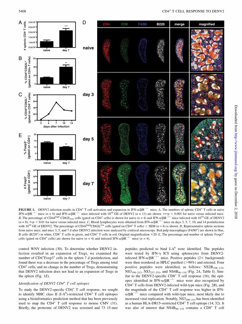
control WNV infection (30). To determine whether DENV2 in-fection resulted in an expansion of Tregs, we examined thenumber of CD4+Foxp3+ cells in the spleen 7 d postinfection, andfound there was a decrease in the percentage of Tregs among totalCD4+ cells, and no change in the number of Tregs, demonstratingthat DENV2 infection does not lead to an expansion of Tregs inthe spleen (Fig. 1E).
Identification of DENV2 CD4+ T cell epitopes
To study the DENV2-specific CD4+ T cell response, we soughtto identify MHC class II (I-Ab)-restricted CD4+ T cell epitopesusing a bioinformatics prediction method that has been previouslyused to map the CD4+ T cell response to mouse CMV (31).Briefly, the proteome of DENV2 was screened and 73 15-mer
peptides predicted to bind I-Ab were identified. The peptideswere tested by IFN-g ICS using splenocytes from DENV2-infected IFN-a/bR2/2 mice. Positive peptides (23 background)were then reordered as HPLC-purified (.90%) and retested. Fourpositive peptides were identified, as follows: NS2B108–122,NS3198–212, NS3237–251, and NS4B96–110 (Fig. 2A, Table I). Sim-ilar to the DENV2-specific CD8+ T cell response (16), the epit-opes identified in IFN-a/bR2/2 mice were also recognized byCD4+ T cells from DENV2-infected wild-type mice (Fig. 2B), andthe magnitude of the CD4+ T cell response was higher in IFN-a/bR2/2 mice compared with wild-type mice, most likely due toincreased viral replication. Notably, NS3200–214 has been identifiedas a human HLA-DR15–restricted CD4+ T cell epitope (14, 32). Itwas also of interest that NS4B96–110 contains a CD8+ T cell
FIGURE 1. DENV2 infection results in CD4+ T cell activation and expansion in IFN-a/bR2/2 mice. A, The numbers of splenic CD4+ T cells in naive
IFN-a/bR2/2 mice (n = 6) and IFN-a/bR2/2 mice infected with 1010 GE of DENV2 (n = 11) are shown. pppp , 0.001 for naive versus infected mice.
B, The percentage of CD44high CD62Llow cells (gated on CD4+ cells) is shown for naive (n = 4) and IFN-a/bR2/2 mice infected with 1010 GE of DENV2
(n = 8). ppp , 0.01 for naive versus infected mice. C, Blood lymphocytes were obtained from IFN-a/bR2/2 mice on days 3, 5, 7, 10, and 14 postinfection
with 1010 GE of DENV2. The percentage of CD44highCD62Llow cells (gated on CD4+ T cells) 6 SEM (n = 6) is shown. D, Representative spleen sections
from naive mice, and mice 3, 5, and 7 d after DENV2 infection were analyzed by confocal microscopy. Red pulp macrophages (F4/80+) are shown in blue,
B cells (B220+) in white, CD8+ T cells in green, and CD4+ T cells in red. Original maginification 320. E, The percentage and number of splenic Foxp3+
cells (gated on CD4+ cells) are shown for naive (n = 4) and infected IFN-a/bR2/2 mice (n = 4).
5408 CD4+ T CELL RESPONSE TO DENV2
by guest on Decem
ber 2, 2018http://w
ww
.jimm
unol.org/D
ownloaded from

epitope (NS4B99–107) that we had previously identified as theimmunodominant epitope in both wild-type and IFN-a/bR2/2
C57BL/6 mice infected with DENV2 (16).
Phenotype of DENV2-specific CD4+ T cells
Multicolor flow cytometry was performed to study the phenotypeof DENV2-specific CD4+ T cells. These cells produced IFN-g,TNF, and IL-2 (Fig. 3). No intracellular IL-4, IL-5, IL-17, or IL-10was detected (data not shown). The DENV2-specific CD4+ T cellsalso expressed CD40L, suggesting they are capable of activatingCD40-expressing cells, which include dendritic cells (DCs) andB cells. The four DENV2-derived CD4+ T cell epitopes inducedresponses that differed in magnitude, but were similar in termsof phenotype. The most polyfunctional cells (those expressingIFN-g, TNF, IL-2, and CD40L) also expressed the highest levelsof the cytokines and CD40L. These results demonstrate thatDENV2 infection elicits a virus-specific Th1 CD4+ T cell responsein IFN-a/bR2/2 mice.
Effects of CD4+ and/or CD8+ T cell depletions on DENV2viral RNA levels
To determine how CD4+ T cells contribute to controlling DENV2infection, we depleted CD4+ T cells, CD8+ T cells, or bothfrom IFN-a/bR2/2 mice and measured DENV2 RNA levels5 d postinfection with 1010 GE of DENV2. We found no differ-ence in viral RNA levels between control undepleted mice andCD4-depleted mice in the serum, kidney, small intestine, spleen,or brain (Fig. 4). As we observed previously (16), CD8-depletedmice had significantly higher viral loads than control mice. De-pletion of both CD4+ and CD8+ T cells resulted in viral RNAlevels that were significantly higher than those in control mice inall tissues examined, and equivalent to the viral RNA levels inCD8-depleted mice. These data show that CD4+ T cells are notrequired to control primary DENV2 infection in IFN-a/bR2/2
mice, and confirm an important role for CD8+ T cells in viralclearance.
CD4+ T cells are not required for the anti-DENV2 Ab response
Although CD4+ T cells were not required for controlling DENV2infection, we wondered whether they made any contribution to theanti-DENV immune response, for example, by helping the B celland/or CD8+ T cell responses. CSR, the process by which the Ig Hchain C region is switched so the B cell expresses a new isotype ofAb, can be induced when CD40L-expressing CD4+ T cells engage
FIGURE 2. Identification of DENV2-derived epitopes recognized by CD4+ T cells. A, Splenocytes were obtained from IFN-a/bR2/2 mice 7 d post-
infection with 1010 GE of DENV2 and restimulated in vitro with DENV2-derived 15-mer peptides predicted to bind I-Ab. Cells were then stained
for surface CD4 and intracellular IFN-g and analyzed by flow cytometry. The four positive peptides identified are shown. In the dot plots, the percentage
of CD4+ T cells producing IFN-g is indicated. The responses of individual mice as well as the mean and SEM are also shown (n = 7–11). The response
of unstimulated cells was subtracted from the response to each DENV2 peptide, and the net percentage and number of splenic CD4+ T cells producing
IFN-g are indicated. B, Splenocytes were obtained from wild-type C57BL/6 mice 7 d postinfection with 1010 GE of DENV2 and stimulated and stained
as in A (n = 6).
Table I. DENV2-derived CD4+ T cell epitopes
Epitope Sequence
NS2B108–122 GLFPVSLPITAAAWYNS3198–212 GKTKRYLPAIVREAINS3237–251 GLPIRYQTPAIRAEHNS4B96–110 IGCYSQVNPITLTAA
The Journal of Immunology 5409
by guest on Decem
ber 2, 2018http://w
ww
.jimm
unol.org/D
ownloaded from

CD40 on B cells (20). However, CSR can also occur in the ab-sence of CD4+ T cell help. To determine whether the anti-DENV2Ab response depends on CD4+ T cells, we first measured DENV2-specific IgM and IgG titers in the sera of control and CD4-depleted mice 7 d postinfection with 1010 GE of DENV2. Asexpected, we found no difference in IgM titers at day 7 betweencontrol and CD4-depleted mice (Fig. 5A). There was also nodifference in IgG titers between control and CD4-depleted mice.To measure the functionality of these DENV2-specific Abs, a flowcytometry-based neutralization assay was performed, in which C6/36 mosquito cells were infected with DENV2 in the presence ofheat-inactivated sera obtained from control and CD4-depletedmice 7 d postinfection. The sera from control and CD4-depletedmice could neutralize DENV2 equally well (Fig. 5B). As reportedpreviously (8), naive serum was able to prevent DENV infection of
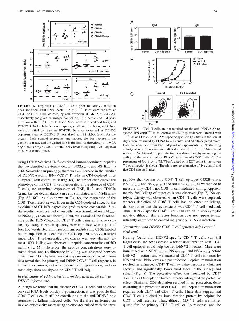
C6/36 cells, although not as efficiently as DENV-immune serum.We also looked for the presence of GC B cells, as the GC reactionis generally thought to be CD4+ T cell dependent (33). Asexpected, we found GC B cells were absent in the CD4-depletedmice (Fig. 5C). Based on the lack of GC B cells in the DENV2-infected CD4-depleted mice, we conclude that the early anti-DENV2 Ab response is CD4- and GC-independent.
CD4+ T cells are not necessary for the primary DENV2-specific CD8+ T cell response
Next, we assessed the role of CD4+ T cells in helping the CD8+
T cell response by examining the DENV2-specific CD8+ T cellresponse in control and CD4-depleted DENV2-infected mice. Thenumbers of splenic CD8+ T cells were equivalent in control andCD4-depleted mice (data not shown). IFN-g ICS was performed
FIGURE 3. DENV2-specific CD4+ T cells are polyfunctional. Splenocytes were obtained from IFN-a/bR2/2 mice 7 d postinfection with 1010 GE of
DENV2 and stimulated in vitro with individual peptides. Cells were then stained for surface CD4, and intracellular IFN-g, TNF, IL-2, and CD40L, and
analyzed by flow cytometry. A, Cells expressing four, three, two, one, or none of the molecules are color coded. Representative dot plots are shown. B, The
response of unstimulated cells was subtracted from the response to each DENV2 peptide, and the net percentages of the CD4+ T cells that are expressing at
least one molecule are indicated. The mean and SEM of three mice are shown.
5410 CD4+ T CELL RESPONSE TO DENV2
by guest on Decem
ber 2, 2018http://w
ww
.jimm
unol.org/D
ownloaded from
using DENV2-derived H-2b–restricted immunodominant peptidesthat we identified previously (M60–67, NS2A8–15, and NS4B99–107)(16). Somewhat surprisingly, there was an increase in the numberof DENV2-specific IFN-g+CD8+ T cells in CD4-depleted micecompared with control mice (Fig. 6A). To further characterize thephenotype of the CD8+ T cells generated in the absence of CD4+
T cells, we examined expression of TNF, IL-2, and CD107a(a marker for degranulation) in cells stimulated with NS4B99–107
(Fig. 6B, 6C). As also shown in Fig. 6A, the magnitude of theCD8+ T cell response was larger in the CD4-depleted mice, but thecytokine and CD107a expression profiles were comparable. Sim-ilar results were observed when cells were stimulated with M60–67
or NS2A8–15 (data not shown). Next, we examined the function-ality of the DENV2-specific CD8+ T cells using an in vivo cyto-toxicity assay, in which splenocytes were pulsed with a pool offour H-2b–restricted immunodominant peptides and CFSE labeledbefore injection into control or CD4-depleted DENV2-infectedmice. CD8+ T cell-mediated cytotoxicity was very efficient; al-most 100% killing was observed at peptide concentrations of 500ng/ml (Fig. 6D). Therefore, the peptide concentrations were ti-trated down, and no difference in killing was observed betweencontrol and CD4-depleted mice at any concentration tested. Thesedata reveal that the primary anti-DENV2 CD8+ T cell response, interms of expansion, cytokine production, degranulation, and cy-totoxicity, does not depend on CD4+ T cell help.
In vivo killing of I-Ab–restricted peptide-pulsed target cells inDENV2-infected mice
Although we found that the absence of CD4+ T cells had no effecton viral RNA levels on day 5 postinfection, it was possible thatCD4+ T cells could still be contributing to the anti-DENV2 hostresponse by killing infected cells. We therefore performed anin vivo cytotoxicity assay using splenocytes pulsed with the three
peptides that contain only CD4+ T cell epitopes (NS2B108–122,NS3198–212, and NS3237–251) and not NS4B96–110, as we wanted tomeasure only CD4+, not CD8+ T cell-mediated killing. Approxi-mately 30% killing of target cells was observed (Fig. 7). No cy-tolytic activity was observed when CD4+ T cells were depleted,whereas depletion of CD8+ T cells had no effect on killing,demonstrating that the cytotoxicity was CD4+ T cell mediated.Thus, DENV2-specific CD4+ T cells can exhibit in vivo cytolyticactivity, although this effector function does not appear to sig-nificantly contribute to controlling primary DENV2 infection.
Vaccination with DENV2 CD4+ T cell epitopes helps controlviral load
Having found that DENV2-specific CD4+ T cells can killtarget cells, we next assessed whether immunization with CD4+
T cell epitopes could help control DENV2 infection. Mice wereimmunized with NS2B108–122, NS3198–212, and NS3237–251 beforeDENV2 infection, and we measured CD4+ T cell responses byICS and viral RNA levels 4 d postinfection. Peptide immunizationresulted in enhanced CD4+ T cell cytokine responses (data notshown), and significantly lower viral loads in the kidney andspleen (Fig. 8). The protective effect was mediated by CD4+
T cells, as CD4 depletion before infection abrogated the protectiveeffect. Similarly, CD8 depletion resulted in no protection, dem-onstrating that protection after CD4+ T cell peptide immunizationrequires both CD4+ and CD8+ T cells. These data suggest thatCD4+ T cells elicited by immunization protect by helping theCD8+ T cell response. Thus, although CD4+ T cells are not re-quired for the primary CD8+ T cell or Ab response, and the
FIGURE 4. Depletion of CD4+ T cells prior to DENV2 infection
does not affect viral RNA levels. IFN-a/bR2/2 mice were depleted of
CD4+ or CD8+ cells, or both, by administration of GK1.5 or 2.43 Ab,
respectively (or given an isotype control Ab), 2 d before and 1 d post-
infection with 1010 GE of DENV2. Mice were sacrificed 5 d later, and
DENV2 RNA levels in the serum, spleen, small intestine, brain, and kidney
were quantified by real-time RT-PCR. Data are expressed as DENV2
copies/ml sera, or DENV2 U normalized to 18S rRNA levels for the
organs. Each symbol represents one mouse, the bar represents the
geometric mean, and the dashed line is the limit of detection. pp , 0.05;
ppp , 0.01; pppp , 0.001 for viral RNA levels comparing T cell-depleted
mice with control mice.
FIGURE 5. CD4+ T cells are not required for the anti-DENV2 Ab re-
sponse. IFN-a/bR2/2 mice (control or CD4 depleted) were infected with
1010 GE of DENV2. A, DENV2-specific IgM and IgG titers in the sera at
day 7 were measured by ELISA (n = 5 control and 6 CD4-depleted mice).
Data are combined from two independent experiments. B, Neutralizing
activity of sera from naive (n = 4) and control (n = 6) or CD4-depleted
mice (n = 6) obtained 7 d postinfection was determined by measuring the
ability of the sera to reduce DENV2 infection of C6/36 cells. C, The
percentage of GC B cells (GL7+Fas+, gated on B220+ cells) in the spleen
7 d postinfection is shown. The plots are representative of five control and
five CD4-depleted mice.
The Journal of Immunology 5411
by guest on Decem
ber 2, 2018http://w
ww
.jimm
unol.org/D
ownloaded from

absence of CD4+ T cells had no effect on viral RNA levels, vac-cination with CD4+ T cell epitopes can reduce viral loads.
DiscussionNumerous studies have investigated the phenotype of DENV se-rotype cross-reactive T cells, which have been hypothesized tocontribute to the pathogenesis of secondary heterologous infec-tions, yet the actual contribution of T cells during DENV infectionis unknown. The findings presented in this study, and in our pre-vious study, reveal that CD8+ T cells play an important protectiverole in the response to primary DENV2 infection, whereas CD4+
T cells do not. CD4+ T cells expanded, were activated, and werelocated near CD8+ T cells and B cells in the spleen after DENV2infection, yet they did not seem to affect the induction of theDENV2-specific CD8+ T cell or Ab responses. In fact, CD4+
T cell depletion had no effect on viral clearance. However, ourdata demonstrate that vaccination with CD4+ T cell epitopes prior
to DENV infection can provide significant protection, supportingT cell peptide vaccination as a strategy for DENV immunizationwithout the risk of ADE.To the best of our knowledge, this is the first study to identify
CD4+ T cell epitopes from DENV-infected mice. We found theDENV2-specific CD4+ T cells recognized epitopes from theNS2B, NS3, and NS4B proteins, and displayed a Th1 phenotype.CD4+ T cell epitopes have been identified in mice infected withother flaviviruses, including yellow fever virus, for which an I-Ab–restricted peptide from the E protein was identified (34), andWNV, for which six epitopes from the E and NS3 proteins wereidentified (35). DENV-derived epitopes recognized by humanCD4+ T cells have been identified, primarily from NS proteins,including the highly conserved NS3 (10). One study identifiednumerous epitopes from the NS3200–324 region, and alignment ofconsensus sequences for DENV1–4 revealed that this region ismore conserved (78%) than NS3 as a whole (68%) (14), sug-
FIGURE 6. CD4+ T cells are not required for the primary DENV2-specific CD8+ T cell response. A, Splenocytes were obtained from IFN-a/bR2/2 mice
(control or CD4-depleted) 7 d postinfection with 1010 GE of DENV2, and stimulated in vitro with immunodominant DENV2-derived H-2b-restricted CD8+ T
cell epitopes. Cells were then stained for CD8 and IFN-g and analyzed by flow cytometry, and the number of CD8+ T cells producing IFN-g is shown. Results
are expressed as the mean and SEM of four mice per group. ppp , 0.01. B and C, Splenocytes were obtained as in A and stimulated with NS4B99–107 in the
presence of an anti-CD107 Ab, and then stained for CD8, IFN-g, TNF, and IL-2. B, Cells expressing four, three, two, one, or none of the molecules are color
coded. Representative dot plots are shown. C, The response of unstimulated cells was subtracted from the response to each DENV2 peptide, and the net
percentages of the CD8+ T cells that are expressing at least one molecule are indicated. The mean and SEM of three mice are shown.D, CD8+ T cell-mediated
killing. IFN-a/bR2/2mice (control or CD4 depleted) infected 7 d previously with 1010 GE of DENV2were injected i.v. with CFSE-labeled target cells pulsed
with a pool of DENV2-derived immunodominant H-2b–restricted peptides (M60–67, NS2A8–15, NS4B99–107, and NS5237–245) at the indicated concentrations
(n = 3–6 mice per group). After 4 h, splenocytes were harvested and analyzed by flow cytometry, and the percentage of killing was calculated.
5412 CD4+ T CELL RESPONSE TO DENV2
by guest on Decem
ber 2, 2018http://w
ww
.jimm
unol.org/D
ownloaded from

gesting that the region contains good candidates for a DENVT cell epitope-based vaccine. Interestingly, one of the NS3-derivedepitopes we identified has also been described as a human CD4+
T cell epitope. This species cross-reactive NS3 peptide may bindhuman HLAs promiscuously, making it a good potential vaccinecandidate. Another intriguing finding was that one of the CD4+
T cell epitopes identified in this study contained the mostimmunodominant of the CD8+ T cell epitopes we had identifiedpreviously. Overlapping epitopes have also been found in lym-phocytic choriomeningitis virus (LCMV) (36–38). The signifi-cance of overlapping epitopes is unknown, but is most likelyrelated to homology between MHC class I and MHC class II, andmay be associated with proteasomal processing. Overlapping epi-topes may turn out to be common once the complete CD4+ andCD8+ T cell responses to other pathogens are mapped.CD4+ T cells are classically defined as helper cells, as they help
B cell and CD8+ T cell responses. However, inflammatory stimulican override the need for CD4+ T cell help, and therefore, theresponses to many acute infections are CD4-independent (17).DENV2 replicates to high levels in IFN-a/bR2/2 mice, the miceappear hunched and ruffled at the time of peak viremia, and theyhave intestinal inflammation (T. Prestwood, unpublished obser-
vations), suggesting that there is a significant inflammatory re-sponse to DENV2 in these mice. Accordingly, we found CD4+
T cells did not play a critical role in the immune response to primaryDENV2 infection. The contribution of CD4+ T cells has been ex-amined during infections with other flaviviruses. Adoptive transferof primed CD4+ and CD8+ T cells, only in combination, protectedmice from a lethal challenge with Japanese encephalitis virus whenthe cells and virus were administered intracerebrally (39). A pro-tective role has been demonstrated for CD4+ T cells in response toWNV infection. One study showed CD4+ T cells contributed toprotection by helping the Ab response and maintaining (but notpriming) the CD8+ T cell response, and were important for clear-ance of virus from the CNS, but not the periphery (40). Anotherstudy found WNV-specific CD4+ T cells directly contributed toprotection in the absence of CD8+ T cells and B cells, producedIFN-g and IL-2, and could kill peptide-pulsed cells (35). Thus, thecontribution of CD4+ T cells to protection against flavivirus in-fection depends on the virus and experimental system.Ab responses can be T cell dependent or T cell independent. In
particular, the formation of GCs is thought to be CD4+ T cell de-pendent, and is where high-affinity plasma cells andmemory B cellsare generated and where CSR can occur (20, 33). Classical CSRoccurs in B cells after CD40L on activated CD4+ T cells bindsCD40 on B cells. However, it is well established that CSR and Abresponses can occur in the absence of CD4+ T cells and orga-nized secondary lymphoid structures (41). T-independent Ab re-sponses to viruses have been demonstrated for vesicular stomatitisvirus (42), rotavirus (43), and polyomavirus (44). In addition, EBV(via latent membrane protein 1) can induce CD40-independent CSR(45), and mice deficient for CD40 or CD4+ T cells are able tomount an influenza-specific IgG response that is protective (46).Similar to the studies mentioned above, our results demonstrate
that the DENV2-specific IgG response at day 7 is CD4-inde-pendent. The lack of GC B cells in CD4-depleted mice shows thatour CD4 depletions have a functional effect, and indicate anti-DENV IgG is being produced by extrafollicular B cells. It ispossible that the absence of CD4+ T cells would have an effect onDENV2-specific Ab titers and/or neutralizing activity at later timepoints; however, the goal of our study was to determine how CD4+
T cells contribute to clearance of primary DENV2 infection,and we clearly show that the early anti-DENV2 Ab response isCD4-independent. The mechanisms by which T-independent Abresponses are induced are beginning to be elucidated. B lym-phocyte stimulator, also known as B cell-activating factor, anda proliferation-inducing ligand are induced on DCs after activa-tion with IFN-a, IFN-g, LPS, or CD40L, and mediate CD40-independent CSR in human B cells (47). B cell-activating factorand a proliferation-inducing ligand also induce CSR in mouseB cells in the absence of CD40 (48). T-independent CSR can alsobe induced via TLR signaling. For example, T-independent CSRto human papillomavirus virus-like particles occurs via TLR4 andMyD88 in mouse B cells (49), and TLR3 or TLR9 signaling caninduce CSR in human B cells (50, 51). The pathways involved inmediating the CD4-independent CSR in DENV2-infected miceremain to be determined.Like pathogen-specific Ab responses, the primary CD8+ T cell
responses to many acute infections are also CD4-independent.CD4-independent CD8+ T cell responses have been demonstratedfor Listeria monocytogenes (52, 53), LCMV (54), and influenza(55). Recently, a mechanism for how DCs can activate CD8+
T cells in the absence of CD4+ T cell help has been described.DCs activated with TLR3 or TLR9 agonists, or by influenza in-fection, upregulated CD40L and activated naive CD8+ T cells inthe absence of CD4+ T cells (56). In accordance with the studies
FIGURE 7. Cytotoxicity mediated by DENV2-specific CD4+ T cells.
In vivo killing of DENV2-derived I-Ab–restricted peptide-pulsed cells.
IFN-a/bR2/2 mice (control, CD4-depleted, or CD8-depleted) infected 7 d
previously with 1010 GE of DENV2 were injected i.v. with CFSE-labeled
target cells pulsed with the three epitopes that contain only CD4+ T cell
epitopes (NS2B108–122, NS3198–212, and NS3237–251) (n = 6 control, 3 CD4-
depleted, and 3 CD8-depleted mice). After 16 h, splenocytes were har-
vested and analyzed by flow cytometry, and the percentage of killing was
calculated.
FIGURE 8. Peptide immunization with CD4+ T cell epitopes results in
enhanced DENV2 clearance. IFN-a/bR2/2 mice were immunized s.c. with
50 mg each of the three DENV peptides that contain only CD4+ T cell
epitopes (NS2B108–122, NS3198–212, NS3237–251) in CFA, or mock immu-
nized with DMSO in CFA. Mice were boosted 11 d later with peptide in
IFA, then challenged with 1011 GE of DENV2 13 d later, and sacrificed 4 d
postinfection. Separate groups of peptide-immunized mice were depleted
of CD4+ or CD8+ T cells prior to infection. DENV2 RNA levels in the
tissues were quantified by real-time RT-PCR and are expressed as DENV2
U normalized to 18S rRNA. Each symbol represents one mouse, and the
bar represents the geometric mean. pp , 0.05; ppp , 0 0.01.
The Journal of Immunology 5413
by guest on Decem
ber 2, 2018http://w
ww
.jimm
unol.org/D
ownloaded from

mentioned above, we found that the primary CD8+ T cell responseto DENV2 did not depend on CD4+ T cells. In fact, we observedan enhanced DENV2-specific CD8+ T cell response in CD4-deficient mice compared with control mice at day 7, which hasalso been reported for influenza (55)- and WNV (40)-specificCD8+ T cell responses. In our study, this could be due to thedepletion of Tregs, or an increased availability of cytokines (e.g.,IL-2) in mice lacking CD4+ T cells. This enhanced CD8+ T cellresponse may explain why CD4-depleted mice have no differencesin viral titers despite the fact that DENV2-specific CD4+ T cellsdemonstrate in vivo cytotoxicity.Although CD4+ T cells did not play an important role in helping
the Ab or CD8+ T cell responses, we found DENV2-specific CD4+
T cells could kill peptide-pulsed target cells in vivo. CD4+ T cellsspecific for other pathogens, including HIV (57) and influenza(58), demonstrate in vitro cytotoxicity. In vivo cytotoxicity assayshave been used to show CD4+ T cell-mediated killing followinginfection with LCMV (59) and WNV (35). DENV-specific cyto-lytic human CD4+ T cell clones (60, 61) and a mouse (H-2d) CD4+
T cell clone (62) have been reported. Whether CD4+ T cells ac-tually kill infected cells during DENV infection remains to bedetermined, but is possible, as MHC class II-expressing macro-phages are targets of DENV infection (8). Based on the fact thatCD4 depletion did not have a significant effect on viral clearance,it is unlikely that CD4+ T cell-mediated killing plays a major rolein the anti-DENV2 response in this model.An obvious caveat to using the IFN-a/bR2/2 mice is that type I
IFNs are known to help T cell and B cell responses through theiractions on DCs, and can act directly on T cells (63). Type I IFNswere found to contribute to the expansion of CD4+ T cells fol-lowing infection with LCMV, but not L. monocytogenes (64). TypeI IFNs can induce the development of Th1 IFN-g responses inhuman CD4+ T cells, but cannot substitute for IL-12 in promotingTh1 responses in mouse CD4+ T cells (65). Following Listeriainfection, IL-12 synergized with type I IFN to induce IFN-gproduction by CD4+ T cells (66). Although DENV does not rep-licate to detectable levels in wild-type mice, examining the CD4+
T cell response in these mice revealed that the same epitopes wererecognized as in the IFN-a/bR2/2 mice, but the magnitude of theepitope-specific response was greater in the IFN-a/bR2/2 mice.This suggests that the high levels of viral replication in the IFN-a/bR2/2 mice are sufficient to drive a DENV2-specific CD4+
IFN-g response. Our results demonstrate a DENV2-specific CD4+
T cell response, including Th1-type cytokine production and cy-totoxicity, in the absence of IFN-a/bR signaling, yet this responseis not required for clearance of infection. It is, however, possiblethat CD4+ T cells contribute to protection during DENV infectionof hosts with intact IFN responses.We have previously shown that immunization with CD8+ T cell
epitopes resulted in enhanced viral clearance (16), and the re-sults presented in this study show that immunization with CD4+
T epitopes is also protective. Peptide immunization with CD4+
T cell epitopes has also been found to protect against WNV (35)and HSV-1 (67). Our results have significant implications forDENV vaccine development. Although a DENV vaccine is aglobal public health priority, designing a vaccine has been chal-lenging, as it needs to induce protection against all four serotypes.The DENV vaccine candidates in development, some of whichare in phase II trials, focus on eliciting an Ab response. The chal-lenge is to induce and maintain a neutralizing Ab response againstall four serotypes, as it is becoming increasingly clear that non-neutralizing Ab (or subneutralizing quantities of Ab) can actuallyenhance dengue disease (8, 9). A different approach would bea peptide vaccine that induces cell-mediated immunity, including
both CD4+ and CD8+ T cell responses, which would not be able toprevent infection, but could reduce viral loads and disease sever-ity, and would eliminate the risk of ADE. Such a vaccine shouldtarget highly conserved regions of the proteome, for example,NS3, NS4B, and/or NS5, and ideally include epitopes conservedacross all four serotypes. A vaccine containing only peptides fromthese particular NS proteins would also preclude the induction ofany Ab against epitopes on the virion, which could enhance in-fection, or Ab against NS1, which could potentially contribute topathogenesis (68). Our peptide vaccination was given along withCFA, which is commonly used in mice to induce Th1 responses(69), which was the type of response observed after natural DENVinfection. CFA is not a vaccine adjuvant approved for human use,and thus, any peptide vaccine developed against DENV will haveto be formulated with an adjuvant that is approved for human useand promotes a Th1 response. Numerous vaccine adjuvants arecurrently under development and can be tested in our experimentalsystem in the future.Although our results indicate CD4+ T cells do not make a sig-
nificant contribution to controlling primary DENV2 infection, thecharacterization of the primary CD4+ T cell response and epitopeidentification will allow us to determine the role of CD4+ T cellsduring secondary homologous and heterologous infections. CD4+
T cells are often dispensable for the primary CD8+ T cell responseto infection, but have been shown to be required for the mainte-nance of memory CD8+ T cell responses after acute infection (70).Finally, our findings support a DENV vaccine strategy that indu-ces CD4+ T cell, in addition to CD8+ T cell, responses.
AcknowledgmentsWe thank Steven Lada for technical assistance, Dr. Carl Ware for mice, and
Dr. Shane Crotty for reagents.
DisclosuresThe authors have no financial conflicts of interest.
References1. Fu, J., B. H. Tan, E. H. Yap, Y. C. Chan, and Y. H. Tan. 1992. Full-length
cDNA sequence of dengue type 1 virus (Singapore strain S275/90). Virology188: 953–958.
2. WHO. 2009. Dengue: Guidelines for Diagnosis, Treatment, Prevention andControl. World Health Organization Press, Geneva, Switzerland.
3. Whitehead, S. S., J. E. Blaney, A. P. Durbin, and B. R. Murphy. 2007. Prospectsfor a dengue virus vaccine. Nat. Rev. Microbiol. 5: 518–528.
4. Sangkawibha, N., S. Rojanasuphot, S. Ahandrik, S. Viriyapongse, S. Jatanasen,V. Salitul, B. Phanthumachinda, and S. B. Halstead. 1984. Risk factors in dengueshock syndrome: a prospective epidemiologic study in Rayong, Thailand. I. The1980 outbreak. Am. J. Epidemiol. 120: 653–669.
5. Guzman, M. G., G. Kouri, L. Valdes, J. Bravo, M. Alvarez, S. Vazques,I. Delgado, and S. B. Halstead. 2000. Epidemiologic studies on dengue inSantiago de Cuba, 1997. Am. J. Epidemiol. 152: 793–799, discussion 804.
6. Morens, D. M. 1994. Antibody-dependent enhancement of infection and thepathogenesis of viral disease. Clin. Infect. Dis. 19: 500–512.
7. Halstead, S. B. 2003. Neutralization and antibody-dependent enhancement ofdengue viruses. Adv. Virus Res. 60: 421–467.
8. Zellweger, R. M., T. R. Prestwood, and S. Shresta. 2010. Enhanced infection ofliver sinusoidal endothelial cells in a mouse model of antibody-induced severedengue disease. Cell Host Microbe 7: 128–139.
9. Balsitis, S. J., K. L. Williams, R. Lachica, D. Flores, J. L. Kyle, E. Mehlhop,S. Johnson, M. S. Diamond, P. R. Beatty, and E. Harris. 2010. Lethal antibodyenhancement of dengue disease in mice is prevented by Fc modification. PLoSPathog. 6: e1000790.
10. Mathew, A., and A. L. Rothman. 2008. Understanding the contribution of cellularimmunity to dengue disease pathogenesis. Immunol. Rev. 225: 300–313.
11. Mangada, M. M., and A. L. Rothman. 2005. Altered cytokine responses ofdengue-specific CD4+ T cells to heterologous serotypes. J. Immunol. 175: 2676–2683.
12. Mongkolsapaya, J., W. Dejnirattisai, X. N. Xu, S. VasanawathanaN. Tangthawornchaikul, A. Chairunsri, S. Sawasdivorn, T. Duangchinda,
5414 CD4+ T CELL RESPONSE TO DENV2
by guest on Decem
ber 2, 2018http://w
ww
.jimm
unol.org/D
ownloaded from
T. Dong, S. Rowland-Jones, et al. 2003. Original antigenic sin and apoptosis inthe pathogenesis of dengue hemorrhagic fever. Nat. Med. 9: 921–927.
13. Mongkolsapaya, J., T. Duangchinda, W. Dejnirattisai, S. Vasanawathana,P. Avirutnan, A. Jairungsri, N.Khemnu,N. Tangthawornchaikul, P. Chotiyarnwong,K. Sae-Jang, et al. 2006. T cell responses in dengue hemorrhagic fever: are cross-reactive T cells suboptimal? J. Immunol. 176: 3821–3829.
14. Simmons, C. P., T. Dong, N. V. Chau, N. T. Dung, T. N. Chau, T. T. Thao Le,N. T. Dung, T. T. Hien, S. Rowland-Jones, and J. Farrar. 2005. Early T-cellresponses to dengue virus epitopes in Vietnamese adults with secondary denguevirus infections. J. Virol. 79: 5665–5675.
15. Yauch, L. E., and S. Shresta. 2008. Mouse models of dengue virus infection anddisease. Antiviral Res. 80: 87–93.
16. Yauch, L. E., R. M. Zellweger, M. F. Kotturi, A. Qutubuddin, J. Sidney,B. Peters, T. R. Prestwood, A. Sette, and S. Shresta. 2009. A protective role fordengue virus-specific CD8+ T cells. J. Immunol. 182: 4865–4873.
17. Bevan, M. J. 2004. Helping the CD8(+) T-cell response. Nat. Rev. Immunol. 4:595–602.
18. Elgueta, R., M. J. Benson, V. C. de Vries, A. Wasiuk, Y. Guo, and R. J. Noelle.2009. Molecular mechanism and function of CD40/CD40L engagement in theimmune system. Immunol. Rev. 229: 152–172.
19. Nakanishi, Y., B. Lu, C. Gerard, and A. Iwasaki. 2009. CD8(+) T lymphocytemobilization to virus-infected tissue requires CD4(+) T-cell help. Nature 462:510–513.
20. Stavnezer, J., J. E. Guikema, and C. E. Schrader. 2008. Mechanism and regu-lation of class switch recombination. Annu. Rev. Immunol. 26: 261–292.
21. Lin, Y. L., C. L. Liao, L. K. Chen, C. T. Yeh, C. I. Liu, S. H. Ma, Y. Y. Huang,Y. L. Huang, C. L. Kao, and C. C. King. 1998. Study of dengue virus infection inSCID mice engrafted with human K562 cells. J. Virol. 72: 9729–9737.
22. Prestwood, T. R., D. M. Prigozhin, K. L. Sharar, R. M. Zellweger, and S. Shresta.2008. A mouse-passaged dengue virus strain with reduced affinity for heparansulfate causes severe disease in mice by establishing increased systemic viralloads. J. Virol. 82: 8411–8421.
23. Wang, P., J. Sidney, C. Dow, B. Mothe, A. Sette, and B. Peters. 2008. A sys-tematic assessment of MHC class II peptide binding predictions and evaluationof a consensus approach. PLoS Comput. Biol. 4: e1000048.
24. Zhang, Q., P. Wang, Y. Kim, P. Haste-Andersen, J. Beaver, P. E. Bourne,H. H. Bui, S. Buus, S. Frankild, J. Greenbaum, et al. 2008. Immune epitopedatabase analysis resource (IEDB-AR). Nucleic Acids Res. 36(Web Server is-sue): W513–518.
25. Lambeth, C. R., L. J. White, R. E. Johnston, and A. M. de Silva. 2005. Flowcytometry-based assay for titrating dengue virus. J. Clin. Microbiol. 43: 3267–3272.
26. Houng, H. H., D. Hritz, and N. Kanesa-thasan. 2000. Quantitative detection ofdengue 2 virus using fluorogenic RT-PCR based on 39-noncoding sequence.J. Virol. Methods 86: 1–11.
27. Lacher, M. D., M. I. Tiirikainen, E. F. Saunier, C. Christian, M. Anders, M. Oft,A. Balmain, R. J. Akhurst, and W. M. Korn. 2006. Transforming growth factor-beta receptor inhibition enhances adenoviral infectability of carcinoma cells viaup-regulation of Coxsackie and adenovirus receptor in conjunction with reversalof epithelial-mesenchymal transition. Cancer Res. 66: 1648–1657.
28. Josefowicz, S. Z., and A. Rudensky. 2009. Control of regulatory T cell lineagecommitment and maintenance. Immunity 30: 616–625.
29. Lund, J. M., L. Hsing, T. T. Pham, and A. Y. Rudensky. 2008. Coordination ofearly protective immunity to viral infection by regulatory T cells. Science 320:1220–1224.
30. Lanteri, M. C., K. M. O’Brien, W. E. Purtha, M. J. Cameron, J. M. Lund,R. E. Owen, J. W. Heitman, B. Custer, D. F. Hirschkorn, L. H. Tobler, et al. 2009.Tregs control the development of symptomatic West Nile virus infection inhumans and mice. J. Clin. Invest. 119: 3266–3277.
31. Arens, R., P. Wang, J. Sidney, A. Loewendorf, A. Sette, S. P. Schoenberger,B. Peters, and C. A. Benedict. 2008. Cutting edge: murine cytomegalovirusinduces a polyfunctional CD4 T cell response. J. Immunol. 180: 6472–6476.
32. Zeng, L., I. Kurane, Y. Okamoto, F. A. Ennis, and M. A. Brinton. 1996. Iden-tification of amino acids involved in recognition by dengue virus NS3-specific,HLA-DR15-restricted cytotoxic CD4+ T-cell clones. J. Virol. 70: 3108–3117.
33. Allen, C. D., T. Okada, and J. G. Cyster. 2007. Germinal-center organizationand cellular dynamics. Immunity 27: 190–202.
34. van der Most, R. G., L. E. Harrington, V. Giuggio, P. L. Mahar, and R. Ahmed.2002. Yellow fever virus 17D envelope and NS3 proteins are major targets of theantiviral T cell response in mice. Virology 296: 117–124.
35. Brien, J. D., J. L. Uhrlaub, and J. Nikolich-Zugich. 2008. West Nile virus-specific CD4 T cells exhibit direct antiviral cytokine secretion and cytotoxicityand are sufficient for antiviral protection. J. Immunol. 181: 8568–8575.
36. Homann, D., H. Lewicki, D. Brooks, J. Eberlein, V. Mallet-Designe, L. Teyton,and M. B. Oldstone. 2007. Mapping and restriction of a dominant viral CD4+T cell core epitope by bothMHC class I andMHC class II. Virology 363: 113–123.
37. Mothe, B. R., B. S. Stewart, C. Oseroff, H. H. Bui, S. Stogiera, Z. Garcia,C. Dow, M. P. Rodriguez-Carreno, M. Kotturi, V. Pasquetto, et al. 2007. Chroniclymphocytic choriomeningitis virus infection actively down-regulates CD4+T cell responses directed against a broad range of epitopes. J. Immunol. 179:1058–1067.
38. Dow, C., C. Oseroff, B. Peters, C. Nance-Sotelo, J. Sidney, M. Buchmeier,A. Sette, and B. R. Mothe. 2008. Lymphocytic choriomeningitis virus infectionyields overlapping CD4+ and CD8+ T-cell responses. J. Virol. 82: 11734–11741.
39. Murali-Krishna, K., V. Ravi, and R. Manjunath. 1996. Protection of adult but notnewborn mice against lethal intracerebral challenge with Japanese encephalitis
virus by adoptively transferred virus-specific cytotoxic T lymphocytes: re-quirement for L3T4+ T cells. J. Gen. Virol. 77: 705–714.
40. Sitati, E. M., and M. S. Diamond. 2006. CD4+ T cell responses are required forclearance of West Nile virus from the central nervous system. J. Virol. 80:12060–12069.
41. Fagarasan, S., and T. Honjo. 2000. T-Independent immune response: new aspectsof B cell biology. Science 290: 89–92.
42. Freer, G., C. Burkhart, I. Ciernik, M. F. Bachmann, H. Hengartner, andR. M. Zinkernagel. 1994. Vesicular stomatitis virus Indiana glycoprotein asa T-cell-dependent and -independent antigen. J. Virol. 68: 3650–3655.
43. Franco, M. A., and H. B. Greenberg. 1997. Immunity to rotavirus in T celldeficient mice. Virology 238: 169–179.
44. Szomolanyi-Tsuda, E., J. D. Brien, J. E. Dorgan, R. L. Garcea, R. T. Woodland,and R. M. Welsh. 2001. Antiviral T-cell-independent type 2 antibody responsesinduced in vivo in the absence of T and NK cells. Virology 280: 160–168.
45. He, B., N. Raab-Traub, P. Casali, and A. Cerutti. 2003. EBV-encoded latentmembrane protein 1 cooperates with BAFF/BLyS and APRIL to induce T cell-independent Ig heavy chain class switching. J. Immunol. 171: 5215–5224.
46. Lee, B. O., J. Rangel-Moreno, J. E. Moyron-Quiroz, L. Hartson, M. Makris,F. Sprague, F. E. Lund, and T. D. Randall. 2005. CD4 T cell-independent anti-body response promotes resolution of primary influenza infection and helps toprevent reinfection. J. Immunol. 175: 5827–5838.
47. Litinskiy, M. B., B. Nardelli, D. M. Hilbert, B. He, A. Schaffer, P. Casali, andA. Cerutti. 2002. DCs induce CD40-independent immunoglobulin classswitching through BLyS and APRIL. Nat. Immunol. 3: 822–829.
48. Castigli, E., S. A. Wilson, S. Scott, F. Dedeoglu, S. Xu, K. P. Lam, R. J. Bram,H. Jabara, and R. S. Geha. 2005. TACI and BAFF-R mediate isotype switchingin B cells. J. Exp. Med. 201: 35–39.
49. Yang, R., F. M. Murillo, M. J. Delannoy, R. L. Blosser, W. H. Yutzy, 4th,S. Uematsu, K. Takeda, S. Akira, R. P. Viscidi, and R. B. Roden. 2005.B lymphocyte activation by human papillomavirus-like particles directly inducesIg class switch recombination via TLR4-MyD88. J. Immunol. 174: 7912–7919.
50. He, B., X. Qiao, and A. Cerutti. 2004. CpG DNA induces IgG class switch DNArecombination by activating human B cells through an innate pathway thatrequires TLR9 and cooperates with IL-10. J. Immunol. 173: 4479–4491.
51. Xu, W., P. A. Santini, A. J. Matthews, A. Chiu, A. Plebani, B. He, K. Chen, andA. Cerutti. 2008. Viral double-stranded RNA triggers Ig class switching by ac-tivating upper respiratory mucosa B cells through an innate TLR3 pathway in-volving BAFF. J. Immunol. 181: 276–287.
52. Sun, J. C., and M. J. Bevan. 2003. Defective CD8 T cell memory following acuteinfection without CD4 T cell help. Science 300: 339–342.
53. Shedlock, D. J., J. K.Whitmire, J. Tan, A. S. MacDonald, R. Ahmed, and H. Shen.2003. Role of CD4 T cell help and costimulation in CD8 T cell responses duringListeria monocytogenes infection. J. Immunol. 170: 2053–2063.
54. Ahmed, R., L. D. Butler, and L. Bhatti. 1988. T4+ T helper cell function in vivo:differential requirement for induction of antiviral cytotoxic T-cell andantibody responses. J. Virol. 62: 2102–2106.
55. Belz, G. T., D. Wodarz, G. Diaz, M. A. Nowak, and P. C. Doherty. 2002.Compromised influenza virus-specific CD8(+)-T-cell memory in CD4(+)-T-cell-deficient mice. J. Virol. 76: 12388–12393.
56. Johnson, S., Y. Zhan, R. M. Sutherland, A. M. Mount, S. Bedoui, J. L. Brady,E. M. Carrington, L. E. Brown, G. T. Belz, W. R. Heath, and A. M. Lew. 2009. Se-lected Toll-like receptor ligands and viruses promote helper-independent cytotoxicT cell priming by upregulating CD40L on dendritic cells. Immunity 30: 218–227.
57. Norris, P. J., H. F. Moffett, O. O. Yang, D. E. Kaufmann, M. J. Clark,M. M. Addo, and E. S. Rosenberg. 2004. Beyond help: direct effector functionsof human immunodeficiency virus type 1-specific CD4(+) T cells. J. Virol. 78:8844–8851.
58. Taylor, S. F., and B. S. Bender. 1995. Beta 2-microglobulin-deficient micedemonstrate class II MHC restricted anti-viral CD4+ but not CD8+ CTL againstinfluenza-sensitized autologous splenocytes. Immunol. Lett. 46: 67–73.
59. Jellison, E. R., S. K. Kim, and R. M. Welsh. 2005. Cutting edge: MHC classII-restricted killing in vivo during viral infection. J. Immunol. 174: 614–618.
60. Gagnon, S. J., W. Zeng, I. Kurane, and F. A. Ennis. 1996. Identification of twoepitopes on the dengue 4 virus capsid protein recognized by a serotype-specificand a panel of serotype-cross-reactive human CD4+ cytotoxic T-lymphocyteclones. J. Virol. 70: 141–147.
61. Kurane, I., A. Meager, and F. A. Ennis. 1989. Dengue virus-specific humanT cell clones: serotype crossreactive proliferation, interferon gamma production,and cytotoxic activity. J. Exp. Med. 170: 763–775.
62. Rothman, A. L., I. Kurane, and F. A. Ennis. 1996. Multiple specificities in themurine CD4+ and CD8+ T-cell response to dengue virus. J. Virol. 70: 6540–6546.
63. Iwasaki, A., and R. Medzhitov. 2004. Toll-like receptor control of the adaptiveimmune responses. Nat. Immunol. 5: 987–995.
64. Havenar-Daughton, C., G. A. Kolumam, and K. Murali-Krishna. 2006. Cuttingedge: the direct action of type I IFN on CD4 T cells is critical for sustainingclonal expansion in response to a viral but not a bacterial infection. J. Immunol.176: 3315–3319.
65. Rogge, L., D. D’Ambrosio, M. Biffi, G. Penna, L. J. Minetti, D. H. Presky,L. Adorini, and F. Sinigaglia. 1998. The role of Stat4 in species-specific regu-lation of Th cell development by type I IFNs. J. Immunol. 161: 6567–6574.
66. Way, S. S., C. Havenar-Daughton, G. A. Kolumam, N. N. Orgun, and K. Murali-Krishna. 2007. IL-12 and type-I IFN synergize for IFN-gamma production byCD4 T cells, whereas neither are required for IFN-gamma production by CD8T cells after Listeria monocytogenes infection. J. Immunol. 178: 4498–4505.
The Journal of Immunology 5415
by guest on Decem
ber 2, 2018http://w
ww
.jimm
unol.org/D
ownloaded from

67. BenMohamed, L., G. Bertrand, C. D. McNamara, H. Gras-Masse, J. Hammer,S. L. Wechsler, and A. B. Nesburn. 2003. Identification of novel immunodo-minant CD4+ Th1-type T-cell peptide epitopes from herpes simplex virus gly-coprotein D that confer protective immunity. J. Virol. 77: 9463–9473.
68. Lin, C. F., S. W. Wan, H. J. Cheng, H. Y. Lei, and Y. S. Lin. 2006. Autoimmunepathogenesis in dengue virus infection. Viral Immunol. 19: 127–132.
69. Billiau, A., and P. Matthys. 2001. Modes of action of Freund’s adjuvantsin experimental models of autoimmune diseases. J. Leukoc. Biol. 70: 849–860.
70. Sun, J. C., M. A. Williams, and M. J. Bevan. 2004. CD4+ T cells are required forthe maintenance, not programming, of memory CD8+ T cells after acute in-fection. Nat. Immunol. 5: 927–933.
5416 CD4+ T CELL RESPONSE TO DENV2
by guest on Decem
ber 2, 2018http://w
ww
.jimm
unol.org/D
ownloaded from

![Dengue Fever/Severe Dengue Fever/Chikungunya Fever · Dengue fever and severe dengue (dengue hemorrhagic fever [DHF] and dengue shock syndrome [DSS]) are caused by any of four closely](https://img.pdfslide.us/doc/110x75/5e87bf3e7a86e85d3b149cd7/dengue-feversevere-dengue-feverchikungunya-dengue-fever-and-severe-dengue-dengue.jpg)