Embed Size (px)
Citation preview
7 6 | H O R E N S T E I N E T A L . | M O L M E D 1 5 ( 3 - 4 ) 7 6 - 8 4 , M A R C H - A P R I L 2 0 0 9
INTRODUCTIONThe avascular and transparent human
cornea is composed of epithelial, stromal,and endothelial layers. The corneal ep-ithelium, which plays vital functionalroles both as a physical barrier at the oc-ular surface and in wound repair and in-flammation, must undergo continuousself-renewal through the action of stemcells (1,2). Epithelial stem cells reside inspecialized niches at the corneoscleraljunction, or limbus (3). A number of sig-
naling pathways have been shown to beinvolved in the regulation of self-renewing tissues; the pathways involvedin corneal epithelium regenerationmostly are unexplored (4).
In many tissues, extracellular NAD+
induces a variety of physiological re-sponses in epithelial cells (5). NAD+ ismade available either by passive releasefrom dying cells or by active secretionthrough connexin 43 (Cx43) channels in aparacrine and in an autocrine way (6). In
humans, CD38 and CD157 are the mainNAD+-modifying enzymes, synthesizingcompounds critical for cell homeostasisand metabolism (7). CD38, a 45-kDatransmembrane glycoprotein, functionsboth as an ectoenzyme and as a receptor.CD38 is a multifunctional enzyme whichconverts NAD+ to adenosine diphos-phate ribose (ADPR) in neutral pH con-ditions, through its NADase activity andto cyclic ADPR (cADPR) through itsADP-ribosyl cyclase (ARC) activity.CD38 hydrolyzes cADPR to ADPRthrough its cADPR hydrolase activity.These end products are active biologi-cally as second messengers: cADPR gatesCa2+ release from intracellular stores, andADPR can activate the transient receptorpotential (TRP) channel-2 leading to Ca2+
influx (8,9). As a receptor, CD38 is in-volved in the transduction of activationand proliferation signals, thus cooperat-
CD38 and CD157 Ectoenzymes Mark Cell Subsets in theHuman Corneal Limbus
Alberto L Horenstein,1,2* Federico Sizzano,3* Riccardo Lusso,1,2 Federico Genzano Besso,3 Enza Ferrero,1,2
Silvia Deaglio,1,2 Franco Corno,4 and Fabio Malavasi1,2
1Laboratory of Immunogenetics, Department of Genetics, Biology and Biochemistry, University of Torino Medical School, Torino,Italy; 2Research Center for Experimental Medicine (CeRMS), University of Torino Medical School, Torino, Italy; 3Transplant Immunol-ogy Service, San Giovanni Battista Hospital, Torino, Italy; and 4Department of Surgical and Medical Sciences, University of TorinoMedical School, Torino, Italy
Nicotinamide adenine dinucleotide (NAD+), a precursor of molecules involved in cell regulatory processes, is released in extra-cellular compartments after stress or inflammation.This study investigates the expression in the human cornea of CD38 and CD157,two NAD+-consuming ectoenzymes and surface receptors. The analysis in corneal epithelial and stromal cells was performed bymeans of multiple approaches, which included immunofluorescence, reverse transcriptase polymerase chain reaction (RT-PCR),Western blot, and confocal microscopy. The presence of enzymatically active NAD+-consumers in intact corneal cells was ana-lyzed by high performance liquid chromatography (HPLC)-based assays. The results obtained show that CD38 and CD157 are ex-pressed constitutively by corneal cells: CD38 appears as a 45-kDa monomer, while CD157 is a 42- to 45-kDa doublet. The mole-cules are enzymatically active, with features reminiscent of those observed in human leukocytes. CD38 is expressed by cells of thesuprabasal limbal epithelium, whereas it is not detectable in central corneal epithelium and stroma. CD157 is expressed by basallimbal clusters, a p63+/cytokeratin 19+ cell subset reported to contain corneal stem cells, and by stromal cells. The results of thework indicates that the human cornea is equipped with molecular tools capable of consuming extracellular NAD+, and thatCD157 is a potential marker of corneal limbal cells in the stem cell niche. The presence and characteristics of these ectoenzymesmay be exploited to design drugs for wound repair or for applications in tissue transplantation.© 2009 The Feinstein Institute for Medical Research, www.feinsteininstitute.orgOnline address: http://www.molmed.orgdoi: 10.2119/molmed.2008.00108
*ALH and FS contributed equally to the experimental work.Address correspondence and reprint requests to Alberto L Horenstein, Laboratory of Im-munogenetics, Department of Genetics, Biology and Biochemistry, University of TorinoMedical School, via Santena 19, 10126 Torino, Italy. Phone: + 39-011-696 1734; Fax: + 39-011-696 6155; E-mail: [email protected] November 14, 2008; Accepted for publication November 19, 2008; Epub (www.molmed.org) ahead of print November 19, 2008.
R E S E A R C H A R T I C L E
M O L M E D 1 5 ( 3 - 4 ) 7 6 - 8 4 , M A R C H - A P R I L 2 0 0 9 | H O R E N S T E I N E T A L . | 7 7
ing in the adhesion of leukocytes to en-dothelium through binding to its non-substrate ligand CD31 (10). CD38 is ex-pressed in a unique pattern by cells ofthe immune system, and shows wide-spread distribution and cellular functionsin non-hematopoietic tissues (7).
CD157, a 42- to 45-kDa surface mole-cule with a glycosyl-phosphatidylinositol(GPI)-anchor, was identified originally inhumans as bone marrow stromal cell-1(BST-1) antigen (11). CD157 is a secondmammalian member of the NADase/ARC family, and shares 36% amino acididentity and 53% similarity to humanCD38. The tissue distribution of CD157differs significantly from that of CD38(12); despite their probably differentphysiological roles, the two glycopro-teins nonetheless have similar enzymaticfunctions. Like CD38, CD157 also is a re-ceptor that transduces activation signals,although a non-substrate ligand has notyet been identified (13).
Increasing evidence indicates that thebioactive molecules cADPR and ADPRare regulators of Ca2+-dependent func-tions (8). As variations in Ca2+ concentra-tion in ocular compartments also mayunderlie pathological changes leading toblindness, an investigation into the func-tional role of the ARC family in cornealtissue is of the utmost importance(14,15). Although reported in the retinain vertebrate eyes (16–18), the expressionof NAD+-metabolizing ectoenzymes inthe human cornea has been unexploreduntil now. This work was designed to fillthis gap, to add to our body of knowl-edge concerning the tissue distributionof CD38 and CD157, and to shed light onthe possible functional role of ARCs inthe health and disease of the human eye.
MATERIALS AND METHODS
Antibodies and ReagentsThe anti-CD38 monoclonal antibodies
(mAbs) used were IB4 (IgG2a) (19) andAT13/5 (IgG1) (20). The anti-CD157 mAbused was SY/11B5 (IgG1) and the anti-HLA Class I mAb was O1.65 (IgG2a),used as a positive control. All mAbs
were produced in-house and purified byHPLC (Beckman Gold 126/166NM)affinity chromatography on Protein A(MAb Select, GE Healthcare, Piscataway,NJ, USA), followed by hydroxyapatitecolumn (BioRad, Milan, Italy), as de-scribed (21). Other reagents were poly-clonal goat anti-human cytokeratin 3/12(CK3/12), anti-cytokeratin 19 (CK19),anti-connexin 43 (Cx43), and anti-p63mAbs, purchased from Santa CruzBiotech (Santa Cruz, CA, USA). Sec-ondary antibodies for immunofluores-cence were: donkey anti-mouse IgG con-jugated with Alexa 488 (DαMIgG-Alexa488) and donkey anti-goat Ig labeledwith Alexa 555 (DαGIg-Alexa 555) fromInvitrogen (Milan, Italy). All otherreagents were of analytical grade and ob-tained from Sigma (Milan, Italy).
Isolation and Culture of Corneal CellsViable epithelial and stromal cells were
obtained from human corneoscleral tis-sues from 10 cadaver eye donors (Pied-mont Cornea Bank, San Giovanni Bat-tista Hospital, Torino, Italy) deemedunsuitable for transplantation due to un-acceptably low endothelial cell counts orstromal wounds. Epithelial cells wereisolated by scraping the ocular surfaceafter detaching the endothelial layerfrom the stroma, or were cultured fromcorneal explants. Surface-scraped epithe-lial cells were counted under a micro-scope and washed twice in phosphatebuffered saline (PBS). Cultured epithelialcells were obtained by outgrowing.Corneas were cut into 12 single sectionsand placed epithelial side down in 6-wellplates (Corning Costar, Milan, Italy) witha drop of Corneal Epithelial Cell EpiLifeMedium (Invitrogen), followed 24 h laterby an additional 1 mL of medium. Afterspreading, cells were cultured at 2 × 10⁴
cell/cm2 for 3 to 5 passages. Cornealstromal cells were derived from the en-dothelial and epithelial layers scrapedfrom the donor corneas. The stromalcomponent was released after treatment(3 to 4 h, with gentle shaking) with colla-genase IV (2.5 mg/mL in Hank’s bal-anced salt solution without Ca2+ and
Mg2+), and then cultured (5% CO2 , 37°C)in DMEM (all from Gibco, Milan, Italy)supplemented with 2 mM L-glutamine(Invitrogen), 10% FCS (Hyclone, Logan,UT, USA), and 50 IU/mL streptomycin(Sigma).
ImmunofluorescenceSurface expression of CD38 and CD157
in corneal cells was determined by indi-rect immunofluorescence (IIF). Primarycells scraped from the epithelium (2 × 10⁵
cells/sample) or from cultured cornealepithelial and stromal cells were fixed in2% paraformaldehyde (PAF) for 15 min.Non-specific protein binding wasblocked (30 min at 4°C) by incubationwith 1% BSA. Cells were washed withPBS and then incubated (1 h at 4°C forsurface-scraped cells or 2 h at room tem-perature for cultured cells) with anti-CD38, anti-CD157, or anti-HLA Class ImAbs. Staining was obtained by incubat-ing the cells (30 min for surface-scrapedcells or 90 min for cultured cells at 4°C)with DαMIgG-Alexa 488. The sectionswere coverslipped in Mounting Medium(Sigma) and viewed under a wide-fieldor by confocal microscope (Leica TCSSP2, Heidelberg, Germany), using 488nm and 543 nm laser excitation lines andcollected by Leica confocal software.Background fluorescence was deter-mined by means of isotype-matched con-trol mAbs in parallel experiments. Flowcytometry was performed with scrapedand cultured epithelial cells analyzed ona FACSort (Becton-Dickinson, San Jose,CA, USA) with CellQuest software.
RNA Extraction and ReverseTranscriptase-PCR (RT-PCR)
Total RNA was extracted from isolatedprimary corneal cell populations. For eachsample, 1.5 × 10⁶ cells were resuspendedin TRIzol solution (Invitrogen) andprocessed according to the manufac-turer’s instructions. For cDNA synthesis,1 to 5 µg of total RNA was reverse tran-scribed (1 h at 37°C) in the presence ofoligo-dT primers. One-fifth of the samplewas used per PCR reaction. Full-lengthtranscripts for CD38 and CD157 were am-
7 8 | H O R E N S T E I N E T A L . | M O L M E D 1 5 ( 3 - 4 ) 7 6 - 8 4 , M A R C H - A P R I L 2 0 0 9
C D 3 8 A N D C D 1 5 7 E X P R E S S I O N I N C O R N E A L L I M B U S
plified using primers that correspondedto their respective 5′ and 3′ UTRs: F38 5′ TCTCTCTTGCTGCCTAGC 3′; R38 5′ AACCACAAGGAGTCAAGG 3′;pBST 5′ GAGATATCCGAGCGAGAG 3′;mBST 5′ AGGACATCGTTTTCCCAG.After 2 min denaturation at 94°C, the 1 kbCD38 transcript was amplified following40 cycles at 94°C for 30 s, 55°C for 30 s,and 68°C for 1.5 min. The 1.1 kb CD157amplicon was obtained following 40 cy-cles at 94°C for 30 s, 60°C for 30 s, and68°C for 1.5 min. PCR products were ana-lyzed by electrophoresis on a 1% agarosegel. Gels were stained with ethidium bro-mide and photographed under an ultravi-olet (UV) lamp. A negative control lackingthe cDNA template was used routinely torule out contamination.
Immunoprecipitation and Western BlotPrimary isolated epithelial and stromal
cells were harvested and lysed as re-ported (22). Before immunoprecipitation,cell lysates were precleared by incubationwith anti-mouse IgG-agarose (Sigma, 1 to2 h at 4°C) followed by centrifugation(11,000g , 5 s at 4°C). The antibody bind-ing reaction was performed (12 h at 4°C)with anti-CD38 IB4 or anti-CD157SY/11B5 mAbs (0.2 µg/100 µg of lysate)diluted in PBS containing 1 mg/mL BSA.After incubation (1 to 2 h at 4°C) withanti-mouse IgG-agarose, the reaction mix-ture was centrifuged (3,000g, 15 min at4°C). The immunocomplexes then wereheat denaturated for 5 min in samplebuffer (NuPAGE, Invitrogen). Samples(45 µg/lane) were loaded on a NovexNuPAGE 4% to 12% Bis-Tris precast gel(Invitrogen) and run under non-reducingand reducing conditions. Positive con-trols (CD38+ Raji B cells and murine NIH-3T3 fibroblasts transfected with thehuman CD157 gene, NIH-3T3/CD157+),and molecular weight protein markers(Bio-Rad) were run simultaneously withthe samples.
For Western blotting, proteins resolvedas above were blotted electrophoreticallyonto PVDF membranes (BioRad) in Tris-glycine buffer. Membranes were stainedwith 0.2% Ponceau S to assess elec-
trophoretic transfer. The membrane wasblocked by 5% BLOTTO (Bio-Rad) in im-munoblot buffer (10 mM Tris, pH 7.4,0.9% NaCl, 0.05% Tween-20, and 1 mMEDTA) for 1 h and then incubated (12 hat 4°C) with primary anti-CD38 (AT13/5,10 µg/mL) or anti-CD157 (SY/11B5,5 µg/mL) mAbs in 0.5% BLOTTO. Afterthree washes in immunoblot buffer, goatanti-mouse IgG labeled with horseradishperoxidase (HRP) (Perkin Elmer, Life Sci-ence, Milan, Italy) was added (30 min atroom temperature). Immunoreactivebands were visualized by ECL (Chemilu-minescence Reagent Plus, Perkin Elmer).
Enzymatic Activities: NADase andGDP-Ribosyl Cyclase Assays
The enzymatic activities of intactcorneal cells were measured by anHPLC-based assay (23) in terms of degra-dation of the substrate NAD+ to ADPR(NADase) and by measuring the cycliza-tion of nicotinamide guanine dinucleotide(NGD+), an NAD+ surrogate in the super-natant, to its cell impermeant end-productcGDPR (GDP-ribosyl cyclase). Intactcorneal cells (1 × 10⁶ cell/mL) were di-luted in a final volume of 1 mL PBS, 5mM glucose, and either 0.2 mM NAD+ orNGD+. At different incubation times at37°C, aliquots were withdrawn and su-pernatants filtered on 0.22 µm-microcen-trifuge filters (Sigma). The amount of re-action products was determined on aBeckman Gold NM166 HPLC system,using a reversed-phase C18 column runat room temperature at a flow rate of0.8 mL/min. Nucleotides were eluted iso-cratically in 15 mM potassium phosphatebuffer, pH 6.7, containing 2.5% (vol/vol)HPLC-grade acetonitrile (Merck, Darm-stadt, Germany) and detected by UV ab-sorbance at 254 nm. The elution times (inmin) of standard nucleotides (Sigma)were: NAD+, 6.5; NGD+, 8.0; ADPR, 12.5;and cGDPR, 18. Raji lymphoma (CD38+/CD157–) and NIH-3T3/CD157+ transfec-tants were used as positive cell controls.
Immunohistochemical AnalysisCompartmentalization of CD38 and
CD157 was determined after IIF tests on
frozen corneal cryostat sections. Corneaswere included in optimal cutting temper-ature (OCT) compound (Tissue-Tek;Sakura, Zoeterwoude, the Netherlands),frozen in liquid nitrogen, and stored at–80°C; 7 to 10 µm sections were collectedon Superfrost Slides, fixed in 2% PAF for30 min, and then washed in PBS. Whennecessary, sections were permeabilizedwith Triton X-100 (0.3% in PBS) for 5 min.Non-specific protein binding wasblocked (1 h at room temperature) by in-cubation with 1% BSA. Slices then wereincubated (12 h at 4°C) with the follow-ing mAbs diluted in PBS: anti-CD38(AT13/5, 10 µg/mL), anti-CD157 (SY/11B5, 10 µg/mL), anti-Cx43 (2 µg/mL),anti-CK3/12, anti-CK19 (2 µg/mL), anti-p63 (2 µg/mL), and anti-HLA Class I(10 µg/mL). Control isotype-matched ir-relevant mAbs were incubated on paral-lel slides. After multiple washings, slideswere incubated (2 h at room tempera-ture) with DαMIgG-Alexa 488 or DαGIg-Alexa 555 (Invitrogen) as secondaryantibodies, mounted, and preserved at–20°C.
RESULTS
Expression of CD38 and CD157 inHuman Corneal Cells
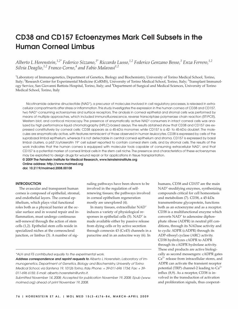
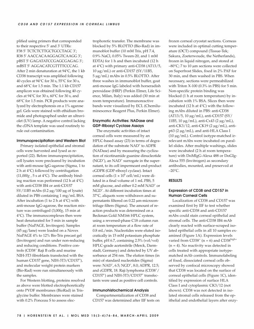
Localization of CD38 and CD157 wasexamined first by IIF to test whetherspecific anti-CD38 and anti-CD157mAbs could stain corneal epithelial andstromal cells. The anti-CD38 IB4 mAbclearly reacted with surface-scraped iso-lated epithelial cells in all 10 samples ex-amined (Figure 1A). Expression levelsvaried from CD38+ (n = 6) and CD38high+
(n = 4). No reactivity was detected incells treated with appropriate isotype-matched mAb controls. Immunolabelingof fixed, dissociated corneal cells ob-served by confocal microscopy showedthat CD38 was located on the surface ofcorneal epithelial cells (Figure 1C), iden-tified by expression of surface HLAClass I and cytoplasmic CK3/12 (notshown). CD38 was not detected in iso-lated stromal cells released from the ep-ithelial and endothelial layers after enzy-
R E S E A R C H A R T I C L E
M O L M E D 1 5 ( 3 - 4 ) 7 6 - 8 4 , M A R C H - A P R I L 2 0 0 9 | H O R E N S T E I N E T A L . | 7 9
matic treatment (Figure 1B), excludingthe possibility that the signal might de-rive from contaminating stromal cells.These results confirm that CD38 was lo-calized in a discrete subpopulation ofhuman corneal epithelial cells, as re-ported previously (24). Bearing in mindthe discontinuous pattern of expressionexhibited by CD38 in lymphoid cells, weanalyzed modulation of CD38 expres-sion in corneal epithelial cells main-tained in culture. The expression de-scribed above in Figure 1A was found todecrease over time from the initial 50%
to 60% to ~10% after 7 d in culture (datanot shown).
Strong CD157 expression was ob-served by IIF in both primary epithelialand stromal cells (see Figure 1A). Un-like CD38, CD157 expression was main-tained over time in cultured cells (seeFigure 1B). Confocal microscopy re-vealed that CD157 expression resem-bled that of CD38, although in a dis-tinct pattern: CD157 displayed adot-like pattern of expression of GPI-linked molecules in the plasma cellmembranes (see Figure 1A,1C), gener-
ally reported in other human cells andtissues (12).
Differential Expression of CD38 andCD157 Transcripts and Proteins inCorneal Epithelium and Stroma
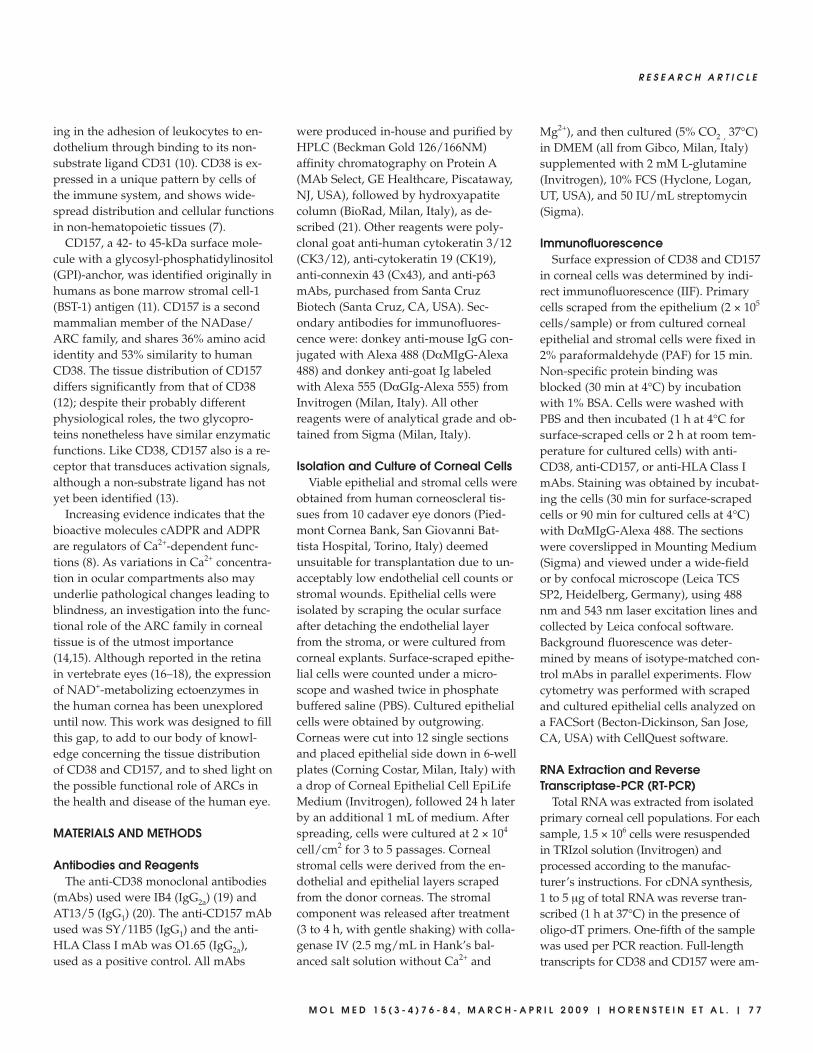
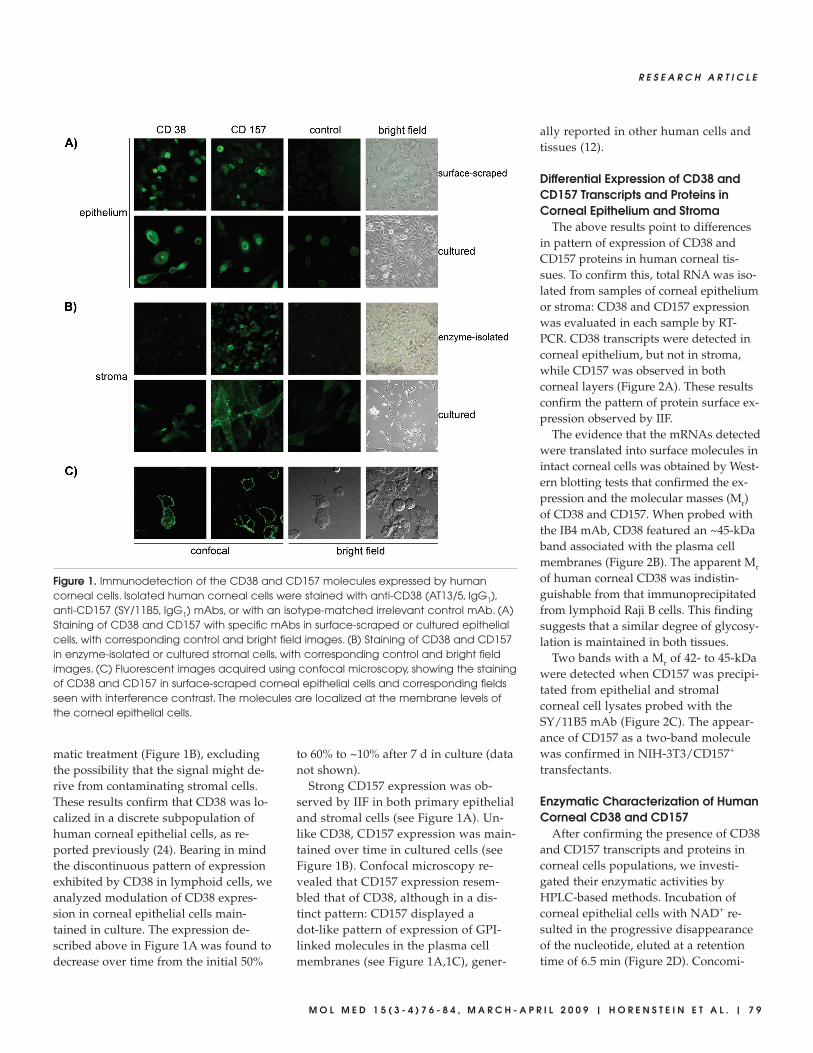
The above results point to differencesin pattern of expression of CD38 andCD157 proteins in human corneal tis-sues. To confirm this, total RNA was iso-lated from samples of corneal epitheliumor stroma: CD38 and CD157 expressionwas evaluated in each sample by RT-PCR. CD38 transcripts were detected incorneal epithelium, but not in stroma,while CD157 was observed in bothcorneal layers (Figure 2A). These resultsconfirm the pattern of protein surface ex-pression observed by IIF.
The evidence that the mRNAs detectedwere translated into surface molecules inintact corneal cells was obtained by West-ern blotting tests that confirmed the ex-pression and the molecular masses (Mr)of CD38 and CD157. When probed withthe IB4 mAb, CD38 featured an ~45-kDaband associated with the plasma cellmembranes (Figure 2B). The apparent Mrof human corneal CD38 was indistin-guishable from that immunoprecipitatedfrom lymphoid Raji B cells. This findingsuggests that a similar degree of glycosy-lation is maintained in both tissues.
Two bands with a Mr of 42- to 45-kDawere detected when CD157 was precipi-tated from epithelial and stromalcorneal cell lysates probed with theSY/11B5 mAb (Figure 2C). The appear-ance of CD157 as a two-band moleculewas confirmed in NIH-3T3/CD157+
transfectants.
Enzymatic Characterization of HumanCorneal CD38 and CD157
After confirming the presence of CD38and CD157 transcripts and proteins incorneal cells populations, we investi-gated their enzymatic activities byHPLC-based methods. Incubation ofcorneal epithelial cells with NAD+ re-sulted in the progressive disappearanceof the nucleotide, eluted at a retentiontime of 6.5 min (Figure 2D). Concomi-
Figure 1. Immunodetection of the CD38 and CD157 molecules expressed by humancorneal cells. Isolated human corneal cells were stained with anti-CD38 (AT13/5, IgG1),anti-CD157 (SY/11B5, IgG1) mAbs, or with an isotype-matched irrelevant control mAb. (A)Staining of CD38 and CD157 with specific mAbs in surface-scraped or cultured epithelialcells, with corresponding control and bright field images. (B) Staining of CD38 and CD157in enzyme-isolated or cultured stromal cells, with corresponding control and bright fieldimages. (C) Fluorescent images acquired using confocal microscopy, showing the stainingof CD38 and CD157 in surface-scraped corneal epithelial cells and corresponding fieldsseen with interference contrast. The molecules are localized at the membrane levels ofthe corneal epithelial cells.
8 0 | H O R E N S T E I N E T A L . | M O L M E D 1 5 ( 3 - 4 ) 7 6 - 8 4 , M A R C H - A P R I L 2 0 0 9
C D 3 8 A N D C D 1 5 7 E X P R E S S I O N I N C O R N E A L L I M B U S
tant with NAD+ hydrolysis, ADPReluted 6.0 min after NAD+ (see Figure 2D,upper panel). The NAD+ peak area wascorrelated with nmoles of NAD+ in-jected as control, and the specific activitydetermined from the area under thecurve of the NAD+ peak. The NADaseactivity measured was 0.25 ± 0.04 nmol/min/106 cells for corneal epithelial cells.NADase activity in control Raji cells(CD38+/CD157–) was measured as 2.51 ±0.04 nmol/min/106 cells.
The use of NAD+ as substrate may re-sult in an underestimation of ARC activ-ity, due to the subsequent hydrolysis ofcADPR (8). Interference of cADPR hydro-lase was avoided by using NGD+, aNAD+ surrogate, to assay ARC activity,given that the cGDPR is resistant to hy-drolysis (23). This system initially yieldeda modest NGD+ cyclization, which wasresolved after longer incubations (see Fig-ure 2D, lower panel). ARC activity was0.05 ± 0.1 nmol/min/106 cells for cornealepithelial cells. Control Raji cells showedmarked ARC activity, while K562 (CD38–)cells neither generated cGDPR nor con-sumed NAD+ (not shown). The ratio be-tween NADase and ARC activities (5:1)for viable intact epithelial corneal cellswas comparable with that reported forRaji cells, for CD38 purified from humanerythrocytes and for molecules in solubleform in biological fluids (25,26). These re-sults suggest that CD38 and/or CD157present on the surface of corneal tissueswere active enzymatically.
The finding that stromal corneal cellsexpress CD157, but not CD38, promptedus to test their capacity to catalyze thecyclase reaction in the presence of NGD+.Stromal cells (1.0 × 10⁶/sample) yieldedlow amounts of cGDPR. Thus, stromalcells were intrinsically inefficient at usingNGD+ for generating cGDPR at concen-trations higher than the threshold sensi-tivity of the HPLC method adopted. Sim-ilar experiments performed withNIH-3T3/CD157+ cells demonstratedthat human CD157 could metabolizeNGD+. These results are in line with thelow catalytic activities reported forCD157 (12).
Figure 2. Biochemical analysis of the CD38 and CD157 molecules expressed by humancorneal cells. (A) RT-PCR profile of CD38 and CD157 transcripts in corneal cells. PCR prod-ucts were amplified from cDNA obtained from epithelial and stromal cells using CD38- orCD157-specific primer sets. Negative controls lacking cDNA template were used to ruleout contamination. (B–C) Western blot analysis of corneal stromal and epithelial proteinsimmunoprecipitated with mAbs to CD38 and CD157. Precipitates of epithelial and stromalcells from biopsied corneas with anti-CD38 (IB4, IgG2a) or with anti-CD157 (SY/11B5, IgG1)mAbs were analyzed by 4% to 12% SDS-PAGE under non-reducing conditions and blottedonto PVDF membranes. Proteins with Mr of 45-kDa were immunodetected in the cornealepithelia and control Raji (CD38+) cells when blots were probed with anti-CD38 (AT13/5,IgG1) mAb and developed by ECL (B). Proteins with Mr of 42- to 45-kDa were immunode-tected in the stroma and corneal epithelia when blots were probed with anti-CD157(SY/11B5, IgG1) mAb and developed by ECL (C). NIH-3T3/CD157+ transfectants were usedas positive control. Isotype-matched irrelevant mAbs were used as negative controls. (D)Ectoenzyme activity molecules assessed in epithelial corneal cells. Cells were incubatedwith 0.2 mM NAD+ or NGD+ at 37°C and samples collected after 0, 15, 60, and 120 min in-cubations. NADase (upper panel) and GDP-ribosyl cyclase (lower panel) enzymatic activ-ities were determined by HPLC. Raji (CD38+) cells and NIH-3T3/CD157+ transfectants wereused as positive controls. K562 (CD38–) cells and NIH-3T3 mock transfectants were thenegative controls.
R E S E A R C H A R T I C L E
M O L M E D 1 5 ( 3 - 4 ) 7 6 - 8 4 , M A R C H - A P R I L 2 0 0 9 | H O R E N S T E I N E T A L . | 8 1
Topographical Localization of CornealCD38 and CD157
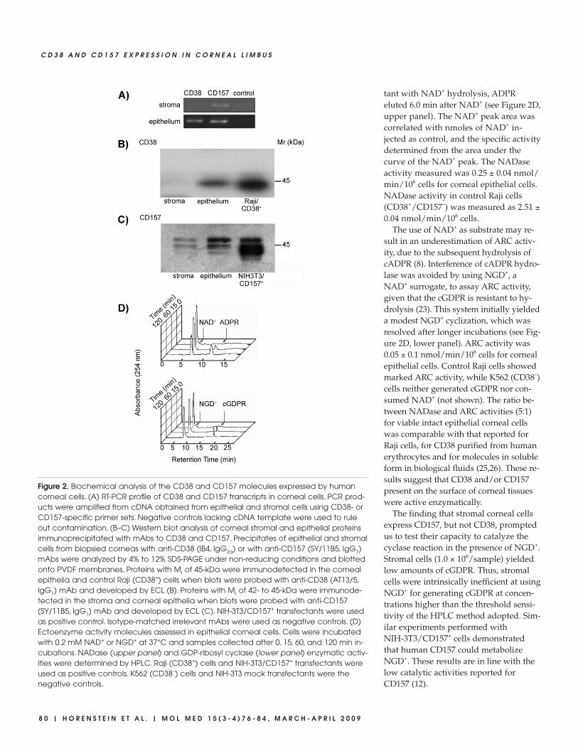
Having demonstrated that functionallyactive CD38 and CD157 ectoenzymes areexpressed constitutively in the cornea,next we turned to fine-mapping the dis-tribution of both proteins by IIF in frozencorneal cryostat sections. The resultsshowed that CD38 was localized in thesuprabasal and superficial epitheliumlayers, limited to the limbal area at theexternal peripheral structure of thecornea (Figure 3A). CD157 was ex-pressed by the epithelia of the basal lim-bal zone (Figure 3B), progressively de-creasing in the corneal-limbal epithelium(Figure 3C), located above Bowman’slayer.
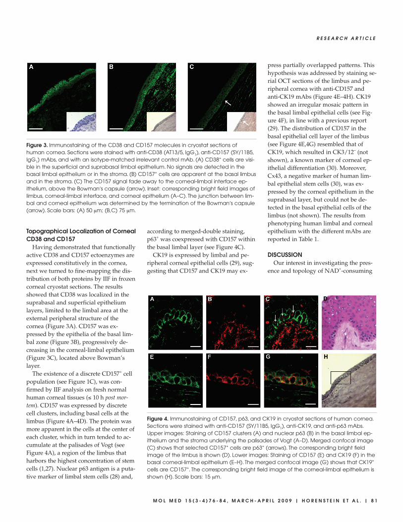
The existence of a discrete CD157+ cellpopulation (see Figure 1C), was con-firmed by IIF analysis on fresh normalhuman corneal tissues (≤ 10 h post mor-tem). CD157 was expressed by discretecell clusters, including basal cells at thelimbus (Figure 4A–4D). The protein wasmore apparent in the cells at the center ofeach cluster, which in turn tended to ac-cumulate at the palisades of Vogt (seeFigure 4A), a region of the limbus thatharbors the highest concentration of stemcells (1,27). Nuclear p63 antigen is a puta-tive marker of limbal stem cells (28) and,
according to merged-double staining,p63+ was coexpressed with CD157 withinthe basal limbal layer (see Figure 4C).
CK19 is expressed by limbal and pe-ripheral corneal epithelial cells (29), sug-gesting that CD157 and CK19 may ex-
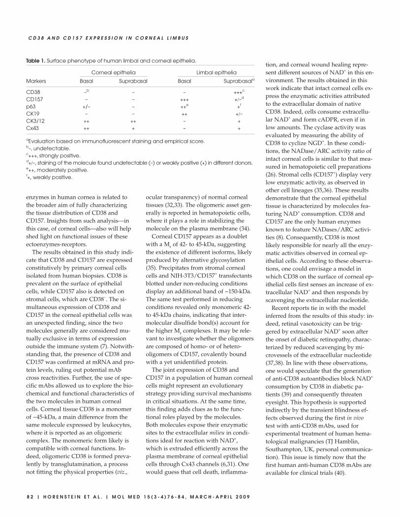
press partially overlapped patterns. Thishypothesis was addressed by staining se-rial OCT sections of the limbus and pe-ripheral cornea with anti-CD157 andanti-CK19 mAbs (Figure 4E–4H). CK19showed an irregular mosaic pattern inthe basal limbal epithelial cells (see Fig-ure 4F), in line with a previous report(29). The distribution of CD157 in thebasal epithelial cell layer of the limbus(see Figure 4E,4G) resembled that ofCK19, which resulted in CK3/12– (notshown), a known marker of corneal ep-ithelial differentiation (30). Moreover,Cx43, a negative marker of human lim-bal epithelial stem cells (30), was ex-pressed by the corneal epithelium in thesuprabasal layer, but could not be de-tected in the basal epithelial cells of thelimbus (not shown). The results fromphenotyping human limbal and cornealepithelium with the different mAbs arereported in Table 1.
DISCUSSIONOur interest in investigating the pres-
ence and topology of NAD+-consuming
Figure 4. Immunostaining of CD157, p63, and CK19 in cryostat sections of human cornea.Sections were stained with anti-CD157 (SY/11B5, IgG1), anti-CK19, and anti-p63 mAbs.Upper images: Staining of CD157 clusters (A) and nuclear p63 (B) in the basal limbal ep-ithelium and the stroma underlying the palisades of Vogt (A–D). Merged confocal image(C) shows that selected CD157+ cells are p63+ (arrows). The corresponding bright fieldimage of the limbus is shown (D). Lower images: Staining of CD157 (E) and CK19 (F) in thebasal corneal-limbal epithelium (E–H). The merged confocal image (G) shows that CK19+
cells are CD157+. The corresponding bright field image of the corneal-limbal epithelium isshown (H). Scale bars: 15 µm.
Figure 3. Immunostaining of the CD38 and CD157 molecules in cryostat sections ofhuman cornea. Sections were stained with anti-CD38 (AT13/5, IgG1), anti-CD157 (SY/11B5,IgG1) mAbs, and with an isotype-matched irrelevant control mAb. (A) CD38+ cells are visi-ble in the superficial and suprabasal limbal epithelium. No signals are detected in thebasal limbal epithelium or in the stroma. (B) CD157+ cells are apparent at the basal limbusand in the stroma. (C) The CD157 signal fade away to the corneal-limbal interface ep-ithelium, above the Bowman′s capsule (arrow). Inset: corresponding bright field images oflimbus, corneal-limbal interface, and corneal epithelium (A–C). The junction between lim-bal and corneal epithelium was determined by the termination of the Bowman′s capsule(arrow). Scale bars: (A) 50 µm; (B,C) 75 µm.
8 2 | H O R E N S T E I N E T A L . | M O L M E D 1 5 ( 3 - 4 ) 7 6 - 8 4 , M A R C H - A P R I L 2 0 0 9
C D 3 8 A N D C D 1 5 7 E X P R E S S I O N I N C O R N E A L L I M B U S
enzymes in human cornea is related tothe broader aim of fully characterizingthe tissue distribution of CD38 andCD157. Insights from such analysis—inthis case, of corneal cells—also will helpshed light on functional issues of theseectoenzymes-receptors.
The results obtained in this study indi-cate that CD38 and CD157 are expressedconstitutively by primary corneal cellsisolated from human biopsies. CD38 isprevalent on the surface of epithelialcells, while CD157 also is detected onstromal cells, which are CD38–. The si-multaneous expression of CD38 andCD157 in the corneal epithelial cells wasan unexpected finding, since the twomolecules generally are considered mu-tually exclusive in terms of expressionoutside the immune system (7). Notwith-standing that, the presence of CD38 andCD157 was confirmed at mRNA and pro-tein levels, ruling out potential mAbcross reactivities. Further, the use of spe-cific mAbs allowed us to explore the bio-chemical and functional characteristics ofthe two molecules in human cornealcells. Corneal tissue CD38 is a monomerof ~45-kDa, a main difference from thesame molecule expressed by leukocytes,where it is reported as an oligomericcomplex. The monomeric form likely iscompatible with corneal functions. In-deed, oligomeric CD38 is formed preva-lently by transglutamination, a processnot fitting the physical properties (viz.,
ocular transparency) of normal cornealtissues (32,33). The oligomeric asset gen-erally is reported in hematopoietic cells,where it plays a role in stabilizing themolecule on the plasma membrane (34).
Corneal CD157 appears as a doubletwith a Mr of 42- to 45-kDa, suggestingthe existence of different isoforms, likelyproduced by alternative glycosylation(35). Precipitates from stromal cornealcells and NIH-3T3/CD157+ transfectantsblotted under non-reducing conditionsdisplay an additional band of ~150-kDa.The same test performed in reducingconditions revealed only monomeric 42-to 45-kDa chains, indicating that inter-molecular disulfide bond(s) account forthe higher Mr complexes. It may be rele-vant to investigate whether the oligomersare composed of homo- or of hetero-oligomers of CD157, covalently boundwith a yet unidentified protein.
The joint expression of CD38 andCD157 in a population of human cornealcells might represent an evolutionarystrategy providing survival mechanismsin critical situations. At the same time,this finding adds clues as to the func-tional roles played by the molecules.Both molecules expose their enzymaticsites to the extracellular milieu in condi-tions ideal for reaction with NAD+,which is extruded efficiently across theplasma membrane of corneal epithelialcells through Cx43 channels (6,31). Onewould guess that cell death, inflamma-
tion, and corneal wound healing repre-sent different sources of NAD+ in this en-vironment. The results obtained in thiswork indicate that intact corneal cells ex-press the enzymatic activities attributedto the extracellular domain of nativeCD38. Indeed, cells consume extracellu-lar NAD+ and form cADPR, even if inlow amounts. The cyclase activity wasevaluated by measuring the ability ofCD38 to cyclize NGD+. In these condi-tions, the NADase/ARC activity ratio ofintact corneal cells is similar to that mea-sured in hematopoietic cell preparations(26). Stromal cells (CD157+) display verylow enzymatic activity, as observed inother cell lineages (35,36). These resultsdemonstrate that the corneal epithelialtissue is characterized by molecules fea-turing NAD+ consumption. CD38 andCD157 are the only human enzymesknown to feature NADases/ARC activi-ties (8). Consequently, CD38 is mostlikely responsible for nearly all the enzy-matic activities observed in corneal ep-ithelial cells. According to these observa-tions, one could envisage a model inwhich CD38 on the surface of corneal ep-ithelial cells first senses an increase of ex-tracellular NAD+ and then responds byscavenging the extracellular nucleotide.
Recent reports tie in with the modelinferred from the results of this study: in-deed, retinal vasotoxicity can be trig-gered by extracellular NAD+ soon afterthe onset of diabetic retinopathy, charac-terized by reduced scavenging by mi-crovessels of the extracellular nucleotide(37,38). In line with these observations,one would speculate that the generationof anti-CD38 autoantibodies block NAD+
consumption by CD38 in diabetic pa-tients (39) and consequently threateneyesight. This hypothesis is supportedindirectly by the transient blindness ef-fects observed during the first in vivotest with anti-CD38 mAbs, used forexperimental treatment of human hema-tological malignancies (TJ Hamblin,Southampton, UK, personal communica-tion). This issue is timely now that thefirst human anti-human CD38 mAbs areavailable for clinical trials (40).
Table 1. Surface phenotype of human limbal and corneal epithelia.
Corneal epithelia Limbal epithelia
Markers Basal Suprabasal Basal Suprabasala
CD38 –b – – +++c
CD157 – – +++ +/–d
p63 +/– – ++e +f
CK19 – – ++ +/–CK3/12 ++ ++ – +Cx43 ++ + – +
aEvaluation based on immunofluorescent staining and empirical score.b–, undetectable.c+++, strongly positive.d+/–, staining of the molecule found undetectable (–) or weakly positive (+) in different donors.e++, moderately positive.f+, weakly positive.
R E S E A R C H A R T I C L E
M O L M E D 1 5 ( 3 - 4 ) 7 6 - 8 4 , M A R C H - A P R I L 2 0 0 9 | H O R E N S T E I N E T A L . | 8 3
Epithelium regenerates during woundrepair by migration of cells from thecorneal limbus (1,28). However, an openissue in corneal regeneration is the lackof a dependable surface marker of thelimbal stem cell population (41). The re-sults of this work show that CD38 andCD157 mark subsets of human cornealepithelial cells. The corneal epitheliumdoes not express CD38, on the contrary,detectable on the suprabasal and superfi-cial limbal epithelium. CD157 is ex-pressed by the basal epithelial layer ofthe limbus, where it appears in clustersof highly stained cells coexpressing nu-clear p63, a molecule apparently requiredfor the migration of limbal cells (28,42).Recent studies support the hypothesisthat CD157 is one of a coordinated seriesof events linked to hematopoietic cell mi-gration (7). The inference that could beextrapolated from the present results isthat CD157 may be a useful marker forthe identification and isolation of humanlimbal epithelial cells that share charac-teristics attributed to corneal stem cells.We thus conclude that the basal epithe-lial cells of the human limbus are ofCD157+, CD38–, p63+, CK19+, CK3/12–,and Cx43– phenotype. Therefore, a pecu-liar combination of markers of cornealepithelial differentiation (for example,CK3/12, Cx43, and CD38) and limbalcell-associated markers (for example,p63, CK19, and CD157) may allow iden-tification of human corneal stem cells.
These results show that the normalhuman cornea is equipped with a molec-ular tool to actively metabolize NAD+,helping maintain corneal homeostasis.The presence of these ectoenzymes maypave the way to the design of noveldrugs to control wound repair. Lastly,the role of CD38 and CD157 as cornealsurface receptors is currently being ex-amined to obtain sound experimentalevidence to translate biological informa-tion back to a clinical set, such as tissuetransplantation.
ACKNOWLEDGMENTSWork supported by grants from
Telethon (ALH) and by the special proj-
ect “Oncologia” Compagnia SanPaolo(Torino, Italy). FIRMS (Fondazione Inter-nazionale Ricerca in Medicina Sperimen-tale) provided valuable financial contri-butions. ALH and RL were supported byFIRMS Research Fellowships.
We thank George Stevenson (TenovusResearch Laboratory, Southampton Uni-versity Hospital, UK) for providing theAT13/5 anti-CD38 mAb. We also thankG Mazzucco (Electronic Microscopy Lab,Department of Biomedical Science,Torino) for advice with corneal cryostatsectioning.
DISCLOSUREWe declare that the authors have no
competing interests as defined by Molec-ular Medicine, or other interests thatmight be perceived to influence the re-sults and discussion reported in thispaper.
REFERENCES1. Cotsarelis G, Cheng SZ, Dong G, Sun TT, Lavker
RM. (1989) Existence of slow-cycling limbal ep-ithelial basal cells that can be preferentially stim-ulated to proliferate: implications on epithelialstem cells. Cell 57:201–9.
2. Lavker RM, Tseng SC, Sun TT. (2004) Corneal ep-ithelial stem cells at the limbus: looking at someold problems from a new angle. Exp. Eye Res.78:433–46.
3. Lindberg K, Brown ME, Chaves HV, Kenyon KR,Rheinwald JG. (1993) In vitro propagation ofhuman ocular surface epithelial cells for trans-plantation. Invest. Ophthalmol. Vis. Sci.. 34:2672–9.
4. Fuchs E, Tumbar T, Guasch G. (2004) Socializingwith the neighbors: stem cells and their niche.Cell. 116:769–78.
5. Ying W. (2008) NAD+/NADH andNADP+/NADPH in cellular functions and celldeath: regulation and biological consequences.Antioxid. Redox Signal. 10:179–206.
6. Bruzzone S, Guida L, Zocchi E, Franco L, DeFlora A. (2001) Connexin 43 hemi channels medi-ate Ca2+-regulated transmembrane NAD+ fluxesin intact cells. Faseb. J. 15:10–2.
7. Malavasi F, et al. (2008) Evolution and function ofthe ADP ribosyl cyclase/CD38 gene family inphysiology and pathology. Physiol. Rev.88:841–86.
8. Lee HC. (2006) Structure and enzymatic func-tions of human CD38. Mol. Med. 12:317–323.
9. Massullo P, Sumoza-Toledo A, Bhagat H, Partida-Sanchez S. (2006) TRPM channels, calcium andredox sensors during innate immune responses.Semin. Cell Dev. Biol. 17:654–66.
10. Deaglio S, et al. (1996) Human CD38 ligand. A
120-kDa protein predominantly expressed on en-dothelial cells. J. Immunol. 156:727–34.
11. Kaisho T, et al. (1994) BST-1, a surface moleculeof bone marrow stromal cell lines that facilitatespre-B-cell growth. Proc. Natl. Acad. Sci. U. S. A.91:5325–9.
12. Ortolan E, et al. (2002) CD157, the Janus of CD38but with a unique personality. Cell Biochem.Funct. 20:309–22.
13. Malavasi F, et al. (2006) CD38 and CD157 as re-ceptors of the immune system: a bridge betweeninnate and adaptive immunity. Mol. Med.12:334–41.
14. Duncan G, Williams MR, Riach RA. (1994). Cal-cium, cell signalling and cataract. Prog. Retin. EyeRes. 13:623–52.
15. Chattopadhyay N, et al. (1997) Expression of ex-tracellular calcium-sensing receptor by humanlens epithelial cells. Biochem. Biophys. Res. Com-mun. 233:801–5.
16. Khoo KM, Chang CF. (1999) Characterizationand localization of CD38 in the vertebrate eye.Brain Res. 821:17–25.
17. Esguerra M, Miller RF. (2002) CD38 expression andNAD+-induced intracellular Ca+ mobilization inisolated retinal Muller cells. Glia. 39:314–9.
18. Panfoli I, et al. (2007) Localization of the cyclicADP-ribose-dependent calcium signaling path-way in bovine rod outer segments. Invest. Oph-thalmol. Vis. Sci. 48:978–84.
19. Malavasi F, et al. (1984) Characterization of amurine monoclonal antibody specific for humanearly lymphohemopoietic cells. Hum. Immunol.9:9–20.
20. Ausiello CM, et al. (2000) Functional topographyof discrete domains of human CD38. Tissue Anti-gens. 56:539–47.
21. Horenstein AL, Durelli I, Malavasi F. (2005) Pu-rification of clinical-grade monoclonal antibodiesby chromatographic methods. Methods Mol. Biol.308:191–208.
22. Horenstein AL, Stockinger H, Imhof BA,Malavasi F. (1998) CD38 binding to humanmyeloid cells is mediated by mouse and humanCD31. Biochem. J. 330:1129–35.
23. Graeff RM, Walseth TF, Fryxell K, Branton WD,Lee HC. (1994) Enzymatic synthesis and charac-terizations of cyclic GDP-ribose. A procedure fordistinguishing enzymes with ADP-ribosyl cy-clase activity. J. Biol. Chem. 269:30260–7.
24. Sizzano F, et al. (2007) Identification of theectoenzyme-receptor CD38 on human cornealepithelial cells [abstract]. Tissue Antigens 69:390.21st European Immunogenetics and Histocom-patibility Conference; 2007 May 5–8; Barcelona,Spain.
25. Zocchi E, et al. (1993) A single protein immuno-logically identified as CD38 displays NAD+ gly-cohydrolase, ADP-ribosyl cyclase and cyclicADP-ribose hydrolase activities at the outer sur-face of human erythrocytes. Biochem. Biophys. Res.Commun.196:1459–65.
26. Funaro A, et al. (1996) Identification and charac-
8 4 | H O R E N S T E I N E T A L . | M O L M E D 1 5 ( 3 - 4 ) 7 6 - 8 4 , M A R C H - A P R I L 2 0 0 9
C D 3 8 A N D C D 1 5 7 E X P R E S S I O N I N C O R N E A L L I M B U S
terization of an active soluble form of humanCD38 in normal and pathological fluids. Int. Im-munol. 8:1643–50.
27. Chen Z, et al. (2004) Characterization of putativestem cell phenotype in human limbal epithelia.Stem Cells 22:355–66.
28. Di Iorio E, et al. (2005) Isoforms of DeltaNp63and the migration of ocular limbal cells inhuman corneal regeneration. Proc. Natl. Acad. Sci.U. S. A. 102:9523–8.
29. Lauweryns B, van den Oord JJ, De Vos R, Missot-ten L (1993) A new epithelial cell type in the humancornea. Invest. Ophthalmol. Vis. Sci. 34:1983–90.
30. Harkin DG, Barnard Z, Gillies P, Ainscough SL,Apel AJ. (2004) Analysis of p63 and cytokeratinexpression in a cultivated limbal autograft usedin the treatment of limbal stem cell deficiency. Br.J. Ophthalmol. 88:1154–8.
31. Shurman DL, Glazewski L, Gumpert A, ZieskeJD, Richard G. (2005) In vivo and in vitro expres-sion of connexins in the human corneal epithe-lium. Invest. Ophthalmol. Vis. Sci.. 46:1957–65.
32. Umar S, Malavasi F, Mehta K. (1996) Post-translational modification of CD38 protein into ahigh molecular weight form alters its catalyticproperties. J. Biol. Chem. 271:15922–7.
33. Tong L, et al. (2006) Transglutaminase partici-pates in UVB-induced cell death pathways inhuman corneal epithelial cells. Invest. Ophthalmol.Vis. Sci. 47:4295–301.
34. Moreno-Garcia ME, et al. (2004) CD38 is ex-pressed as noncovalently associated homodimerson the surface of murine B lymphocytes. Eur. J.Biochem. 271:1025–34.
35. Hirata Y, et al. (1994) ADP ribosyl cyclase activityof a novel bone marrow stromal cell surface mol-ecule, BST-1. FEBS Lett. 356:244–8.
36. Hussain AM, Lee HC, Chang CF. (1998) Func-tional expression of secreted mouse BST-1 inyeast. Protein Expr. Purif. 12:133–7.
37. Liao SD, Puro DG. (2006) NAD+-induced vaso-toxicity in the pericyte-containing microvascula-ture of the rat retina: effect of diabetes. Invest.Ophthalmol. Vis. Sci. 47:5032–8.
38. Dianzani U, et al. (1994) Interaction between en-dothelium and CD4+CD45RA+ lymphocytes.Role of the human CD38 molecule. J. Immunol.153:952–9.
39. Antonelli A, et al. (2001) Human anti-CD38 au-toantibodies raise intracellular calcium and stim-ulate insulin release in human pancreatic islets.Diabetes 50:985–91.
40. Tesar M. (2007) Fully human antibody MOR202against CD38 for the treatment of multiple my-eloma and other blood-borne malignancies [ab-stract]. J. Clin. Oncol. 25:8106 (abstract no.).2007 ASCO Annual Meeting proceedings (post-meeting edition) of the American Society ofClinical Oncology.
41. Chee KY, Kicic A, Wiffen SJ. (2006) Limbal stemcells: the search for a marker. Clin. ExperimentOphthalmol. 34:64–73.
42. Pellegrini G, et al. (1999) Location and clonal
analysis of stem cells and their differentiatedprogeny in the human ocular surface. J. Cell Biol.145:769–82.