Embed Size (px)
Citation preview
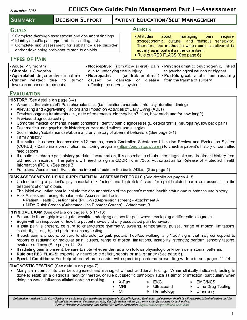
Attitudes about managing pain require socioeconomic, cultural, and religious sensitivity. Therefore, the method in which care is delivered is equally as important as the care itself.
Rule out RED FLAGS (See page 6)
Complete thorough assessment and document findings Identify specific pain type and clinical diagnosis Complete risk assessment for substance use disorder
and/or developing problems related to opioids
GOALS ALERTS
EVALUATION
CCHCS Care Guide: Pain Management Part 1—Assessment September 2018
SUMMARY DECISION SUPPORT PATIENT EDUCATION/SELF MANAGEMENT
TYPES OF PAIN
HISTORY (See details on page 3-4) When did the pain start? Pain characteristics (i.e., location, character, intensity, duration, timing) Alleviating and Aggravating Factors and Impact on Activities of Daily Living (ADLs) Previous/ongoing treatments (i.e., date of treatments, did they help? If so, how much and for how long?) Previous diagnostic testing Comorbid medical or mental health conditions; identify pain diagnoses (e.g., osteoarthritis, neuropathy, low back pain) Past medical and psychiatric histories; current medications and allergies Social history/substance use/abuse and any history of aberrant behaviors (See page 3-4) Family history If a patient has been incarcerated <12 months, check Controlled Substance Utilization Review and Evaluation System
(CURES) - California’s prescription monitoring program (https://oag.ca.gov/cures) to check a patient’s history of controlled medications
If a patient’s chronic pain history predates incarceration, it is essential to obtain prior diagnostic and treatment history from old medical records. The patient will need to sign a CDCR Form 7385, Authorization for Release of Protected Health Information (ROI). (See page 3)
Functional Assessment: Evaluate the impact of pain on the basic ADLs. (See page 4)
RISK ASSESSMENTS USING SUPPLEMENTAL ASSESSMENT TOOLS (See details on pages 4- 5) Understanding a patient’s psychosocial risk factors and high risk factors for opioid-related harm are essential in the
treatment of chronic pain. The initial evaluation should include the documentation of the patient’s mental health status and substance use history. Risk Assessment using Supplemental Assessment Tools:
Patient Health Questionnaire (PHQ-9) (Depression screen) - Attachment A NIDA Quick Screen (Substance Use Disorder Screen) - Attachment B
PHYSICAL EXAM (See details on pages 6 & 11-13) Be sure to thoroughly investigate possible underlying causes for pain when developing a differential diagnosis. Begin with an inspection of how the patient moves and any associated pain behaviors. If joint pain is present, be sure to characterize symmetry, swelling, temperature, pulses, range of motion, limitations,
instability, strength, and perform sensory testing. If back pain is present, be sure to characterize gait, posture, heel/toe walking, any “root” signs that may correspond to
reports of radiating or radicular pain, pulses, range of motion, limitations, instability, strength; perform sensory testing, evaluate reflexes (See pages 12-13).
If radiating pain is present, be sure to note whether the radiation follows physiologic or known dermatomal patterns. Rule out RED FLAGS: especially neurologic deficit, sepsis or malignancy (See page 6). Special Conditions: For helpful tools/tips to assist with specific problems presenting with pain see pages 11-14.
DIAGNOSTIC TESTING (See details on page 7) Many pain complaints can be diagnosed and managed without additional testing. When clinically indicated, testing is
done to establish a diagnosis, monitor therapy, or rule out specific pathology such as tumor or infection, particularly when doing so would influence clinical decision making.
• Acute: < 3 months • Chronic: > 3 months • Age-related: degenerative in nature • Cancer related: due to tumor
invasion or cancer treatments
• Nociceptive: (somatic/visceral) pain due to underlying tissue injury
• Neuropathic: (central/peripheral) caused by damage or disease affecting the nervous system
• Psychosomatic: psychogenic, linked to psychological causes or triggers
• Post-Surgical: acute pain resulting from the trauma of surgery
EMG/NCS Urine Drug Testing Chemistry
1
EKG Ultrasound Hematology
X-Ray MRI CT
Information contained in the Care Guide is not a substitute for a health care professional's clinical judgment. Evaluation and treatment should be tailored to the individual patient and the clinical circumstances. Furthermore, using this information will not guarantee a specific outcome for each patient. Refer to “Disclaimer Regarding Care Guides” for further clarification. https://cchcs.ca.gov/clinical-resources/
September 2018
SUMMARY DECISION SUPPORT PATIENT EDUCATION/SELF MANAGEMENT
2
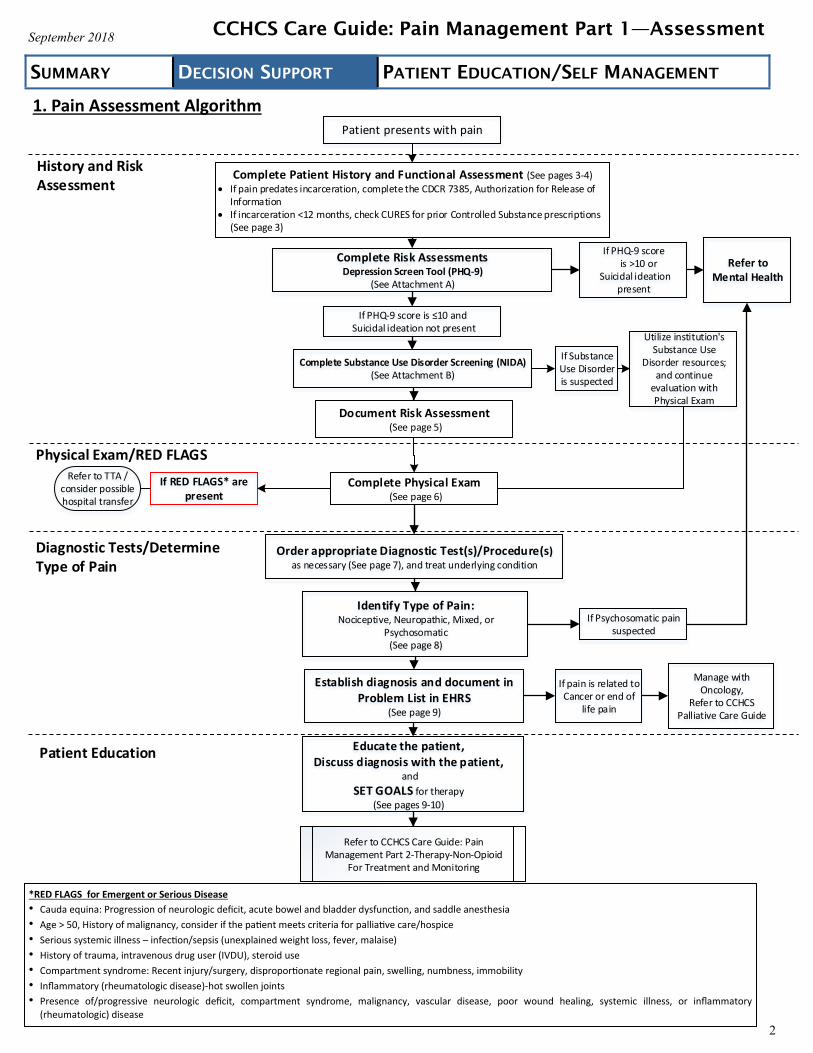
1. Pain Assessment Algorithm
CCHCS Care Guide: Pain Management Part 1—Assessment
Patient presents with pain
Refer to CCHCS Care Guide: Pain Management Part 2-Therapy-Non-Opioid
For Treatment and Monitoring
Refer to TTA / consider possible hospital transfer
Complete Patient History and Functional Assessment (See pages 3-4) If pain predates incarceration, complete the CDCR 7385, Authorization for Release of
Information If incarceration <12 months, check CURES for prior Controlled Substance prescriptions (See page 3)
Complete Physical Exam (See page 6)
Complete Substance Use Disorder Screening (NIDA)(See Attachment B)
Order appropriate Diagnostic Test(s)/Procedure(s) as necessary (See page 7), and treat underlying condition
Refer to Mental Health
Educate the patient, Discuss diagnosis with the patient,
and
SET GOALS for therapy(See pages 9-10)
Utilize institution's Substance Use
Disorder resources;and continue
evaluation with Physical Exam
Establish diagnosis and document in Problem List in EHRS
(See page 9)
Complete Risk AssessmentsDepression Screen Tool (PHQ-9)
(See Attachment A)
Identify Type of Pain:Nociceptive, Neuropathic, Mixed, or
Psychosomatic(See page 8)
Manage with Oncology,
Refer to CCHCS Palliative Care Guide
If PHQ-9 score is >10 or
Suicidal ideation present
If PHQ-9 score is ≤10 and Suicidal ideation not present
If RED FLAGS* are present
If Substance Use Disorder is suspected
If Psychosomatic pain suspected
If pain is related to Cancer or end of
life pain
Document Risk Assessment (See page 5)
History and Risk Assessment
Physical Exam/RED FLAGS
Diagnostic Tests/Determine Type of Pain
Patient Education
*RED FLAGS for Emergent or Serious Disease
• Cauda equina: Progression of neurologic deficit, acute bowel and bladder dysfunction, and saddle anesthesia
• Age > 50, History of malignancy, consider if the patient meets criteria for palliative care/hospice
• Serious systemic illness – infection/sepsis (unexplained weight loss, fever, malaise)
• History of trauma, intravenous drug user (IVDU), steroid use
• Compartment syndrome: Recent injury/surgery, disproportionate regional pain, swelling, numbness, immobility
• Inflammatory (rheumatologic disease)-hot swollen joints • Presence of/progressive neurologic deficit, compartment syndrome, malignancy, vascular disease, poor wound healing, systemic illness, or inflammatory
(rheumatologic) disease
September 2018
SUMMARY DECISION SUPPORT PATIENT EDUCATION/SELF MANAGEMENT
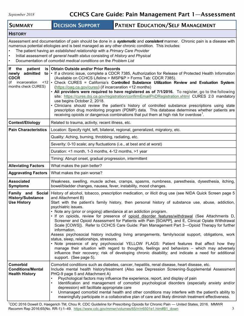
If the patient is newly admitted to CDCR (if incarceration <12 months check CURES)
Obtain Outside and/or Prior Records • If a chronic issue, complete a CDCR 7385, Authorization for Release of Protected Health Information
(Available on CCHCS Lifeline > IMSP&P > Forms Tab: CDCR 7385). • Check CURES = California’s Controlled Substance Utilization Review and Evaluation System:
(https://oag.ca.gov/cures) (if incarceration <12 months) • All providers were required to have registered as of 7/1/2016. To register, go to the following
site: https://cures.doj.ca.gov/registration/confirmEmailPnDRegistration.xhtml CURES 2.0 mandatory use begins October 2, 2018.
• Clinicians should review the patient’s history of controlled substance prescriptions using state prescription drug monitoring program (PDMP) data. This database determines whether patients are receiving opioids or dangerous combinations that put them at high risk for overdose
1.
Context/Etiology Related to trauma, activity, recent illness, etc.
Pain Characteristics Location: Specify right, left, bilateral, regional, generalized, migratory, etc.
Quality: Aching, burning, throbbing, radiating, etc.
Severity: 0-10 scale; any fluctuations (i.e., at best and at worst)
Duration: <1 month, 1-3 months, 4-12 months, >1 year
Timing: Abrupt onset, gradual progression, intermittent
Alleviating Factors What makes the pain better?
Aggravating Factors What makes the pain worse?
Associated Symptoms
Weakness, swelling, muscle aches, cramps, spasms, numbness, paresthesia, dysesthesia, itching, bowel/bladder changes, nausea, fever, instability, mood changes.
Family and Social History/Substance Use History
History of alcohol, tobacco, prescription medication, or illicit drug use (see NIDA Quick Screen page 5 and Attachment B) Start with the patient’s family history, then personal history of substance use, abuse, addiction, psychiatric issues. • Note any (prior or ongoing) attendance at an addiction program. • If on opioids, review for presence of opioid disorder features/withdrawal (See Attachments D,
Screener and Opioid Assessment for Patients with Pain [SOAPP], and E, Clinical Opiate Withdrawal Scale [COWS]). Refer to CCHCS Care Guide: Pain Management Part 3—Opioid Therapy for further information.
Assess psychosocial history including living arrangements, family/social support, obligations, work status, sleep, relationships, stressors. • Note presence of any psychosocial YELLOW FLAGS: Patient features that affect how they
manage their situation with regard to thoughts, feelings and behaviors – which may adversely influence their recovery; risk of developing chronic disability; and indicate a need for additional support. (See page 5).
Comorbid Conditions/Mental Health History
Comorbid conditions such as diabetes, cancer, hepatitis, renal disease, heart disease, etc. Include mental health history/treatment (Also see Depression Screening-Supplemental Assessment PHQ-9 page 5 and Attachment A) • Psychological factors may influence the experience, report, and display of pain • Identification and management of comorbid psychological disorders (especially anxiety and/or
depression) will facilitate appropriate care • Unmanaged comorbid mental health and other conditions may interfere with the patient's ability to
meaningfully participate in a collaborative plan of care and likely diminish treatment effectiveness.
Assessment and documentation of pain should be done in a systematic and consistent manner. Chronic pain is a disease with numerous potential etiologies and is best managed as any other chronic condition. This includes:
• The patient having an established relationship with a Primary Care Provider
• Initial assessment of general health status consisting of History and Physical
• Documentation of comorbid medical conditions on the Problem List
3
1CDC 2016 Dowell D, Haegerich TM, Chou R. CDC Guideline for Prescribing Opioids for Chronic Pain — United States, 2016. MMWR
Recomm Rep 2016;65(No. RR-1):1–49. https://www.cdc.gov/mmwr/volumes/65/rr/rr6501e1.htm#B1_down
CCHCS Care Guide: Pain Management Part 1—Assessment
HISTORY
September 2018
SUMMARY DECISION SUPPORT PATIENT EDUCATION/SELF MANAGEMENT
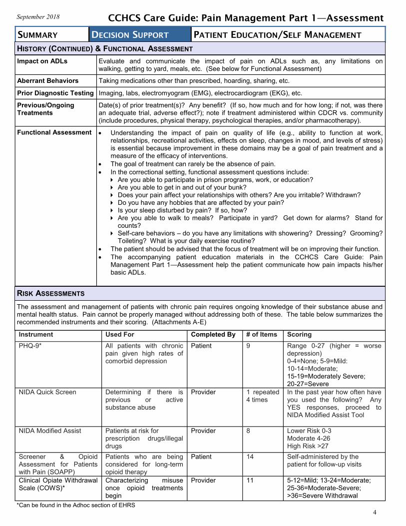
Impact on ADLs Evaluate and communicate the impact of pain on ADLs such as, any limitations on walking, getting to yard, meals, etc. (See below for Functional Assessment)
Aberrant Behaviors Taking medications other than prescribed, hoarding, sharing, etc.
Prior Diagnostic Testing Imaging, labs, electromyogram (EMG), electrocardiogram (EKG), etc.
Previous/Ongoing Treatments
Date(s) of prior treatment(s)? Any benefit? (If so, how much and for how long; if not, was there an adequate trial, adverse effect?); note if treatment administered within CDCR vs. community (include procedures, physical therapy, psychological therapies, and/or pharmacotherapy).
Functional Assessment Understanding the impact of pain on quality of life (e.g., ability to function at work, relationships, recreational activities, effects on sleep, changes in mood, and levels of stress) is essential because improvement in these domains may be a goal of pain treatment and a measure of the efficacy of interventions.
The goal of treatment can rarely be the absence of pain.
In the correctional setting, functional assessment questions include: Are you able to participate in prison programs, work, or education? Are you able to get in and out of your bunk? Does your pain affect your relationships with others? Are you irritable? Withdrawn? Do you have any hobbies that are affected by your pain? Is your sleep disturbed by pain? If so, how? Are you able to walk to meals? Participate in yard? Get down for alarms? Stand for
counts? Self-care behaviors – do you have any limitations with showering? Dressing? Grooming?
Toileting? What is your daily exercise routine?
The patient should be advised that the focus of treatment will be on improving their function.
The accompanying patient education materials in the CCHCS Care Guide: Pain Management Part 1—Assessment help the patient communicate how pain impacts his/her basic ADLs.
4
The assessment and management of patients with chronic pain requires ongoing knowledge of their substance abuse and mental health status. Pain cannot be properly managed without addressing both of these. The table below summarizes the recommended instruments and their scoring. (Attachments A-E)
Instrument Used For # of Items Scoring Completed By
PHQ-9* All patients with chronic pain given high rates of comorbid depression
9 Range 0-27 (higher = worse depression) 0-4=None; 5-9=Mild: 10-14=Moderate; 15-19=Moderately Severe; 20-27=Severe
Patient
NIDA Quick Screen Determining if there is previous or active substance abuse
1 repeated 4 times
In the past year how often have you used the following? Any YES responses, proceed to NIDA Modified Assist Tool
Provider
NIDA Modified Assist Patients at risk for prescription drugs/illegal drugs
8 Lower Risk 0-3 Moderate 4-26 High Risk >27
Provider
Screener & Opioid Assessment for Patients with Pain (SOAPP)
Patients who are being considered for long-term opioid therapy
14 Self-administered by the patient for follow-up visits
Patient
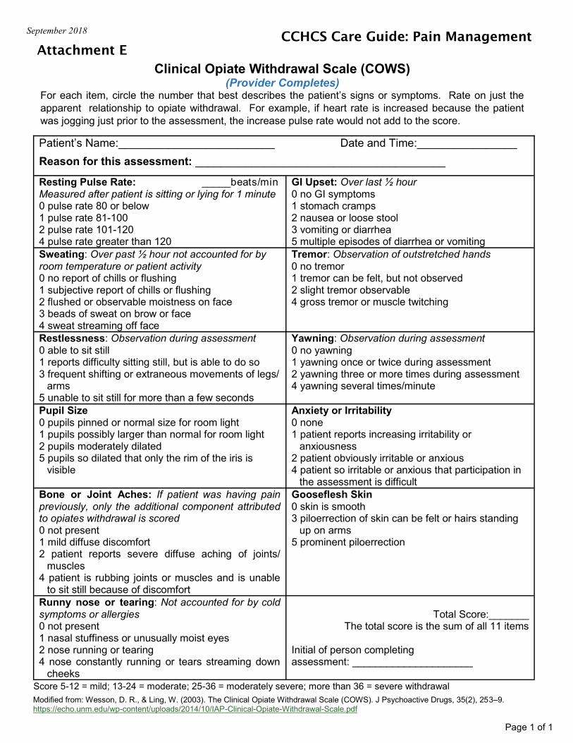
Clinical Opiate Withdrawal Scale (COWS)*
Characterizing misuse once opioid treatments begin
11 5-12=Mild; 13-24=Moderate; 25-36=Moderate-Severe; >36=Severe Withdrawal
Provider
CCHCS Care Guide: Pain Management Part 1—Assessment
HISTORY (CONTINUED) & FUNCTIONAL ASSESSMENT
RISK ASSESSMENTS
*Can be found in the Adhoc section of EHRS
September 2018
SUMMARY DECISION SUPPORT PATIENT EDUCATION/SELF MANAGEMENT
5
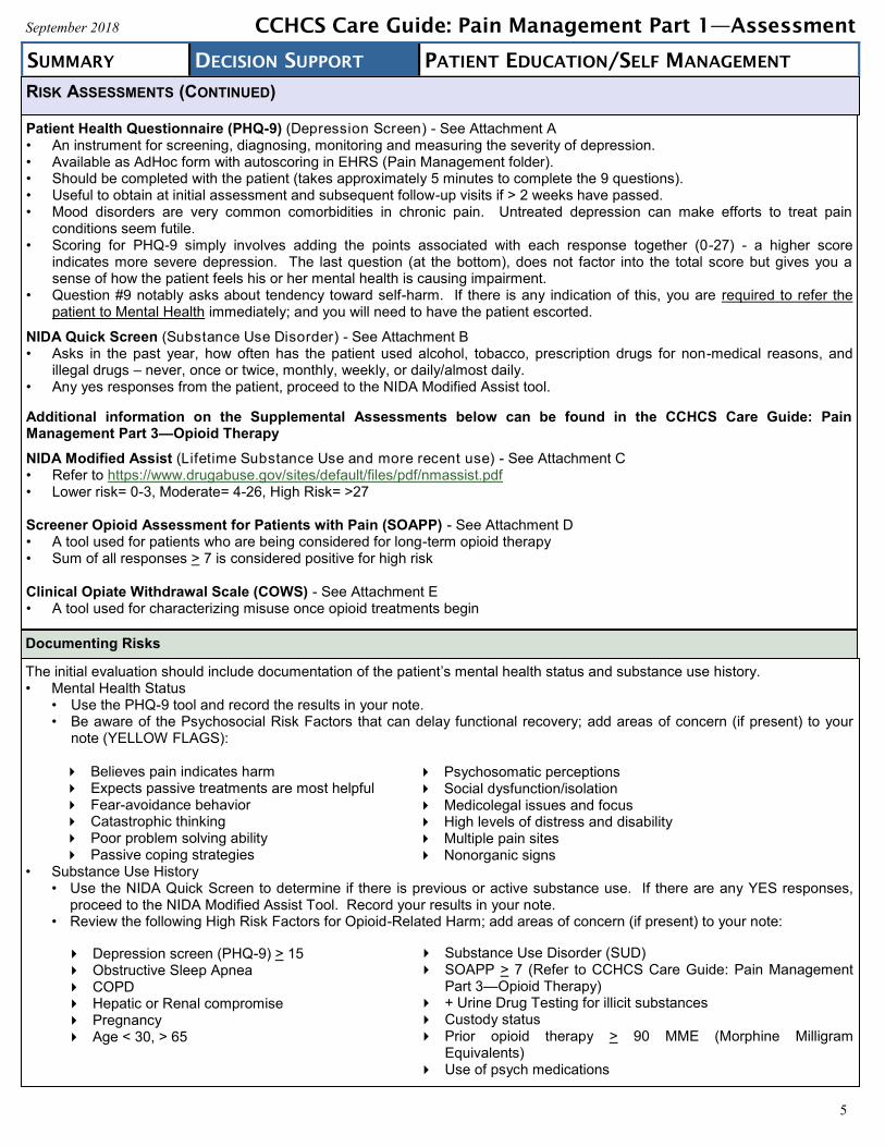
Patient Health Questionnaire (PHQ-9) (Depression Screen) - See Attachment A • An instrument for screening, diagnosing, monitoring and measuring the severity of depression. • Available as AdHoc form with autoscoring in EHRS (Pain Management folder). • Should be completed with the patient (takes approximately 5 minutes to complete the 9 questions). • Useful to obtain at initial assessment and subsequent follow-up visits if > 2 weeks have passed. • Mood disorders are very common comorbidities in chronic pain. Untreated depression can make efforts to treat pain
conditions seem futile. • Scoring for PHQ-9 simply involves adding the points associated with each response together (0-27) - a higher score
indicates more severe depression. The last question (at the bottom), does not factor into the total score but gives you a sense of how the patient feels his or her mental health is causing impairment.
• Question #9 notably asks about tendency toward self-harm. If there is any indication of this, you are required to refer the patient to Mental Health immediately; and you will need to have the patient escorted.
NIDA Quick Screen (Substance Use Disorder) - See Attachment B • Asks in the past year, how often has the patient used alcohol, tobacco, prescription drugs for non-medical reasons, and
illegal drugs – never, once or twice, monthly, weekly, or daily/almost daily. • Any yes responses from the patient, proceed to the NIDA Modified Assist tool.
Additional information on the Supplemental Assessments below can be found in the CCHCS Care Guide: Pain Management Part 3—Opioid Therapy
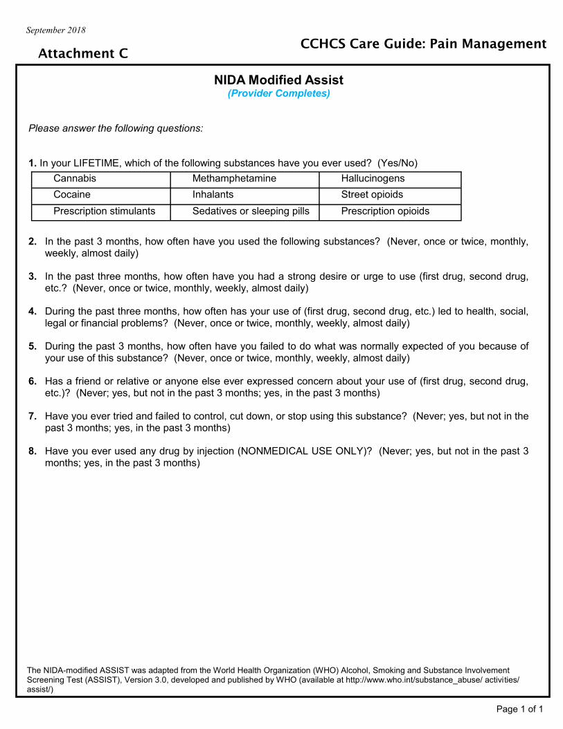
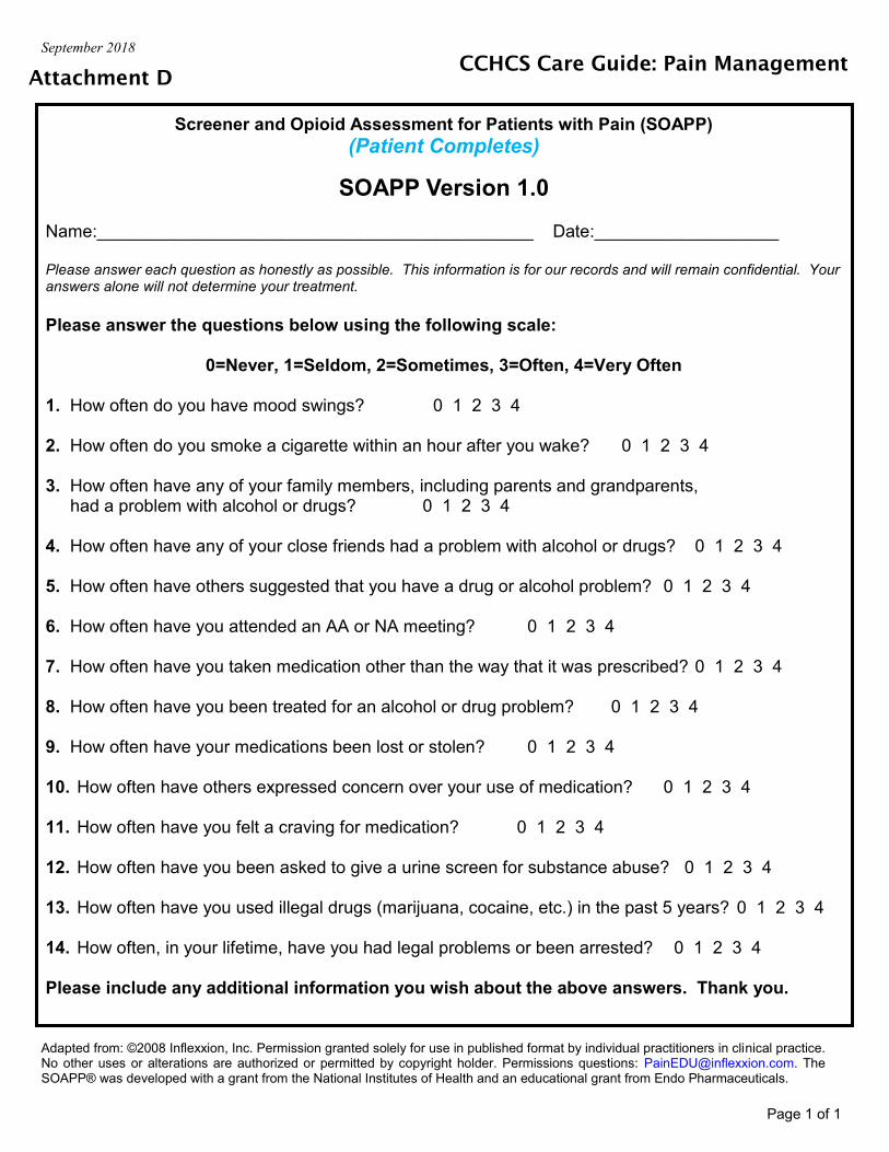
NIDA Modified Assist (Lifetime Substance Use and more recent use) - See Attachment C • Refer to https://www.drugabuse.gov/sites/default/files/pdf/nmassist.pdf • Lower risk= 0-3, Moderate= 4-26, High Risk= >27 Screener Opioid Assessment for Patients with Pain (SOAPP) - See Attachment D • A tool used for patients who are being considered for long-term opioid therapy • Sum of all responses > 7 is considered positive for high risk Clinical Opiate Withdrawal Scale (COWS) - See Attachment E • A tool used for characterizing misuse once opioid treatments begin
The initial evaluation should include documentation of the patient’s mental health status and substance use history. • Mental Health Status
• Use the PHQ-9 tool and record the results in your note. • Be aware of the Psychosocial Risk Factors that can delay functional recovery; add areas of concern (if present) to your
note (YELLOW FLAGS): Believes pain indicates harm Expects passive treatments are most helpful Fear-avoidance behavior Catastrophic thinking Poor problem solving ability Passive coping strategies
• Substance Use History • Use the NIDA Quick Screen to determine if there is previous or active substance use. If there are any YES responses,
proceed to the NIDA Modified Assist Tool. Record your results in your note. • Review the following High Risk Factors for Opioid-Related Harm; add areas of concern (if present) to your note:
CCHCS Care Guide: Pain Management Part 1—Assessment
RISK ASSESSMENTS (CONTINUED)
Documenting Risks
Psychosomatic perceptions Social dysfunction/isolation Medicolegal issues and focus High levels of distress and disability Multiple pain sites Nonorganic signs
Substance Use Disorder (SUD) SOAPP > 7 (Refer to CCHCS Care Guide: Pain Management
Part 3—Opioid Therapy) + Urine Drug Testing for illicit substances Custody status Prior opioid therapy > 90 MME (Morphine Milligram
Equivalents) Use of psych medications
Depression screen (PHQ-9) > 15 Obstructive Sleep Apnea COPD Hepatic or Renal compromise Pregnancy Age < 30, > 65
September 2018
SUMMARY DECISION SUPPORT PATIENT EDUCATION/SELF MANAGEMENT
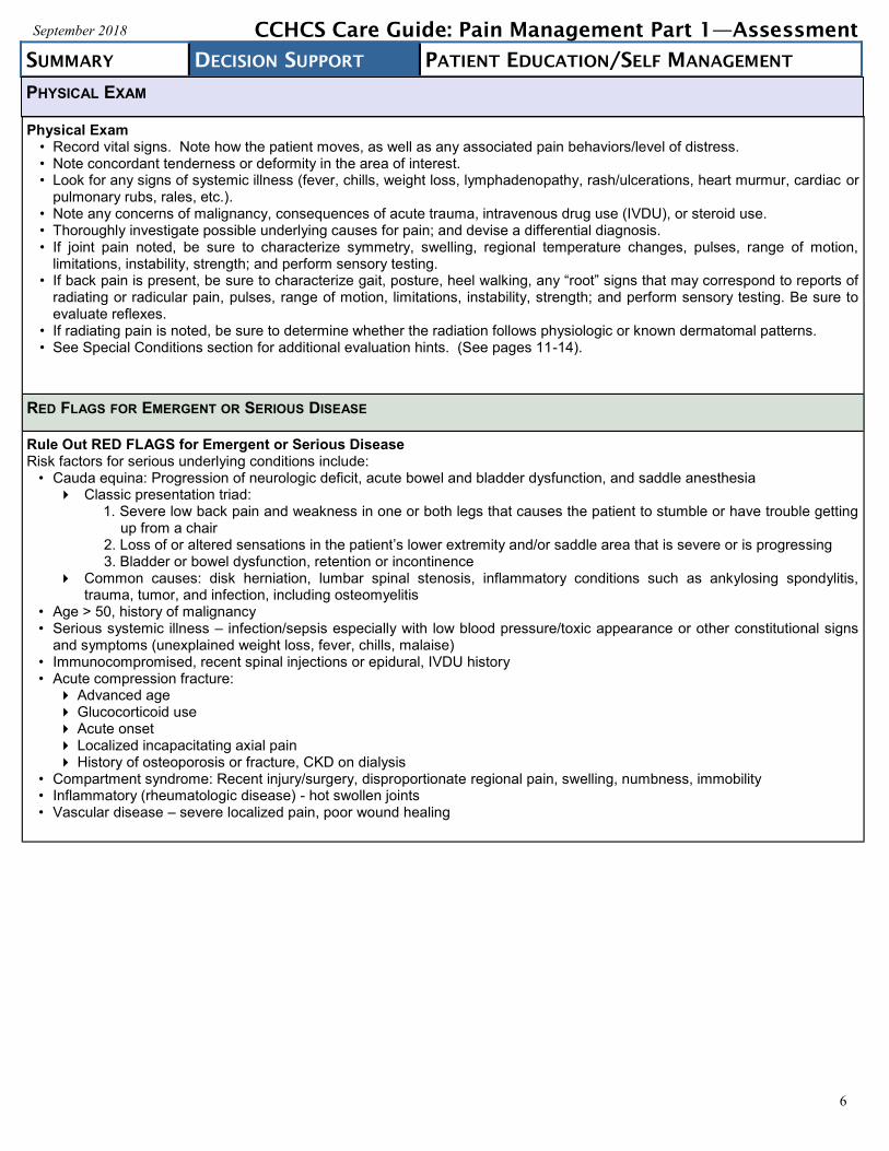
Physical Exam • Record vital signs. Note how the patient moves, as well as any associated pain behaviors/level of distress. • Note concordant tenderness or deformity in the area of interest. • Look for any signs of systemic illness (fever, chills, weight loss, lymphadenopathy, rash/ulcerations, heart murmur, cardiac or
pulmonary rubs, rales, etc.). • Note any concerns of malignancy, consequences of acute trauma, intravenous drug use (IVDU), or steroid use. • Thoroughly investigate possible underlying causes for pain; and devise a differential diagnosis. • If joint pain noted, be sure to characterize symmetry, swelling, regional temperature changes, pulses, range of motion,
limitations, instability, strength; and perform sensory testing. • If back pain is present, be sure to characterize gait, posture, heel walking, any “root” signs that may correspond to reports of
radiating or radicular pain, pulses, range of motion, limitations, instability, strength; and perform sensory testing. Be sure to evaluate reflexes.
• If radiating pain is noted, be sure to determine whether the radiation follows physiologic or known dermatomal patterns. • See Special Conditions section for additional evaluation hints. (See pages 11-14).
RED FLAGS FOR EMERGENT OR SERIOUS DISEASE
Rule Out RED FLAGS for Emergent or Serious Disease Risk factors for serious underlying conditions include:
• Cauda equina: Progression of neurologic deficit, acute bowel and bladder dysfunction, and saddle anesthesia Classic presentation triad:
1. Severe low back pain and weakness in one or both legs that causes the patient to stumble or have trouble getting up from a chair
2. Loss of or altered sensations in the patient’s lower extremity and/or saddle area that is severe or is progressing 3. Bladder or bowel dysfunction, retention or incontinence
Common causes: disk herniation, lumbar spinal stenosis, inflammatory conditions such as ankylosing spondylitis, trauma, tumor, and infection, including osteomyelitis
• Age > 50, history of malignancy • Serious systemic illness – infection/sepsis especially with low blood pressure/toxic appearance or other constitutional signs
and symptoms (unexplained weight loss, fever, chills, malaise) • Immunocompromised, recent spinal injections or epidural, IVDU history • Acute compression fracture:
Advanced age Glucocorticoid use Acute onset Localized incapacitating axial pain History of osteoporosis or fracture, CKD on dialysis
• Compartment syndrome: Recent injury/surgery, disproportionate regional pain, swelling, numbness, immobility • Inflammatory (rheumatologic disease) - hot swollen joints • Vascular disease – severe localized pain, poor wound healing
6
CCHCS Care Guide: Pain Management Part 1—Assessment
PHYSICAL EXAM
September 2018
SUMMARY DECISION SUPPORT PATIENT EDUCATION/SELF MANAGEMENT
7
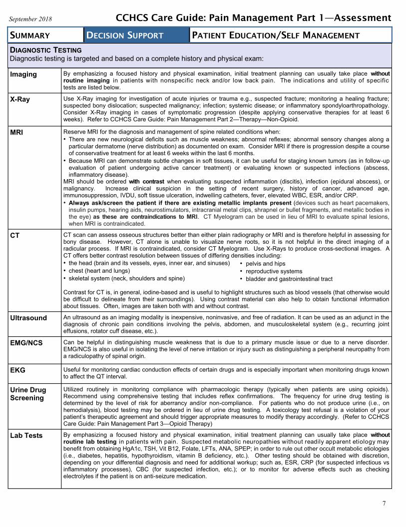
Imaging By emphasizing a focused history and physical examination, initial treatment planning can usually take place without routine imaging in patients with nonspecific neck and/or low back pain. The indications and utility of specific tests are listed below.
X-Ray Use X-Ray imaging for investigation of acute injuries or trauma e.g., suspected fracture; monitoring a healing fracture; suspected bony dislocation; suspected malignancy; infection; systemic disease; or inflammatory spondyloarthropathology. Consider X-Ray imaging in cases of symptomatic progression (despite applying conservative therapies for at least 6 weeks). Refer to CCHCS Care Guide: Pain Management Part 2—Therapy—Non-Opioid.
MRI Reserve MRI for the diagnosis and management of spine related conditions when:
• There are new neurological deficits such as muscle weakness; abnormal reflexes; abnormal sensory changes along a
particular dermatome (nerve distribution) as documented on exam. Consider MRI if there is progression despite a course of conservative treatment for at least 6 weeks within the last 6 months.
• Because MRI can demonstrate subtle changes in soft tissues, it can be useful for staging known tumors (as in follow-up evaluation of patient undergoing active cancer treatment) or evaluating known or suspected infections (abscess, inflammatory disease).
MRI should be ordered with contrast when evaluating suspected inflammation (discitis), infection (epidural abscess), or malignancy. Increase clinical suspicion in the setting of recent surgery, history of cancer, advanced age, immunosuppression, IVDU, soft tissue ulceration, indwelling catheters, fever, elevated WBC, ESR, and/or CRP.
• Always ask/screen the patient if there are existing metallic implants present (devices such as heart pacemakers, insulin pumps, hearing aids, neurostimulators, intracranial metal clips, shrapnel or bullet fragments, and metallic bodies in the eye) as these are contraindications to MRI. CT Myelogram can be used in lieu of MRI to evaluate spinal lesions, when MRI is contraindicated.
CT CT scan can assess osseous structures better than either plain radiography or MRI and is therefore helpful in assessing for bony disease. However, CT alone is unable to visualize nerve roots, so it is not helpful in the direct imaging of a radicular process. If MRI is contraindicated, consider CT Myelogram. Use X-Rays to produce cross-sectional images. A CT offers better contrast resolution between tissues of differing densities including:
• the head (brain and its vessels, eyes, inner ear, and sinuses)
• chest (heart and lungs)
• skeletal system (neck, shoulders and spine) Contrast for CT is, in general, iodine-based and is useful to highlight structures such as blood vessels (that otherwise would be difficult to delineate from their surroundings). Using contrast material can also help to obtain functional information about tissues. Often, images are taken both with and without contrast.
Ultrasound An ultrasound as an imaging modality is inexpensive, noninvasive, and free of radiation. It can be used as an adjunct in the diagnosis of chronic pain conditions involving the pelvis, abdomen, and musculoskeletal system (e.g., recurring joint effusions, rotator cuff disease, etc.).
EMG/NCS Can be helpful in distinguishing muscle weakness that is due to a primary muscle issue or due to a nerve disorder. EMG/NCS is also useful in isolating the level of nerve irritation or injury such as distinguishing a peripheral neuropathy from a radiculopathy of spinal origin.
EKG Useful for monitoring cardiac conduction effects of certain drugs and is especially important when monitoring drugs known to affect the QT interval.
Urine Drug Screening
Utilized routinely in monitoring compliance with pharmacologic therapy (typically when patients are using opioids). Recommend using comprehensive testing that includes reflex confirmations. The frequency for urine drug testing is determined by the level of risk for aberrancy and/or non-compliance. For patients who do not produce urine (i.e., on hemodialysis), blood testing may be ordered in lieu of urine drug testing. A toxicology test refusal is a violation of your patient’s therapeutic agreement and should trigger appropriate measures to modify therapy accordingly. (Refer to CCHCS Care Guide: Pain Management Part 3—Opioid Therapy)
Lab Tests By emphasizing a focused history and physical examination, initial treatment planning can usually take place without routine lab testing in patients with pain. Suspected metabolic neuropathies without readily apparent etiology may benefit from obtaining HgA1c, TSH, Vit B12, Folate, LFTs, ANA, SPEP; in order to rule out other occult metabolic etiologies (i.e., diabetes, hepatitis, hypothyroidism, vitamin B deficiency, etc.). Other testing should be obtained with discretion, depending on your differential diagnosis and need for additional workup; such as, ESR, CRP (for suspected infectious vs inflammatory processes), CBC (for suspected infection, etc.); or to monitor for adverse effects such as checking electrolytes if the patient is on anti-seizure medication.
CCHCS Care Guide: Pain Management Part 1—Assessment
• pelvis and hips
• reproductive systems
• bladder and gastrointestinal tract
DIAGNOSTIC TESTING Diagnostic testing is targeted and based on a complete history and physical exam:
September 2018
SUMMARY DECISION SUPPORT PATIENT EDUCATION/SELF MANAGEMENT
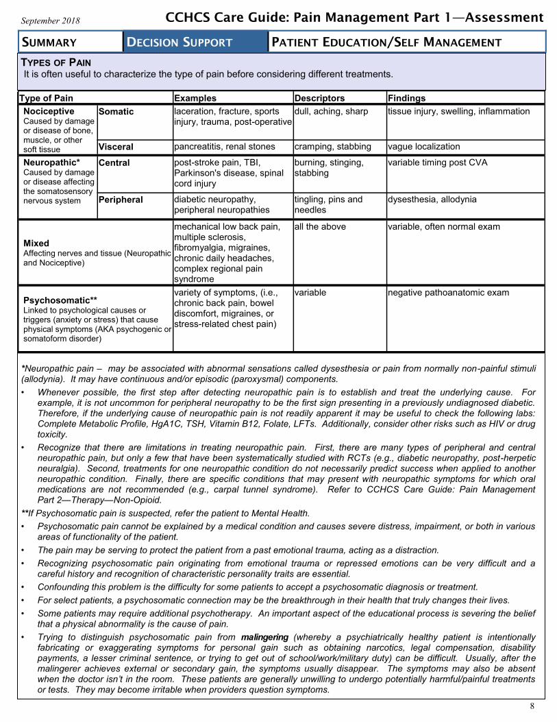
*Neuropathic pain – may be associated with abnormal sensations called dysesthesia or pain from normally non-painful stimuli (allodynia). It may have continuous and/or episodic (paroxysmal) components.
• Whenever possible, the first step after detecting neuropathic pain is to establish and treat the underlying cause. For example, it is not uncommon for peripheral neuropathy to be the first sign presenting in a previously undiagnosed diabetic. Therefore, if the underlying cause of neuropathic pain is not readily apparent it may be useful to check the following labs: Complete Metabolic Profile, HgA1C, TSH, Vitamin B12, Folate, LFTs. Additionally, consider other risks such as HIV or drug toxicity.
• Recognize that there are limitations in treating neuropathic pain. First, there are many types of peripheral and central neuropathic pain, but only a few that have been systematically studied with RCTs (e.g., diabetic neuropathy, post-herpetic neuralgia). Second, treatments for one neuropathic condition do not necessarily predict success when applied to another neuropathic condition. Finally, there are specific conditions that may present with neuropathic symptoms for which oral medications are not recommended (e.g., carpal tunnel syndrome). Refer to CCHCS Care Guide: Pain Management Part 2—Therapy—Non-Opioid.
**If Psychosomatic pain is suspected, refer the patient to Mental Health.
• Psychosomatic pain cannot be explained by a medical condition and causes severe distress, impairment, or both in various areas of functionality of the patient.
• The pain may be serving to protect the patient from a past emotional trauma, acting as a distraction.
• Recognizing psychosomatic pain originating from emotional trauma or repressed emotions can be very difficult and a careful history and recognition of characteristic personality traits are essential.
• Confounding this problem is the difficulty for some patients to accept a psychosomatic diagnosis or treatment.
• For select patients, a psychosomatic connection may be the breakthrough in their health that truly changes their lives.
• Some patients may require additional psychotherapy. An important aspect of the educational process is severing the belief that a physical abnormality is the cause of pain.
• Trying to distinguish psychosomatic pain from malingering (whereby a psychiatrically healthy patient is intentionally fabricating or exaggerating symptoms for personal gain such as obtaining narcotics, legal compensation, disability payments, a lesser criminal sentence, or trying to get out of school/work/military duty) can be difficult. Usually, after the malingerer achieves external or secondary gain, the symptoms usually disappear. The symptoms may also be absent when the doctor isn’t in the room. These patients are generally unwilling to undergo potentially harmful/painful treatments or tests. They may become irritable when providers question symptoms.
Type of Pain Examples Descriptors Findings
Nociceptive Caused by damage or disease of bone, muscle, or other soft tissue
Somatic laceration, fracture, sports injury, trauma, post-operative
dull, aching, sharp tissue injury, swelling, inflammation
Visceral pancreatitis, renal stones cramping, stabbing vague localization
Neuropathic* Caused by damage or disease affecting the somatosensory nervous system
Central post-stroke pain, TBI, Parkinson's disease, spinal cord injury
burning, stinging, stabbing
variable timing post CVA
Peripheral diabetic neuropathy, peripheral neuropathies
tingling, pins and needles
dysesthesia, allodynia
Mixed Affecting nerves and tissue (Neuropathic and Nociceptive)
mechanical low back pain, multiple sclerosis, fibromyalgia, migraines, chronic daily headaches, complex regional pain syndrome
all the above variable, often normal exam
Psychosomatic** Linked to psychological causes or triggers (anxiety or stress) that cause physical symptoms (AKA psychogenic or somatoform disorder)
variety of symptoms, (i.e., chronic back pain, bowel discomfort, migraines, or stress-related chest pain)
variable negative pathoanatomic exam
8
CCHCS Care Guide: Pain Management Part 1—Assessment
TYPES OF PAIN It is often useful to characterize the type of pain before considering different treatments.
September 2018
SUMMARY DECISION SUPPORT PATIENT EDUCATION/SELF MANAGEMENT
9
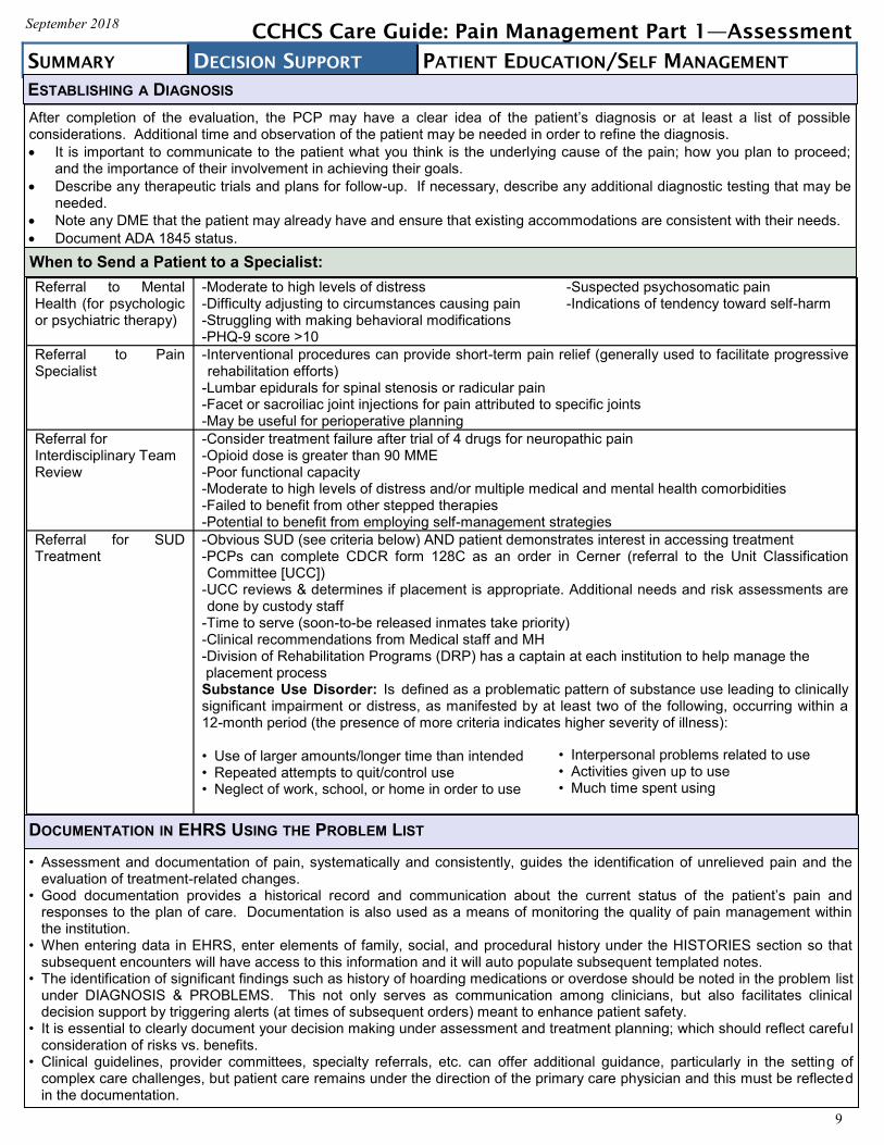
After completion of the evaluation, the PCP may have a clear idea of the patient’s diagnosis or at least a list of possible considerations. Additional time and observation of the patient may be needed in order to refine the diagnosis.
It is important to communicate to the patient what you think is the underlying cause of the pain; how you plan to proceed; and the importance of their involvement in achieving their goals.
Describe any therapeutic trials and plans for follow-up. If necessary, describe any additional diagnostic testing that may be needed.
Note any DME that the patient may already have and ensure that existing accommodations are consistent with their needs.
Document ADA 1845 status.
Referral to Mental Health (for psychologic or psychiatric therapy)
-Moderate to high levels of distress -Suspected psychosomatic pain -Difficulty adjusting to circumstances causing pain -Indications of tendency toward self-harm -Struggling with making behavioral modifications -PHQ-9 score >10
Referral to Pain Specialist
-Interventional procedures can provide short-term pain relief (generally used to facilitate progressive rehabilitation efforts)
-Lumbar epidurals for spinal stenosis or radicular pain -Facet or sacroiliac joint injections for pain attributed to specific joints -May be useful for perioperative planning
Referral for Interdisciplinary Team Review
-Consider treatment failure after trial of 4 drugs for neuropathic pain -Opioid dose is greater than 90 MME -Poor functional capacity -Moderate to high levels of distress and/or multiple medical and mental health comorbidities -Failed to benefit from other stepped therapies -Potential to benefit from employing self-management strategies
Referral for SUD Treatment
-Obvious SUD (see criteria below) AND patient demonstrates interest in accessing treatment -PCPs can complete CDCR form 128C as an order in Cerner (referral to the Unit Classification Committee [UCC])
-UCC reviews & determines if placement is appropriate. Additional needs and risk assessments are done by custody staff
-Time to serve (soon-to-be released inmates take priority) -Clinical recommendations from Medical staff and MH -Division of Rehabilitation Programs (DRP) has a captain at each institution to help manage the placement process Substance Use Disorder: Is defined as a problematic pattern of substance use leading to clinically significant impairment or distress, as manifested by at least two of the following, occurring within a 12-month period (the presence of more criteria indicates higher severity of illness): • Use of larger amounts/longer time than intended • Repeated attempts to quit/control use • Neglect of work, school, or home in order to use
• Interpersonal problems related to use • Activities given up to use • Much time spent using
CCHCS Care Guide: Pain Management Part 1—Assessment
ESTABLISHING A DIAGNOSIS
When to Send a Patient to a Specialist:
DOCUMENTATION IN EHRS USING THE PROBLEM LIST
• Assessment and documentation of pain, systematically and consistently, guides the identification of unrelieved pain and the evaluation of treatment-related changes.
• Good documentation provides a historical record and communication about the current status of the patient’s pain and responses to the plan of care. Documentation is also used as a means of monitoring the quality of pain management within the institution.
• When entering data in EHRS, enter elements of family, social, and procedural history under the HISTORIES section so that subsequent encounters will have access to this information and it will auto populate subsequent templated notes.
• The identification of significant findings such as history of hoarding medications or overdose should be noted in the problem list under DIAGNOSIS & PROBLEMS. This not only serves as communication among clinicians, but also facilitates clinical decision support by triggering alerts (at times of subsequent orders) meant to enhance patient safety.
• It is essential to clearly document your decision making under assessment and treatment planning; which should reflect careful consideration of risks vs. benefits.
• Clinical guidelines, provider committees, specialty referrals, etc. can offer additional guidance, particularly in the setting of complex care challenges, but patient care remains under the direction of the primary care physician and this must be reflected in the documentation.
September 2018
SUMMARY DECISION SUPPORT PATIENT EDUCATION/SELF MANAGEMENT
Clinicians should establish treatment goals with all patients including realistic goals for pain and function. Any therapy should be avoided/discontinued if benefits do not outweigh risks.
Carefully assessing the functional impact of pain is useful when establishing goals for therapy.
Engage the patient in identifying areas of significant functional impact.
Remind the patient that even with therapy, pain may still occur. In fact, this is more common than not.
There are multiple domains assessed for impact on pain including, stress, exercise, activity, sleep, fear of pain, appetite, mood, socializing; and any adverse effects caused by therapies (including constipation).
Establishing specific changes in these areas as treatment goals accomplishes at least two important things: 1. The focus of the discussion is redirected away from pain and onto functional landmarks. 2. The treatment goals become more observable by you and others (nursing, custody, etc.).
Document the patient’s expectations and help them reframe, if necessary, toward realistic goals.
Document agreed upon functional goals. Use specific examples: Taking walks (define distance/# laps), attendance at programming sessions, returning to job duties, participation in recreational activities, etc. Be realistic and establish goals that are Specific, Measurable, Achievable, Relevant and Time-based (SMART); to help with assessing progress.
It is important to also establish your expectations with the patient. For example, therapeutic trials require ongoing follow-up and assessment. Therefore, refusal of visits and/or testing – while certainly the prerogative of the patient – undermines your ability to assess therapeutic responses, and/or adverse reactions; and therefore must lead to modification of therapy.
10
Patient beliefs and expectations regarding pain and its treatment are major determinants of treatment outcomes.
It is essential to work with the patient and engage them in their treatment process and emphasize their role on the care team.
The Patient Education materials included in this care guide provide interactive tools to assist the provider in communicating with their patients and actively engage patients to participate in their own care.
Providers are encouraged to take the time to go through each Patient Education handout with the patient and answer any questions they may have.
Patients should be encouraged to prepare for their visits by filling out the PE-2 and PE-3 forms, Chronic Pain: Preparing for Your Health Care Visit before each visit.
Providers should:
Ongoing Assessment and Monitoring – Identify new/related symptoms and any significant changes at follow-up visits. Timing for scheduling follow-up visits Document the 4 A’s: 1-4 weeks: Initial therapeutic trials/diagnostic testing Analgesia - pain relieving effect 4-8 weeks: Later stages of therapeutic titration Adverse effects - related to treatment 1-3 months: Monitor for adverse effects, emerging Opiate Use ADLs - treatment effect on function, quality of life, mood Disorder Aberrant behaviors - cheeking, hoarding, not taking 3-6 months: Stable therapy (opioid therapy maximum duration medication as prescribed, no show for visits/classes is 3 months)
CCHCS Care Guide: Pain Management Part 1—Assessment
Instruct the patient on how to fill out each section. Explain why the tools are helpful in monitoring their treatment progress. Answer any questions the patients might have.
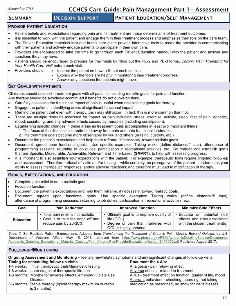
Goal Pain Reduction Improved Function Minimize Side Effects
Education
• Total pain relief is not realistic
• Goal is to take the edge off and reduce pain by 20-30%
• Ultimate goal is to improve quality of life (QOL)
• Degree of pain that interferes with QOL is highly personal
• Educate on potential side effects and risks associated with the chosen treatment(s)
Complete pain relief is not a realistic goal.
Focus on function.
Document the patient’s expectations and help them reframe, if necessary, toward realistic goals.
Document agreed upon functional goals. Use specific examples: Taking walks (define distance/# laps), attendance at programming sessions, returning to job duties, participation in recreational activities, etc.
Table 3. Set Realistic Patient Expectations. Adapted from Transforming the Treatment of Chronic Pain, Moving Beyond Opioids, by U.S. Department of Veterans Affairs, May 15, 2018, retrieved from https://www.pbm.va.gov/PBM/AcademicDetailingService/Documents/Academic_Detailing_Educational_Material_Catalog/Pain_ChronicPainProviderEducationalGuide_IB101000.pdf Published August 2017.
PROVIDE PATIENT EDUCATION
SET GOALS WITH PATIENTS
GOALS, EXPECTATIONS, AND EDUCATION
FOLLOW-UP/MONITORING
Spinal Pain: Pain originating from the spine, such as low back pain or neck pain is a dynamic process with high rates of incidence, recurrence, and recovery. General health, physical, and psychosocial factors can influence both the incidence and recurrence of spinal pain. Clinical disorders affecting the spine can be categorized as those that predominantly cause axial pain and those that most often cause radicular pain and/or neurologic dysfunction.
September 2018
SUMMARY DECISION SUPPORT PATIENT EDUCATION/SELF MANAGEMENT
Axial Pain: Also called mechanical pain and commonly caused by degenerative changes
Cervical or Lumbar strain — Strain injury is a nonspecific diagnosis used to describe an injury to the paraspinal muscles and ligaments with associated spasm of the surrounding muscles. May result from the physical stresses of everyday life, including poor posture and sleeping habits. Typically experienced as localized pain, stiffness, and tightness and is usually self-limited in nature resolving in 1-2 months.
Cervical or Lumbar spondylosis — Describes a continuum of degenerative changes from normal aging to the overtly pathologic state, primarily in the spinal structures. Correlation between the degree of radiographic change and the presence or severity of pain is poor. The degenerative condition can be ascribed to the discs or the facets or both, and can generally be associated with muscle tightness and spasm. Exam findings may show axial discomfort and limitations in spinal range of motion, but is usually a benign neurological exam. Facet pain can be referred to local regions, but does not typically follow a radicular or dermatomal pattern. A fluoroscopically guided intra-articular facet injection with local anesthetic resulting in relief is considered the definitive diagnostic test.
Myofascial Pain — Regional pain with associated trigger points (palpable tender fibrous bands of muscle) is called myofascial pain. Myofascial pain can be a nonspecific manifestation of any pathologic condition that causes muscular irritability. Myofascial pain can also be associated with muscle sensitivity, depression, anxiety, or insomnia.
Radicular Pain – involving extremity pain and/or neurologic deficit
Spondylotic Myelopathy — is defined by degenerative changes narrowing the spinal canal, resulting in spinal cord injury or dysfunction. Patients may present with a variety of neurologic complaints: weakness, coordination impairment, gait disturbance, bowel or bladder retention or incontinence, and sexual dysfunction. Symptoms usually begin insidiously. Gait impairment is a common early symptom.
Cervical or Lumbar Radiculopathy — Refers to dysfunction of a spinal nerve root that may manifest with pain, weakness, reflex changes, or sensory changes. Multiple conditions can give rise to radiculopathy most commonly degenerative changes in the spine including foraminal stenosis and herniated disc. Onset of symptoms is most frequently acute when caused by a herniated disc, but may be more indolent when due to spondylosis. In the presence of pain, it can be difficult to perform an accurate motor examination. In such cases, the reflex examination is a more objective test of nerve root function. However, not all spinal levels have a corresponding reflex to test.
Spinal Stenosis — Characterized by neuroclaudication described as intense pain (burning, tingling, numbness, cramping, radiating) down extremities; associated with activity and generally improves with flexion and/or rest. Affects upper extremities in cervical and lower extremities in lumbar.
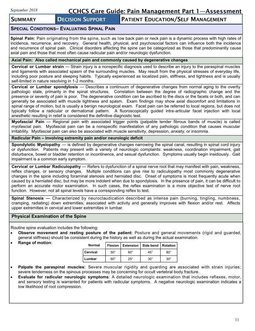
Physical Examination of the Spine
Routine spine evaluation includes the following:
Observe movement and resting posture of the patient: Posture and general movements (rigid and guarded, general stiffness) should be consistent during the history as well as during the actual examination.
Range of motion:
Palpate the paraspinal muscles: Severe muscular rigidity and guarding are associated with strain injuries; severe tenderness on the spinous processes may be concerning for occult vertebral body fracture.
Evaluate for radicular neurologic symptoms: A detailed neurologic examination that includes reflexes, motor, and sensory testing is warranted for patients with radicular symptoms. A negative neurologic examination indicates a low likelihood of root compression.
Normal Flexion Extension Side bend Rotation
Cervical 50° 60° 45° 80°
Lumbar 60° 25° 30° 30°
11
CCHCS Care Guide: Pain Management Part 1—Assessment
SPECIAL CONDITIONS– EVALUATING SPINAL PAIN
September 2018
SUMMARY DECISION SUPPORT PATIENT EDUCATION/SELF MANAGEMENT
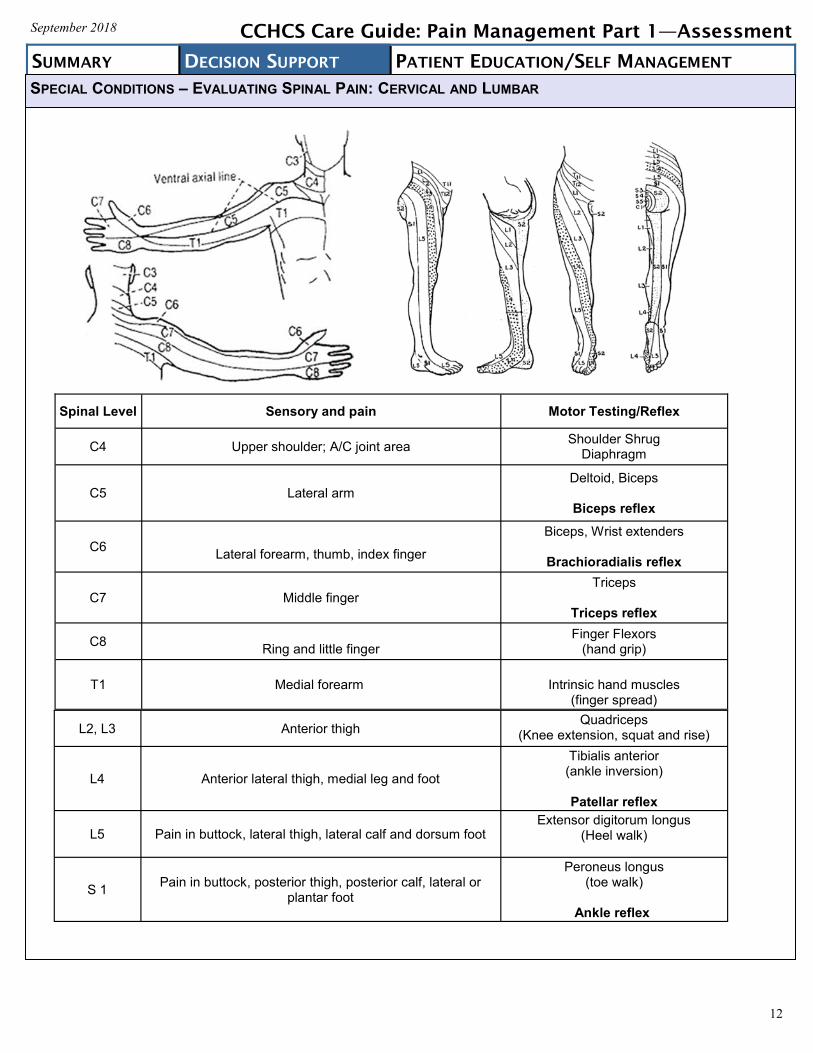
Spinal Level Sensory and pain Motor Testing/Reflex
C4 Upper shoulder; A/C joint area Shoulder Shrug
Diaphragm
C5 Lateral arm
Deltoid, Biceps
Biceps reflex
C6
Lateral forearm, thumb, index finger
Biceps, Wrist extenders
Brachioradialis reflex
C7 Middle finger Triceps
Triceps reflex
C8
Ring and little finger Finger Flexors
(hand grip)
T1 Medial forearm
Intrinsic hand muscles (finger spread)
L2, L3 Anterior thigh Quadriceps
(Knee extension, squat and rise)
L4 Anterior lateral thigh, medial leg and foot
Tibialis anterior (ankle inversion)
Patellar reflex
L5 Pain in buttock, lateral thigh, lateral calf and dorsum foot Extensor digitorum longus
(Heel walk)
S 1 Pain in buttock, posterior thigh, posterior calf, lateral or
plantar foot
Peroneus longus (toe walk)
Ankle reflex
12
CCHCS Care Guide: Pain Management Part 1—Assessment
SPECIAL CONDITIONS – EVALUATING SPINAL PAIN: CERVICAL AND LUMBAR
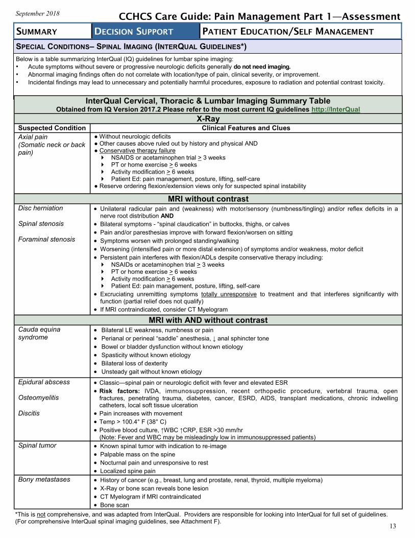
Below is a table summarizing InterQual (IQ) guidelines for lumbar spine imaging:
• Acute symptoms without severe or progressive neurologic deficits generally do not need imaging.
• Abnormal imaging findings often do not correlate with location/type of pain, clinical severity, or improvement.
• Incidental findings may lead to unnecessary and potentially harmful procedures, exposure to radiation and potential contrast toxicity.
September 2018
SUMMARY DECISION SUPPORT PATIENT EDUCATION/SELF MANAGEMENT
13
InterQual Cervical, Thoracic & Lumbar Imaging Summary Table Obtained from IQ Version 2017.2 Please refer to the most current IQ guidelines http://InterQual
X-Ray
Suspected Condition Clinical Features and Clues
Axial pain (Somatic neck or back pain)
● Without neurologic deficits
● Other causes above ruled out by history and physical AND
● Conservative therapy failure NSAIDS or acetaminophen trial > 3 weeks
PT or home exercise > 6 weeks
Activity modification > 6 weeks
Patient Ed: pain management, posture, lifting, self-care
● Reserve ordering flexion/extension views only for suspected spinal instability
MRI without contrast Disc herniation Spinal stenosis Foraminal stenosis
Unilateral radicular pain and (weakness) with motor/sensory (numbness/tingling) and/or reflex deficits in a nerve root distribution AND
Bilateral symptoms - “spinal claudication” in buttocks, thighs, or calves
Pain and/or paresthesias improve with forward flexion/worsen on sitting
Symptoms worsen with prolonged standing/walking
Worsening (intensified pain or more distal extension) of symptoms and/or weakness, motor deficit
Persistent pain interferes with flexion/ADLs despite conservative therapy including:
NSAIDs or acetaminophen trial > 3 weeks
PT or home exercise > 6 weeks
Activity modification > 6 weeks
Patient Ed: pain management, posture, lifting, self-care
Excruciating unremitting symptoms totally unresponsive to treatment and that interferes significantly with
function (partial relief does not qualify)
If MRI contraindicated, consider CT Myelogram
MRI with AND without contrast
Cauda equina syndrome
Bilateral LE weakness, numbness or pain
Perianal or perineal “saddle” anesthesia, ↓ anal sphincter tone
Bowel or bladder dysfunction without known etiology
Spasticity without known etiology
Bilateral loss of dexterity
Unsteady gait without known etiology
Epidural abscess Osteomyelitis Discitis
Classic—spinal pain or neurologic deficit with fever and elevated ESR
Risk factors: IVDA, immunosuppression, recent orthopedic procedure, vertebral trauma, open
fractures, penetrating trauma, diabetes, cancer, ESRD, AIDS, transplant medications, chronic indwelling catheters, local soft tissue ulceration
Pain increases with movement
Temp > 100.4° F (38° C)
Positive blood culture, ↑WBC ↑CRP, ESR >30 mm/hr (Note: Fever and WBC may be misleadingly low in immunosuppressed patients)
Spinal tumor Known spinal tumor with indication to re-image
Palpable mass on the spine
Nocturnal pain and unresponsive to rest
Localized spine pain
Bony metastases History of cancer (e.g., breast, lung and prostate, renal, thyroid, multiple myeloma)
X-Ray or bone scan reveals bone lesion
CT Myelogram if MRI contraindicated
Bone scan
CCHCS Care Guide: Pain Management Part 1—Assessment
*This is not comprehensive, and was adapted from InterQual. Providers are responsible for looking into InterQual for full set of guidelines. (For comprehensive InterQual spinal imaging guidelines, see Attachment F).
SPECIAL CONDITIONS– SPINAL IMAGING (INTERQUAL GUIDELINES*)
Gun Shot Wounds (GSW)– ballistic trauma whereby tissue damage is dependent on the firearm, bullet, velocity, entry point, and trajectory. Management can range from observation and local wound care to urgent surgical intervention. Not all GSWs result in chronic pain symptoms just as other forms of puncture wounds can heal over time. GSWs can be particularly devastating compared to other penetrating injuries because the trajectory and fragmentation of
bullets can be unpredictable after entry. Additionally, gunshot wounds typically involve a large degree of nearby tissue disruption and destruction due to the
physical effects of the projectile correlated with the bullet velocity classification. Non-fatal GSWs frequently have severe and long-lasting effects, typically some form of major disfigurement, and/or
permanent disability. It is vital to have records of any previous surgeries and information about retained bullet fragments in order to make
judgements about the need for pain management. Do not order MRI testing in a patient with retained bullet fragments. When considering pain that has resulted from a traumatic event, be aware of the impact that the lingering psychological
trauma may have on the manifestation of symptoms. Be sure to explore the psychological trauma in the course of your history, evaluations, and supplemental assessments.
September 2018
SUMMARY DECISION SUPPORT PATIENT EDUCATION/SELF MANAGEMENT
14
CCHCS Care Guide: Pain Management Part 1—Assessment
ADDITIONAL SPECIAL CONDITIONS– TRAUMA / GUN SHOT WOUNDS
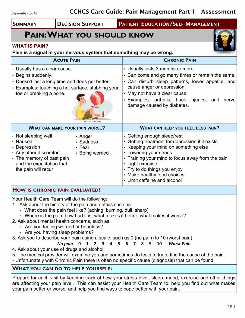
PAIN: WHAT YOU SHOULD KNOW WHAT IS PAIN?
Pain is a signal in your nervous system that something may be wrong.
ACUTE PAIN CHRONIC PAIN
• Usually has a clear cause.
• Begins suddenly.
• Doesn't last a long time and does get better.
• Examples: touching a hot surface, stubbing your toe or breaking a bone.
• Usually lasts 3 months or more.
• Can come and go many times or remain the same.
• Can disturb sleep patterns, lower appetite, and cause anger or depression.
• May not have a clear cause.
• Examples: arthritis, back injuries, and nerve damage caused by diabetes.
WHAT CAN MAKE YOUR PAIN WORSE? WHAT CAN HELP YOU FEEL LESS PAIN?
• Not sleeping well • Nausea • Depression • Any other discomfort • The memory of past pain
and the expectation that the pain will recur
• Getting enough sleep/rest • Getting treatment for depression if it exists • Keeping your mind on something else • Lowering your stress • Training your mind to focus away from the pain • Light exercise • Try to do things you enjoy • Make healthy food choices • Limit caffeine and alcohol
HOW IS CHRONIC PAIN EVALUATED?
Your Health Care Team will do the following: 1. Ask about the history of the pain and details such as:
• What does the pain feel like? (aching, burning, dull, sharp) • Where is the pain, how bad it is, what makes it better, what makes it worse?
2. Ask about mental health concerns, such as: • Are you feeling worried or hopeless? • Are you having sleep problems?
3. Ask you to describe your pain using a scale, such as 0 (no pain) to 10 (worst pain).
No pain 0 1 2 3 4 5 6 7 8 9 10 Worst Pain 4. Ask about your use of drugs and alcohol. 5. The medical provider will examine you and sometimes do tests to try to find the cause of the pain. - Unfortunately with Chronic Pain there is often no specific cause (diagnosis) that can be found.
WHAT YOU CAN DO TO HELP YOURSELF:
Prepare for each visit by keeping track of how your stress level, sleep, mood, exercise and other things are affecting your pain level. This can assist your Health Care Team to: help you find out what makes your pain better or worse; and help you find ways to cope better with your pain.
• Anger • Sadness • Fear • Being worried
SUMMARY DECISION SUPPORT PATIENT EDUCATION/SELF MANAGEMENT
PE-1
September 2018 CCHCS Care Guide: Pain Management Part 1—Assessment
No Pain Worst Pain 1 2 3 4 5 6 7 8 9 10
Stress
September 2018 CCHCS Care Guide: Pain Management
SUMMARY DECISION SUPPORT PATIENT EDUCATION/SELF MANAGEMENT
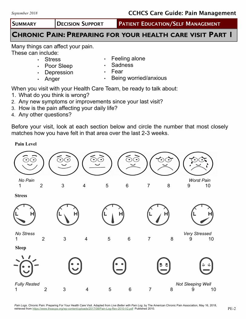
CHRONIC PAIN: PREPARING FOR YOUR HEALTH CARE VISIT PART 1
Many things can affect your pain. These can include:
• Stress • Poor Sleep • Depression • Anger
When you visit with your Health Care Team, be ready to talk about: 1. What do you think is wrong? 2. Any new symptoms or improvements since your last visit?
3. How is the pain affecting your daily life?
4. Any other questions?
Before your visit, look at each section below and circle the number that most closely matches how you have felt in that area over the last 2-3 weeks.
• Feeling alone • Sadness • Fear • Being worried/anxious
Pain Level
Sleep
No Stress Very Stressed 1 2 3 4 5 6 7 8 9 10
Fully Rested Not Sleeping Well 1 2 3 4 5 6 7 8 9 10
Pain Logs. Chronic Pain: Preparing For Your Health Care Visit. Adapted from Live Better with Pain Log, by The American Chronic Pain Association, May 16, 2018, retrieved from https://www.theacpa.org/wp-content/uploads/2017/08/Pain-Log-Rev-2010-V2.pdf Published 2010. PE-2
SUMMARY DECISION SUPPORT PATIENT EDUCATION/SELF MANAGEMENT
PE-3
September 2018
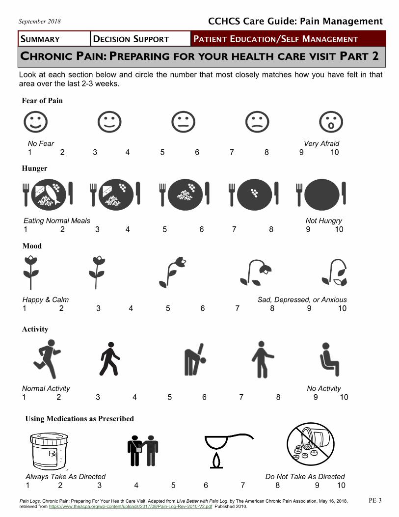
Look at each section below and circle the number that most closely matches how you have felt in that area over the last 2-3 weeks.
CHRONIC PAIN: PREPARING FOR YOUR HEALTH CARE VISIT PART 2
Using Medications as Prescribed
Activity
Normal Activity No Activity
1 2 3 4 5 6 7 8 9 10
CCHCS Care Guide: Pain Management
Pain Logs. Chronic Pain: Preparing For Your Health Care Visit. Adapted from Live Better with Pain Log, by The American Chronic Pain Association, May 16, 2018, retrieved from https://www.theacpa.org/wp-content/uploads/2017/08/Pain-Log-Rev-2010-V2.pdf Published 2010.
Hunger
Fear of Pain
Mood
Happy & Calm Sad, Depressed, or Anxious
1 2 3 4 5 6 7 8 9 10
Eating Normal Meals Not Hungry
1 2 3 4 5 6 7 8 9 10
No Fear Very Afraid
1 2 3 4 5 6 7 8 9 10
Always Take As Directed Do Not Take As Directed
1 2 3 4 5 6 7 8 9 10
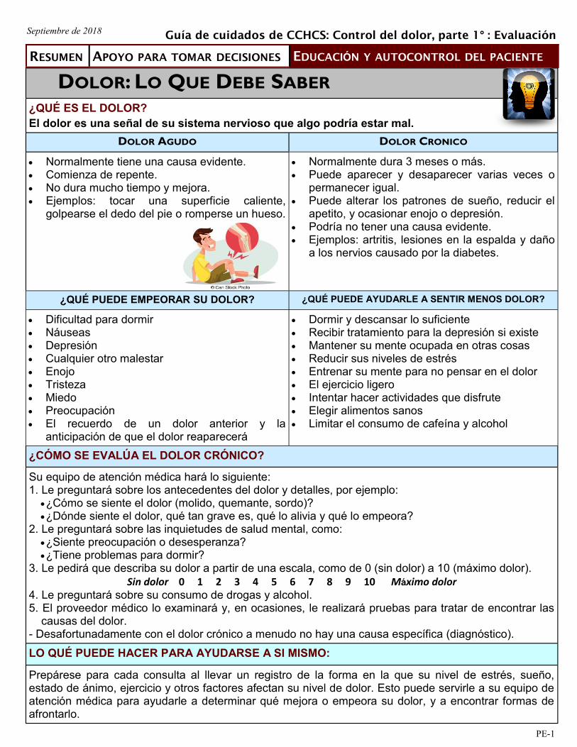
DOLOR: LO QUE DEBE SABER ¿QUÉ ES EL DOLOR?
El dolor es una señal de su sistema nervioso que algo podría estar mal.
DOLOR AGUDO DOLOR CRONICO
Normalmente tiene una causa evidente. Comienza de repente. No dura mucho tiempo y mejora. Ejemplos: tocar una superficie caliente,
golpearse el dedo del pie o romperse un hueso.
Normalmente dura 3 meses o más. Puede aparecer y desaparecer varias veces o
permanecer igual. Puede alterar los patrones de sueño, reducir el
apetito, y ocasionar enojo o depresión. Podría no tener una causa evidente. Ejemplos: artritis, lesiones en la espalda y daño
a los nervios causado por la diabetes.
¿QUÉ PUEDE EMPEORAR SU DOLOR? ¿QUÉ PUEDE AYUDARLE A SENTIR MENOS DOLOR?
Dificultad para dormir Náuseas Depresión Cualquier otro malestar Enojo Tristeza Miedo Preocupación El recuerdo de un dolor anterior y la
anticipación de que el dolor reaparecerá
Dormir y descansar lo suficiente Recibir tratamiento para la depresión si existe Mantener su mente ocupada en otras cosas Reducir sus niveles de estrés Entrenar su mente para no pensar en el dolor El ejercicio ligero Intentar hacer actividades que disfrute Elegir alimentos sanos Limitar el consumo de cafeína y alcohol
¿CÓMO SE EVALÚA EL DOLOR CRÓNICO?
Su equipo de atención médica hará lo siguiente: 1. Le preguntará sobre los antecedentes del dolor y detalles, por ejemplo:
¿Cómo se siente el dolor (molido, quemante, sordo)? ¿Dónde siente el dolor, qué tan grave es, qué lo alivia y qué lo empeora?
2. Le preguntará sobre las inquietudes de salud mental, como: ¿Siente preocupación o desesperanza? ¿Tiene problemas para dormir?
3. Le pedirá que describa su dolor a partir de una escala, como de 0 (sin dolor) a 10 (máximo dolor).
Sin dolor 0 1 2 3 4 5 6 7 8 9 10 Máximo dolor 4. Le preguntará sobre su consumo de drogas y alcohol. 5. El proveedor médico lo examinará y, en ocasiones, le realizará pruebas para tratar de encontrar las
causas del dolor. - Desafortunadamente con el dolor crónico a menudo no hay una causa específica (diagnóstico).
LO QUÉ PUEDE HACER PARA AYUDARSE A SI MISMO:
Prepárese para cada consulta al llevar un registro de la forma en la que su nivel de estrés, sueño, estado de ánimo, ejercicio y otros factores afectan su nivel de dolor. Esto puede servirle a su equipo de atención médica para ayudarle a determinar qué mejora o empeora su dolor, y a encontrar formas de afrontarlo.
RESUMEN APOYO PARA TOMAR DECISIONES EDUCACIÓN Y AUTOCONTROL DEL PACIENTE
PE-1
Septiembre de 2018 Guía de cuidados de CCHCS: Control del dolor, parte 1° : Evaluación
PE-2
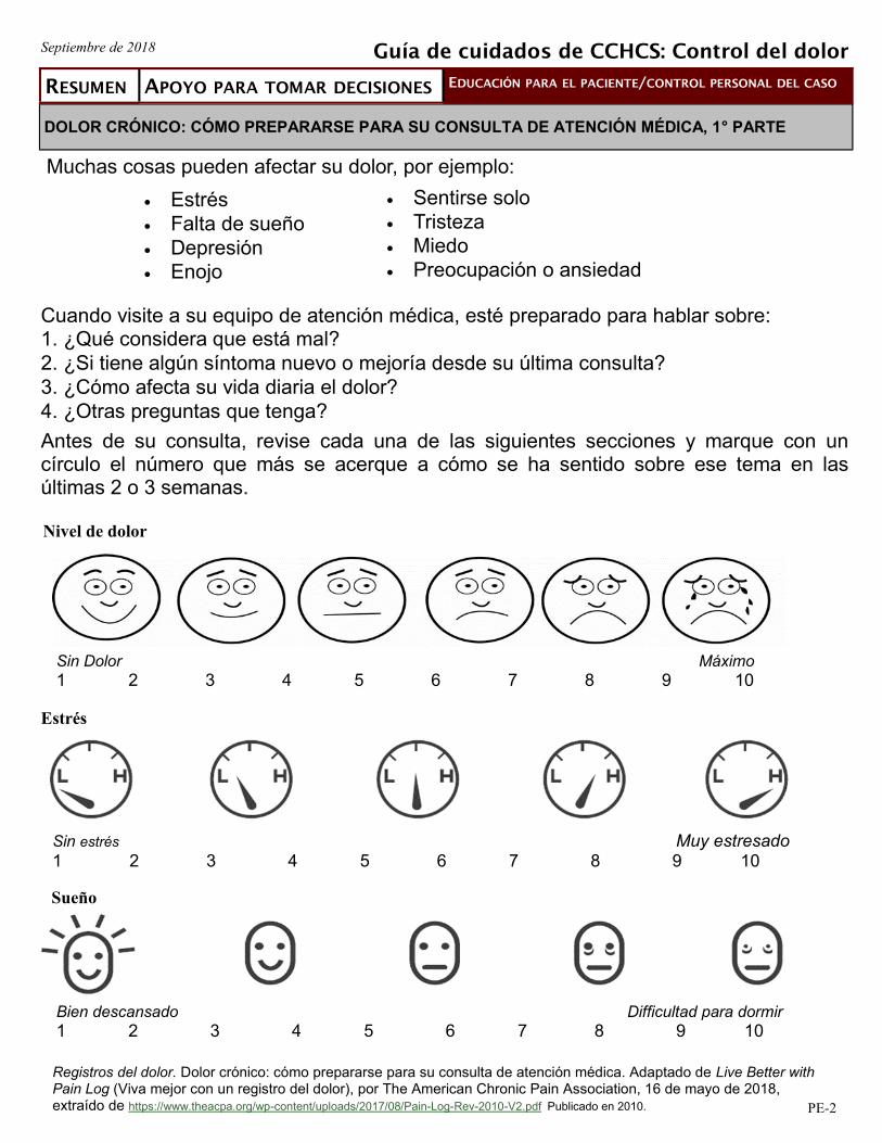
Septiembre de 2018
Muchas cosas pueden afectar su dolor, por ejemplo:
Estrés
Falta de sueño
Depresión
Enojo
Cuando visite a su equipo de atención médica, esté preparado para hablar sobre: 1. ¿Qué considera que está mal?
2. ¿Si tiene algún síntoma nuevo o mejoría desde su última consulta?
3. ¿Cómo afecta su vida diaria el dolor?
4. ¿Otras preguntas que tenga?
Antes de su consulta, revise cada una de las siguientes secciones y marque con un círculo el número que más se acerque a cómo se ha sentido sobre ese tema en las últimas 2 o 3 semanas.
DOLOR CRÓNICO: CÓMO PREPARARSE PARA SU CONSULTA DE ATENCIÓN MÉDICA, 1° PARTE
Sentirse solo
Tristeza
Miedo
Preocupación o ansiedad
Guía de cuidados de CCHCS: Control del dolor
Registros del dolor. Dolor crónico: cómo prepararse para su consulta de atención médica. Adaptado de Live Better with Pain Log (Viva mejor con un registro del dolor), por The American Chronic Pain Association, 16 de mayo de 2018, extraído de https://www.theacpa.org/wp-content/uploads/2017/08/Pain-Log-Rev-2010-V2.pdf Publicado en 2010.
RESUMEN APOYO PARA TOMAR DECISIONES EDUCACIÓN PARA EL PACIENTE/CONTROL PERSONAL DEL CASO
Nivel de dolor
Sin Dolor Máximo 1 2 3 4 5 6 7 8 9 10
Estrés
Sin estrés Muy estresado 1 2 3 4 5 6 7 8 9 10
Sueño
Bien descansado Difficultad para dormir 1 2 3 4 5 6 7 8 9 10
PE-3
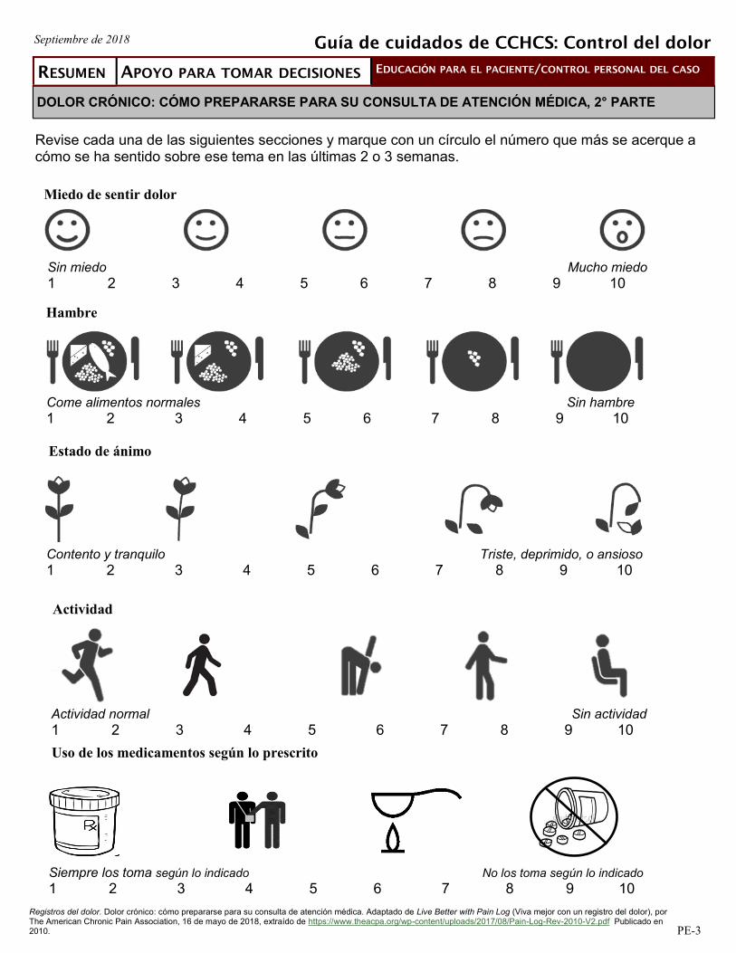
Septiembre de 2018
Revise cada una de las siguientes secciones y marque con un círculo el número que más se acerque a cómo se ha sentido sobre ese tema en las últimas 2 o 3 semanas.
DOLOR CRÓNICO: CÓMO PREPARARSE PARA SU CONSULTA DE ATENCIÓN MÉDICA, 2° PARTE
Uso de los medicamentos según lo prescrito
Actividad
Registros del dolor. Dolor crónico: cómo prepararse para su consulta de atención médica. Adaptado de Live Better with Pain Log (Viva mejor con un registro del dolor), por The American Chronic Pain Association, 16 de mayo de 2018, extraído de https://www.theacpa.org/wp-content/uploads/2017/08/Pain-Log-Rev-2010-V2.pdf Publicado en 2010.
RESUMEN APOYO PARA TOMAR DECISIONES EDUCACIÓN PARA EL PACIENTE/CONTROL PERSONAL DEL CASO
Guía de cuidados de CCHCS: Control del dolor
Miedo de sentir dolor
Sin miedo Mucho miedo
1 2 3 4 5 6 7 8 9 10
Hambre
Come alimentos normales Sin hambre
1 2 3 4 5 6 7 8 9 10
Contento y tranquilo Triste, deprimido, o ansioso
1 2 3 4 5 6 7 8 9 10
Estado de ánimo
Siempre los toma según lo indicado No los toma según lo indicado 1 2 3 4 5 6 7 8 9 10
Actividad normal Sin actividad
1 2 3 4 5 6 7 8 9 10
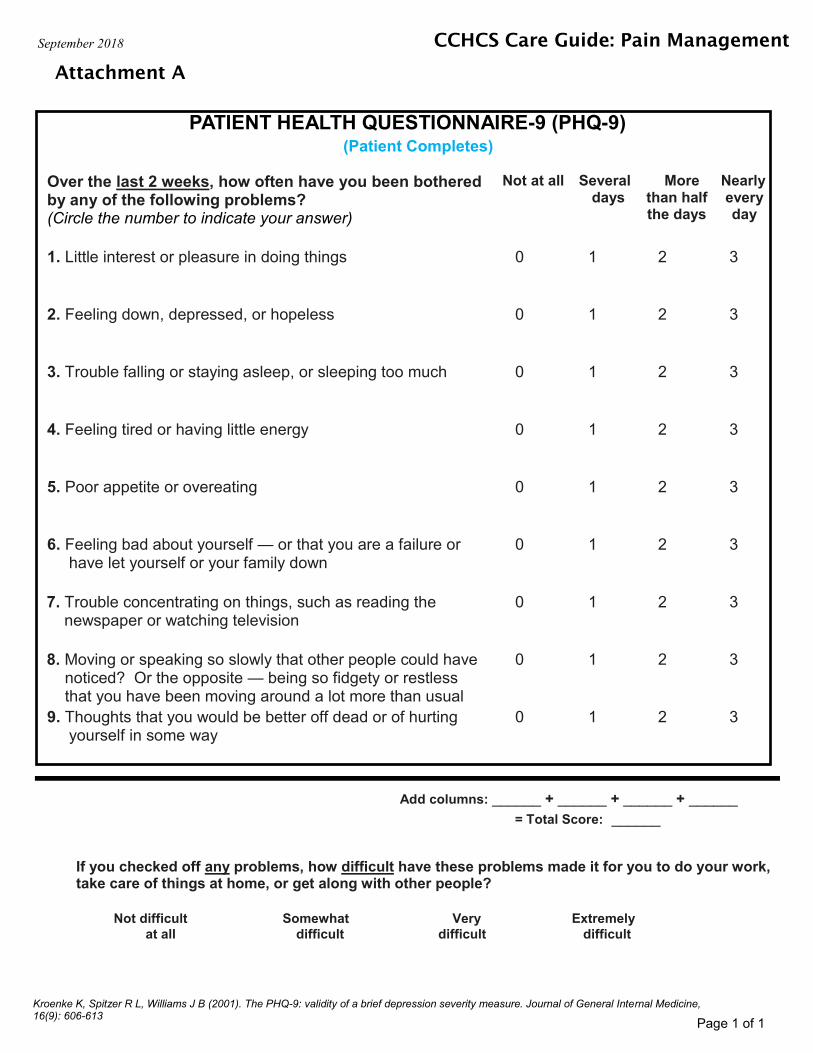
PATIENT HEALTH QUESTIONNAIRE-9 (PHQ-9)
Over the last 2 weeks, how often have you been bothered by any of the following problems? (Circle the number to indicate your answer)
Not at all Several days
More than half the days
Nearly every day
1. Little interest or pleasure in doing things 0 1 2 3
2. Feeling down, depressed, or hopeless 0 1 2 3
3. Trouble falling or staying asleep, or sleeping too much 0 1 2 3
4. Feeling tired or having little energy 0 1 2 3
5. Poor appetite or overeating 0 1 2 3
6. Feeling bad about yourself — or that you are a failure or have let yourself or your family down
0 1 2 3
7. Trouble concentrating on things, such as reading the newspaper or watching television
0 1 2 3
8. Moving or speaking so slowly that other people could have noticed? Or the opposite — being so fidgety or restless that you have been moving around a lot more than usual
0 1 2 3
9. Thoughts that you would be better off dead or of hurting yourself in some way
0 1 2 3
Add columns: ______ + ______ + ______ + ______ = Total Score: ______
If you checked off any problems, how difficult have these problems made it for you to do your work, take care of things at home, or get along with other people?
Not difficult at all
Somewhat difficult
Very difficult
Extremely
difficult
Attachment A
September 2018
(Patient Completes)
CCHCS Care Guide: Pain Management
Kroenke K, Spitzer R L, Williams J B (2001). The PHQ-9: validity of a brief depression severity measure. Journal of General Internal Medicine, 16(9): 606-613
Page 1 of 1
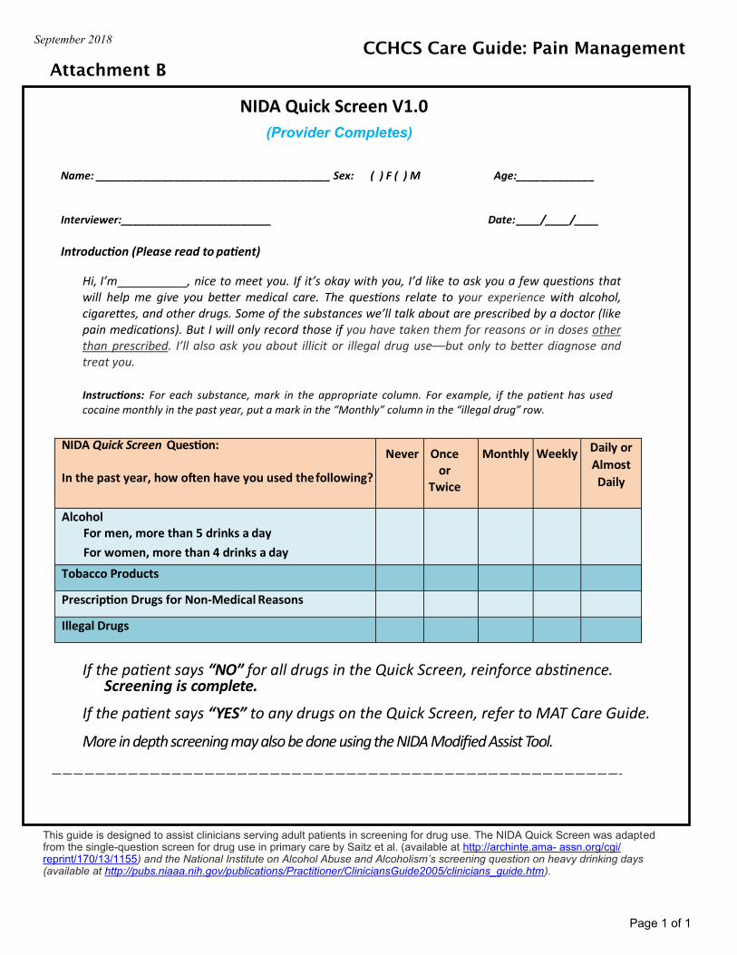
Name: _______________________________________ Sex: ( ) F ( ) M Age:_____________
Interviewer:_________________________ Date: ____/____/____
Introduction (Please read to patient)
Hi, I’m , nice to meet you. If it’s okay with you, I’d like to ask you a few questions that will help me give you better medical care. The questions relate to your experience with alcohol, cigarettes, and other drugs. Some of the substances we’ll talk about are prescribed by a doctor (like pain medications). But I will only record those if you have taken them for reasons or in doses other than prescribed. I’ll also ask you about illicit or illegal drug use––but only to better diagnose and treat you.
Instructions: For each substance, mark in the appropriate column. For example, if the patient has used cocaine monthly in the past year, put a mark in the “Monthly” column in the “illegal drug” row.
If the patient says “NO” for all drugs in the Quick Screen, reinforce abstinence. Screening is complete.
If the patient says “YES” to any drugs on the Quick Screen, refer to MAT Care Guide.
More in depth screening may also be done using the NIDA Modified Assist Tool. ————————————————————————————————————————————————————-
NIDA Quick Screen Question:
In the past year, how often have you used the following?
Never Once or
Twice
Monthly Weekly Daily or
Almost
Daily
Alcohol For men, more than 5 drinks a day
For women, more than 4 drinks a day
Tobacco Products
Prescription Drugs for Non-Medical Reasons
Illegal Drugs
Attachment B
September 2018
This guide is designed to assist clinicians serving adult patients in screening for drug use. The NIDA Quick Screen was adapted from the single-question screen for drug use in primary care by Saitz et al. (available at http://archinte.ama- assn.org/cgi/reprint/170/13/1155) and the National Institute on Alcohol Abuse and Alcoholism’s screening question on heavy drinking days (available at http://pubs.niaaa.nih.gov/publications/Practitioner/CliniciansGuide2005/clinicians_guide.htm).
Page 1 of 1
(Provider Completes)
CCHCS Care Guide: Pain Management
Attachment C
September 2018
Page 1 of 1
CCHCS Care Guide: Pain Management
NIDA Modified Assist (Provider Completes)
Please answer the following questions: 1. In your LIFETIME, which of the following substances have you ever used? (Yes/No)
2. In the past 3 months, how often have you used the following substances? (Never, once or twice, monthly,
weekly, almost daily) 3. In the past three months, how often have you had a strong desire or urge to use (first drug, second drug,
etc.? (Never, once or twice, monthly, weekly, almost daily) 4. During the past three months, how often has your use of (first drug, second drug, etc.) led to health, social,
legal or financial problems? (Never, once or twice, monthly, weekly, almost daily) 5. During the past 3 months, how often have you failed to do what was normally expected of you because of
your use of this substance? (Never, once or twice, monthly, weekly, almost daily) 6. Has a friend or relative or anyone else ever expressed concern about your use of (first drug, second drug,
etc.)? (Never; yes, but not in the past 3 months; yes, in the past 3 months) 7. Have you ever tried and failed to control, cut down, or stop using this substance? (Never; yes, but not in the
past 3 months; yes, in the past 3 months) 8. Have you ever used any drug by injection (NONMEDICAL USE ONLY)? (Never; yes, but not in the past 3
months; yes, in the past 3 months)
Cannabis Methamphetamine Hallucinogens
Cocaine Inhalants Street opioids
Prescription stimulants Sedatives or sleeping pills Prescription opioids
The NIDA-modified ASSIST was adapted from the World Health Organization (WHO) Alcohol, Smoking and Substance Involvement Screening Test (ASSIST), Version 3.0, developed and published by WHO (available at http://www.who.int/substance_abuse/ activities/assist/)
Attachment D
September 2018
Page 1 of 1
CCHCS Care Guide: Pain Management
Screener and Opioid Assessment for Patients with Pain (SOAPP)
(Patient Completes)
SOAPP Version 1.0 Name:_____________________________________________ Date:___________________ Please answer each question as honestly as possible. This information is for our records and will remain confidential. Your answers alone will not determine your treatment.
Please answer the questions below using the following scale:
0=Never, 1=Seldom, 2=Sometimes, 3=Often, 4=Very Often
1. How often do you have mood swings? 0 1 2 3 4 2. How often do you smoke a cigarette within an hour after you wake? 0 1 2 3 4 3. How often have any of your family members, including parents and grandparents,
had a problem with alcohol or drugs? 0 1 2 3 4 4. How often have any of your close friends had a problem with alcohol or drugs? 0 1 2 3 4 5. How often have others suggested that you have a drug or alcohol problem? 0 1 2 3 4 6. How often have you attended an AA or NA meeting? 0 1 2 3 4 7. How often have you taken medication other than the way that it was prescribed? 0 1 2 3 4 8. How often have you been treated for an alcohol or drug problem? 0 1 2 3 4 9. How often have your medications been lost or stolen? 0 1 2 3 4 10. How often have others expressed concern over your use of medication? 0 1 2 3 4 11. How often have you felt a craving for medication? 0 1 2 3 4 12. How often have you been asked to give a urine screen for substance abuse? 0 1 2 3 4 13. How often have you used illegal drugs (marijuana, cocaine, etc.) in the past 5 years? 0 1 2 3 4 14. How often, in your lifetime, have you had legal problems or been arrested? 0 1 2 3 4
Please include any additional information you wish about the above answers. Thank you.
Adapted from: ©2008 Inflexxion, Inc. Permission granted solely for use in published format by individual practitioners in clinical practice. No other uses or alterations are authorized or permitted by copyright holder. Permissions questions: [email protected]. The SOAPP® was developed with a grant from the National Institutes of Health and an educational grant from Endo Pharmaceuticals.
Attachment E
September 2018
Page 1 of 1
CCHCS Care Guide: Pain Management
Clinical Opiate Withdrawal Scale (COWS) (Provider Completes)
For each item, circle the number that best describes the patient’s signs or symptoms. Rate on just the
apparent relationship to opiate withdrawal. For example, if heart rate is increased because the patient
was jogging just prior to the assessment, the increase pulse rate would not add to the score.
Patient’s Name:_________________________ Date and Time:________________
Reason for this assessment: ________________________________________
Resting Pulse Rate: _____beats/min Measured after patient is sitting or lying for 1 minute 0 pulse rate 80 or below 1 pulse rate 81-100 2 pulse rate 101-120 4 pulse rate greater than 120
GI Upset: Over last ½ hour 0 no GI symptoms 1 stomach cramps 2 nausea or loose stool 3 vomiting or diarrhea 5 multiple episodes of diarrhea or vomiting
Sweating: Over past ½ hour not accounted for by room temperature or patient activity 0 no report of chills or flushing 1 subjective report of chills or flushing 2 flushed or observable moistness on face 3 beads of sweat on brow or face 4 sweat streaming off face
Tremor: Observation of outstretched hands 0 no tremor 1 tremor can be felt, but not observed 2 slight tremor observable 4 gross tremor or muscle twitching
Restlessness: Observation during assessment 0 able to sit still 1 reports difficulty sitting still, but is able to do so 3 frequent shifting or extraneous movements of legs/
arms 5 unable to sit still for more than a few seconds
Yawning: Observation during assessment 0 no yawning 1 yawning once or twice during assessment 2 yawning three or more times during assessment 4 yawning several times/minute
Pupil Size 0 pupils pinned or normal size for room light 1 pupils possibly larger than normal for room light 2 pupils moderately dilated 5 pupils so dilated that only the rim of the iris is
visible
Anxiety or Irritability 0 none 1 patient reports increasing irritability or
anxiousness 2 patient obviously irritable or anxious 4 patient so irritable or anxious that participation in
the assessment is difficult
Bone or Joint Aches: If patient was having pain previously, only the additional component attributed to opiates withdrawal is scored 0 not present 1 mild diffuse discomfort 2 patient reports severe diffuse aching of joints/
muscles 4 patient is rubbing joints or muscles and is unable
to sit still because of discomfort
Gooseflesh Skin 0 skin is smooth 3 piloerrection of skin can be felt or hairs standing
up on arms 5 prominent piloerrection
Runny nose or tearing: Not accounted for by cold symptoms or allergies 0 not present 1 nasal stuffiness or unusually moist eyes 2 nose running or tearing 4 nose constantly running or tears streaming down
cheeks
Total Score:_______
The total score is the sum of all 11 items Initial of person completing assessment: _____________________
Score 5-12 = mild; 13-24 = moderate; 25-36 = moderately severe; more than 36 = severe withdrawal
Modified from: Wesson, D. R., & Ling, W. (2003). The Clinical Opiate Withdrawal Scale (COWS). J Psychoactive Drugs, 35(2), 253–9. https://echo.unm.edu/wp-content/uploads/2014/10/IAP-Clinical-Opiate-Withdrawal-Scale.pdf
Attachment F
September 2018
Page 1 of 3
CCHCS Care Guide: Pain Management
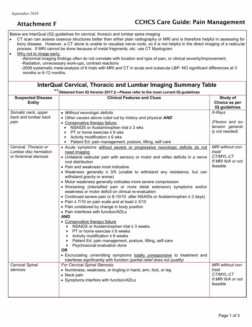
Below are InterQual (IQ) guidelines for cervical, thoracic and lumbar spine imaging
CT scan can assess osseous structures better than either plain radiography or MRI and is therefore helpful in assessing for bony disease. However, a CT alone is unable to visualize nerve roots, so it is not helpful in the direct imaging of a radicular process. If MRI cannot be done because of metal fragments, etc. use CT Myelogram
Why not to image early: -Abnormal imaging findings often do not correlate with location and type of pain, or clinical severity/improvement. -Radiation, unnecessary work-ups, contrast reactions -2009 systematic meta-analysis of 6 trials with MRI and CT in acute and subacute LBP- NO significant differences at 3 months or 6-12 months.
InterQual Cervical, Thoracic and Lumbar Imaging Summary Table **Obtained from IQ Version 2017.2—Please refer to the most current IQ guidelines
Suspected Disease Entity
Clinical Features and Clues Study of Choice as per IQ guidelines
Somatic neck, upper back and lumbar back pain
Without neurologic deficits Other causes above ruled out by history and physical AND Conservative therapy failure: NSAIDS or Acetaminophen trial ≥ 3 wks PT or home exercise ≥ 6 wks Activity modification ≥ 6 wks Patient Ed: pain management, posture, lifting, self-care
X-Rays (Flexion and ex-tension general-ly not needed)
Cervical, Thoracic or Lumbar disc herniation or foraminal stenosis
Acute symptoms without severe or progressive neurologic deficits do not need imaging
Unilateral radicular pain with sensory or motor and reflex deficits in a nerve root distribution
Pain and weakness most indicative Weakness generally ≤ 3/5 (unable to withstand any resistance, but can
withstand gravity or worse) Motor weakness generally indicates more severe compression Worsening (intensified pain or more distal extension) symptoms and/or
weakness or motor deficit on clinical re-evaluation Continued severe pain (≥ 8-10/10 after NSAIDs or Acetaminophen ≥ 3 days) Pain ≥ 7/10 on pain scale and at least ≥ 3/10 Pain unrelieved by change in body position Pain interferes with function/ADLs AND Conservative therapy failure NSAIDS or Acetaminophen trial ≥ 3 weeks PT or home exercise ≥ 6 weeks Activity modification ≥ 6 weeks Patient Ed: pain management, posture, lifting, self-care Psychosocial evaluation done
OR Excruciating unremitting symptoms totally unresponsive to treatment and
interferes significantly with function (partial relief does not qualify)
MRI without con-trast/ CT/MYL-CT If MRI N/A or not feasible
Cervical Spinal stenosis
For Cervical Spinal Stenosis:
Numbness, weakness, or tingling in hand, arm, foot, or leg
Neck pain
Symptoms interfere with function/ADLs
MRI without con-trast CT/MYL-CT If MRI N/A or not feasible
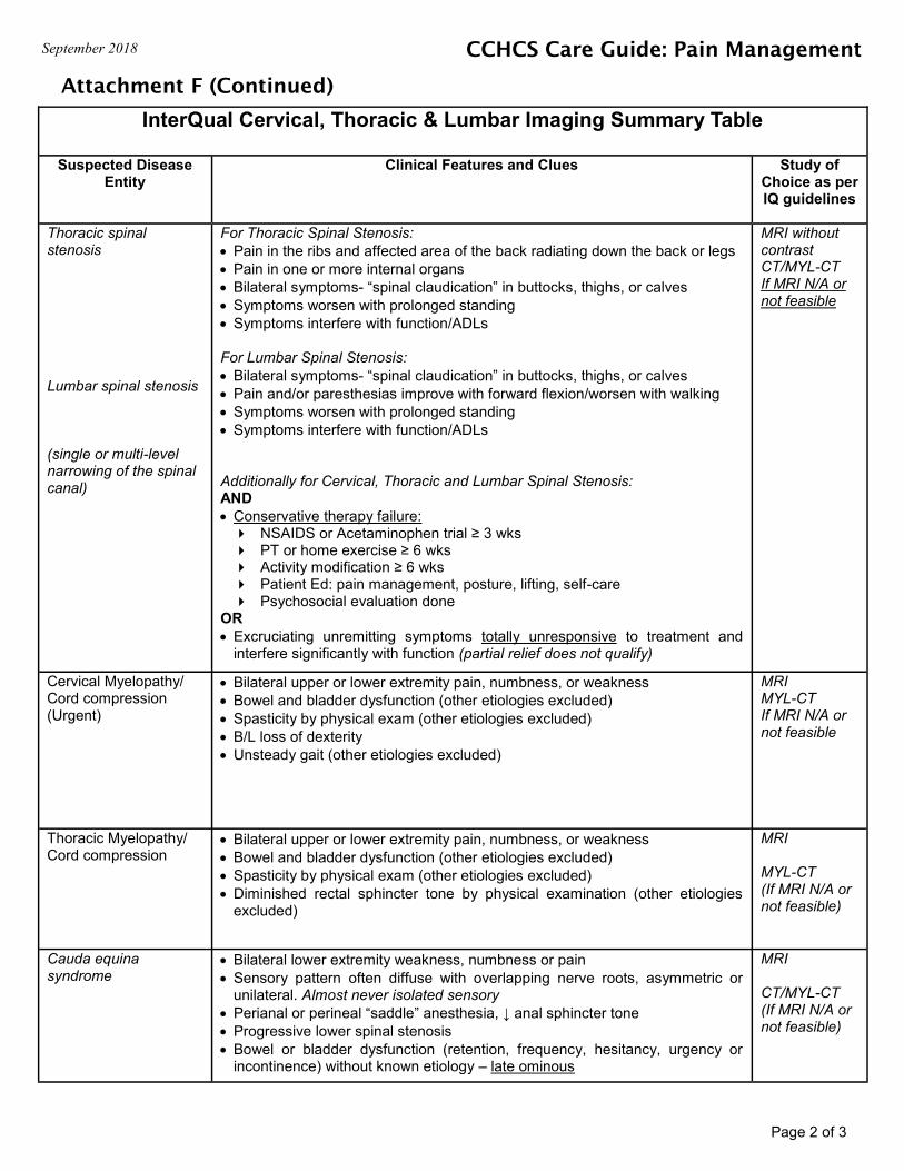
Attachment F (Continued)
September 2018 CCHCS Care Guide: Pain Management
InterQual Cervical, Thoracic & Lumbar Imaging Summary Table
Suspected Disease Entity
Clinical Features and Clues Study of Choice as per IQ guidelines
Thoracic spinal stenosis Lumbar spinal stenosis (single or multi-level narrowing of the spinal canal)
For Thoracic Spinal Stenosis:
Pain in the ribs and affected area of the back radiating down the back or legs Pain in one or more internal organs
Bilateral symptoms- “spinal claudication” in buttocks, thighs, or calves Symptoms worsen with prolonged standing Symptoms interfere with function/ADLs For Lumbar Spinal Stenosis:
Bilateral symptoms- “spinal claudication” in buttocks, thighs, or calves Pain and/or paresthesias improve with forward flexion/worsen with walking
Symptoms worsen with prolonged standing Symptoms interfere with function/ADLs Additionally for Cervical, Thoracic and Lumbar Spinal Stenosis: AND Conservative therapy failure: NSAIDS or Acetaminophen trial ≥ 3 wks PT or home exercise ≥ 6 wks Activity modification ≥ 6 wks Patient Ed: pain management, posture, lifting, self-care Psychosocial evaluation done
OR Excruciating unremitting symptoms totally unresponsive to treatment and
interfere significantly with function (partial relief does not qualify)
MRI without contrast CT/MYL-CT If MRI N/A or not feasible
Cervical Myelopathy/Cord compression (Urgent)
Bilateral upper or lower extremity pain, numbness, or weakness
Bowel and bladder dysfunction (other etiologies excluded)
Spasticity by physical exam (other etiologies excluded)
B/L loss of dexterity
Unsteady gait (other etiologies excluded)
MRI MYL-CT If MRI N/A or not feasible
Thoracic Myelopathy/Cord compression
Bilateral upper or lower extremity pain, numbness, or weakness
Bowel and bladder dysfunction (other etiologies excluded)
Spasticity by physical exam (other etiologies excluded)
Diminished rectal sphincter tone by physical examination (other etiologies excluded)
MRI MYL-CT (If MRI N/A or not feasible)
Cauda equina syndrome
Bilateral lower extremity weakness, numbness or pain Sensory pattern often diffuse with overlapping nerve roots, asymmetric or
unilateral. Almost never isolated sensory Perianal or perineal “saddle” anesthesia, ↓ anal sphincter tone Progressive lower spinal stenosis
Bowel or bladder dysfunction (retention, frequency, hesitancy, urgency or incontinence) without known etiology – late ominous
MRI CT/MYL-CT (If MRI N/A or not feasible)
Page 2 of 3
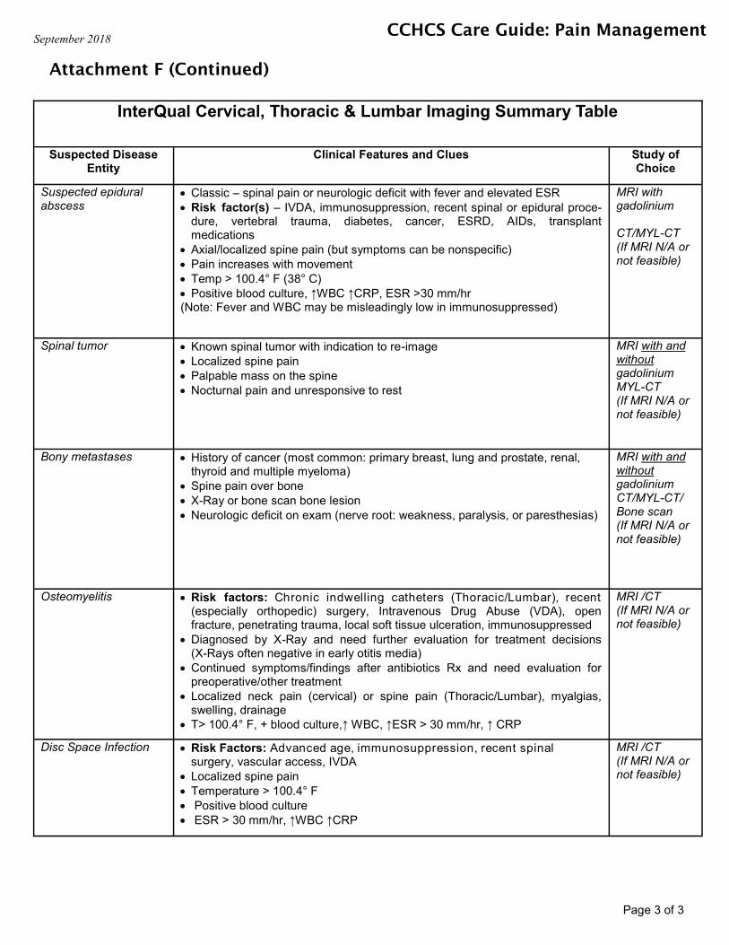
Attachment F (Continued)
September 2018 CCHCS Care Guide: Pain Management
InterQual Cervical, Thoracic & Lumbar Imaging Summary Table
Suspected Disease Entity
Clinical Features and Clues Study of Choice
Suspected epidural abscess
Classic – spinal pain or neurologic deficit with fever and elevated ESR
Risk factor(s) – IVDA, immunosuppression, recent spinal or epidural proce-dure, vertebral trauma, diabetes, cancer, ESRD, AIDs, transplant medications
Axial/localized spine pain (but symptoms can be nonspecific)
Pain increases with movement
Temp > 100.4° F (38° C)
Positive blood culture, ↑WBC ↑CRP, ESR >30 mm/hr (Note: Fever and WBC may be misleadingly low in immunosuppressed)
MRI with gadolinium CT/MYL-CT (If MRI N/A or not feasible)
Spinal tumor Known spinal tumor with indication to re-image Localized spine pain Palpable mass on the spine Nocturnal pain and unresponsive to rest
MRI with and without gadolinium MYL-CT (If MRI N/A or not feasible)
Bony metastases History of cancer (most common: primary breast, lung and prostate, renal, thyroid and multiple myeloma)
Spine pain over bone X-Ray or bone scan bone lesion Neurologic deficit on exam (nerve root: weakness, paralysis, or paresthesias)
MRI with and without gadolinium CT/MYL-CT/Bone scan (If MRI N/A or not feasible)
Osteomyelitis Risk factors: Chronic indwelling catheters (Thoracic/Lumbar), recent (especially orthopedic) surgery, Intravenous Drug Abuse (VDA), open fracture, penetrating trauma, local soft tissue ulceration, immunosuppressed
Diagnosed by X-Ray and need further evaluation for treatment decisions (X-Rays often negative in early otitis media)
Continued symptoms/findings after antibiotics Rx and need evaluation for preoperative/other treatment
Localized neck pain (cervical) or spine pain (Thoracic/Lumbar), myalgias, swelling, drainage
T> 100.4° F, + blood culture,↑ WBC, ↑ESR > 30 mm/hr, ↑ CRP
MRI /CT (If MRI N/A or not feasible)
Disc Space Infection
Risk Factors: Advanced age, immunosuppression, recent spinal surgery, vascular access, IVDA
Localized spine pain Temperature > 100.4° F Positive blood culture ESR > 30 mm/hr, ↑WBC ↑CRP
MRI /CT (If MRI N/A or not feasible)
Page 3 of 3