Embed Size (px)
Citation preview

CCAAT/Enhancer-binding Protein � DNA Binding IsAuto-inhibited by Multiple Elements That Also MediateAssociation with p300/CREB-binding Protein (CBP)*�
Received for publication, March 31, 2010, and in revised form, April 29, 2010 Published, JBC Papers in Press, May 7, 2010, DOI 10.1074/jbc.M110.128413
Sook Lee‡, Maria Miller§, Jon D. Shuman‡1, and Peter F. Johnson‡2
From the ‡Laboratory of Cancer Prevention and §Macromolecular Crystallography Laboratory, NCI-Frederick, National Institutesof Health, Frederick, Maryland 21702-1201
Signaling throughRasGTPases controls the activity ofmany tran-scription factors including CCAAT/enhancer-binding protein(C/EBP�), which regulates oncogenic H-RasV12-induced senescenceandgrowtharrest.HerewereportthatC/EBP� (LAP)DNAbindingisinhibitedbyN-terminal sequencesandderepressedbyoncogenicRassignaling. Sequence and mutational analyses showed that auto-repression involves two LXXLF (�XX��)-likemotifs (LX1 andLX2)and a third element, auto-inhibitory domain (AID), located withinconserved regionCR5.LX1 is a critical componentof the transactiva-tion domain and has been shown tomediate C/EBP� binding to theTAZ2 region of p300/CREB-binding protein coactivators. C/EBP�auto-repression also involves a C-terminal regulatory domain (CRD)adjacent to the leucine zipper. CRD contains a third �XX�� motif(LX3) and a short sequence, KQL, which has similarity to a region inthe protein-binding site of TAZ2. The C/EBP� N- and C-terminaldomains physically associate in a manner that requires the basicregion and CRD. We propose a model in which the regulatorysequences form a hydrophobic core that reciprocally inhibits DNAbinding and transactivation. We also suggest a mechanism forC/EBP� derepression involving several recently identified modifica-tions within AID and CRD. Finally, we show that association of acti-vatedC/EBP�withp300/CREB-bindingproteinrequirestheLX2andAIDauto-inhibitory elements. Thus, theN-terminal regulatory ele-ments have dual roles in auto-inhibition and coactivator binding.
Post-translational regulation of transcription factors (TFs)3is a common means of controlling gene expression in responseto extrinsic and intrinsic cellular signals (1). Several activities of
TFs, including DNA binding, dimerization, transactivation,subcellular localization, and protein stability, can be influencedby signal-dependent modifications. For example, the associa-tion of CREBwith its coactivator p300/CBP requires phosphor-ylation on Ser-133 (2), and phosphorylation of steroid receptorsregulates their DNA binding and transactivation functions (3).Stimulus-induced increases in DNA binding have beenobserved for several TFs, suggesting that their binding activityis intrinsically repressed (auto-inhibited) in the absence of anactivating signal (4). Auto-inhibition serves as a mechanism tostringently control the activity of proteins involved in criticalbiological processes such as signal transduction, proliferation,development, and tumorigenesis. In some cases, specific auto-inhibitory sequences have been identified. For instance, Gravesand colleagues (5) have shown that Ets-1 DNA binding isrepressed by inhibitory regions flanking the DNA-bindingdomain.Tumor cells frequently contain activating mutations in Ras
GTPases, growth factor receptors that signal through Ras, ordownstream kinases such as Raf. These dysregulated signals areultimately transmitted to TFs such as AP-1 that alter geneexpression and promote oncogenic transformation (6).C/EBP� is a member of the CCAAT/enhancer-binding proteinfamily of bZIP TFs (7) that has emerged as another physiologi-cal target of Ras signaling. C/EBP� can mediate either pro-oncogenic or anti-oncogenic responses to activated Ras,depending on the cellular context (8). The pro-oncogenic func-tions of C/EBP� include suppression of apoptosis (9), which insome cases has been shown to involve regulation of the growthand survival factor, IGF-1 (10). In contrast, C/EBP� has an anti-oncogenic role in primary fibroblasts where it promotes onco-gene-induced senescence (11, 12). Oncogene-induced senes-cence is a permanent state of cell cycle arrest induced byoncogenic stress, such as expression of constitutively activatedforms of Ras and Raf, and provides a barrier to tumor develop-ment (13). Oncogene-induced senescence is bypassed inCebpb�/�mouse embryonic fibroblasts (11) and in human dip-loid fibroblasts lacking C/EBP� (12), demonstrating the impor-tance of C/EBP� in cell cycle arrest and senescence.
In view of the key role C/EBP� plays in cellular responses tooncogenic and physiological Ras signals, we wish to understandthe pathways and mechanisms that control its activity. We andothers (14, 15) previously showed that C/EBP� is an intrinsi-cally repressed protein that contains two regions, termed regu-latory domains 1 and 2 (RD1 and RD2), which inhibit transac-
* This work was supported by the Intramural Research Program of theNational Institutes of Health, National Cancer Institute, Center for CancerResearch.
� This article was selected as a Paper of the Week.1 Present address: Brewton-Parker College, Mount Vernon, GA 30445.2 To whom correspondence should be addressed: Bldg. 539, Rm. 122, NCI-
Frederick, National Institutes of Health, Frederick, MD 21702-1201. Fax:301-846-5991; E-mail: [email protected].
3 The abbreviations used are: TF, transcription factor; C/EBP, CCAAT/en-hancer-binding protein; EMSA, electrophoretic mobility shift assay; bZIP,basic region-leucine zipper; DBD, DNA-binding domain; BR, basic region;LZ, leucine zipper; TAD, transactivation domain; RD, regulatory domain;CRD, C-terminal regulatory domain; TLS, TAZ2-like sequence; CBP, CREB-binding protein; CREB, cAMP-response element-binding protein; AID,auto-inhibitory domain; HA, hemagglutinin; GST, glutathione S-transfer-ase; WT, wild type; ERK, extracellular signal-regulated kinase; RSK, riboso-mal S6 kinase; MEK, mitogen-activated protein kinase/ERK kinase; aa,amino acids; m, mutant; LAP, liver-enriched transcriptional activator pro-tein; LIP, liver-enriched inhibitory protein.
THE JOURNAL OF BIOLOGICAL CHEMISTRY VOL. 285, NO. 28, pp. 21399 –21410, July 9, 2010Printed in the U.S.A.
JULY 9, 2010 • VOLUME 285 • NUMBER 28 JOURNAL OF BIOLOGICAL CHEMISTRY 21399
at National Institutes of H
ealth Library, on July 13, 2010w
ww
.jbc.orgD
ownloaded from
http://www.jbc.org/content/suppl/2010/07/01/285.28.21399.DC1.htmlSupplemental Material can be found at:

tivation. RD1 includes a conserved SUMOylation site thatinfluences the transcriptional activity of several C/EBP familymembers (15). Leutz and colleagues (16, 17) also reported thattwo phylogenetically conserved regions in chicken C/EBP�(aka NF-M), CR5 and CR7, inhibit transactivation of a reportergene and suppress NF-M-mediated activation of endogenousmyelomonocytic-specific target genes. Auto-inhibition wasovercome by expression of kinase oncogenes such as ras, raf, orv-erbB, indicating that oncogenic signals stimulate the tran-scriptional functions of C/EBP�.
Here we have extended these observations by showing thatC/EBP� DNA binding is intrinsically repressed and becomesactivated by oncogenic Ras signaling (see alsoRef. 18).We iden-tified three short sequences in the N-terminal portion ofC/EBP� that mediate auto-inhibition: a region of high hydro-phobicity and predicted secondary structure termed the “auto-inhibitory domain” (AID) that resides within CR5 and two�XX��-likemotifs (�denotes a hydrophobic residue).�XX��or LXXLL/LXXLF motifs are �-helical sequences involved ininter- and intraprotein interactions and that are critical forauto-inhibition of steroid receptors and other regulatory pro-teins (19, 20). A third �XX�� motif was identified near theC/EBP� C terminus, which is also involved in auto-repression.We propose that intramolecular interactions involving thesemotifs and the bZIP region maintain C/EBP� in a repressedstate that inhibits both DNA binding and transactivation. Wefurther show that the three N-terminal auto-inhibitory ele-ments mediate association of C/EBP� with p300/CBPcoactivators.
EXPERIMENTAL PROCEDURES
Plasmid Constructs—The G5E1b-luc reporter, pcDNA3.1expression vectors for rat C/EBP�, and GAL4-C/EBP� con-structs have been described (21). pcDNA3-H-RasV12 wasobtained from C. Der. The 2� C/EBP-Luc reporter was a giftfrom P. Rorth (Carnegie Institution of Washington) and con-tains two repeats of the consensus C/EBP-binding site, 5�-TGC-AGATTGCGCAATCTGCA-3�, upstream of the minimal thy-midine kinase promoter (22). C/EBP� LXXLF motif mutants(mLX1, mLX2, and mLX3) and other point mutants were gen-erated by site-directed mutagenesis (Stratagene). Serial C-ter-minal deletions (STOP mutants) were generated by placing atermination codon at each repeated leucine position in theleucine zipper. pcDNA3-CBP1680–2441-2�FLAG (23) waskindly provided by J.-R. Cardinaux. p300 constructs wereobtained from Addgene: pCMVb-HA-p300 (Addgene ID10718; W. Sellers) and TAZ2 mutant d33 (24) (Addgene ID10719).Cell Transfection and Preparation of Lysates—HEK293T
cells were maintained in Dulbecco’s modified Eagle’s mediumwith 10% fetal bovine serum. 1.5 � 105 cells were cultured in60-mm plates for 24 h and transfected with 500 ng ofpcDNA3.1-C/EBP� without or with 100 ng of H-RasV12 plas-mid using 2 �l of FuGENE (Roche Applied Science) per �g ofDNA. After transfection, cells were cultured in completemedium for 24 h and serum-starved overnight prior to harvest,and nuclear extractswere prepared for electrophoreticmobilityshift assay (EMSA). C/EBP� levels were analyzed by immuno-
blotting using antibody C-19 (Santa Cruz Biotechnology). Fortransactivation assays, cells were transfected with 100 ng of 2�C/EBP-luc or G5E1b-luc reporter plasmid and 10 ng of C/EBP�or GAL4-C/EBP� vector, without or with 10 ng of RasV12 plas-mid. Where appropriate, 50 ng of pCMVb-HA-p300 wasincluded. After overnight starvation, cells were lysed in 1� pas-sive lysis buffer, and lysates were analyzed using the luciferaseassay system (Promega). Luciferase activity was normalized tototal protein in the lysates.EMSAs—The EMSA probe was a double-stranded oligonu-
cleotide containing a consensus C/EBP-binding site (under-lined) (5�-GATCCATATCCCTGATTGCGCAATAGGCTCA-AAA-3�) labeled with [�-32P]ATP and T4 polynucleotidekinase. The probewas incubatedwith nuclear extract in a 25-�lreaction containing 20 mM HEPES, pH 7.5, 200 mM NaCl, 5%Ficoll, 5 mM dithiothreitol, 5 mM EDTA, 1 �g of poly[d(I-C)], 1�g of bovine serum albumin, and 4 �l of radioimmune precip-itation buffer at room temperature for 30 min. DNA�proteincomplexes were separated on 6% polyacrylamide/1� Tris-bo-rate-EDTA gels.Recombinant Proteins—His-tagged C/EBP� constructs con-
taining a tobacco etch virus cleavage site after the His6 tag (25)were cloned using Gateway recombination technology(Invitrogen). After isopropyl-1-thio-�-D-galactopyranosideinduction in Escherichia coli, cells were collected and resus-pended in lysis buffer (20mMTris�HCl, pH 6.8, 150mMNaCl, 1mM EDTA, 1 mM benzamidine, 0.2 mM phenylmethylsulfonylfluoride, 1�g/ml antipain, 0.1%�-mercaptoethanol, 0.5%Non-idet P-40) containing 50�g/ml lysozyme. Aftermild sonicationand centrifugation, the pellet containing insoluble C/EBP� wasresuspended in solubilization buffer (5 M urea, 50mMTris�HCl,pH 8.0, 0.1% �-mercaptoethanol, 1 mM benzamidine, 0.2 mM
phenylmethylsulfonyl fluoride, 5 �g/ml antipain) and rotatedovernight. The solubilized protein was clarified and bound to anickel-nitrilotriacetic acid column (Promega). The columnwaswashed serially with solubilization and washing buffer (5 M
urea, 50 mM Tris�HCl, pH 8.0, 0.1% �-mercaptoethanol, 5 mM
imidazole, 5 �g/ml antipain). Protein was eluted with 50 mM
Tris�HCl, pH 6.5, 0.3 M NaCl, 0.1% �-mercaptoethanol,0.5 M imidazole and dialyzed against 50 mM Tris�HCl, pH 8.0,0.3 M NaCl, 0.1% �-mercaptoethanol, 50 mM imidazole, with0.5 M arginine included to prevent aggregation. 25 �g/ml puri-fied tobacco etch virus protease (25) was added to the dialyzedeluate and incubated overnight at room temperature, afterwhich the sample was applied to a nickel-nitrilotriacetic acidcolumn three times to remove theHis6 fragment. Tobacco etchvirus-cleaved C/EBP� in the flow-through fraction was dia-lyzed against 50 mM Tris�HCl, pH 8.0, 200 mM NaCl, 5% glyc-erol, 1mMdithiothreitol for 2 h at room temperature, applied toa MonoS fast protein liquid chromatography column, andeluted using a linear NaCl gradient. N-terminal C/EBP� frag-ments 22–104 and 22–192 were prepared similarly except thatMonoQ chromatography was used for the final purificationstep and the dialysis/loading buffer contained 10 mM NaCl.GST Fusion Proteins and Pulldown Assays—The Gateway
recombination system was used for cloning of GST fusion pro-teins. Isopropyl-1-thio-�-D-galactopyranoside-induced bacte-rial cell pellet was resuspended in 10 mM Tris�HCl, pH 8.0, 150
C/EBP� Auto-inhibition
21400 JOURNAL OF BIOLOGICAL CHEMISTRY VOLUME 285 • NUMBER 28 • JULY 9, 2010
at National Institutes of H
ealth Library, on July 13, 2010w
ww
.jbc.orgD
ownloaded from
http://www.jbc.org/content/suppl/2010/07/01/285.28.21399.DC1.htmlSupplemental Material can be found at:

mM NaCl, 1 mM EDTA with protease inhibitors and incubatedwith 100 �g/ml lysozyme for 15 min on ice. After adding 5 mM
dithiothreitol and 1.5% sarcosyl(N-lauroylsarcosine), the lysatewas sonicated for 1 min (six pulses for 10 s with a 20-s rest).After addingTritonX-100 to a final concentration of 2–4%, thesolubilized GST protein was immobilized on glutathione-agar-ose beads (BD Biosciences). For pulldown assays, GST orGST�DBD beads (normalized for the amount of GST protein)were incubated with purified C/EBP� N-terminal fragments(22–104 or 22–192) in binding buffer (20mMHEPES, pH7.5, 50mMKCl, 20�MZnSO4, 10% glycerol, and 0.1%TritonX-100) atroom temperature for 2 h. After washing six times with bindingbuffer, the bound material was eluted in sample buffer, sepa-rated by 16%SDS-PAGE, and transferred to Immobilon P. Blotswere developed using an antibody against the N terminus ofp34 C/EBP� (LAP) (26). The membranes were subsequentlystained with Ponceau S to verify equal loading of GST proteins.Synthetic Peptides and Recombinant TAZ2 Fragment—Puri-
fied human p300 TAZ2 polypeptide has been described (27).The C-terminal regulatory domain (CRD) peptide correspondsto aa 273–296 of rat C/EBP� and was purchased from GlobalPeptide Services; p53 peptide (aa 9–25 of human p53) was a giftfrom E. Appella.p300/CBP Binding Assays—C/EBP� (WT, mLX1, mLX2,
�AID) and p300/CBP (FLAG-tagged CBP1680–2441, HA-taggedp300 WT, and d33) were expressed separately in 293T cells.Nuclear extracts were prepared, diluted in binding buffer (20mMHEPES, pH 7.9, 100mMNaCl, 0.1mMEDTA, 0.1%NonidetP-40), and mixed for 2 h at 4 °C prior to immunoprecipitationwith anti-FLAG M2-agarose (Sigma) or mono HA.11 affinitymatrix (Covance) overnight. The beads were washed six timeswith binding buffer and boiled in sample buffer, and the eluatesanalyzed for C/EBP� by Western blotting.In Silico Studies—The C/EBP� primary structure was ana-
lyzed using several web-based servers available via the ExPASysite. Assessment of secondary structure and solvent accessibil-ity for individual residues in the sequencewas performed by thePHDmethod (28) included in the Predict-Protein package (29).The hydrophobic profile was obtained using ProtScale (30) runwith the default window size (5 residues). Pre-Link (31) wasused to predict flexible linker regions. Hydrophobic clusteranalysis (32) uses a representation of protein sequence on an�-helical two-dimensional pattern to better visualize structuralfeatures and the environment of each amino acid in the proteinsequence. Sets of adjacent hydrophobic residues in the patternare termed hydrophobic clusters. The shapes of these clustersand their distribution along the sequence provide usefulguidance for predictions of regular secondary structure ele-ments and protein tertiary fold (reviewed in Ref. 33).
RESULTS
C/EBP� DNA Binding Is Auto-repressed by N-terminal Se-quences and Becomes Activated by Oncogenic Ras Signaling—We recently reported that DNA binding of the major C/EBP�isoform, p34 LAP (aa 22–297), is suppressed and becomes stim-ulated by oncogenic Ras signaling (18). To further investigateregulation of C/EBP� DNA binding, we addressed the mecha-nism of auto-inhibition. C/EBP� was expressed in HEK293T
cells without or with H-RasV12, and nuclear extracts normal-ized for C/EBP� levels were analyzed by EMSA using a consen-sus C/EBP site probe. As shown in Fig. 1A, Ras induced a sig-nificant increase inC/EBP�DNAbindingwhen comparedwithits activity without Ras. The shifted protein species in lane 2 ofthe Western blot corresponds to phospho-Ser-64 (21), whichdoes not regulate DNA binding, but its presence confirmsC/EBP� activation by Ras signaling. The binding activity ofendogenous C/EBP� in primary mouse fibroblasts is alsoinduced by H-RasV12 (18), showing that stimulation of C/EBP�DNA binding is a physiological response to Ras.A truncated protein containing only the C-terminal bZIP
DNA-binding domain (DBD) displayed much higher basalactivity that was not further stimulated by Ras (Fig. 1A, lanes 5and 6). Thus, C/EBP� DNA binding in unstimulated cells isconstrained by sequences located in theN-terminal region, andthis auto-repression is overcome by Ras signaling. A constructcontaining the N-terminal transactivation domain (TAD; aa22–104) fused to theDBD (aa 212–297), denoted 22–104/DBD,was also partially inhibited and derepressed by Ras (lanes 3 and4). Thus, the TAD region itself has inhibitory effects on DNAbinding. Transactivation assays using a C/EBP-driven reporter(2� C/EBP-Luc) showed that C/EBP� (LAP) possesses lowbasal transcriptional activity that is strongly increased (�20-fold) by RasV12 (Fig. 1B). 22–104/DBD displayed higher basaltransactivation, and its Ras-induced activity was also increasedrelative to LAP, whereasDBDdid not appreciably activate tran-scription because it lacks a TAD.To provide additional evidence for intrinsic repression of
C/EBP�, we analyzed DNA binding of purified, bacteriallyexpressed proteins (Fig. 1C). C/EBP� (LAP) binding was unde-tectable, whereas 22–104/DBD binding was low but measura-ble, and DBD exhibited very high activity. These results aresimilar to those observed for the same proteins expressed inmammalian cells without Ras, strongly suggesting that lowbasal DNA binding is due to intrinsic inhibition by the N-ter-minal region.To further define C/EBP� auto-inhibitory sequences, we
analyzed a set of N-terminal deletion mutants (Fig. 2). 64–297,which lacks the N-terminal part of the TAD, was auto-re-pressed and was activated by Ras similarly to WT LAP (com-pare lanes 1–4). However, 102–297 (lacking the entire TAD)showed increased basal activity that was further stimulated byRas. The enhanced basal activity observed upon removal of aa64–102 indicates the presence of a repressive sequence withinthis interval. Deletion to aa 122 further derepressed basal activ-ity, revealing another inhibitory sequence in the 102–122interval.Figs. 1A and 2 demonstrate that at least two elements are
involved in auto-repression because sequences both N-termi-nal and C-terminal to aa 104 confer partial inhibition. Interest-ingly, the naturally occurring LIP isoform (153–297) displayednearly undetectable binding activity and was refractory to Rasactivation (Fig. 2, lanes 9 and 10). Thus, LIP apparently containsan auto-inhibitory element but lacks sequences required foractivation by Ras. The differential regulation of LAP and LIPDNA binding was consistently observed and is currently underfurther investigation. Removal of sequences to aa 170 resulted
C/EBP� Auto-inhibition
JULY 9, 2010 • VOLUME 285 • NUMBER 28 JOURNAL OF BIOLOGICAL CHEMISTRY 21401
at National Institutes of H
ealth Library, on July 13, 2010w
ww
.jbc.orgD
ownloaded from
http://www.jbc.org/content/suppl/2010/07/01/285.28.21399.DC1.htmlSupplemental Material can be found at:
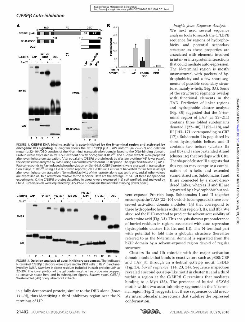
in a fully derepressed protein, similar to the DBD alone (lanes11–14), thus identifying a third inhibitory region near the Nterminus of LIP.
Insights from Sequence Analysis—We next used several sequenceanalysis tools to search the C/EBP�sequence for regions of hydropho-bicity and potential secondarystructure as these properties areassociated with elements involvedin inter- or intraprotein interactionsthat could mediate auto-repression.The N-terminal region is mostlyunstructured, with pockets of hy-drophobicity and a few short seg-ments of possible secondary struc-ture, mainly �-helix (Fig. 3A). Someof the structured segments overlapwith functional elements in theTAD. Prediction of linker regionsand hydrophobic cluster analysis(Fig. 3B) suggested that the N-ter-minal region of LAP (aa 22–211)contains three folded subdomainsdenoted I (22–40), II (52–118), andIII (143–171, corresponding to CR7(17)). Subdomain I is populated byshort hydrophobic helices, and IIcontains two helices (clusters IIaand IIb) and a short extended strand(cluster IIc) that overlaps with CR5.The shape of cluster III suggests thatthis region may comprise a combi-nation of �-helix and extendedstrand structure. Subdomains I andII are connected by a short disor-dered linker, whereas II and III areseparated by a hydrophobic but sol-
vent-exposed Pro-rich loop. Subdomains I and II togetherencompass theTAD (22–104), which is composed of three con-served activation domain modules (14) that correspond tothree hydrophobic heliceswithin this region (I, IIa, and IIb).Wealso used the PHDmethod to predict the solvent accessibility ofeach amino acid (Fig. 3A). This analysis shows a preponderanceof buried residues in regions associated with auto-repression(hydrophobic clusters IIb, IIc, and III). The N-terminal partwith potential to fold into a globular structure (hereafterreferred to as the N-terminal domain) is separated from thebZIP domain by a solvent-exposed region devoid of regularstructure.Clusters IIa and IIb coincide with the major activation
domain module that binds to coactivators such as p300/CBPand TAFII31 through an �-helical �XX�� motif, LSDLF(Fig. 3A, boxed sequence) (14, 23, 34). Sequence inspectionrevealed a second �XX��-like motif in cluster III and a thirdwithin a region at the C/EBP� C terminus that mediatesbinding to c-Myb (35). The presence of buried �XX��motifs within two auto-inhibitory segments in the N-termi-nal region (Fig. 2) suggests that these sequences could medi-ate intramolecular interactions that stabilize the repressedconformation.
FIGURE 1. C/EBP� DNA binding activity is auto-inhibited by the N-terminal region and activated byoncogenic Ras signaling. A, diagram shows the rat C/EBP� p34 (LAP) isoform (aa 22–297) and deletionmutants; 22–104/DBD consists of the N-terminal transactivation domain fused to the DNA-binding domain.Proteins were expressed in 293T cells without or with oncogenic H-RasV12, and nuclear extracts were preparedafter overnight serum starvation. After equalizing C/EBP� protein levels by Western blotting (WB, lower panel),the extracts were analyzed by EMSA using a radiolabeled consensus C/EBP probe. The upper band in lane 2 (LAP �Ras) corresponds to Ras-induced phosphorylation on Ser-64. B, C/EBP� proteins were analyzed in transactiva-tion assays � RasV12 using a C/EBP-driven reporter, 2� C/EBP-luc. Cells were harvested for luciferase assaysafter overnight serum starvation. Normalized activity of the reporter alone was set to one, and all other valuesare expressed as -fold activation relative to the reporter. Data are the average (� S.E.) of three independentexperiments. C, the C/EBP� proteins described in panel A were expressed in E. coli, purified, and analyzed byEMSA. Protein levels were equalized by SDS-PAGE/Coomassie Brilliant Blue staining (lower panel).
FIGURE 2. Deletion analysis of auto-inhibitory sequences. The indicatedN-terminal C/EBP� deletions were expressed in 293T cells � RasV12 and ana-lyzed by EMSA. Numbers indicate residues included in each protein; LAP, aa22–297. The lower portion of the gel containing the free probe was croppedto conserve space here and in subsequent figures. Bottom panel, C/EBP�Western blot (WB) of equalized cell extracts.
C/EBP� Auto-inhibition
21402 JOURNAL OF BIOLOGICAL CHEMISTRY VOLUME 285 • NUMBER 28 • JULY 9, 2010
at National Institutes of H
ealth Library, on July 13, 2010w
ww
.jbc.orgD
ownloaded from
http://www.jbc.org/content/suppl/2010/07/01/285.28.21399.DC1.htmlSupplemental Material can be found at:

Mutational Analysis of Auto-inhibitory Motifs—To assessthe roles of the putative protein interaction elements in auto-repression,we generated site-directedmutations.Hydrophobicresidues at positions 1, 4, and 5 of the �XX�� motifs in theN-terminal region (LX1, aa 88–92; LX2, aa 161–165) were con-verted to Ala, and hydrophobic cluster IIc (aa 108–114),termed the AID, was deleted (Fig. 4A). mLX1 and mLX2 wereconstructed singly and in tandem, and �AID was created aloneor with mLX1/2. Each of the single mutations caused a modestincrease in basal DNA binding, with mLX2 having the largesteffect, but they did not affect the Ras-induced binding (Fig. 4A).The double mutant mLX1/2 was slightly more active than
mLX2. Notably, the mLX1/2�AIDtriple mutant was fully derepressedand was not further activated byRas. Thus, the LX1, LX2, and AIDmotifs mediate auto-inhibition bythe N-terminal region.Transactivation assays showed
that mutants containing mLX1were very poor transcriptional acti-vators, as expected because LX1 is acritical component of the TAD (Fig.4B). The Ras-stimulated activity ofmLX2 was nearly 3-fold higher thanWT LAP, and �AID was at least4-fold greater. Plotting the lucifer-ase data on a different scale (inset)shows that these twomutations alsoenhanced basal activity. However,Ras-induced transcription was in-variably much higher than the basalvalue, even for mutants whoseunstimulated DNA binding activitywas significantly derepressed. Thissuggests that Ras signaling alsoenhances the transcriptional poten-tial of C/EBP� independently ofDNA binding.We next analyzed DNA binding
of purified recombinant proteinscontaining a subset of the �XX��-like (LX) and AID mutations (Fig.4C). mLX2 had a small enhancingeffect, whereas mLX1/2 furtherincreased DNA binding, and theactivity of the triple mutant waseven higher. Thus, the results of Fig.4, A–C, show that C/EBP� is intrin-sically repressed by three elementslocated in the N-terminal half of theprotein.
�XX�� �-helices often includecharged residues that contribute tothe specificity and stability of inter-actions with binding partners (36).LX1 is flanked by 3 Asp residuesthat form a negatively charged edge
on the hydrophobic core (Figs. 3B and 4D). To investigate thefunction of these residues in auto-repression, we analyzedAsp3Arg substitutionmutants (D86R and D94R/D95R). Bothmutants were auto-repressed; however, binding by the D86Rmutant was only weakly activated by Ras, whereas D94R/D95Rwas completely resistant to activation (Fig. 4D). Transactiva-tion by both of these mutants was also severely decreased (Fig.4E). Thus, negatively charged residues flanking LX1 are notcritical for auto-inhibition but play an important role inC/EBP� activation byRas signaling, in agreementwith previousdata (37). The reduction in Ras-induced transcriptional activityby these mutants probably involves not only decreased DNA
FIGURE 3. In silico analysis of the C/EBP� sequence. A, selected features of the rat C/EBP� amino acidsequence. Stretches of residues characterized by normalized hydrophobicity � 0.5 (obtained with ProtScale(30)) are highlighted in green. Functional domains are indicated by colored bars: blue, TAD; yellow, BR; red, LZ.Predictions of secondary structure (H, helix; E, extended strand) and relative solvent accessibility (e, residuesexposed with more than 36% of their surface; b, residues with less than 9% of surface exposed) as determinedby the PHD method (28) are shown below the sequence. Boxes indicate �XX��-like motifs. Boundaries of theCR5/7 and RD1/2 auto-inhibitory domains are depicted. B, two-dimensional helical representation of the LAPN-terminal region (hydrophobic cluster analysis plot). The sequence is displayed on a cylinder representing an�-helix with 3.6 amino acids per turn. The cylinder is then cut parallel to its axis, unrolled, and duplicated tobetter visualize the environment of each amino acid. Hydrophobic residues are colored green; acidic residuesare red; and basic residues are blue. Red star, prolines; diamond, glycines; open square, threonines; dotted square,serines. Hydrophobic clusters are encircled. Brackets below the hydrophobic cluster analysis plot indicate bor-ders of the putative folded regions (designated I, II (a– c), and III) defined by the PreLink program (31).
C/EBP� Auto-inhibition
JULY 9, 2010 • VOLUME 285 • NUMBER 28 JOURNAL OF BIOLOGICAL CHEMISTRY 21403
at National Institutes of H
ealth Library, on July 13, 2010w
ww
.jbc.orgD
ownloaded from
http://www.jbc.org/content/suppl/2010/07/01/285.28.21399.DC1.htmlSupplemental Material can be found at:

binding but also impaired interactions with coactivators (e.g. inthe p300 TAZ2�p53 complex, p53 Glu-17, which is equivalentto C/EBP� Asp-86, forms a salt bridge interaction with TAZ2Arg-1731 (38)).Identification of Sequences in the bZIP Region Mediating
Auto-inhibition—To identify elements in the DBD that partic-ipate in auto-inhibition, we generated a nested set of deletionsthat remove sequences from the C terminus and the end of theLZ (Fig. 5A). Deletion of aa 286–297 (STOP6) had little effect
onDNAbinding. Resection to aa 279 (STOP5) led to reproduc-ibly higher basal activity, implicating sequences between aa 279and 286 in auto-repression. A protein ending at residue 272(STOP4) displayed very low basal and Ras-induced bindingactivity, and further deletion completely eliminatedDNAbind-ing (data not shown). The latter results are probably due todisruption of LZ function and loss of dimerization. The CRDidentified by these deletions also mediates cooperative interac-tions with c-Myb (35). We noted that it contains a �XX��
FIGURE 4. Mutational analysis of putative auto-inhibitory motifs. A, proposed regulatory elements and their mutant versions are depicted in the diagram.Two �XX�� motifs and the AID were mutated by Ala substitution (mLX1, mLX2) or deletion (�AID), singly and in combination. Mutant proteins were expressedin 293T cells and tested by EMSA for basal and Ras-induced DNA binding. The mLX1 mutation consistently reduces the mobility of C/EBP� in SDS-PAGE gels (seealso panel C and Fig. 6C), most likely due to a conformational change. WB, Western blot. B, mutants were analyzed in transactivation assays using 2� C/EBP-Lucreporter. The inset shows the basal activities graphed on a different scale. C, the indicated proteins were expressed in E. coli, purified, and tested for DNAbinding by EMSA. Bottom, protein levels used for EMSA. D and E, effects of mutating acidic residues flanking LX1. Mutants depicted in the diagram were assayedfor DNA binding (panel D) and transactivation (panel E) in 293T cells. Results of three independent assays were averaged (� S.E.).
C/EBP� Auto-inhibition
21404 JOURNAL OF BIOLOGICAL CHEMISTRY VOLUME 285 • NUMBER 28 • JULY 9, 2010
at National Institutes of H
ealth Library, on July 13, 2010w
ww
.jbc.orgD
ownloaded from
http://www.jbc.org/content/suppl/2010/07/01/285.28.21399.DC1.htmlSupplemental Material can be found at:

motif (LX3) as well as an amino acid triplet, KQL, which is alsopresent in the TF-binding site of p300/CBP TAZ2. We namedthis element “TAZ-like sequence” or TLS (Fig. 5A, diagram).The region encompassing LX3 and TLS shows similar hydro-phobicity to theTAZ2helix thatmediates p300/CBPbinding tothe p53 TAD and other TFs (Ref. 39 and references therein),
including to C/EBP� via LX1. Significantly, Gln of the p300KQL motif forms a buried hydrogen bond with aspartic acidfrom the p53 TAD “signature helix” (FSDLW) (38). Therefore,by analogy to the p53-TAZ2 interaction, we hypothesized thatthe C/EBP� C terminus may participate in binding to N-termi-nal inhibitory sequences such as LX1 (LSDLF).
FIGURE 5. Identification of bZIP sequences mediating auto-inhibition. A, effects of mutations in putative C-terminal regulatory elements. The diagramshows the sequence of the C-terminal region indicating LX3 and the TLS and mutations in these elements and an alignment of the C/EBP� C terminus and theTAZ2 domains of human p300 and mouse CBP. STOP mutants contain termination codons at the repeated Leu residues, creating nested C-terminal deletions.DNA binding (left) and transactivation assays (right) were performed. WB, Western blot. The inset shows basal activity on a different scale. B, effect of aheterologous LZ on C/EBP� activation. C/EBP�-GLZ is a chimeric LAP protein containing the GCN4 leucine zipper; GLZ-(170 –297) is a truncated version. Left,DNA binding; right, transactivation assays of C/EBP� and C/EBP�-GLZ. C, mutations in the bZIP domain were introduced into 22–104/DBD fused at its Nterminus to GAL4 DBD. Constructs were co-transfected � RasV12 with a GAL4-dependent reporter, G5E1b-luc. GAL4-(22–104) contains the C/EBP� TAD alonefused to GAL4. Data are the average (�S.E.) of three independent experiments.
C/EBP� Auto-inhibition
JULY 9, 2010 • VOLUME 285 • NUMBER 28 JOURNAL OF BIOLOGICAL CHEMISTRY 21405
at National Institutes of H
ealth Library, on July 13, 2010w
ww
.jbc.orgD
ownloaded from
http://www.jbc.org/content/suppl/2010/07/01/285.28.21399.DC1.htmlSupplemental Material can be found at:

Mutation of LX3 and TLS caused a modest but reproducibleincrease in basal DNA binding and enhanced activation by Ras(Fig. 5A, lanes 9–12), indicating a role in auto-inhibition. TheSTOP5, mLX3, and mTLS mutants showed similar derepres-sion when introduced into 22–104/DBD (data not shown). Wefurther examined the role of the LZ region by analyzing a chi-meric protein containing the GCN4 LZ (C/EBP�-GLZ) (14).C/EBP�-GLZ DNA binding was completely repressed and wasrefractory to Ras activation (Fig. 5B, lanes 1 and 2); transactiva-tion was similarly impaired. The reduced activity of C/EBP�-GLZ is not due to an inability of the chimeric DBD to bind DNAbecause an N-terminally truncated version of this protein(170–297) showed robust DNA binding (lanes 3 and 4). Weconclude that the GCN4 LZ contains sequences capable ofmaking inhibitory interactions with the C/EBP� N-terminalregion but lacks elements required for activation, such as theRSK phosphorylation site (Ser-273) recently identified in theC/EBP� LZ (18).Previous studies showed that the C/EBP� TAD is inhibited
by the bZIP region and by central regulatory domains (14, 16).To further investigate the basis for this inhibition and to iden-tify repressive sequences in the bZIP domain, we fusedWT andmutant 22–104/DBD constructs to the GAL4 DBD and ana-lyzed the ability of these chimeras to activate a GAL4 reportergene (Fig. 5C). GAL4-(22–104/DBD) activity was auto-re-pressed and was stimulated nearly 30-fold by Ras, much likenative C/EBP� (LAP), whereas GAL4-(22–104) displayed con-stitutively high activity. GAL4-LAP was also repressed andcould be activated by Ras (data not shown). Thus, the bZIPregion inhibits the TAD, and this repression can be reversed byRas signaling. Deletion of the basic region in GAL4-(22–104/DBD) caused a 5-fold increase in basal transactivation and a3-fold increase in Ras-induced activity when compared withWT. Therefore, the BR plays a critical role in auto-inhibitoryinteractions between the TAD and DBD. Mutating LX3 orTLS also slightly increased basal activity. Interestingly, Ras-induced transcription by these mutants was decreased rela-tive to GAL4-(22–104/DBD), in contrast to the increasedDNA binding and unaltered transactivation observed for thesame mutations in native C/EBP� (Fig. 5A). These findingssuggest that the auto-inhibitory LX3 and TLS elements canalso play a positive role in transcription depending on thepromoter context.Physical Interactions Involving the N-terminal Auto-inhibi-
tory Domain and the DBD—We next used GST pulldownassays to assess physical interactions between N-terminalsequences and the DBD (bZIP region). For these experiments,we analyzed binding of GST�DBD to two N-terminal C/EBP�polypeptides, 22–104 and 22–192. Both fragments bound toGST�DBD but not to GST alone (Fig. 6A). The addition of var-ious divalent cations to the pulldown reaction showed that22–104 binding to GST�DBD was relatively unaffected by theaddition of Ca2�, Mg2�, or Zn2�, whereas 22–192 binding wasstrongly enhanced by Zn2� (Fig. 6B). It is possible that Zn2�
ions enhance auto-inhibitory interactions by cross-linking res-idues from 104–192 to the bZIP region. However, the molecu-lar/structural basis for the Zn2� effect and the divalent cationspecificity remains to be determined.
To determine whether the auto-inhibitory motifs arerequired for binding of the N-terminal sequences to theDBD, we analyzed several mutants. Introduction of mLX1into 22–104 prevented its association with GST�DBD, andthe mLX1/mLX2/�AID 22–192 mutant also showed greatlyreduced binding (Fig. 6C). Thus, sequences required forC/EBP� auto-repression mediate physical association withthe DBD region. We next analyzed mutant versions ofGST�DBD to determine which sequences in the bZIP domainare required for interaction with the N terminus. Deletion ofthe basic region strongly impaired binding to 22–192 (Fig.6D), whereas STOP5, mLX3, and mTLS also reduced theassociation, albeit less severely. Because RSK-mediatedphosphorylation of Ser-273 in the LZ is required for Ras-induced DNA binding (18), we examined S273A and S273Dmutations. The S273A mutation had little effect on bindingto 22–192 (lane 5), whereas the phosphomimetic S273D sub-stitution decreased this interaction (lane 6). These findingssupport the idea that Ser-273 phosphorylation disruptsintramolecular interactions with the N-terminal region thatmediate auto-inhibition.Our results suggest that C/EBP� auto-repression involves
contacts between the CRD region and the TAD LX1 motif,which also binds to CBP/p300 via TAZ2. If this model iscorrect, auto-inhibition should be disrupted by competitivebinding of peptides that interact with LX1. To test this pos-sibility, we incubated purified TAZ2 domain of p300 (27)with bacterially expressed 22–104/DBD and analyzed DNAbinding. Binding was strongly enhanced by TAZ2, whereas acontrol p53 peptide had no effect (Fig. 7A, top panel). Dere-pression was also induced by a synthetic CRD peptide fromC/EBP� that encompasses LX3 and TLS. In contrast, DBDDNA binding was not stimulated by any of the peptides,ruling out nonspecific effects (Fig. 7A, bottom panel). TheTAZ2 and CRD peptides also stimulated the binding ofrepressed C/EBP� (LAP) expressed in 293T cells, whereasthe p53 peptide had no effect (Fig. 7B). Again, binding of theconstitutively active DBD protein was unaffected by thecompetitor peptides. These data further support the ideathat LX1 interacts with sequences at the C terminus toinhibit C/EBP� activity.C/EBP� Binding to p300/CBP Coactivators Requires the
N-terminal Auto-inhibitory Elements—C/EBP� was reportedto interact with p300/CBP coactivators via LX1 (34, 40, 41).Subsequently, the region C-terminal to the TAD was shown toalso have a role in CBP binding (23). Because AID and LX2 arewithin this region and exhibit features of protein interactiondomains, we hypothesized that they might participate in asso-ciationwith p300/CBP. Therefore, we used co-immunoprecipi-tation to assess binding ofWT andmutantC/EBP� proteins to aC-terminal segment of CBP encompassing TAZ2 (CBP1680–2441)(23). WT C/EBP� was expressed without or with RasV12 in293T cells, and extracts were incubated with a lysate from cellsexpressing FLAG-CBP1680–2441. Following immunoprecipita-tion with FLAG antibody, C/EBP� in the bound fraction wasanalyzed by Western blotting (Fig. 8A). Efficient binding toCBP1680–2441 occurred onlywhenC/EBP�was expressed in thepresence of RasV12 (compare lanes 1 and 2), demonstrating that
C/EBP� Auto-inhibition
21406 JOURNAL OF BIOLOGICAL CHEMISTRY VOLUME 285 • NUMBER 28 • JULY 9, 2010
at National Institutes of H
ealth Library, on July 13, 2010w
ww
.jbc.orgD
ownloaded from
http://www.jbc.org/content/suppl/2010/07/01/285.28.21399.DC1.htmlSupplemental Material can be found at:

activated C/EBP� is uniquely capable of making this associa-tion. Notably, each of the auto-inhibitory domain mutants(mLX1, mLX2, �AID) expressed with Ras displayed greatlyreduced CBP binding (lanes 3–5). Identical results wereobtained using HA-tagged p300; i.e. interaction with C/EBP�required activation by Ras, and the auto-inhibitory domainmutants failed to bind p300. Furthermore, a p300 mutant lack-ing critical sequences in the TAZ2 domain (p300 d33) (24) dis-played reduced binding to WT C/EBP�, confirming that asso-ciation with p300 involves TAZ2.These findings were further corroborated in transactivation
assays (Fig. 8B). Co-expression of p300 stimulated C/EBP�-driven transcription of the 2� C/EBP-luc reporter by �2-fold(p � 0.02). This augmentation occurred only in the presence ofRas signaling and did not affect basal C/EBP� activity. How-ever, none of the N-terminal auto-inhibitory domain mutantsshowed significant stimulation by p300 despite increased Ras-induced transcriptional activity ofmLX2 and�AIDdue to theirimpaired auto-inhibition (see also Fig. 4B). Collectively, thedata of Fig. 8, A and B, strongly suggest that activated C/EBP�
binds to p300/CBP via the LX1, LX2, and AID elements, whichalso play critical roles in auto-repression.
DISCUSSION
The involvement of C/EBP� in cellular responses to onco-genic Ras or Raf (9–12) suggested that its activity may be reg-ulated by Ras signaling. Previous work demonstrated that thetranscriptional potential of C/EBP� is stimulated by activatedRas or other kinase oncogenes (9, 16, 42). In the present study,we have extended these findings and show that C/EBP� DNAbinding activity is auto-repressed and becomes activated incells expressing oncogenic RasV12. Earlier studies identifiedauto-inhibitory regions CR5/7 (16) and RD1/2 (14) and roughlymapped their boundaries based on sequence conservation ordeletion analysis. Here we show that three distinct N-terminalsequences that are predicted to form secondary structuresinhibit C/EBP� DNA binding. These include AID, which islocated within CR5, and two LX motifs. In addition, the LX3and TLS motifs in the CRD region contribute to auto-inhibi-
FIGURE 6. C/EBP� auto-inhibitory sequences physically interact with the bZIP domain. A, C/EBP� 22–104 or 22–192 fragments were expressed in E. coli,purified, and analyzed for binding to GST�DBD (aa 212–297) in pulldown assays. GST alone was used as a control. Bound proteins were eluted and analyzed byWestern blotting (WB) using an antibody against the C/EBP� (LAP) N terminus (upper panel). 1% of input is shown in lanes 1 and 2. The membrane was stainedwith Ponceau S to visualize GST proteins (lower panel). B, effects of various divalent cations on binding of 22–104 or 22–192 to the DBD. Proteins were analyzedin GST�DBD pulldown assays without (lanes 2 and 10) or with the indicated concentrations of Ca2�, Mg2�, or Zn2�. C, mutation of auto-inhibitory elementsdisrupts binding to the DBD. The indicated mutations were introduced into the 22–104 and 22–192 polypeptides, and the recombinant proteins wereanalyzed for binding to GST�DBD in the presence of 20 �M Zn2�. D, effects of mutations in the bZIP domain on binding to the N-terminal region. DBDmutants were fused to GST, and the expressed proteins were absorbed to glutathione beads, equalized for GST levels, and analyzed for binding to22–192 in the presence of 20 �M Zn2�.
C/EBP� Auto-inhibition
JULY 9, 2010 • VOLUME 285 • NUMBER 28 JOURNAL OF BIOLOGICAL CHEMISTRY 21407
at National Institutes of H
ealth Library, on July 13, 2010w
ww
.jbc.orgD
ownloaded from
http://www.jbc.org/content/suppl/2010/07/01/285.28.21399.DC1.htmlSupplemental Material can be found at:

tion. Thus, our study now provides a more detailed model ofC/EBP� auto-inhibition.We propose that the repressed conformation of C/EBP�
involves a hydrophobic core formedby association of theN-ter-minal inhibitory elements and the CRD, as depicted in Fig. 8C.The TAD alone also confers partial repression (Fig. 1), and LIP,which contains only LX2/cluster III, is strongly auto-inhibited(Fig. 2).4 These results suggest that an inhibited structure canbe formed by subsets of the N-terminal repressive elements.However, optimal repression of DNA binding and transactiva-tion requires all three sequences.The basic region appears to be a critical target of inhibition
by the N-terminal region. We hypothesize that intramolecularinteractions between theN-terminal domain andCRD induce astable fold that generates a binding site for the BR, which isunstructured in the absence of DNA (43, 44), and this interac-tion prevents the BR from binding to DNA. Association of theBR with the N-terminal domain may involve electrostaticattractions between basic residues and negatively chargedamino acids in the TAD region and/or hydrophobic interac-tions involving aliphatic portions of specific basic amino acidside chains. This mode of interactions involving both hydro-phobic and electrostatic components mediates binding of Lys/Arg clusters from bipartite nuclear localization sequences toimportin-�/Kap� (45). X-ray crystallography of the C/EBP�
bZIP domain bound to DNA showed that a single conservedArg residue in the BR plays a critical role in the DNA-proteininterface, andmutating this amino acid abrogatesDNAbinding(46). Thus, sequestration of the analogous residue (Arg-230) inauto-repressed C/EBP� may be sufficient to inhibit its bindingactivity. Further structural and mutagenesis studies arerequired to elucidate the detailed intramolecular interactionsthat repress C/EBP� DNA binding.
X-ray crystallography of the C/EBP� DBD in complexwith c-Myb, which cooperates with C/EBP� to activate mye-loid-specific genes such as mim-1 (47–49), showed that theCRD region adjacent to the canonical LZ domain interactswith a subdomain of c-Myb to form a four-helix bundle (35).However, in the absence of c-Myb, the CRD segment is dis-ordered. Thus, CRD is available to participate in formationof the stable hydrophobic core of auto-inhibited C/EBP�, asdepicted in Fig. 8C. It is interesting to consider that interac-tion of CRD with cooperating TFs such as c-Myb on specificpromoters could stimulate C/EBP� DNA binding and trans-activation by disrupting the hydrophobic core and stabiliz-ing the activated form of the protein. This model offers anappealing mechanism to explain the functional cooperativitybetween these two TFs (47–49).Although mutating LX3 and TLS was expected to increase
the transcriptional activity of C/EBP� by disrupting auto-re-pression, GAL4-(22–104/DBD) chimeras bearing these muta-tions instead showed decreased transactivation potential (Fig.5C). Therefore, the CRD may positively regulate transcriptionfrom certain promoters. It is possible that CRDmediates inter-action with coactivators or cooperating TFs such as c-Myb inspecific promoter contexts. In support of this idea, the C/EBP�LZ/C-terminal region is known to play a key role in lipopolysac-charide-induced activation of pro-inflammatory cytokinegenes (50, 51), and a sequence at the C terminus specifies theability of C/EBP�, but not C/EBP�, to activate the CYP2D5cytochrome P450 gene in hepatic cells (52). Thus, the CRDregions of C/EBP family members, all of which harbor �XX��motifs, may contribute to the distinct regulatory functions ofeach protein.The inability of LIP to bind DNA, even in response to Ras,
was unexpected but was observed in multiple cell lines.4 Theresistance to Ras activation is highly specific to LIP becausedeletions ending before and after the LIP start site displayRas-induced or constitutive DNA binding (Fig. 2). Interest-ingly, a similar pattern of activity was seen for a panel ofC/EBP� deletion mutants analyzed for lipopolysaccharide-induced activation of the MCP-1 gene (53). These resultssuggest that LIP DNA binding is indeed regulated differentlyfrom LAP and may contribute to its distinct biological func-tions. Themechanistic basis for differential regulation of LIPand LAP is presently unclear and is under further investiga-tion. Although LIP is a transcriptional inhibitor, it can alsofunction as an activator in conjunction with other TFs onspecific target promoters (54). The fact that LIP containsLX2 raises the possibility that this C/EBP� isoform providesa partial p300/CBP-binding site and thus could functioncooperatively with other TFs to recruit coactivators to cer-tain promoters.4 S. Lee and P. F. Johnson, unpublished data.
FIGURE 7. C/EBP� DNA binding is activated by the presence of p300 TAZ2or a C/EBP� C-terminal peptide. A, bacterially expressed 22–104/DBD andDBD were incubated with 0, 0.1, or 1 �g of the competitor protein/peptide,and DNA binding was assessed by EMSA. CRD is a synthetic peptide encom-passing C/EBP� aa 273–296; TAZ2 is a purified protein corresponding to thep300 TAZ2 domain. A peptide containing aa 9 –25 of human p53 was used asa negative control. B, a similar peptide competition experiment was per-formed using nuclear extracts from 293T cells expressing C/EBP� (LAP) (lanes1– 4) or DBD (lanes 5– 8) in the absence of RasV12.
C/EBP� Auto-inhibition
21408 JOURNAL OF BIOLOGICAL CHEMISTRY VOLUME 285 • NUMBER 28 • JULY 9, 2010
at National Institutes of H
ealth Library, on July 13, 2010w
ww
.jbc.orgD
ownloaded from
http://www.jbc.org/content/suppl/2010/07/01/285.28.21399.DC1.htmlSupplemental Material can be found at:
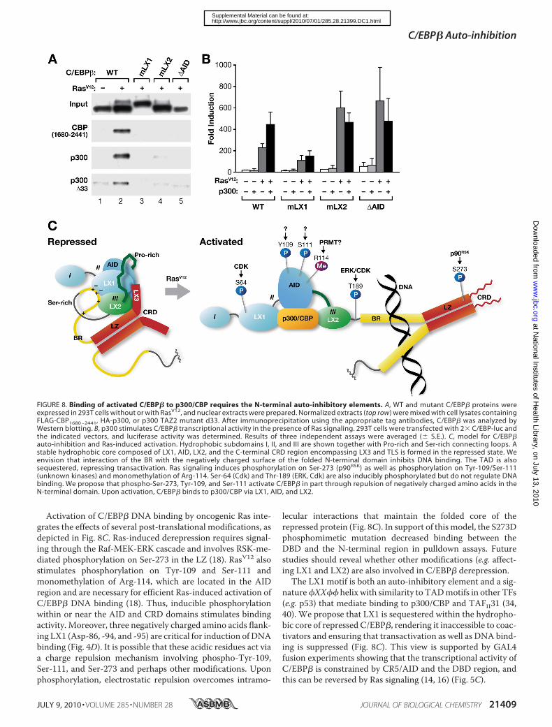
Activation of C/EBP� DNA binding by oncogenic Ras inte-grates the effects of several post-translational modifications, asdepicted in Fig. 8C. Ras-induced derepression requires signal-ing through the Raf-MEK-ERK cascade and involves RSK-me-diated phosphorylation on Ser-273 in the LZ (18). RasV12 alsostimulates phosphorylation on Tyr-109 and Ser-111 andmonomethylation of Arg-114, which are located in the AIDregion and are necessary for efficient Ras-induced activation ofC/EBP� DNA binding (18). Thus, inducible phosphorylationwithin or near the AID and CRD domains stimulates bindingactivity. Moreover, three negatively charged amino acids flank-ing LX1 (Asp-86, -94, and -95) are critical for induction of DNAbinding (Fig. 4D). It is possible that these acidic residues act viaa charge repulsion mechanism involving phospho-Tyr-109,Ser-111, and Ser-273 and perhaps other modifications. Uponphosphorylation, electrostatic repulsion overcomes intramo-
lecular interactions that maintain the folded core of therepressed protein (Fig. 8C). In support of thismodel, the S273Dphosphomimetic mutation decreased binding between theDBD and the N-terminal region in pulldown assays. Futurestudies should reveal whether other modifications (e.g. affect-ing LX1 and LX2) are also involved in C/EBP� derepression.
The LX1 motif is both an auto-inhibitory element and a sig-nature�XX�� helix with similarity to TADmotifs in other TFs(e.g. p53) that mediate binding to p300/CBP and TAFII31 (34,40). We propose that LX1 is sequestered within the hydropho-bic core of repressed C/EBP�, rendering it inaccessible to coac-tivators and ensuring that transactivation as well as DNA bind-ing is suppressed (Fig. 8C). This view is supported by GAL4fusion experiments showing that the transcriptional activity ofC/EBP� is constrained by CR5/AID and the DBD region, andthis can be reversed by Ras signaling (14, 16) (Fig. 5C).
FIGURE 8. Binding of activated C/EBP� to p300/CBP requires the N-terminal auto-inhibitory elements. A, WT and mutant C/EBP� proteins wereexpressed in 293T cells without or with RasV12, and nuclear extracts were prepared. Normalized extracts (top row) were mixed with cell lysates containingFLAG-CBP1680 –2441, HA-p300, or p300 TAZ2 mutant d33. After immunoprecipitation using the appropriate tag antibodies, C/EBP� was analyzed byWestern blotting. B, p300 stimulates C/EBP� transcriptional activity in the presence of Ras signaling. 293T cells were transfected with 2� C/EBP-luc andthe indicated vectors, and luciferase activity was determined. Results of three independent assays were averaged (� S.E.). C, model for C/EBP�auto-inhibition and Ras-induced activation. Hydrophobic subdomains I, II, and III are shown together with Pro-rich and Ser-rich connecting loops. Astable hydrophobic core composed of LX1, AID, LX2, and the C-terminal CRD region encompassing LX3 and TLS is formed in the repressed state. Weenvision that interaction of the BR with the negatively charged surface of the folded N-terminal domain inhibits DNA binding. The TAD is alsosequestered, repressing transactivation. Ras signaling induces phosphorylation on Ser-273 (p90RSK) as well as phosphorylation on Tyr-109/Ser-111(unknown kinases) and monomethylation of Arg-114. Ser-64 (Cdk) and Thr-189 (ERK, Cdk) are also inducibly phosphorylated but do not regulate DNAbinding. We propose that phospho-Ser-273, Tyr-109, and Ser-111 activate C/EBP� in part through repulsion of negatively charged amino acids in theN-terminal domain. Upon activation, C/EBP� binds to p300/CBP via LX1, AID, and LX2.
C/EBP� Auto-inhibition
JULY 9, 2010 • VOLUME 285 • NUMBER 28 JOURNAL OF BIOLOGICAL CHEMISTRY 21409
at National Institutes of H
ealth Library, on July 13, 2010w
ww
.jbc.orgD
ownloaded from
http://www.jbc.org/content/suppl/2010/07/01/285.28.21399.DC1.htmlSupplemental Material can be found at:

The fact that CR5/AID and LX2 are essential for p300/CBPbinding shows that the interaction with these coactivators ismore complex than previously envisioned and probablyinvolves multiple contacts in both proteins. Indeed, a previousstudy showed that sequences in the C/EBP� TAD cluster IIaregion (aka “Box A” (55)) are also involved in CBP binding (23).The CR5/AID and LX2 regions have not been associated withactivating functions, and it is possible that the systems previ-ously used to assess C/EBP� transcriptional activity, includingthe reporter assays employed in our study, do not require p300/CBP as a coactivator. The critical role of the LX1 motif in tran-sient reporter assays suggests that another coactivator oradapter protein, possibly TAFII31, that interacts primarily viaLX1 is essential for transcription in this setting. It is conceivablethat p300/CBP recruitment is more relevant for activation ofendogenous genes in a native chromatin environment, wherebasal transcription is stringently suppressed and histone acety-lation is critical for gene induction. Thus, it will be of interest todetermine whether AID and LX2 indeed contribute to tran-scription of specific chromatin-embedded target genes.
Acknowledgments—We thank Carlton Briggs for purification ofrecombinant proteins, Nancy Martin for expert technical assistance,William Sellers for p300 plasmids, Jean-Rene Cardinaux for CBPvectors, Allan Kane and Jiro Wada for preparation of figures, andCindy Zahnow for critical reading of the manuscript.
REFERENCES1. Whitmarsh, A. J., and Davis, R. J. (2000) Cell Mol. Life Sci. 57, 1172–11832. Chrivia, J. C., Kwok, R. P., Lamb, N., Hagiwara, M., Montminy, M. R., and
Goodman, R. H. (1993) Nature 365, 855–8593. Weigel, N. L., and Moore, N. L. (2007)Mol. Endocrinol. 21, 2311–23194. Graves, B. J., Cowley, D. O., Goetz, T. L., Petersen, J.M., Jonsen,M.D., and
Gillespie, M. E. (1998) Cold Spring Harb. Symp. Quant. Biol. 63, 621–6295. Jonsen, M. D., Petersen, J. M., Xu, Q. P., and Graves, B. J. (1996)Mol. Cell.
Biol. 16, 2065–20736. Eferl, R., and Wagner, E. F. (2003) Nat. Rev. Cancer 3, 859–8687. Ramji, D. P., and Foka, P. (2002) Biochem. J. 365, 561–5758. Sebastian, T., and Johnson, P. F. (2006) Cell Cycle 5, 953–9579. Zhu, S., Yoon, K., Sterneck, E., Johnson, P. F., and Smart, R. C. (2002) Proc.
Natl. Acad. Sci. U.S.A. 99, 207–21210. Wessells, J., Yakar, S., and Johnson, P. F. (2004) Mol. Cell. Biol. 24,
3238–325011. Sebastian, T., Malik, R., Thomas, S., Sage, J., and Johnson, P. F. (2005)
EMBO J. 24, 3301–331212. Kuilman, T., Michaloglou, C., Vredeveld, L. C., Douma, S., van Doorn, R.,
Desmet, C. J., Aarden, L. A., Mooi,W. J., and Peeper, D. S. (2008)Cell 133,1019–1031
13. Collado, M., Blasco, M. A., and Serrano, M. (2007) Cell 130, 223–23314. Williams, S. C., Baer, M., Dillner, A. J., and Johnson, P. F. (1995) EMBO J.
14, 3170–318315. Kim, J., Cantwell, C. A., Johnson, P. F., Pfarr, C. M., and Williams, S. C.
(2002) J. Biol. Chem. 277, 38037–3804416. Kowenz-Leutz, E., Twamley, G., Ansieau, S., and Leutz, A. (1994) Genes
Dev. 8, 2781–279117. Katz, S., Kowenz-Leutz, E.,Muller, C.,Meese, K., Ness, S. A., and Leutz, A.
(1993) EMBO J. 12, 1321–133218. Lee, S., Shuman, J. D., Guszczynski, T., Sakchaisri, K., Sebastian, T., Copel-
and, T. D., Miller, M., Cohen, M. S., Taunton, J., Smart, R. C., Xiao, Z., Yu,L. R., Veenstra, T. D., and Johnson, P. F. (2010) Mol. Cell. Biol. 30,2621–2635
19. Darimont, B. D., Wagner, R. L., Apriletti, J. W., Stallcup, M. R., Kushner,
P. J., Baxter, J. D., Fletterick, R. J., and Yamamoto, K. R. (1998) Genes Dev.12, 3343–3356
20. Yamamoto, K. R., Darimont, B. D., Wagner, R. L., and Iniguez-Lluhí, J. A.(1998) Cold Spring Harb. Symp. Quant. Biol. 63, 587–598
21. Shuman, J. D., Sebastian, T., Kaldis, P., Copeland, T. D., Zhu, S., Smart,R. C., and Johnson, P. F. (2004)Mol. Cell. Biol. 24, 7380–7391
22. McKnight, S. L., and Kingsbury, R. (1982) Science 217, 316–32423. Kovacs, K. A., Steinmann,M.,Magistretti, P. J., Halfon, O., andCardinaux,
J. R. (2003) J. Biol. Chem. 278, 36959–3696524. Eckner, R., Ewen, M. E., Newsome, D., Gerdes, M., DeCaprio, J. A., Law-
rence, J. B., and Livingston, D. M. (1994) Genes Dev. 8, 869–88425. Kapust, R. B., Tozser, J., Fox, J. D., Anderson, D. E., Cherry, S., Copeland,
T. D., and Waugh, D. S. (2001) Protein Eng. 14, 993–100026. Williams, S. C., Cantwell, C. A., and Johnson, P. F. (1991) Genes Dev. 5,
1553–156727. Jenkins, L. M., Yamaguchi, H., Hayashi, R., Cherry, S., Tropea, J. E., Miller,
M., Wlodawer, A., Appella, E., and Mazur, S. J. (2009) Biochemistry 48,1244–1255
28. Rost, B. (1996)Methods Enzymol. 266, 525–53929. Rost, B., Yachdav, G., and Liu, J. (2004) Nucleic Acids Res. 32,W321–32630. Gasteiger, E., Hoogland, C., Gattiker, A., Duvaud, S.,Wilkins,M. R., Appel,
R. D., and Bairoch, A. (2005) in The Proteomics Protocols Handbook(Walker, J. M., ed) pp. 571–607, Humana Press, Totowa, NJ
31. Coeytaux, K., and Poupon, A. (2005) Bioinformatics 21, 1891–190032. Gaboriaud, C., Bissery, V., Benchetrit, T., and Mornon, J. P. (1987) FEBS
Lett. 224, 149–15533. Callebaut, I., Labesse, G., Durand, P., Poupon, A., Canard, L., Chomilier, J.,
Henrissat, B., and Mornon, J. P. (1997) Cell Mol. Life Sci. 53, 621–64534. Choi, Y., Asada, S., andUesugi,M. (2000) J. Biol. Chem. 275, 15912–1591635. Tahirov, T. H., Sato, K., Ichikawa-Iwata, E., Sasaki, M., Inoue-Bungo, T.,
Shiina, M., Kimura, K., Takata, S., Fujikawa, A., Morii, H., Kumasaka, T.,Yamamoto, M., Ishii, S., and Ogata, K. (2002) Cell 108, 57–70
36. He, B., and Wilson, E. M. (2003)Mol. Cell. Biol. 23, 2135–215037. Trautwein, C., Walker, D. L., Plumpe, J., and Manns, M. P. (1995) J. Biol.
Chem. 270, 15130–1513638. Feng, H., Jenkins, L. M., Durell, S. R., Hayashi, R., Mazur, S. J., Cherry, S.,
Tropea, J. E., Miller, M., Wlodawer, A., Appella, E., and Bai, Y. (2009)Structure 17, 202–210
39. Wojciak, J. M., Martinez-Yamout, M. A., Dyson, H. J., and Wright, P. E.(2009) EMBO J. 28, 948–958
40. Joerger, A. C., and Fersht, A. R. (2008) Annu. Rev. Biochem. 77, 557–58241. Mink, S., Haenig, B., and Klempnauer, K. H. (1997) Mol. Cell. Biol. 17,
6609–661742. Nakajima, T., Kinoshita, S., Sasagawa, T., Sasaki, K., Naruto, M., Kishi-
moto, T., and Akira, S. (1993) Proc. Natl. Acad. Sci. U.S.A. 90, 2207–221143. Shuman, J. D., Vinson, C. R., and McKnight, S. L. (1990) Science 249,
771–77444. Podust, L. M., Krezel, A. M., and Kim, Y. (2001) J. Biol. Chem. 276,
505–51345. Fontes, M. R., Teh, T., and Kobe, B. (2000) J. Mol. Biol. 297, 1183–119446. Miller, M., Shuman, J. D., Sebastian, T., Dauter, Z., and Johnson, P. F.
(2003) J. Biol. Chem. 278, 15178–1518447. Burk, O., Mink, S., Ringwald, M., and Klempnauer, K. H. (1993) EMBO J.
12, 2027–203848. Burk, O., Worpenberg, S., Haenig, B., and Klempnauer, K. H. (1997)
EMBO J. 16, 1371–138049. Ness, S. A., Kowenz-Leutz, E., Casini, T., Graf, T., and Leutz, A. (1993)
Gene Dev. 7, 749–75950. Gao, H., Parkin, S., Johnson, P. F., and Schwartz, R. C. (2002) J. Biol. Chem.
277, 38827–3883751. Hu, H.M., Tian, Q., Baer,M., Spooner, C. J.,Williams, S. C., Johnson, P. F.,
and Schwartz, R. C. (2000) J. Biol. Chem. 275, 16373–1638152. Lee, Y. H., Williams, S. C., Baer, M., Sterneck, E., Gonzalez, F. J., and
Johnson, P. F. (1997)Mol. Cell. Biol. 17, 2038–204753. Spooner, C. J., Guo, X., Johnson, P. F., and Schwartz, R. C. (2007) Mol.
Immunol. 44, 1384–139254. Zahnow, C. A. (2009) Expert. Rev. Mol. Med. 11, e1255. Nerlov, C., and Ziff, E. B. (1995) EMBO J. 14, 4318–4328
C/EBP� Auto-inhibition
21410 JOURNAL OF BIOLOGICAL CHEMISTRY VOLUME 285 • NUMBER 28 • JULY 9, 2010
at National Institutes of H
ealth Library, on July 13, 2010w
ww
.jbc.orgD
ownloaded from
http://www.jbc.org/content/suppl/2010/07/01/285.28.21399.DC1.htmlSupplemental Material can be found at:


![P.F. Lemkin LECB, CCR, NCI/FCRDC mail: lemkin@ncifcrf.gov [This document is under construction] Revised: 06-19-2002 Software Design of the MicroArray Explorer](https://img.pdfslide.us/doc/110x75/56649eac5503460f94bb25e5/pf-lemkin-lecb-ccr-ncifcrdc-mail-lemkinncifcrfgov-this-document-is.jpg)