Embed Size (px)
Citation preview

Cavalcanti, I. M. G., Nobbs, A. H., Ricomini-Filho, A. P., Jenkinson, H. F.,& Cury, A. A. D. B. (2016). Interkingdom cooperation between Candidaalbicans, Streptococcus oralis and Actinomyces oris modulates early biofilmdevelopment on denture material. Pathogens and Disease, 74(3), [ftw002].DOI: 10.1093/femspd/ftw002
Peer reviewed version
Link to published version (if available):10.1093/femspd/ftw002
Link to publication record in Explore Bristol ResearchPDF-document
This is a pre-copyedited, author-produced PDF of an article accepted for publication in Pathogens and Diseasefollowing peer review. The version of record "Interkingdom cooperation between Candida albicans,Streptococcus oralis and Actinomyces oris modulates early biofilm development on denture material" is availableonline at: http://femspd.oxfordjournals.org/content/74/3/ftw002.abstract.
University of Bristol - Explore Bristol ResearchGeneral rights
This document is made available in accordance with publisher policies. Please cite only the publishedversion using the reference above. Full terms of use are available:http://www.bristol.ac.uk/pure/about/ebr-terms.html

1
Interkingdom cooperation between Candida albicans,
Streptococcus oralis and Actinomyces oris modulates early
biofilm development on denture material
Indira M.G. Cavalcanti1,2, Angela H. Nobbs2, Antônio Pedro Ricomini-Filho3,
Howard F. Jenkinson2* and Altair A. Del Bel Cury1
1Department of Prosthodontics and Periodontology, Piracicaba Dental School -
University of Campinas, Limeira Avenue, 901, 13414-903 Piracicaba, SP, Brazil,
2School of Oral and Dental Sciences, University of Bristol, Lower Maudlin Street,
Bristol, BS1 2LY, United Kingdom, 3Department Physiological Sciences, Piracicaba
Dental School - University of Campinas, Avenida Limeira, 901, 13414-903 Piracicaba,
SP, Brazil.
Running header: Interkingdom biofilms on denture acrylic
*Correspondence: Professor Howard F. Jenkinson, School of Oral and Dental
Sciences, University of Bristol, Lower Maudlin Street, Bristol BS1 2LY, United
Kingdom. Tel: +44-117-342-4424. Fax: +44-117-342-4313. E-mail:
Pathogens and Disease Advance Access published January 10, 2016 by guest on February 10, 2016
http://femspd.oxfordjournals.org/
Dow
nloaded from

2
ABSTRACT
Candida-associated stomatitis affects up to 60% of denture wearers, and Candida
albicans remains the most commonly isolated fungal species. The oral bacteria
Actinomyces oris and Streptococcus oralis are abundant in early dental plaque. The
aims of this study were to determine the effects of S. oralis and A. oris on the
development of C. albicans biofilms on denture material. Resin discs were coated with
saliva and at early (1.5 h) or later (24 h) stages of biofilm development, cell numbers of
each species were determined. Spatial distribution of microorganisms was visualized
by confocal scanning laser microscopy of biofilms labelled by differential fluorescence
or by fluorescence in situ hybridization (FISH). Interkingdom interactions underpinning
biofilm development were also evaluated planktonically utilizing fluorescence
microscopy. Synergistic interactions between all three species occurred within biofilms
and planktonically. Bacterial cells coaggregated with each other and adhered singly or
in coaggregates to C. albicans hyphal filaments. S. oralis appeared to enhance hyphal
filament production and C. albicans biovolume was increased two-fold. Concomitantly,
cell numbers of S. oralis and A. oris were enhanced by C. albicans. Thus cooperative
physical and metabolic processes occurring between these three microbial species
intensify pathogenic plaque communities on denture surfaces.
Keywords: human oral cavity; microbial communities; stomatitis; FISH; coaggregation
Text for graphical abstract:
Interactive processes occurring between Streptococcus oralis, Actinomyces oris, and
Candida albicans intensify pathogenic plaque communities on denture surfaces
by guest on February 10, 2016http://fem
spd.oxfordjournals.org/D
ownloaded from
3
INTRODUCTION
Denture stomatitis is a common disease, often recurring, and is seen in up to
60% of otherwise healthy denture wearers (Arendorf and Walker, 1987; Figueira et al.
2007; Geerts et al. 2008). It is characterized by inflamed mucosa, particularly under an
upper denture (palatal mucosa), causing a burning sensation, soreness and bad taste
(Ramage et al. 2004). Denture samples from subjects with denture stomatitis generally
carry surface biofilms containing networks of hyphal filaments characteristic of Candida
albicans (Douglas, 2003). The fungi have a high propensity for adhesion to oral acrylic
dentures (Pereira-Cenci et al. 2007), and this is affected by the surface roughness of
the denture resin (Verran and Maryan, 1997; Jackson et al. 2014; Mayahara et al.
2014) and by the type of dietary carbohydrates (Santana et al. 2013).
While C. albicans and other Candida species that can co-exist with C. albicans
(Coco et al. 2008) have been a focus in treatment strategies for oral stomatitis
(Redding et al., 2009; Salerno et al. 2011), studies have underlined that the plaque
accumulated on dentures during stomatitis contains both bacterial and fungal
components (Campos et al. 2008; Salerno et al. 2011). This is potentially significant
because it has been shown that oral bacteria, such as streptococci, can form mixed-
species biofilms with C. albicans (Xu et al. 2014a) that are more luxurious and contain
more hyphal filaments than monospecies biofilms (Dutton et al. 2014; Sztajer et al.
2014; Dutton et al. 2015). Streptococcus oralis appears to be able to facilitate invasion
of oral mucosa by C. albicans (Diaz et al. 2012), while growth of S. oralis itself is
promoted by the fungus. In addition, the pathogenic properties and invasive potential of
the bacteria can be elevated by the presence of C. albicans (Falsetta et al. 2014; Xu et
al. 2014b). Less is known about how other bacterial colonizers of hard surfaces, for
example species of Actinomyces or Veillonella, interact with C. albicans and influence
biofilm development.
Mitis/sanguinis group streptococci and Actinomyces species (e.g. oris,
naeslundii, johnsonii) are amongst the first oral bacteria to colonize a fresh salivary
glycoprotein-surface (Nyvad and Kilian, 1987; Al-Ahmad et al. 2009; Dige et al. 2009).
In vitro studies have indicated that mutualism in biofilm formation occurs between
strains of S. oralis and Actinomyces oris (formerly A. naeslundii). Neither organism
alone was able to form a salivary-flow biofilm, but a robust dual-species biofilm could
be formed owing to nutritional cross-feeding (Palmer et al. 2001). Interactions between
these species and C. albicans in triadic conditions has not before been studied, but
would be significant in respect of early colonization of denture materials. The concept
of synergy in biofilm community development rests on the notion that a primary species
by guest on February 10, 2016http://fem
spd.oxfordjournals.org/D
ownloaded from

4
preferentially adheres to the salivary pellicle-coated oral cavity surface and by doing
so, provides additional sites for adhesion of secondary colonizers (Wright et al. 2013).
Based upon this notion, and upon previous work showing synergistic interactions
between streptococci and C. albicans (Bamford et al. 2009; Jack et al. 2015), it was
hypothesized that colonization of salivary glycoprotein pellicle-coated denture material
by C. albicans might be modulated when both streptococci and actinomyces are
present. The results demonstrate that there is synergy in triadic-species biofilm
formation, and that the increased presence of C. albicans enhances the development
of bacterial plaque. This suggests that antibacterial approaches should be adopted in
addition to antifungal strategies to more effectively cleanse dentures of biofilms and
control stomatitis.
MATERIALS AND METHODS
Microbial strains and growth conditions
A. oris T14V and S. oralis 34 were cultivated anaerobically at 37 °C on BHYN agar (per
liter: 37 g Brain Heart Infusion, 5 g yeast extract, 5 g Bacto-Neopeptone and 15 g
agar). Suspension cultures were grown in BHY medium (Brain Heart Infusion medium
containing 5 g L-1 yeast extract), in sealed bottles or tubes, and incubated stationary at
37 °C. C. albicans SC5314 was cultivated aerobically on Sabouraud Dextrose (SD)
agar (LabM, Heywood, Leics. UK) at 37 °C, and suspensions were grown in YMD
medium (per liter: 20 g Oxoid Mycological Peptone, 10 g yeast extract, 20 g dextrose)
in conical flasks at 37 °C with shaking (200 r.p.m.) (Dutton et al. 2014). For preparation
of cells for experiments, cultures were grown for 16 h at 37 °C, centrifuged (5000 g for
7 min), the cell pellets were suspended and washed twice with YPT medium (1 x yeast
nitrogen base, 20 mM NaH2PO4-H3PO4 buffer pH 7.0, 0.1% tryptone) (Silverman et al.,
2010), and suspended in YPT medium at OD600 1.0.
Planktonic interaction assay
C. albicans cell suspension at OD600 1.0 (approximately 1 x 10⁷ cells mL-1) (0.2 mL)
was added to glass tubes containing 1.8 mL warm YPTG medium (YPT supplemented
with 0.4% glucose) and incubated for 2 h at 37 °C with shaking (200 r.p.m.) to initiate
hyphal filament formation. During this period, A. oris cells were fluorescently labelled
with FITC (fluorescein isothiocyanate) as described by Dutton et al. (2014) and S.
oralis cells were labelled with 1.3 mM TRITC (tetramethylrhodamine-5 (and 6)
isothiocyanate) in 0.05 M Na2CO3 containing 0.15 M NaCl for 30 min at 20 oC in the
by guest on February 10, 2016http://fem
spd.oxfordjournals.org/D
ownloaded from

5
dark with gentle shaking. Bacterial cells were washed three times with YPT and
suspended at OD600 0.5 (2 x 10⁸ cells mL-1) in YPT medium. Fluorescently-labelled
bacterial cell suspension (1 ml) was added to C. albicans cell suspension (2 ml) and
incubated for 1 h at 37 °C with gentle shaking. To each cell suspension was then
added fluorescent brightener 28 (Calcofluor white, Sigma-Aldrich Co., St. Louis, MO) (1
µL of 0.2 mg mL-1 stock solution) to fluoresce the C. albicans. Samples of suspension
were applied to glass microscope slides and visualized by fluorescence microscopy
(Leica DMLB) (Silverman et al. 2010).
PMMA disc preparation
Discs were fabricated using poly(methyl methacrylate) (PMMA) resin (QC-20 PMMA,
Dentsply International, York, PA) and polymerized in a hot water bath according to the
manufacturer’s instructions within a metal device (10 mm diameter x 2 mm thick). The
discs were ground progressively with smoother aluminum oxide papers, grids 320, 400
and 600, in a horizontal polisher (Arotec APL-4, São Paulo, SP, Brazil) and then
standardized for surface roughness by profilometry (Verran and Maryan, 1997) to Ra =
0.31 ± 0.02 (Ra value is the arithmetical average of all departures of the surface profile
through the mean sample length). They were cleaned in an ultrasonic bath (Thornton T
740, Thornton-Inpec Eletronica LTDA, Vinhedo, SP, Brazil) for 2 x 10 min to remove
surface contaminants, disinfected with 70% ethanol, washed with sterile distilled water
and allowed to dry under aseptic conditions.
Salivary pellicle formation
Collection of human saliva samples was approved by the National Research Ethics
Committee South Central Oxford C (no. 08/H0606/87+5). Saliva was collected from at
least 6 adult subjects, who provided written informed consent. The samples were
pooled, treated with 0.25 M dithiothreitol on ice for 10 min, and centrifuged (8000 g for
10 min). The supernatant was removed, diluted to 10% with sterile water, filter
sterilized (0.45-µm pore-size membrane), and stored at -20°C. Salivary pellicle was
formed on the resin discs by incubating them in 10% sterile saliva for 2 h at 37 °C.
Biofilm development
Saliva-coated PMMA discs were added to individual wells of a 24-well plate each
containing 1.8 mL YPTG medium at 37 oC. Microbial cell suspensions in YPT medium,
prepared as described above, were then added (75 µL) together with 150 µL, 75 µL or
0 µL YPTG medium as appropriate (total 225 µL) to generate mono-, dual- or triadic-
species biofilms respectively in triplicate. After 1.5 h a number of the resin discs were
by guest on February 10, 2016http://fem
spd.oxfordjournals.org/D
ownloaded from

6
removed, rinsed, and analysed for biofilm formation. For the remaining discs,
unattached cell suspension was aspirated from the wells and replaced with 2 mL fresh
YPTG medium. The plates were incubated at 37 ºC for 24 h with gentle agitation as
previously described (Silverman et al. 2010; Cavalcanti et al. 2014).
Measurement of viable cell numbers
After 1.5 h incubation, or after 24 h, the PMMA discs were removed from the culture
wells, rinsed gently with sterile PBS (50 mM Na2HPO4-KH2PO4, 0.15 M NaCl, pH 7.2),
added to 3 mL PBS in a polypropylene tube, and sonicated at 7 W for 30 s in a
sonication bath to disrupt the biofilms. Portions (200 µL) of the homogenized
suspensions were removed, serially 10-fold diluted and 20 µL aliquots were applied to
BHYN agar plates containing 50 µg nystatin mL-1 (to inhibit growth of C. albicans) for
estimating numbers of bacterial colony-forming units (CFU), or onto SD agar for
estimating CFU of C. albicans. The plates were incubated anaerobically at 37 oC for 24
h (BHYN agar) or aerobically at 37 oC for 16 h (SD agar), and colony counts were
subsequently converted to CFU mL-1. It was possible to differentiate between S. oralis
and A. oris on the basis of colony size and colour. Control experiments determined that
the sonication treatment was sufficient to remove >90% cells from the surfaces of the
discs, and at the same time was sufficiently gentle to result in <5% loss in cell viability.
Fluorescence microscopy and confocal scanning laser microscopy (CSLM)
For visualizing C. albicans monospecies biofilms, 2 µg Calcofluor mL-1 was
incorporated into the YPTG growth medium to fluorescently label the fungal cells.
Monospecies biofilms of bacteria were fluorescently labelled with FITC (10 µM, 20 min)
at the end of the experiment. The resin discs were gently washed twice with PBS and
the biofilms were visualized by fluorescence microscopy (Leica DMLB) or by CSLM
with a Leica SP5-AOBS confocal microscope attached to a Leica DM I6000 inverted
epifluorescence microscope.
Dual-species or triadic-species biofilms were also subjected to fluorescence in
situ hybridization (FISH) analysis by CSLM. Biofilms were fixed in 4%
paraformaldehyde (2 h), permeabilized using lysozyme for 30 min at 37 °C, washed
twice in PBS and then incubated in hybridization buffer (0.9 M NaCl, 20 mM Tris-HCl
pH 8.0, 0.01% SDS, and 10-30% formamide depending upon the probe) containing 5
μg mL-1 fluorescently-labelled 16S rRNA-specific oligonucleotide probe for 150 min at
55 °C. The oligonucleotide probe sequences were as described by Thurnheer et al.
(2004) and were labelled (Eurofins Genomic Services Ltd., Wolverhampton, UK) as
follows: S. oralis probe MIT447_488 (Alexa 488), A. oris probe ANA103_647 (Alexa
by guest on February 10, 2016http://fem
spd.oxfordjournals.org/D
ownloaded from

7
647) and C. albicans EUK516_555 (Alexa 555). Following hybridization the discs were
incubated in washing buffer (20 mM Tris-HCl pH 7.5, 5 mM EDTA, 0.01% SDS, and
between 159 and 636 mM NaCl depending on the formamide concentration used
during hybridization) for 15 min at 55 °C. After rinsing briefly in 0.9% NaCl the discs
were inverted onto a glass slide to be visualized by CSLM. Volocity® software was
utilized to prepare three-dimensional (3D) images and Imaris® v7.5 software (Bitplane
AG, Zurich, Switzerland) was used to calculate biovolumes (µm3).
Statistical analysis
Data were processed by Prism 6 software at a confidence level of 95% using the two-
way ANOVA followed by Tukey test for biovolumes or for total biofilm counts,
respective to the time points or the biofilm combinations.
RESULTS
Planktonic interactions between bacteria and fungi
Approximately 60% of C. albicans cells formed hyphal filaments after 2 h planktonic
incubation at 37 oC in YPTG medium. S. oralis (labelled with TRITC) adhered
principally to hyphal filaments, with small chains of streptococci aligned along the
hyphae (Fig. 1A), or adhered in patches as single cells or doublets (Fig. 1A). The
actinomyces cell pairs showed characteristic whiplash morphology and small
aggregates were visible freely and in association with hyphae (Fig. 1B). In triadic
associations, the actinomyces and streptococci formed large coaggregates and these
adhered principally to the fungal filaments (Fig. 1). Coaggregates of S. oralis and A.
oris were also seen independently of candidal cells (Fig. 1C). Therefore coaggregation
between these three microorganisms occurred pairwise as well as cooperatively.
Early biofilm formation on resin discs
To assess the capacity for the three microbial species, or combinations thereof, to bind
saliva-coated resin, microbial cells were incubated with resin discs for 1.5 h. Non-
adherent cells were then removed, and levels of attachment visualized by fluorescence
microscopy following staining of the discs with FITC. Because of the uneven surfaces
of the resin discs, it was not possible to capture the entire population of attached cells
in a single plane of focus. Nonetheless, all three species could be seen to attach to
saliva-coated resin, both as individual cells or as aggregates (Fig. 2, upper panels).
Overall coverage was uniform, although the natural grooves of the resin surface
by guest on February 10, 2016http://fem
spd.oxfordjournals.org/D
ownloaded from

8
seemed to promote microbial deposition, especially of bacteria, compared to smoother
regions of the resin.
For dual-species or triadic samples, S. oralis or A. oris could be seen interacting
both directly with the salivary pellicle and with candidal cells (Fig. 2, lower panels).
Patterns of association were similar overall to those seen under planktonic conditions
(compare Figs. 1 and 2). For C. albicans cells incubated with resin either alone or in
combination with bacteria, a mixture of blastospores and hyphae was evident attached
to the surface. In dual-species biofilms of C. albicans and A. oris, the A. oris cells
tended to associate with the C. albicans as opposed to adhering to the pellicle (Fig. 2).
This suggested that the pellicle receptors for A. oris might be modified by the presence
of C. albicans, or that A. oris bound preferentially to C. albicans rather than to the
coated resin. In triadic-species biofilms, the C. albicans cells forming hyphal filaments
were distributed more evenly over the surface, while notably the bacteria formed
clusters associated with the C. albicans hyphae (Fig. 2). Although not quantified
directly in these biofilms, a higher proportion of hyphal filaments was present in dual- or
triadic-species samples compared to monospecies C. albicans samples (Fig. 2).
At 1.5 h the viable counts of bacteria recovered from the biofilms were similar
(Table 1), while the C. albicans CFU were ten-fold less than bacterial cell numbers.
However, C. albicans cells are approximately ten-fold larger in size/volume than the
bacterial cells. The CFU of C. albicans were reduced by about 40% in the presence of
A. oris and about 20% by S. oralis (Table 2). By contrast, there were no significant
differences (P ≥ 0.05) in CFU of streptococci or actinomyces in the presence of C.
albicans (Table 2). The numbers of C. albicans CFU were also reduced in the presence
of S. oralis and A. oris together.
To overcome the difficulties of visualizing the roughened surface of the resin
discs by fluorescence microscopy, monospecies biofilm formation was also
investigated by CSLM. Microbial cells were incubated with saliva-coated resin discs for
1.5 h. Non-adherent cells were then removed, and biofilms were visualized by
fluorescent labelling with FITC (S. oralis, A. oris) or Calcofluor (C. albicans). At 1.5 h all
three species exhibited relatively uniform coverage of the resin surface, with areas of
elevated cell density in the resin grooves, particularly for S. oralis (Fig. 3). Overall
levels of coverage were comparable between S. oralis and A. oris, while C. albicans
cells had a more sparse distribution. All three species formed distinct microcolonies.
Later (24 h) biofilms on resin discs
To investigate biofilm formation by these microorganisms in more detail, later stage (24
h) biofilms on resin discs were also examined. Using CSLM, no obvious changes were
by guest on February 10, 2016http://fem
spd.oxfordjournals.org/D
ownloaded from

9
seen in S. oralis biomass levels from 1.5 h to 24 h (Fig. 3). By contrast, biofilms formed
by A. oris or C. albicans increased in density and depth over the 24 h period to form a
thick ‘mat’ on the resin (Fig. 3). Additionally, for C. albicans, a distinct transition from
predominately blastospores and short hyphal filaments to long hyphal filaments was
seen from 1.5 h to 24 h (Fig. 3).
At 24 h the viable counts (CFU) of each of the monospecies biofilms had
increased, with A. oris showing a 3.6-fold increase and C. albicans a 30-fold increase
(Table 1) compared to 1.5 h CFU levels. However, in the dual- or triadic-species
biofilms there were notable interactive effects of the organisms. In the presence of C.
albicans, the CFU of S. oralis and A. oris increased between 1.5 h and 24 h by 14-fold
and 19-fold respectively, while C. albicans exhibited up to 38-fold increased CFU
(Table 2). In the triadic-species biofilm total bacterial CFU increased 20-fold over the
1.5 h to 24 h time period. Therefore, based upon these CFU data, biofilm formation by
S. oralis and A. oris was highly augmented in the presence of C. albicans. It should be
noted that for C. albicans, CFU are not a meaningful indicator of biomass because
multicellular hyphal filaments were the major morphological components of C. albicans
24 h biofilms. We were unable to measure total biomass by conventional staining
methods because the resin adsorbed the stains.
In the experiments so far described the bacteria and fungi were added
simultaneously to the resin discs. To determine if the order of deposition had any effect
upon the biofilms formed, we first incubated the resin discs with bacteria for 1.5 h, then
added C. albicans and measured CFU at 3 h or 24 h. A similar pattern of results was
obtained (Supplemental Table S1) to those when the microorganisms were inoculated
together at the same time (Table 2). S. oralis and A. oris CFU increased 8-fold and 12-
fold over the time period, while C. albicans CFU increased up to 15-fold. The bacterial
components of the biofilm were again augmented, especially bacterial CFU present
within the triadic-species biofilms (Table S1).
Microbial composition of 24 h biofilms determined by FISH
To visualize relative proportions of microbes present in dual- or triadic-species
biofilms, 1.5 h or 24 h biofilms were grown on saliva-coated resin, as above, and the
presence of each microbial species detected using FISH. For all combinations the
microorganisms formed fully integrated biofilms, interacting with one another and with
salivary pellicle (Fig. 4). At 1.5 h, both bacterial species were in close contact with the
pellicle, and this was particularly evident for A. oris. (Fig. 4). Initial deposition of C.
albicans appeared to be enhanced by S. oralis (compare Fig. 4 and Fig. 3) but was
unaffected by A. oris. This appears to contradict the corresponding CFU data (above),
by guest on February 10, 2016http://fem
spd.oxfordjournals.org/D
ownloaded from

10
but there are limitations in CFU determinations for C. albicans undergoing filamentation
(see Discussion). At 24 h, S. oralis cells were integrated from the base to the surface of
the dual-species biofilm, while A. oris cells were particularly associated with the upper
biofilm layers (Fig. 4). These biofilms were composed mainly of hyphal filaments with
integrated patches of bacteria. The triadic-species biofilms were also composed mainly
of hyphal filaments, with integrated groups of S. oralis and fewer A. oris (Fig. 4).The
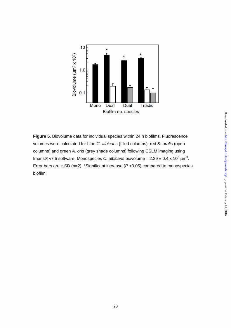
biovolume data at 24 h demonstrated that the presence of S. oralis enhanced C.
albicans biofilm development (biovolume) two-fold (Fig. 5), compared with C. albicans
alone. A. oris had a lesser stimulatory effect on C. albicans (Fig. 5). C. albicans
biovolume was also enhanced in the triadic-species biofilms (Fig. 5), but not as much
as with S. oralis alone. In conclusion, S. oralis and A. oris individually augment biofilm
formation by C. albicans, but S. oralis appears to out-compete A. oris in the presence
of C. albicans.
DISCUSSION
The understanding of multispecies biofilm partnerships and their impact on
human health has grown over recent years. Further investigation is required to take
into account the potential for a wide spectrum of different microbial communities to
develop in the human host and affect health. The interkingdom interactions of C.
albicans and bacteria in the context of human disease have revived attention since one
of the first descriptions of coaggregation between Gram-positive cocci and Candida
(Jenkinson et al. 1990). Some bacterial species produce molecules that kill or inhibit C.
albicans (Morales et al. 2010; 2013), while others promote growth of the fungus (Xu et
al. 2014a). On the other side, the pathogenic properties of some bacteria may be
enhanced (Schlecht et al. 2015) or suppressed (Lopez-Medina et al. 2015) in the
presence of C. albicans. Dual-species biofilms of Streptococcus gordonii and C.
albicans have at least two-fold increased biomass over and above the biomass sum of
the monospecies biofilms, showing synergy in growth of these species (Bamford et al.
2009; Dutton et al. 2014). Moreover, dual-species communities of S. oralis and C.
albicans show pathogenic synergy, and induce an exaggerated inflammatory response
in the host, over and above the inflammatory response levels of the individual species
(Xu et al. 2014b). This latter observation is clearly relevant to the condition of denture
stomatitis, the inflammatory nature of which might therefore be exacerbated by the
presence of specific bacterial species in association with C. albicans in denture biofilms
(Cavalcanti et al. 2015).
by guest on February 10, 2016http://fem
spd.oxfordjournals.org/D
ownloaded from

11
In the current article we investigated whether the interactions of two early-
colonizing bacterial species affected the manner in which C. albicans could be
incorporated into the biofilm and colonize denture acrylic surface. We found that both
species of bacteria interacted with Candida hyphae under planktonic conditions. There
was no evidence that bacteria bound specific regions of the filaments, for example the
septal domain, suggesting that receptors for adhesion were distributed all along the
length. Bacterial cells attached to hyphal filaments individually or in small chains, and
aggregates were visible. A. oris and S. oralis were mixed simultaneously with C.
albicans in the triadic-species combinations, so the patterns of coaggregation indicated
that there were sufficient binding sites on C. albicans available for both bacterial
species. Moreover, the hyphae were able to support larger coaggegates of S. oralis
and A. oris.
In dual- and triadic-species biofilms, C. albicans hyphal filaments appeared to
be incorporated throughout the biofilm layers. S. oralis cells formed a basal layer upon
the salivary pellicle, in dual-species biofilms, colonizing areas not occupied by C.
albicans. This is similar to the stratified appearance of S. oralis-C. albicans biofilms
described recently by Bertolini et al. (2015). Streptococci could also be seen in patches
at all levels within the 24 h biofilm. A. oris on the other hand did not effectively colonize
the pellicle in 24 h dual-species biofilms with C. albicans, and instead was mainly
associated with the hyphal filaments. The dual-species biofilms exhibited synergistic
behaviours and bacterial CFU were higher in the presence of C. albicans than in the
absence. A. oris CFU were increased 3.7-fold, and S. oralis CFU were increased 8.8-
fold, in the presence of C. albicans compared to CFU within the respective
monospecies biofilms. Measurements of C. albicans CFU in biofilms after ~2 h
incubation have limitations as more hyphal filaments are formed. The limitations are
clearly evident in that C. albicans biovolume within the triadic-species biofilms
increased nearly two-fold in the presence of bacteria, while bacteria themselves
comprised <10% of the total biovolume. Interestingly, incorporation of A. oris into the C.
albicans-S. oralis biofilm dampened in part the augmenting effect of S. oralis on C.
albicans. This indicates that A. oris may have some growth inhibitory effect on C.
albicans in the presence of S. oralis. However, we did not observe direct inhibition of C.
albicans proliferation by A. oris, as has been reported recently (Guo et al. 2015). This
might be because we are using different strains of C. albicans and A. oris, as it is
known that coaggregation between C. albicans, actinomyces and streptococci is strain-
dependent (Arzmi et al. 2015).
The biofilm model described could be valuable in evaluating means of
controlling oral candidosis, or growth of denture biofilms, because it contains two early-
by guest on February 10, 2016http://fem
spd.oxfordjournals.org/D
ownloaded from

12
colonizing species of bacteria as well as C. albicans. In vivo, C. albicans forms biofilms
in association with bacteria, and colonization of a surface may require the ability to
successfully compete with or live alongside the early colonizers. Our results indicate
that these two early colonizing strains of bacteria are promoted by the presence of C.
albicans, with the fungus providing an alternative substratum for A. oris. Concomitantly
we see greater levels of C. albicans biofilm development occurring in the presence of
bacteria, especially with S. oralis, compared to C. albicans monospecies biofilm.
Further work is underway to determine the kinds of signals, nutritional or otherwise,
that are produced by the microorganisms in order to mediate these synergistic
interactions. This knowledge would be important for impact on clinical intervention
strategies or antimicrobial therapies to control these polymicrobial infections.
FUNDING
This work was supported by grants 2013/15884-2 from the São Paulo Research
Foundation (FAPESP) and 08808/2013-09 from Science Without Borders, Brazil.
ACKNOWLEDGEMENTS
We thank Lindsay Dutton and Jane Brittan for excellent technical assistance, and the
Wolfson Foundation for establishing the University of Bristol Bioimaging Facility
managed by Dr Mark Jepson.
REFERENCES
Al-Ahmad A, Follo M, Selzer AC et al. Bacterial colonization of enamel in situ
investigated using fluorescence in situ hybridization. J Med Microbiol 2009;58:1359-
66.
Arendorf TM, Walker DM. Denture stomatitis: a review. J Oral Rehabil 1987;14:217-27.
Arzmi MH, Dashper S, Catmull D et al. Coaggregation of Candida albicans,
Actinomyces naeslundii and Streptococcus mutans is Candida albicans strain
dependent. FEMS Yeast Res 2015;15:fov038.
Bamford CV, d'Mello A, Nobbs AH et al. Streptococcus gordonii modulates Candida
albicans biofilm formation through intergeneric communication. Infect Immun
2009;77:3696-704.
by guest on February 10, 2016http://fem
spd.oxfordjournals.org/D
ownloaded from
13
Bertolini MM, Xu H, Sobue T et al. Candida-streptococcal mucosal biofilms display
distinct structural and virulence characteristics depending on growth conditions and
hyphal phenotypes. Mol Oral Microbiol 2015;30:307-22.
Campos MS, Marchini L, Bernardes LAS et al. Biofilm microbial communities of
denture stomatitis. Oral Microbiol Immunol 2008;23:419-24.
Cavalcanti IM, Ricomini Filho AP et al. Salivary pellicle composition and multispecies
biofilm developed on titanium nitrided by cold plasma. Arch Oral Biol 2014;59:695-
703.
Cavalcanti YW, Morse DJ, da Silva WJ et al. Virulence and pathogenicity of Candida
albicans is enhanced in biofilms containing oral bacteria. Biofouling 2015;31:27-38.
Coco BJ, Bagg J, Cross LJ et al. Mixed Candida albicans and Candida glabrata
populations associated with the pathogenesis of denture stomatitis. Oral Microbiol
Immunol 2008;23:377-83.
Douglas LJ. Candida albicans biofilms and their role in infection. Trends Microbiol
2003;11:30-6.
Diaz PI, Xie Z, Sobue T et al. Synergistic interaction between Candida albicans and
commensal oral streptococci in a novel in vitro mucosal model. Infect Immun
2012;80:620-32.
Dige I, Raarup MK, Nyengaard JR et al. Actinomyces naeslundii in initial dental biofilm
formation. Microbiology 2009;155:2116-26.
Dutton LC, Nobbs AH, Jepson K et al. O-mannosylation in Candida albicans enables
development of interkingdom biofilm communities. mBio 2014;5:e00911.
Dutton LC, Paszkiewicz KH, Silverman RJ et al. Transcriptional landscape of trans-
kingdom communication between Candida albicans and Streptococcus gordonii. Mol
Oral Microbiol 2015;30:in press
Falsetta ML, Klein MI, Colonne PM et al. Symbiotic relationship between Streptococcus
mutans and Candida albicans synergizes virulence of plaque biofilms in vivo. Infect
Immun 2014;82:1968-81.
Figueira MH, Azul A, Pinto E et al. Denture-related stomatitis: identification of
aetiological and predisposing factors- a large cohort. J Oral Rehabil 2007;34:448-
455.
Guo Y, Wei C, Liu C et al. Inhibitory effects of oral Actinomyces on the proliferation,
virulence and biofilm formation of Candida albicans. Arch Oral Biol 2015;60:1368-
74.
Geerts GA, Stuhlinger ME, Basson NJ. Effect of an antifungal denture liner on the
saliva yeast count in patients with denture stomatitis: a pilot study. J Oral Rehabil
2008;35:664-9.
by guest on February 10, 2016http://fem
spd.oxfordjournals.org/D
ownloaded from

14
Jack AA, Daniels DE, Jepson MA et al. Streptococcus gordonii comCDE (competence)
operon modulates biofilm formation with Candida albicans. Microbiology
2015;161:411-21.
Jackson S, Coulthwaite L, Loewy Z. Biofilm development by blastospores and hyphae
of Candida albicans on abraded denture acrylic resin surfaces. J Proseth Dent
2014;112:988-993.
Jenkinson HF, Lala HC, Shepherd MG. Coaggregation of Streptococcus sanguis and
other streptococci with Candida albicans. Infect Immun 1990;58:1429-36.
Lopez-Medina E, Fan D, Coughlin LA et al. Candida albicans inhibits Pseudomonas
aeruginosa virulence through suppression of pyochelin and pyoverdine biosynthesis.
PLoS Pathog 2015;11:e1005129.
Mayahara M, Kataoka R, Arimoto T et al. Effects of surface roughness and dimorphism
on the adhesion of Candida albicans to the surface of resins: scanning electron
microscope analyses of mode and number of adhesins. J Investig Clin Dent
2014;5:307-12.
Morales DK, Grahl N, Okegbe C et al. Control of Candida albicans metabolism and
biofilm formation by Pseudomonas aeruginosa phenazines. mBio 2013;4:e00526-
12.
Morales DK, Jacobs NJ, Rajamani S et al. Antifungal mechanisms by which a novel
Pseudomonas aeruginosa phenazine toxin kills Candida albicans in biofilms. Mol
Microbiol 2010;78:1379-92.
Nyvad B and Kilian M. Microbiology of the early colonization of human enamel and root
surfaces in vivo. Scand J Dent Res 1987;96:369-80.
Palmer RJ Jr, Kazmerzak K, Hansen MC et al. Mutualism versus independence:
strategies of mixed-species oral biofilms in vitro using saliva as the sole nitrogen
source. Infect Immun 2001;69:5794-804.
Pereira-Cenci T, Cury AA, Cenci MS et al. In vitro Candida colonization on acrylic
resins and denture liners: influence of surface free energy, roughness, saliva, and
adhering bacteria. Int J Prosthodont 2007;20:308-10.
Ramage G, Tomsett K, Wickes BL et al. Denture stomatitis: a role for Candida biofilms.
Oral Surg Oral Med Oral Pathol Oral Radiol Endod 2004;98:53-9.
Redding S, Bhatt B, Rawls HR et al. Inhibition of Candida albicans biofilm formation on
denture material. Oral Surg Oral Med Oral Pathol Oral Radiol Endod 2009;107:669-
72.
Salerno C1, Pascale M, Contaldo M et al. Candida-associated denture stomatitis. Med
Oral Patol Oral Cir Bucal 2011;16:e139-43.
by guest on February 10, 2016http://fem
spd.oxfordjournals.org/D
ownloaded from

15
Santana IL, Goncalves LM, de Vasconcellos AA et al. Dietary carbohydrates modulate
Candida albicans biofilm development on the denture surface. PloS One
2013;8:e64645.
Schlecht LM, Peters BM, Krom BP et al. Systemic Staphylococcus aureus infection
mediated by Candida albicans hyphal invasion of mucosal tissue. Microbiology
2015;161:168-81.
Silverman RJ, Nobbs AH, Vickerman MM et al. Interaction of Candida albicans cell wall
Als3 protein with Streptococcus gordonii SspB adhesin promotes development of
mixed-species communities. Infect Immun 2010;78:4644-52.
Sztajer H, Szafranski SP, Tomasch J et al. Cross-feeding and interkingdom
communication in dual-species biofilms of Streptococcus mutans and Candida
albicans. ISME J 2014;8:2256-71.
Thurnheer T, Gmür R, Guggenheim B. Multiplex FISH analysis of a six-species
bacterial biofilm. J Microbiol Methods 2004;56:37-47.
Verran J and Maryan CJ. Retention of Candida albicans on acrylic resin and silicone of
different surface topography. J Prosthet Dent 1997;77:535-9.
Wright CJ, Burns LH, Jack AA et al. Microbial interactions in building of communities.
Mol Oral Microbiol 2013;28:83-101.
Xu H, Jenkinson HF, Dongari-Bagtzoglou A. Innocent until proven guilty: mechanisms
and roles of Streptococcus-Candida interactions in oral health and disease. Mol Oral
Microbiol 2014a;29:99-116.
Xu H, Sobue T, Thompson A et al. Streptococcal co-infection augments Candida
pathogenicity by amplifying the mucosal inflammatory response. Cell Microbiol
2014b;16:214-31.
by guest on February 10, 2016http://fem
spd.oxfordjournals.org/D
ownloaded from

16
Table 1. Colony-forming units (CFU) for monospecies biofilms of S. oralis, A. oris or C.
albicans formed on resin discs at 1.5 h or 24 h
Biofilm (h) CFU per biofilm (x 106)
S. oralis A. oris C. albicans
1.5 1.0 ± 0.3 1.0 ± 0.01 0.10 ± 0.003
24 1.3 ± 0.7 3.6 ± 1.6* 3.0 ± 0.8*
n = 2, mean ± SD x 106, * P < 0.05 versus 1.5 h
by guest on February 10, 2016http://fem
spd.oxfordjournals.org/D
ownloaded from

17
Table 2. Colony forming units (CFU x 106) per disc of C. albicans, S. oralis and/or A. oris in dual-
or triadic-species biofilms formed on resin discs at 1.5 h or 24 h
n = 2, mean ± SD x 106, * P < 0.05 versus 1.5 h
Biofilm (h) C. albicans + S. oralis C. albicans + A. oris C. albicans + S. oralis + A. oris
S. oralis C. albicans A. oris C. albicans Bacteria C. albicans
1.5 0.8 ± 0.2 0.08 ± 0.02 0.7 ± 0.04 0.06 ± 0.02 0.7 ± 0.07 0.08 ± 0.02
24 11.5 ± 6.9* 2.6 ± 0.7* 13.4 ± 4.9* 2.3 ± 0.8* 14.4 ± 3.5* 2.2 ± 0.7*
by guest on February 10, 2016http://fem
spd.oxfordjournals.org/D
ownloaded from

18
FIGURES
Figure 1. Fluorescence micrographs of planktonic interactions between C. albicans
hypha-forming cells and S. oralis or A. oris. C. albicans cells were induced to form
hyphae, as described in Materials and Methods, and then incubated with TRITC-
labelled (red) S. oralis (panel A), FITC-labelled (green) A. oris (panel B), or both
bacterial species (panel C). Arrows in panel A show clusters of streptococci associated
by guest on February 10, 2016http://fem
spd.oxfordjournals.org/D
ownloaded from

19
with hyphal filaments, while arrows in panel B show similar accumulations of
actinomyces. In panel C, arrows indicate coaggregated clumps of bacteria adhered to
hyphal filaments. Scale bars 50 µm.
by guest on February 10, 2016http://fem
spd.oxfordjournals.org/D
ownloaded from

20
Figure 2. Fluorescence micrographs of S. oralis, A. oris or C. albicans adhered to
salivary pellicle-coated denture acrylic resin. Cells were incubated with resin discs for
1.5 h and stained with FITC, as described in Materials and Methods. Scale bars 50 µm.
by guest on February 10, 2016http://fem
spd.oxfordjournals.org/D
ownloaded from

21
Figure 3. CSLM images of mono-species biofilms formed on salivary pellicle-coated
denture acrylic resin after 1.5 h or 24 h incubation. Bacteria were stained with FITC
while C. albicans was stained with Calcofluor, as described in Materials and Methods.
Each biofilm is presented in 3D (xyz), side view (xz), and top down view (xy). Note that
S. oralis formed an adhesive early biofilm that was incapable of growing over time,
while the A. oris and C. albicans biofilms continued to develop over the time period.
Scale bars 50 µm.
by guest on February 10, 2016http://fem
spd.oxfordjournals.org/D
ownloaded from

22
Figure 4. CSLM images of dual-species or triadic biofilms formed on salivary pellicle-
coated denture acrylic resin after 1.5 h or 24 h incubation. Microbial cells were labelled
by FISH probes for S. oralis (red), A. oris (green) or C. albicans (blue). At 24 h, S.
oralis cells were integrated from the base to the surface of the dual-species biofilm,
while A. oris cells were mainly associated with the upper biofilm layers. Scale bars 50
µm.
by guest on February 10, 2016http://fem
spd.oxfordjournals.org/D
ownloaded from
23
Figure 5. Biovolume data for individual species within 24 h biofilms. Fluorescence
volumes were calculated for blue C. albicans (filled columns), red S. oralis (open
columns) and green A. oris (grey shade columns) following CSLM imaging using
Imaris® v7.5 software. Monospecies C. albicans biovolume = 2.29 ± 0.4 x 105 µm3.
Error bars are ± SD (n=2). *Significant increase (P <0.05) compared to monospecies
biofilm.
by guest on February 10, 2016http://fem
spd.oxfordjournals.org/D
ownloaded from