Embed Size (px)
Citation preview
Caution for anabolic androgenic steroid use -a case report of multiple organ dysfunction
syndrome.
Shinya Unai, MD1, Joseph Miessau, MS
1, Pawel Karbowski, MS
1, Michael Baram, MD
2, Hitoshi
Hirose, MD1, Nicholas C. Cavarocchi, MD
1.
1. From Division of Cardiothoracic Surgery, Department of Surgery, Thomas Jefferson
University, Philadelphia, PA.
2. From Division of Pulmonary and Critical Care Medicine, Department of Medicine, Thomas
Jefferson University, Philadelphia, PA.
Short running Title: Anabolic Androgenic Steroid and multiple organ dysfunction syndromes
Corresponding author: Hirose Hitoshi MD.
Division of Cardiothoracic Surgery, Department of Surgery, Thomas Jefferson University
1025 Walnut Street Room 605, Philadelphia, PA 19107, USA.
Tel: 215-955-5654 Fax: 215-955-6010
E-mail: [email protected]
Total word count (with reference): 2330
Total word count (without reference): 1847
RESPIRATORY CARE Paper in Press. Published on April 23, 2013 as DOI: 10.4187/respcare.02338
Epub ahead of print papers have been peer-reviewed and accepted for publication but are posted before being copy edited and proofread, and as a result, may differ substantially when published in final version in the online and print editions of RESPIRATORY CARE.
Copyright (C) 2013 Daedalus Enterprises
Abstract
This is a report of a 42 year old male amateur body builder using anabolic androgenic
steroids, who developed acute respiratory distress syndrome, acute kidney injury and refractory
supraventricular tachycardia. He required extracorporeal membrane oxygenation, continuous
veno-venous hemodialysis, and catheter ablation. It was thought that long-term anabolic
androgenic steroid abuse predisposed the patient to developing multiple organ dysfunction
syndromes from its immunomodulatory effects in an otherwise healthy patient. Anabolic
androgenic steroid use should be part of the history taking process since it may complicate patient
outcomes. (Word count of abstract: 86)
Key words: anabolic androgenic steroid, acute respiratory distress syndrome, acute kidney injury,
multiple organ dysfunction syndrome, extracorporeal membrane oxygenation
RESPIRATORY CARE Paper in Press. Published on April 23, 2013 as DOI: 10.4187/respcare.02338
Epub ahead of print papers have been peer-reviewed and accepted for publication but are posted before being copy edited and proofread, and as a result, may differ substantially when published in final version in the online and print editions of RESPIRATORY CARE.
Copyright (C) 2013 Daedalus Enterprises
Introduction
Usage of anabolic androgenic steroid (AAS) has become more common among
professional and amateur athletes. The medical consequence of long term steroid abuse has
remained unclear; however, there are reports of AAS abuse resulting in death.1 We report a 42
year old male amateur body builder using AAS for muscle building who developed multiple
organ dysfunction syndrome including acute respiratory distress syndrome, who required
veno-venous extracorporeal membrane oxygenation (VV-ECMO).
Case Report
The patient was a 42 year old male, well built (180cm, 90kg BMI 27.8 kg/m2) amateur
body builder with a previous history of smoking but otherwise having no other past medical
history. He admitted to injecting himself with AAS complex’s including testosterone acetate,
testosterone cypionate, testosterone decanoate, testosterone propionate, testosterone
phenylpropionate, testosterone enanthate and testosterone isocaproate for a few years. He
presented to a local emergency room during the summer of 2012, with complaints of nausea,
vomiting, diarrhea, as well as 5 days of shortness of breath and productive cough. The patient
was found to be hypoxic and had episodes of supraventricular tachycardia (SVT) and rapid atrial
flutter (AF) at a ventricular rate of 160 to 200 beats per minute, which was thought to be due to
hypoxia. He was volume resuscitated and pharmacological anti-arrhythmic therapy was
initiated, but was unable to maintain sinus rhythm. The initial diagnosis was “pneumonia”, and
he was started on a standard dose of ampicillin / sulbactam, vancomycin, and moxifloxacin.
He became profoundly hypoxic and was subsequently intubated on the following
morning. Arterial blood gas (ABG) showed PaO2/FiO2 (P/F) ratio of 76 and he was diagnosed
RESPIRATORY CARE Paper in Press. Published on April 23, 2013 as DOI: 10.4187/respcare.02338
Epub ahead of print papers have been peer-reviewed and accepted for publication but are posted before being copy edited and proofread, and as a result, may differ substantially when published in final version in the online and print editions of RESPIRATORY CARE.
Copyright (C) 2013 Daedalus Enterprises
with acute respiratory distress syndrome (ARDS). Despite optimal ventilator support, he was
unable to be oxygenated and was transferred to our hospital for further management. Upon
arrival, he was afebrile, was in normal sinus rhythm with a heart rate of 80 beats per minute, and
a blood pressure of 111/50 mmHg. His blood pressure dropped shortly after admission and
required 0.2 to 1.3 mcg/kg/min of phenylephrine to stabilize his hemodynamics. Physical exam
was significant for massive edema and poor air movement; chest x-ray revealed bilateral
infiltrates (Figure 1). He was placed on assist control mode ventilation with 100% FiO2 and
15cmH2O of PEEP. The ABG was pH 7.26, pCO2 40mmHg, PaO2 67mmHg, base deficit 8.2,
SatO2 88%. The peak airway pressure was 36 cmH2O. Other laboratory data showed WBC
10.8 (Bands 15%) B/L, Hb 13.2g/dl, Plt 185B/L, INR 1.39, PTT 24 sec, Ca 3.6mg/dl, Na
140mEq/L, K 5.5mEq/L, Cr 3.8mg/dl, T.bil 0.4mg/dl, AST/ALT 2335/1458 IU, procalcitonin
7.51ng/ml, CRP 26.7mg/dl (Table 1). Urine toxicology screen test was negative;
echocardiography showed normal systolic and diastolic left and right ventricular function without
any evidence of intra-cardiac shunting or valvular disease; abdominal ultrasound was normal.
Due to profound hypoxia, veno-venous extracorporeal membrane oxygenation
(VV-ECMO) was initiated using the Avalon cannula (Avalon Laboratories, Rancho Dominguez,
CA). He was placed on low tidal volume (4-6ml/kg ideal body weight) ventilation based on
ARDSnet protocol2, and the plateau airway pressure was kept at 20-25 cm H2O. His
oxygenation was controlled with ECMO aiming at upper extremity arterial saturation above 85%.
Cerebral saturation was kept over 50% using Fore-sight tissue oximetry (Casmed, Branford, CT)
to ensure cerebral perfusion.3 The antibiotic regimen was changed to piperacillin / tazobactam
3.375g IV q8 hours and moxifloxacin 400mg IV qday. The patient was oliguric and massively
RESPIRATORY CARE Paper in Press. Published on April 23, 2013 as DOI: 10.4187/respcare.02338
Epub ahead of print papers have been peer-reviewed and accepted for publication but are posted before being copy edited and proofread, and as a result, may differ substantially when published in final version in the online and print editions of RESPIRATORY CARE.
Copyright (C) 2013 Daedalus Enterprises
volume overloaded, and was diagnosed with acute kidney injury (AKI) and continuous
veno-veno hemodialysis (CVVHD) was initiated for fluid removal. Multiple cultures of blood,
bronchial lavage and urine, including those from the outside hospital before antibiotic therapy
was initiated, were negative. HIV 1/2 antibody, virus PCR for influenza A, B/ parainfluenza 1-3
/ rhinovirus / enterovirus / metapneumovirus / adenovirus / respiratory syncytial virus, hepatitis B
surface antigen, hepatitis C antibody, antigen tests for legionella pneumophila and streptococcal
pnuemoniae were also negative. Due to these test results, antibiotics were discontinued on day
7. His respiratory status and chest x-ray gradually improved (Figure 2), and VV-ECMO was
discontinued on day 7. Following discontinuation of VV-ECMO, his ABG was pH 7.39, pCO2
43mmHg, PaO2 69mmHg, SatO2 93% on assist controlled ventilation with 60% FiO2 and
12cmH2O of PEEP. The patient started to make urine and CVVHD was discontinued on day 9
and intermittent hemodialysis was discontinued on day 11. During his hospital stay, he had
multiple episodes of SVT and AF. A diltiazem / amiodarone protocol was initiated and IV
metoprolol was given, but the patient failed to maintain in sinus rhythm. Electrical
cardioversion and adenosine were able to convert to sinus rhythm, but he returned to SVT or AF
in a short period of time. After his oxygenation improved, he was taken to the
electrophysiology lab and cava tricuspid isthmus was ablated for AF. Afterwards, further
electrophysiology studies were performed, but SVT was not able to be induced; thus, no
additional ablation was done. He was placed on oral amiodarone and diltiazem, and was able to
maintain sinus rhythm afterwards. Tracheostomy was performed on day 20 due to ventilator
dependency; however, he was able to come off ventilator on day 27. His nausea and vomiting
persisted during the hospital stay, but resolved gradually. The tracheostomy was removed prior
RESPIRATORY CARE Paper in Press. Published on April 23, 2013 as DOI: 10.4187/respcare.02338
Epub ahead of print papers have been peer-reviewed and accepted for publication but are posted before being copy edited and proofread, and as a result, may differ substantially when published in final version in the online and print editions of RESPIRATORY CARE.
Copyright (C) 2013 Daedalus Enterprises
to discharge to home on day 38.
Discussion
The use of AAS among professional athletes and bodybuilders has been reported since
the 1950s.4 Gradually it became more popular among recreational and non- professional
bodybuilders. In 1980s, AAS had gained popularity among young males for purposes to
increase muscle mass and physical appearance.5 Currently, AAS use has spread to casual fitness
enthusiasts and sub-elite sportsmen and sportswomen since AAS can be obtained via the internet
without a prescription. It is estimated that there are three million AAS users in the USA and the
lifetime prevalence of AAS use is 0.9% for males and 0.1% for females in general population.5
The medical effects of long-term AAS abuse are unclear. The known common side
effects include acne, testicular atrophy, gynecomastia and pain at the injection site. Erectile
dysfunction and libido loss may also occur.6 Nausea and vomiting, as seen in our case, has been
reported in several case reports.6, 7
The mechanism of the chronic nausea and vomiting in AAS
users was considered to be related to hypercalcemia, which was induced by AAS8, but it was not
seen in our case. Other less common but severe side effects include cardiovascular
complications, hepatic dysfunction, AKI, psychiatric disorders, reduction of thyroid hormone
production, infertility and immunomodulatory effects.6, 9
Mortality risk among chronic AAS
users is estimated to be 4.6 times higher than normal age-adjusted population.4
Herr reported a 30 year old bodybuilder who developed ARDS secondary to sepsis from
an abscess at the injection site.10
The authors suggested that the long term use of AAS caused
an immunosuppressive effect and resulted in sepsis in an otherwise healthy patient. In our case,
despite extensive workup, the etiology of ARDS remained unknown. One differential diagnosis
RESPIRATORY CARE Paper in Press. Published on April 23, 2013 as DOI: 10.4187/respcare.02338
Epub ahead of print papers have been peer-reviewed and accepted for publication but are posted before being copy edited and proofread, and as a result, may differ substantially when published in final version in the online and print editions of RESPIRATORY CARE.
Copyright (C) 2013 Daedalus Enterprises
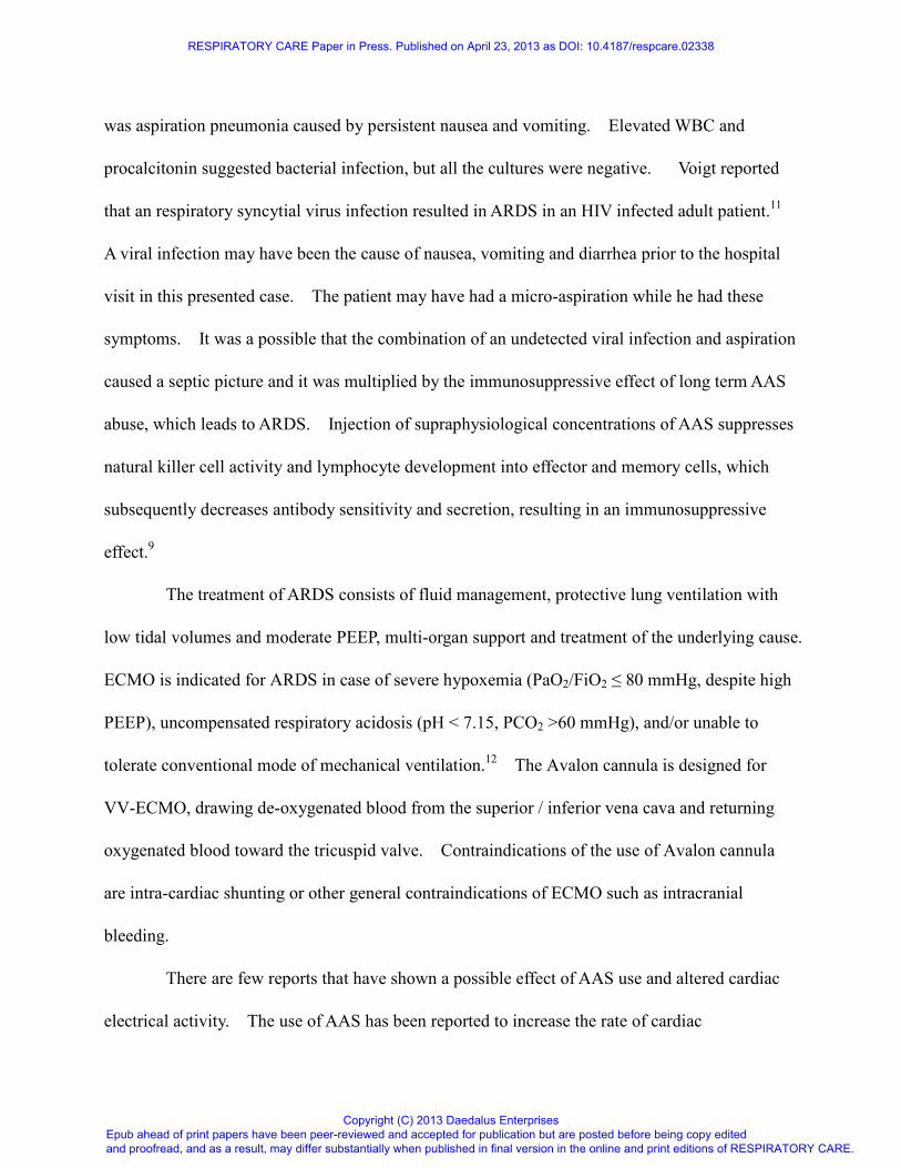
was aspiration pneumonia caused by persistent nausea and vomiting. Elevated WBC and
procalcitonin suggested bacterial infection, but all the cultures were negative. Voigt reported
that an respiratory syncytial virus infection resulted in ARDS in an HIV infected adult patient.11
A viral infection may have been the cause of nausea, vomiting and diarrhea prior to the hospital
visit in this presented case. The patient may have had a micro-aspiration while he had these
symptoms. It was a possible that the combination of an undetected viral infection and aspiration
caused a septic picture and it was multiplied by the immunosuppressive effect of long term AAS
abuse, which leads to ARDS. Injection of supraphysiological concentrations of AAS suppresses
natural killer cell activity and lymphocyte development into effector and memory cells, which
subsequently decreases antibody sensitivity and secretion, resulting in an immunosuppressive
effect.9
The treatment of ARDS consists of fluid management, protective lung ventilation with
low tidal volumes and moderate PEEP, multi-organ support and treatment of the underlying cause.
ECMO is indicated for ARDS in case of severe hypoxemia (PaO2/FiO2 ≤ 80 mmHg, despite high
PEEP), uncompensated respiratory acidosis (pH < 7.15, PCO2 >60 mmHg), and/or unable to
tolerate conventional mode of mechanical ventilation.12
The Avalon cannula is designed for
VV-ECMO, drawing de-oxygenated blood from the superior / inferior vena cava and returning
oxygenated blood toward the tricuspid valve. Contraindications of the use of Avalon cannula
are intra-cardiac shunting or other general contraindications of ECMO such as intracranial
bleeding.
There are few reports that have shown a possible effect of AAS use and altered cardiac
electrical activity. The use of AAS has been reported to increase the rate of cardiac
RESPIRATORY CARE Paper in Press. Published on April 23, 2013 as DOI: 10.4187/respcare.02338
Epub ahead of print papers have been peer-reviewed and accepted for publication but are posted before being copy edited and proofread, and as a result, may differ substantially when published in final version in the online and print editions of RESPIRATORY CARE.
Copyright (C) 2013 Daedalus Enterprises
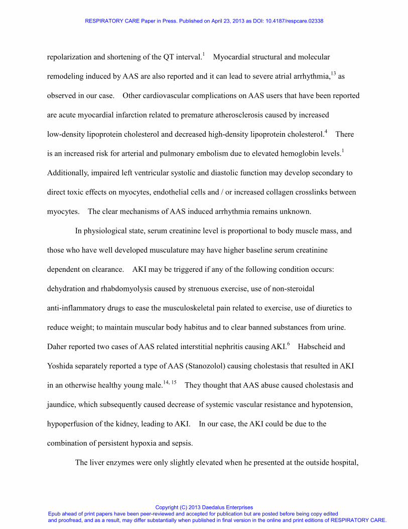
repolarization and shortening of the QT interval.1 Myocardial structural and molecular
remodeling induced by AAS are also reported and it can lead to severe atrial arrhythmia,13
as
observed in our case. Other cardiovascular complications on AAS users that have been reported
are acute myocardial infarction related to premature atherosclerosis caused by increased
low-density lipoprotein cholesterol and decreased high-density lipoprotein cholesterol.4 There
is an increased risk for arterial and pulmonary embolism due to elevated hemoglobin levels.1
Additionally, impaired left ventricular systolic and diastolic function may develop secondary to
direct toxic effects on myocytes, endothelial cells and / or increased collagen crosslinks between
myocytes. The clear mechanisms of AAS induced arrhythmia remains unknown.
In physiological state, serum creatinine level is proportional to body muscle mass, and
those who have well developed musculature may have higher baseline serum creatinine
dependent on clearance. AKI may be triggered if any of the following condition occurs:
dehydration and rhabdomyolysis caused by strenuous exercise, use of non-steroidal
anti-inflammatory drugs to ease the musculoskeletal pain related to exercise, use of diuretics to
reduce weight; to maintain muscular body habitus and to clear banned substances from urine.
Daher reported two cases of AAS related interstitial nephritis causing AKI.6 Habscheid and
Yoshida separately reported a type of AAS (Stanozolol) causing cholestasis that resulted in AKI
in an otherwise healthy young male.14, 15
They thought that AAS abuse caused cholestasis and
jaundice, which subsequently caused decrease of systemic vascular resistance and hypotension,
hypoperfusion of the kidney, leading to AKI. In our case, the AKI could be due to the
combination of persistent hypoxia and sepsis.
The liver enzymes were only slightly elevated when he presented at the outside hospital,
RESPIRATORY CARE Paper in Press. Published on April 23, 2013 as DOI: 10.4187/respcare.02338
Epub ahead of print papers have been peer-reviewed and accepted for publication but are posted before being copy edited and proofread, and as a result, may differ substantially when published in final version in the online and print editions of RESPIRATORY CARE.
Copyright (C) 2013 Daedalus Enterprises
but were elevated when the patient was transferred to our hospital. We believe this transient
liver dysfunction was related to the combination of hypoxia and septic condition, rather than
direct toxicity from AAS abuse. Although his echocardiography was normal, multiple episodes
of SVT and massive fluid resuscitation for low blood pressure, acute kidney injury associated
with low urine output, may have resulted in fluid overload and hepatic congestion. His NT
proBNP was mildly elevated upon admission to the outside hospital (917.8pg/ml) and the
existence of a pleural effusion on echo, support this assumption. However, it would be unusual
that the cause of hypoxia was cardiogenic pulmonary edema, as he was rather
dehydrated/hypovolemic when he arrived at the outside hospital, and echo showed good left
ventricular function. The liver enzymes quickly improved, after correction of the volume status,
and the initiation of VV-ECMO and CVVHD.
Conclusions
Although there is only limited literature regarding AAS use and their side effects, long
term AAS use may lead to serious health consequences. The direct relation of AAS and
multi-organ dysfunction is unclear in our case, it is reasonable to believe that the long standing
history of AAS abuse played a great role in causing systemic inflammatory response syndrome
and multiple organ dysfunction syndromes in an otherwise healthy patient.
RESPIRATORY CARE Paper in Press. Published on April 23, 2013 as DOI: 10.4187/respcare.02338
Epub ahead of print papers have been peer-reviewed and accepted for publication but are posted before being copy edited and proofread, and as a result, may differ substantially when published in final version in the online and print editions of RESPIRATORY CARE.
Copyright (C) 2013 Daedalus Enterprises
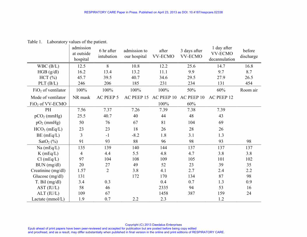
Table 1. Laboratory values of the patient.
admission
at outside
hospital
6 hr after
intubation
admission to
our hospital
after
VV-ECMO
3 days after
VV-ECMO
1 day after
VV-ECMO
decannulation
before
discharge
WBC (B/L) 12.5 8 10.8 12.2 25.6 14.7 16.8
HGB (g/dl) 16.2 13.4 13.2 11.1 9.9 9.7 8.7
HCT (%) 45.7 39.5 40.7 34.6 29.5 27.9 26.5
PLT (B/L) 246 206 185 231 234 131 454
FiO2 of ventilator 100% 100% 100% 100% 50% 60% Room air
Mode of ventilator NR mask AC PEEP 5 AC PEEP 15 AC PEEP 10 AC PEEP 10 AC PEEP 12
FiO2 of VV-ECMO 100% 60%
PH 7.56 7.37 7.26 7.39 7.38 7.39
pCO2 (mmHg) 25.5 40.7 40 44 48 43
pO2 (mmHg) 50 76 67 81 104 69
HCO3 (mEq/L) 23 23 18 26 28 26
BE (mEq/L) 3 -1 -8.2 1.8 3.1 1.3
SatO2 (%) 91 93 88 96 98 93 98
Na (mEq/L) 135 139 140 144 137 137 137
K (mEq/L) 4 4.4 5.5 4.8 4.7 3.8 3.8
Cl (mEq/L) 97 104 108 109 105 101 102
BUN (mg/dl) 20 27 49 52 23 39 35
Creatinine (mg/dl) 1.57 2 3.8 4.1 2.7 2.4 2.2
Glucose (mg/dl) 131 172 170 134 87 98
T. Bil (mg/dl) 3.4 0.3 0.4 0.7 1.3 0.9
AST (IU/L) 58 46 2335 94 53 16
ALT (IU/L) 109 67 1458 387 159 24
Lactate (mmol/L) 1.9 0.7 2.2 2.3 1.2
RESPIRATORY CARE Paper in Press. Published on April 23, 2013 as DOI: 10.4187/respcare.02338
Epub ahead of print papers have been peer-reviewed and accepted for publication but are posted before being copy edited and proofread, and as a result, may differ substantially when published in final version in the online and print editions of RESPIRATORY CARE.
Copyright (C) 2013 Daedalus Enterprises
AC: assist control; NR non-rebreasing mask with 100% oxygen; VV-ECMO: veno-veno extracorporeal membrane oxygenation
RESPIRATORY CARE Paper in Press. Published on April 23, 2013 as DOI: 10.4187/respcare.02338
Epub ahead of print papers have been peer-reviewed and accepted for publication but are posted before being copy edited and proofread, and as a result, may differ substantially when published in final version in the online and print editions of RESPIRATORY CARE.
Copyright (C) 2013 Daedalus Enterprises
Legends of Figures
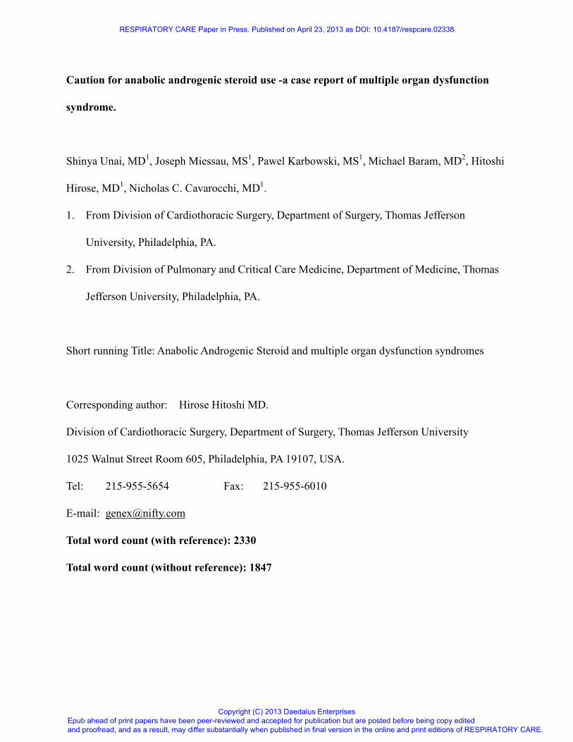
Figure 1: Chest x-ray on admission shows bilateral infiltrates.
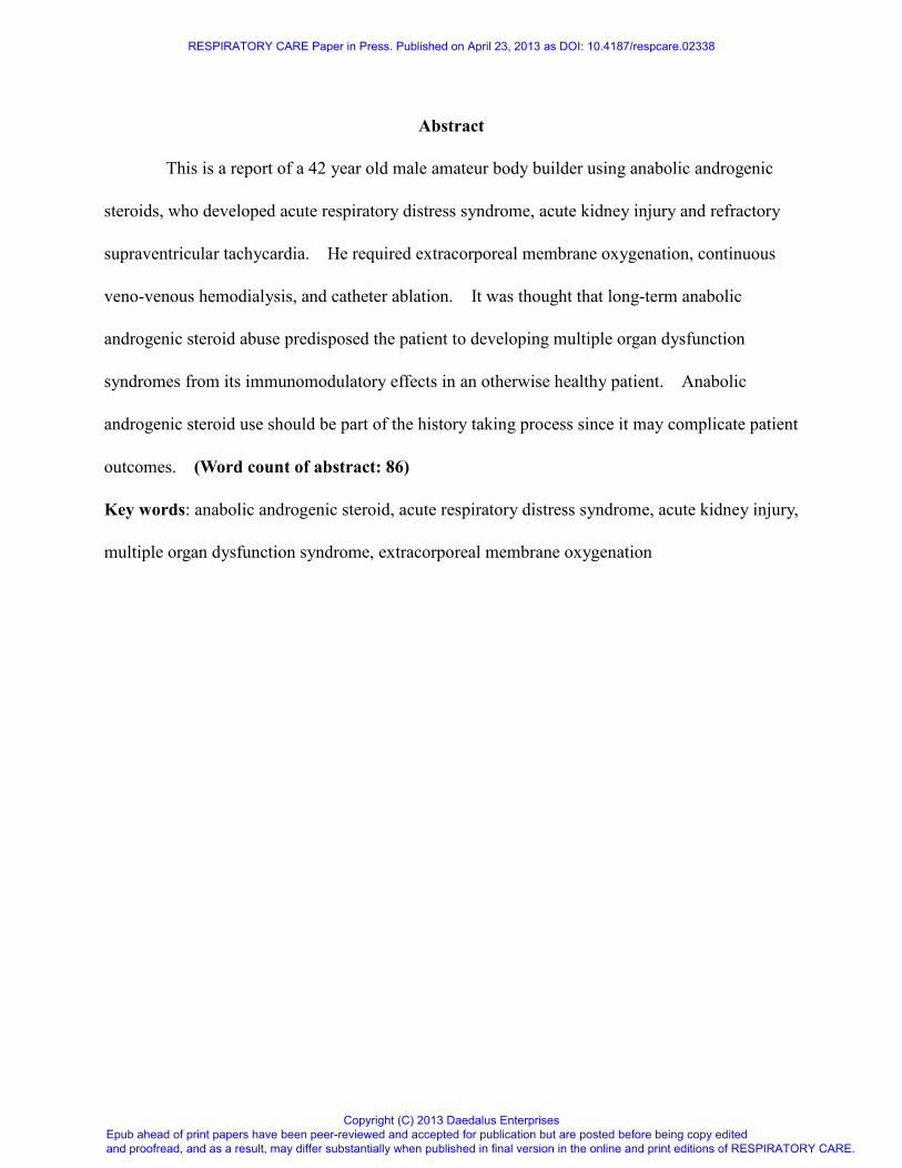
Figure 2: Chest x-ray before discharge shows clear lung fields.
RESPIRATORY CARE Paper in Press. Published on April 23, 2013 as DOI: 10.4187/respcare.02338
Epub ahead of print papers have been peer-reviewed and accepted for publication but are posted before being copy edited and proofread, and as a result, may differ substantially when published in final version in the online and print editions of RESPIRATORY CARE.
Copyright (C) 2013 Daedalus Enterprises
Reference
1. Angell P, Chester N, Green D, Somauroo J, Whyte G, George K. Anabolic steroids and
cardiovascular risk. Sports Med 2012;42(2):119-134.
2. Thompson BT, Bernard GR. ARDS Network (NHLBI) studies: successes and challenges
in ARDS clinical research. Crit Care Clin 2011;27(3):459-468.
3. Wong JK, Smith TN, Pitcher HT, Hirose H, Cavarocchi NC. Cerebral and lower limb
near-infrared spectroscopy in adults on extracorporeal membrane oxygenation. Artif
Organs 2012;36(8):659-667.
4. Kanayama G, Hudson JI, Pope HG, Jr. Long-term psychiatric and medical consequences
of anabolic-androgenic steroid abuse: a looming public health concern? Drug Alcohol
Depend 2008;98(1-2):1-12.
5. Hakansson A, Mickelsson K, Wallin C, Berglund M. Anabolic androgenic steroids in the
general population: user characteristics and associations with substance use. Eur Addict
Res 2012;18(2):83-90.
6. Daher EF, Silva Junior GB, Queiroz AL, Ramos LM, Santos SQ, Barreto DM, et al.
Acute kidney injury due to anabolic steroid and vitamin supplement abuse: report of two
cases and a literature review. Int Urol Nephrol 2009;41(3):717-723.
7. Geraci MJ, Cole M, Davis P. New onset diabetes associated with bovine growth hormone
and testosterone abuse in a young body builder. Hum Exp Toxicol
2011;30(12):2007-2012.
8. Samaha AA, Nasser-Eddine W, Shatila E, Haddad JJ, Wazne J, Eid AH. Multi-organ
damage induced by anabolic steroid supplements: a case report and literature review. J
Med Case Rep 2008;2:340.
9. Brenu EW, McNaughton L, Marshall-Gradisnik SM. Is there a potential immune
RESPIRATORY CARE Paper in Press. Published on April 23, 2013 as DOI: 10.4187/respcare.02338
Epub ahead of print papers have been peer-reviewed and accepted for publication but are posted before being copy edited and proofread, and as a result, may differ substantially when published in final version in the online and print editions of RESPIRATORY CARE.
Copyright (C) 2013 Daedalus Enterprises
dysfunction with anabolic androgenic steroid use?: A review. Mini Rev Med Chem
2011;11(5):438-445.
10. Herr A, Rehmert G, Kunde K, Gust R, Gries A. A thirty-year old bodybuilder with septic
shock and ARDS from abuse of anabolic steroids. Anaesthesist 2002;51(7):557-563.
11. Voigt E, Tillmann RL, Schewe JC, Molitor E, Schildgen O. ARDS in an HIV-positive
patient associated to respiratory syncytial virus. Eur J Med Res 2008;13(3):131-132.
12. Brodie D, Bacchetta M. Extracorporeal membrane oxygenation for ARDS in adults. N
Engl J Med 2011;365(20):1905-1914.
13. Lau DH, Stiles MK, John B, Shashidhar, Young GD, Sanders P. Atrial fibrillation and
anabolic steroid abuse. Int J Cardiol 2007;117(2):e86-87.
14. Yoshida EM, Karim MA, Shaikh JF, Soos JG, Erb SR. At what price, glory? Severe
cholestasis and acute renal failure in an athlete abusing stanozolol. CMAJ
1994;151(6):791-793.
15. Habscheid W, Abele U, Dahm HH. Severe cholestasis with kidney failure from anabolic
steroids in a body builder. Dtsch Med Wochenschr 1999;124(36):1029-1032.
RESPIRATORY CARE Paper in Press. Published on April 23, 2013 as DOI: 10.4187/respcare.02338
Epub ahead of print papers have been peer-reviewed and accepted for publication but are posted before being copy edited and proofread, and as a result, may differ substantially when published in final version in the online and print editions of RESPIRATORY CARE.
Copyright (C) 2013 Daedalus Enterprises
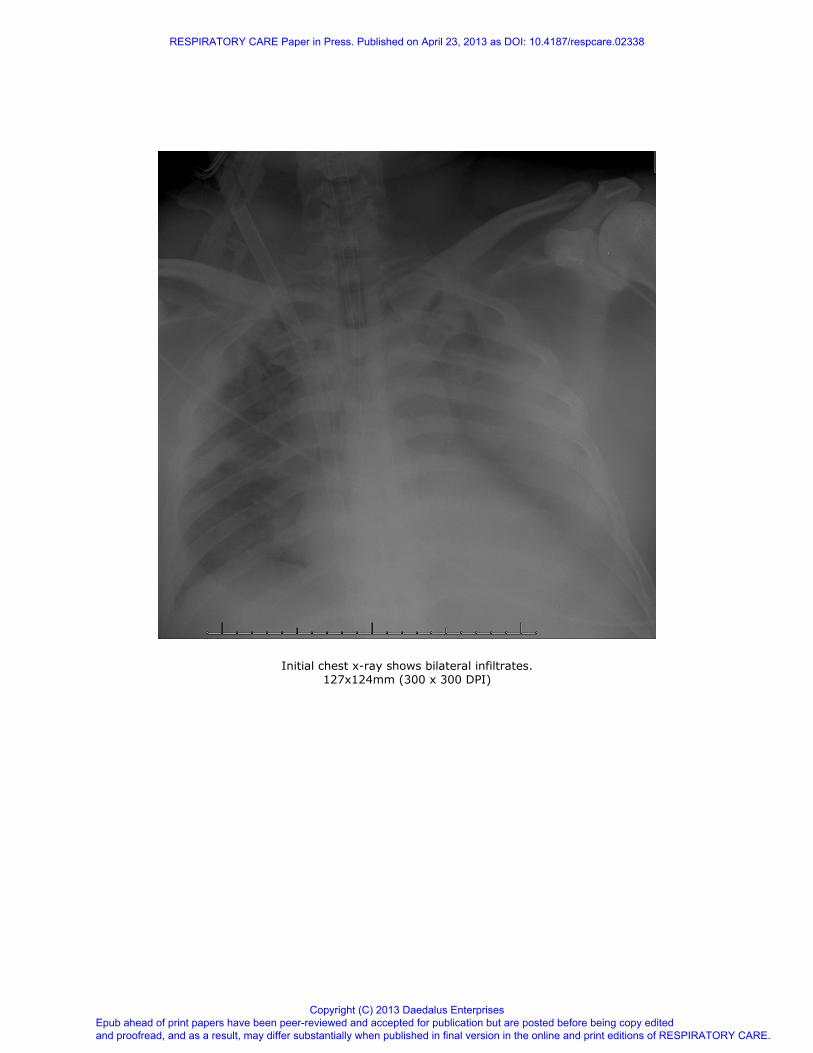
Initial chest x-ray shows bilateral infiltrates.
127x124mm (300 x 300 DPI)
RESPIRATORY CARE Paper in Press. Published on April 23, 2013 as DOI: 10.4187/respcare.02338
Epub ahead of print papers have been peer-reviewed and accepted for publication but are posted before being copy edited and proofread, and as a result, may differ substantially when published in final version in the online and print editions of RESPIRATORY CARE.
Copyright (C) 2013 Daedalus Enterprises
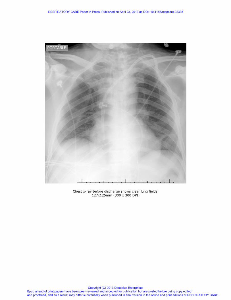
Chest x-ray before discharge shows clear lung fields. 127x125mm (300 x 300 DPI)
RESPIRATORY CARE Paper in Press. Published on April 23, 2013 as DOI: 10.4187/respcare.02338
Epub ahead of print papers have been peer-reviewed and accepted for publication but are posted before being copy edited and proofread, and as a result, may differ substantially when published in final version in the online and print editions of RESPIRATORY CARE.
Copyright (C) 2013 Daedalus Enterprises


![A summary of the health harms of drugs · • Anabolic agents (anabolic-androgenic steroids, growth hormone, clenbuterol, [human and non- human] chorionic gonadotropin [hCG]2) a](https://img.pdfslide.us/doc/110x75/603c267c0af1bf56c7735e66/a-summary-of-the-health-harms-of-drugs-a-anabolic-agents-anabolic-androgenic.jpg)
![STUDIES ON ANABOLIC STEROIDS III. DETECTION AND … · The metabolism of stanozolol (17P-hydroxy-17~-methyl-5a-androstano[3,2-c]pyrazole), an androgenic-anabolic steroid widely used](https://img.pdfslide.us/doc/110x75/5f80a2936657d33deb3dbd42/studies-on-anabolic-steroids-iii-detection-and-the-metabolism-of-stanozolol-17p-hydroxy-17-methyl-5a-androstano32-cpyrazole.jpg)